Targeting the Integrated Stress Response in Cancer Therapy
←
→
Page content transcription
If your browser does not render page correctly, please read the page content below
REVIEW
                                                                                                                                       published: 24 September 2021
                                                                                                                                      doi: 10.3389/fphar.2021.747837

                                           Targeting the Integrated Stress
                                           Response in Cancer Therapy
                                           Xiaobing Tian 1,2,3,4*, Shengliang Zhang 1,2,3,4, Lanlan Zhou 1,2,3,4, Attila A. Seyhan 1,2,3,4,
                                           Liz Hernandez Borrero 1, Yiqun Zhang 1 and Wafik S. El-Deiry 1,2,3,4,5*
                                           1
                                             Laboratory of Translational Oncology and Experimental Cancer Therapeutics, Warren Alpert Medical School, Brown University,
                                           Providence, RI, United States, 2Department of Pathology and Laboratory Medicine, Warren Alpert Medical School, Brown
                                           University, Providence, RI, United States, 3Joint Program in Cancer Biology, Lifespan Health System and Brown University,
                                           Providence, RI, United States, 4Cancer Center at Brown University, Providence, RI, United States, 5Hematology/Oncology
                                           Division, Department of Medicine, Lifespan Health System and Brown University, Providence, RI, United States

                                           The integrated stress response (ISR) is an evolutionarily conserved intra-cellular signaling
                                           network which is activated in response to intrinsic and extrinsic stresses. Various stresses
                                           are sensed by four specialized kinases, PKR-like ER kinase (PERK), general control non-
                                           derepressible 2 (GCN2), double-stranded RNA-dependent protein kinase (PKR) and
                                           heme-regulated eIF2α kinase (HRI) that converge on phosphorylation of serine 51 of
                                           eIF2α. eIF2α phosphorylation causes a global reduction of protein synthesis and triggers
                          Edited by:
                        Anne Lorant,       the translation of specific mRNAs, including activating transcription factor 4 (ATF4).
Laboratoire de Biologie Moléculaire et     Although the ISR promotes cell survival and homeostasis, when stress is severe or
      Cellulaire du Cancer (LBMCC),
                         Luxembourg
                                           prolonged the ISR signaling will shift to regulate cellular apoptosis. We review the ISR
                    Reviewed by:
                                           signaling pathway, regulation and importance in cancer therapy.
                       Souvik Dey,
                                           Keywords: integrated stress responses, ATF4, CHOP, apoptosis, cancer treatment
          Jadavpur University, India
                   Akihiro Tomida,
    Japanese Foundation For Cancer
                  Research, Japan          INTRODUCTION
                  *Correspondence:         ISR is an evolutionarily conserved intra-cellular signal network activated in response to various
                        Xiaobing Tian
                                           intrinsic and extrinsic factors (Figure 1). Extrinsic factors include amino acid depletion, glucose
            xiaobing_tian@brown.edu
                     Wafik S. El-Deiry
                                           deprivation, viral infection, hypoxia, heme deficiency, ROS (reactive oxygen species) and DNA
                    wafik@brown.edu         damage (Pakos-Zebrucka et al., 2016; Clementi et al., 2020; Akman et al., 2021). Cellular intrinsic
                                           stresses, such as ER (endoplasmic reticulum) stress, can also activate the ISR (Pakos-Zebrucka et al.,
                 Specialty section:        2016). In the context of cancer biology, oncogene activation, such as MYC overexpression, can
      This article was submitted to        trigger the ISR (Tameire et al., 2019). Cancer cells with enhanced proliferation have enhanced
Pharmacology of Anti-Cancer Drugs,         protein synthesis which leads to a high basal level of the ISR as compared to normal cells (McConkey,
            a section of the journal       2017; Tameire et al., 2019). This may explain why ISR inducers can selectively target cancer cells.
         Frontiers in Pharmacology             Various stresses are sensed by four specialized kinases (PERK, GCN2, PKR and HRI) that converge on
            Received: 26 July 2021         phosphorylation of serine 51 of eIF2α (Figure 1) (Perkins and Barber, 2004; Wek et al., 2006; Donnelly
     Accepted: 10 September 2021           et al., 2013). Although significant sequence homology exists between these four eIF2α kinases in their
     Published: 24 September 2021
                                           kinase catalytic domains, underlying their common role in phosphorylating eIF2α, each eIF2α kinase
                            Citation:      possesses distinct regulatory domains and additional unique features that determine the regulation of
Tian X, Zhang S, Zhou L, Seyhan AA,        these four kinases by signals that activate them (Donnelly et al., 2013). Each kinase responds to distinct
   Hernandez Borrero L, Zhang Y and
                                           environmental and physiological stresses, which reflects their unique regulatory mechanisms (Donnelly
     El-Deiry WS (2021) Targeting the
        Integrated Stress Response in
                                           et al., 2013). eIF2α phosphorylation causes global reduction of protein synthesis and triggers the
                      Cancer Therapy.      translation of specific mRNAs, including ATF4 to help with cell survival and recovery. However, if
        Front. Pharmacol. 12:747837.       the stress cannot be reduced, ATF4 regulates an apoptosis program to eliminate the damaged cells (Pakos-
     doi: 10.3389/fphar.2021.747837        Zebrucka et al., 2016; Costa-Mattioli and Walter, 2020).

Frontiers in Pharmacology | www.frontiersin.org                                   1                                     September 2021 | Volume 12 | Article 747837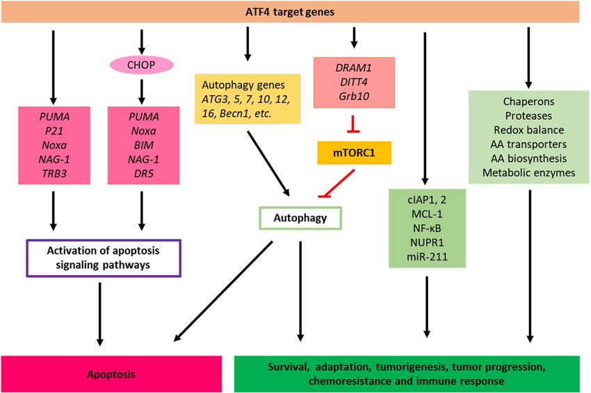
Tian et al.                                                                                                                   ISR Cancer Apoptosis Through ATF4

                                                                                       activation and apoptosis seen in these cells (Figure 2)
                                                                                       (Hamanaka et al., 2009).
                                                                                           ATF4 has also been demonstrated to facilitate anti-neoplastic
                                                                                       agent bortezomib-induced upregulation of anti-apoptotic
                                                                                       myeloid cell leukemia-1 (Mcl-1) protein, which is an anti-
                                                                                       apoptotic Bcl-2 family protein that plays essential roles in
                                                                                       multiple myeloma survival and drug resistance in many tumor
                                                                                       types (Figure 2) (Hu et al., 2012).
                                                                                           It has been shown that both MCL-1 and cIAPs can suppress
                                                                                       apoptosis at different points in the apoptosis pathway that are
                                                                                       upstream and downstream of the release of cytochrome c from
                                                                                       the mitochondria. Mitochondrial cytochrome c plays a dual
                                                                                       function in controlling both cellular energetic metabolism and
                                                                                       apoptosis. It has been shown that, upon interacting with
                                                                                       apoptotic protease activating factors (Apaf), cytochrome c can
                                                                                       trigger the activation cascade of caspases once it is released from
                                                                                       the mitochondria into the cytosol (Cai et al., 1998).
                                                                                           It has also been reported that miR-211 is a pro-survival
                                                                                       microRNA that regulates CHOP expression in a PERK-
                                                                                       dependent manner and thus PERK can mediate a pro-survival
                                                                                       function by suppressing a stress-dependent expression of CHOP
                                                                                       consequently leading to re-establishment of cellular homeostasis
                                                                                       before the initiation of apoptosis (Chitnis et al., 2012). In addition to
                                                                                       its beneficial roles in restoring homeostasis, these ISR mechanisms
                                                                                       may also contribute to tumor development. For example, an
  FIGURE 1 | Integrated stress responses signaling pathway. ER stress,
                                                                                       increased miR-211 expression, found to be PERK-dependent, and
  mitochondria stress or heme depletion, amino acid deficiency and ds-RNA
  virus infection activate PERK, HRI, GCN2 and PKR sensor kinases, leading to          was reported in mammary carcinoma and mouse models of human
  phosphorylation of eIF2α. eIF2α phosphorylation causes global inhibition             B-cell lymphoma (Figure 2) (Chitnis et al., 2012).
  of protein synthesis but selective translation of ATF4 mRNA. ATF4 binds to               Cancer cells use multiple stress response pathways such as the
  DNA targets to regulate the expression of genes that promote cellular                integrated stress response (ISR), cytosolic heat shock response
  adaptation, survival and apoptosis. Feedback regulation of ISR is regulated by
                                                                                       (HSR), and unfolded protein response (UPR) mediated by
  constitutively expressed phosphatase complex CReP-PP1 and inducible
  phosphatase GADD34-PP1, which dephosphorylate eIF2α and attenuate or                 organelles such as the endoplasmic reticulum (ER) and
  terminate ISR. AA, Amino acid; ER, Endoplasmic reticulum.                            mitochondria to respond exogenous and endogenous or
                                                                                       environmental stresses to evade apoptosis, ensure survival,
                                                                                       proliferation, metastatic potential, and maintain cellular
   ATF4 plays an important role in communicating pro-survival                          homoeostasis (OʼMalley et al., 2020). For example, to evade
and pro-apoptotic signals. Once activated, ATF4 regulates                              apoptosis and ensure survival, cancer cells may utilize the
transcriptional programs involved in cell survival (antioxidant                        mitochondrial unfolded protein response (UPRmt) pathway
response, amino acid biosynthesis and autophagy), senescence                           and associated key proteins including chaperones HSP10,
and apoptosis. The final outcome of ATF4 activation is dependent                        HSP60, and mtHSP70 and proteases ClpP and LONP1 to
on the cell type, nature of stressors and duration of the stresses                     eliminate proteotoxic stress (Figure 2) (OʼMalley et al., 2020).
(Figure 1) (Wang et al., 2015; Wortel et al., 2017; Ojha et al., 2019;                 Notably, upregulation of HSP60 expression and its upstream
Tameire et al., 2019).                                                                 regulator ATF5 has been shown to enhance the apoptotic
                                                                                       threshold in cancer cells resulting in therapeutic resistance in
                                                                                       many cancer types. ATF-5 has been reported to regulate
The Integrated Stress Response and Cell                                                expression of Egr-1, BCL-2, and MCL1 to mediate
Survival                                                                               proliferation and survival in cancer (Dluzen et al., 2011; Liu
The ISR promotes cellular survival signaling by negative                               et al., 2011; Karpel-Massler et al., 2016).
regulation of cell death pathways, such as apoptosis. For                                  Moreover, in addition to the genes mentioned above many
instance, as a consequence of ER stress, PERK-induced                                  other genes activated in response to ISR (Costa-Mattioli and
activation of the ISR results in the expression of cIAP1 and                           Walter, 2020), including those encoding ATF4, ATF5 (Zhou
cIAP2 (cellular inhibitor of apoptosis proteins) in tumor and                          et al., 2008); CHOP (C/EBP-homologous protein) (Palam
non-tumor cells (Hamanaka et al., 2009; Hu et al., 2004;                               et al., 2011); GADD34 (Growth Arrest And DNA-Damage-
Warnakulasuriyarachchi et al., 2004). Previously, it was                               Inducible 34) (Lee et al., 2009); and in neurons, OPHN1
demonstrated that restoration of the function of cIAP1 or                              (Oligophrenin-1) (Di Prisco et al., 2014), other genes such as
cIAP2 in PERK−/− murine embryonic fibroblasts during ER                                 IBTKα (the α isoform of inhibitor of Bruton’s tyrosine kinase)
stress delays the early onset of ER stress-induced caspase                             (Baird et al., 2014) and NUPR1 (Nuclear protein-1), also play

Frontiers in Pharmacology | www.frontiersin.org                                    2                                  September 2021 | Volume 12 | Article 747837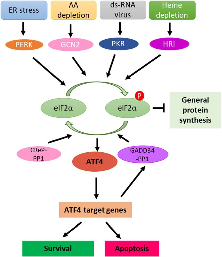
Tian et al.                                                                                                                      ISR Cancer Apoptosis Through ATF4

  FIGURE 2 | Cell death, pro-survival, tumor progression and chemoresistance pathways of ISR. ATF4 directly or indirectly controls the transcription of apoptotic,
  adaptive, tumor progression and chemoresistance genes. When stress persists (for example, drug treatments) and cancer cells are unable to adapt to and reach
  homeostasis though the activation of ISR, ATF4 shifts this balance towards apoptosis by inducing apoptotic genes. AA, Amino acid.

important roles in cell survival. NUPR1 has been found to play an                     found that the eIF2α kinase HRI controls NOD1 (Nucleotide-binding
important role in cell stress and stress-related apoptosis (Martin                    oligomerization domain-containing protein 1) signalosome folding
et al., 2021) and inactivation of NUPR1 promotes cell death by                        and activation through a process requiring eIF2α, ATF4, and the heat
coupling ER-stress responses with necrosis (Santofimia-Castaño                         shock protein HSPB8 (Abdel-Nour et al., 2019). Moreover, HRI/
et al., 2018). More evidences suggest that ATF4 initiates the                         eIF2α signaling pathway was shown to be required for signaling
activity of transcription factor NUPR1. NUPR1 regulates the                           downstream of the innate immune mediators including NOD2,
expression of several metabolic stress-responsive genes, in                           MAVS (Mitochondrial antiviral-signaling protein), and TRIF
particular, genes required in cell cycle regulation and DNA                           (TIR-domain-containing adapter-inducing interferon-β) but
repair, as such, NUPR1 also is regarded as pro-survival factors                       dispensable for signaling pathways that rely on MyD88 (Myeloid
(Figure 2) (Jin et al., 2009; Hamidi et al., 2012).                                   differentiation primary response 88) or STING (Stimulator of
    Another gene activated during the ISR is the IBTKα which is                       interferon genes) (Figure 2) (Abdel-Nour et al., 2019).
activated during ER stress. IBTKα is a major substrate adaptor for
protein ubiquitination and is an essential pro-survival factor
(Baird et al., 2014).                                                                 The Integrated Stress Response and
    Likewise, eIF2α mediated translational repression has been                        Activation of Autophagy
suggested in activated B cell NF-κB pathway induction as a                            Autophagy is a highly regulated eukaryotic cellular pathway that
mechanism to protect cells against ER stress (Deng et al., 2004).                     plays a major role in the lysosomal degradation of cytoplasmic
In a recent study, a pharmacologically activable version of PERK was                  unfolded proteins, peptides, damaged organelles or cytosolic
used to uncouple eIF2α phosphorylation from stress and it was                         components while also serving as a means to replenish depleted
determined that eIF2α phosphorylation is both required and                            amino acids for building proteins and to provide energy to a
adequate to activate both NF-κB DNA binding and an NF-κB                              starved cell. Autophagy can be activated by a variety of cellular
reporter gene (Deng et al., 2004). Also, HRI has been shown to                        stresses such as nutrient or growth factor deprivation, hypoxia,
be involved in NF-κB activation (Abdel-Nour et al., 2019). This study                 reactive oxygen species, DNA damage, protein aggregates,

Frontiers in Pharmacology | www.frontiersin.org                                   3                                     September 2021 | Volume 12 | Article 747837
Tian et al. ISR Cancer Apoptosis Through ATF4
damaged organelles, or intracellular pathogens (Pakos-Zebrucka for the activation of autophagy (Ye et al., 2010). Notably, while
et al., 2016; Clementi et al., 2020; Akman et al., 2021). Autophagy GCN2 knockout cells exhibited decreased LC3 expression, cells
can be activated both via specific, stimulus-dependent manner and with mutant the eIF2α S51A were not able to activate the
more general, stimulus-independent signaling pathways to processing of LC3 (Ye et al., 2010). Likewise, in the regulation
coordinate different phases of autophagy. of autophagy induced by amino acid starvation, phosphorylation
The ISR can modulate cell survival and cell death pathways of eIF2α at S51 was found to be required in yeast and mouse
through the activation of autophagy and the phosphorylation of embryonic fibroblasts (MEFs) (Tallóczy et al., 2002). These
eIF2α at S51 appears to be essential for stress-induced autophagy findings suggest that eIF2α phosphorylation at S51 forms the
(Pakos-Zebrucka et al., 2016). Autophagy can be integrated with central hub between different stresses and activation of
other cellular stress responses through parallel stimulation of autophagy.
autophagy and other stress responses by specific stress stimuli, Downstream of eIF2α phosphorylation, although ATF4 has
through dual regulation of autophagy and other stress responses been implicated to be essential for activation of autophagy, other
by multifunctional stress signaling molecules, and/or through mechanisms directed from eIF2α phosphorylation other than
mutual control of autophagy and other stress responses. selective translation of ATF4 mRNA might also be involved in the
activation of the autophagy process (Kroemer et al., 2010). It was
PERK Regulates Autophagy previously suggested that phosphorylation of eIF2α might affect
Although mechanisms by which phosphorylated eIF2α induces the ER in a manner that promotes the physical formation of the
autophagy are still not completely elucidated, specific extrinsic isolation membrane. Alternatively, eIF2α phosphorylation might
and intrinsic stresses that lead to the phosphorylation of eIF2α stimulate autophagy through its effects on the transactivation of
have been demonstrated to trigger autophagy. For instance, ER autophagy genes. eIF2α phosphorylation stimulates the selective
stress increases phosphorylation of eIF2α and ensuing translation of the ATF4 transcription factor, which stimulates
upregulation of certain autophagy receptors including LC3 expression which is essential for sustained autophagy
SQSTM1, NBR1, and BNIP3L through PERK (Deegan et al., (Milani et al., 2009; Kroemer et al., 2010). Furthermore,
2015). Likewise, inhibition of PERK pharmacologically although autophagy interaction network components play
suppresses transcriptional upregulation of these autophagy important roles in vesicle trafficking, protein or lipid
receptors in mammalian cells (Deegan et al., 2015). phosphorylation and protein ubiquitination and there are
Furthermore, phosphorylation of eIF2α mediated by PERK direct interactions between eIF2α subunits and core autophagy
increases the conversion of ATG12 and LC3 due to the proteins, whether these interactions are biologically significant is
expression of polyQ72 aggregates in C2C5 cells, which is an not clearly understood (Behrends et al., 2010).
essential step for autophagy formation (Kouroku et al., 2007). Under conditions of ER stress or amino acid deprivation, there
This PERK-mediated Unfolded Protein Response (UPR) has is transcriptional upregulation of key autophagy genes mediated
been shown to regulate autophagy from induction, to vesicle by ATF4 including MAP1LC3B and ATG5 which are required for
nucleation, phagophore elongation, and maturation (Deegan autophagosome biogenesis and function (Deegan et al., 2015;
et al., 2013). Rzymski et al., 2010; BʼChir et al., 2013). ATF4 can also
Moreover, it was reported that ER stress due to bluetongue upregulate the DITT4/REDD1 and DRAM1, which represses
virus infection of cells leads to autophagy through the activation the activity of mTORC1, subsequently inducing autophagy
of the PERK-eIF2α pathway (Lv et al., 2015). The UPR which is (Figure 2) (Kazemi et al., 2007; Whitney et al., 2009; Dennis
initiated in response to the accumulation of misfolded proteins in et al., 2013; Tian et al., 2021).
the ER leading to stress is predominantly an adaptive response to Furthermore, ATF4 activation in response to amino acid
the activation of the ISR. It was shown that UPR protects human deprivation also directs an autophagy gene transcriptional
tumor cells during hypoxia through regulation of the autophagy program by upregulating several autophagy genes such as
genes MAP1LC3B and ATG5 (Rouschop et al., 2010) and this was Atg3, Atg5, Atg7, Atg10, Atg12, Atg16, Becn1, Gabarap,
mediated by PERK phosphorylation of eIF2α. Conversely, Gabarapl2, Map1lc3b, and Sqstm1 (Figure 2) (BʼChir et al.,
abrogation of PERK signaling or expression of mutant eIF2α 2013). Through the stimulation of key genes involved in
S51A which cannot be phosphorylated under the condition of autophagy, the ISR mediates the up-regulation of the
hypoxia reduces the transcription of MAP1LC3B and ATG5 autophagic process in an attempt to resolve the stress induced
(Rouschop et al., 2010). by amino acid deprivation. This is accomplished by the increased
IRS-induced autophagy also can lead to cell death. A recent recycling of cytoplasmic components and sustaining the
paper reported that compound SH003 induces autophagy and biosynthetic capacity of the cell and cellular ATP
autophagic cell death through a PERK-eIF2α-ATF4-CHOP concentrations. The increased autophagic function leads to
signaling pathway in human gastric cancer cells (Figure 2) increased amino acid levels in ER required for de novo protein
(Kim et al., 2020). biosynthesis and similarly leads to increased levels of substrates
including free fatty acids and amino acids for the tricarboxylic
General Control Non-Derepressible 2 Regulates acid cycle (Rzymski et al., 2009; Ye et al., 2010).
Autophagy However, it was also shown that a variety of autophagy genes
Similarly, amino acid deprivation in cancer cells leads to the can have a varying degree of reliance on ATF4 and CHOP
phosphorylation of eIF2α mediated by GCN2 which is required signaling and that the transcriptional upregulation of such
Frontiers in Pharmacology | www.frontiersin.org 4 September 2021 | Volume 12 | Article 747837Tian et al. ISR Cancer Apoptosis Through ATF4
genes is regulated by the ratio of ATF4 and CHOP proteins that PKR Regulates Autophagy
are bound to a particular promoter, and thus fine-tuning the Talloczy, Z. et al. report that PKR acts as a potent inducer of
expression of autophagy genes depending on the needs of the cell autophagy during viral infection (Tallóczy et al., 2006). Also, two
(BʼChir et al., 2013). papers indicate that PKR is very important for the autophagic
Studies on the effect of proteasome inhibition on survival degradation of herpes simplex virions both in vitro and in vivo
signaling by the ISR have revealed that suppression of proteasome (Tallóczy et al., 2006; Orvedahl et al., 2007). In these settings, PKR
function pharmacologically using antineoplastic agent was shown to operate upstream of Beclin 1 (Tallóczy et al., 2006).
bortezomib results in depletion of amino acids in the ER Shen, S. et al. report that STAT3 inhibitors (JSI-124, WP1066
required for protein synthesis leading to the activation of the and Stattic) caused the disruption of inhibitory STAT3-PKR
ISR via GCN2 stress sensor (Suraweera et al., 2012). interactions in human osteosarcoma U2OS cells, resulting in
Amino acid depletion as a result of proteasome inhibition also release and activation of PKR. PKR phosphorylates eIF2α,
activates autophagy through mTOR in an attempt to restore which regulates the activity of Beclin 1/Vps34 complex and
amino acid homeostasis (Suraweera et al., 2012). Conversely, facilitates autophagy induction (Figure 3) (Shen et al., 2012).
exogenous supplementation of essential amino acids depleted by Pathogenic bacterium Mycobacterium tuberculosis (Mtb)
the inhibition of proteasome function inhibition attenuates the infection induces the activation of PKR and PKR-mediated
phosphorylation of eIF2α and down-regulates autophagy autophagy in macrophage. Sustained expression and activation
(Suraweera et al., 2012). As such, depletion of amino acids by of PKR reduced the intracellular survival of Mtb, which could be
proteasome inhibition establishes a link between ISR activation enhanced by Interferon gamma (IFNγ) treatment (Smyth et al.,
and induction of autophagy in an attempt to sustain the survival 2020).
of the cell.
Heme-Regulated eIF2α Kinase Regulates Autophagy The Integrated Stress Response and Cell
Although the other eIF2α kinases are present across different Death
tissues, eIF2α kinase HRI is more specific to erythroid cells and The cell death pathways are complex and can be exploited by
plays a major role in erythrocyte differentiation during cancer therapeutic agents (Carneiro and El-Deiry, 2020). When
erythropoiesis (Suraweera et al., 2012). eIF2α kinase HRI stress persists and cells are unable to reach homeostasis despite
mediates the translation of globin mRNAs with the availability the activation of stress response pathways, ATF4 can induce the
of heme for the production of hemoglobin. By doing so, HRI transcriptional activation of apoptotic genes encoding CHOP
protects erythroid cells from the increase of toxic globin (DDIT3) (Harding et al., 2000), TRB3 (Tribbles homolog 3)
aggregates under conditions of iron deficiency (Bruns and (Ohoka et al., 2005), and pro-apoptotic BH3-only proteins
London, 1965; Chefalo et al., 1998; Han et al., 2001; Suragani including PUMA (p53 upregulated modulator of apoptosis),
et al., 2012). Other stresses such as arsenite-induced oxidative Noxa (Phorbol-12-myristate-13-acetate-induced protein 1) and
stress, heat shock, osmotic stress, 26S proteasome inhibition, and BIM (Bcl-2 Interacting mediator of cell death), thus leading to cell
nitric oxide also were shown to activate HRI (Han et al., 2001; Lu death (Galehdar et al., 2010; Altman et al., 2009; Puthalakath
et al., 2001; McEwen et al., 2005; Yerlikaya et al., 2008; Ill-Raga et al., 2007). ATF4 has been shown to regulate Noxa at the
et al., 2015) and activation of HRI by these stresses is independent transcriptional level and this leads to the induction of apoptosis
of heme and heat shock proteins HSP90 and HSP70 facilitates this (Sharma et al., 2018; Núñez-Vázquez et al., 2021). Overall,
process; however, the exact mechanism of HRI activation is still through the induction of ATF4, this transcription factor
being studied (Lu et al., 2001). appears to mainly trigger the intrinsic apoptosis by
A recent report demonstrated that HRI controls autophagy to modulating the expression of pro- and anti-apoptotic BCL-2
clear cytosolic protein aggregates (Mukherjee et al., 2021). In that family members. Interestingly, in the case of CHOP activation,
study, researchers found that the eIF2α kinase HRI induced a induction of DR5 (Death receptor 5) mediated apoptosis
cytosolic unfolded protein response to prevent aggregation of appeared to be DR5 ligand binding independent and involving
innate immune signalosomes. Furthermore, they demonstrated the engagement of FADD (Fas-associated protein with death
that HRI controls autophagy to clear cytosolic protein aggregates domain) and caspase-8 (Figure 2) (Lu et al., 2014; Li et al., 2015).
when the ubiquitin-proteasome system is inhibited (Mukherjee Additional stresses such as those resulting from decreased
et al., 2021). mitochondrial translation (Sasaki et al., 2020) as well as the
Growth factor receptor-bound protein 10 (Grb10) is regulated generation of reactive oxygen species (Kasai et al., 2019) have
by ATF4 (Zhang et al., 2018). the HRI-eIF2αP-ATF4 pathway been shown to induce ATF4 expression. In the case of sustained
suppresses mTORC1 signaling through Grb10 specifically in the mitochondrial deficiency, ATF4 response has been reported to
erythroid lineage (Figure 2) (Zhang et al., 2018). mTORC1 was lead to p53-mediated apoptosis (Evstafieva et al., 2014). Reactive
shown to act as a master regulator of autophagy since inhibition of oxygen species generated by Fenretinide treatment in
mTORC1 was required to initiate the autophagy process (Dossou neuroblastoma cells activates ATF4 leading to the induction of
and Basu, 2019). It was also shown that mTORC1 directly Noxa ultimately leading to apoptosis (Nguyen et al., 2019). In
regulates the downstream steps of the autophagy process, such multiple myeloma cells, sensitivity to bortezomib treatment was
as the nucleation, autophagosome elongation, autophagosome associated with higher expression of ATF4 and loss of its
maturation and termination (Dossou and Basu, 2019). expression lead to lower levels of Noxa, CHOP and DR5
Frontiers in Pharmacology | www.frontiersin.org 5 September 2021 | Volume 12 | Article 747837Tian et al. ISR Cancer Apoptosis Through ATF4
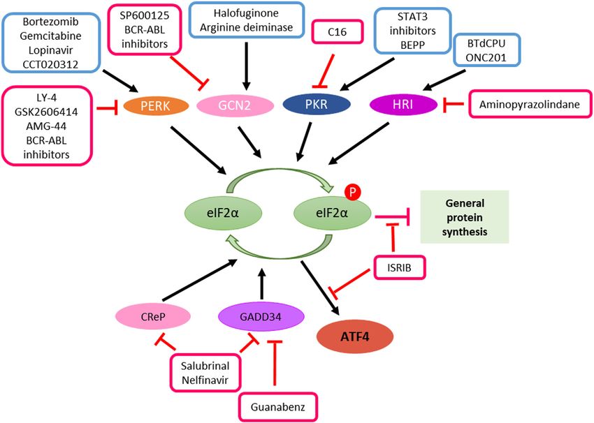
FIGURE 3 | Manipulation of ISR in cancer therapy. ATF4 induction can be achieved either through kinase activators such as bortezomib, gemcitabline, lopinavir,
CCT020312, halofuginone, arginine deiminase, STAT3 inhibitors, BEPP, BTdCPU and ONC201 or the inhibitors of phosphatases such as salubrinal, guanabenz and
nelfinavir. In the case of ISR promotes cancer cell survival and resistant to therapeutic treatments, inhibition of ATF4 can be achieved by kinase inhibitors such as LY-4,
GSK2606414, AMG-44, BCR-ABL inhibitors, SP600125, C16 and aminopyranzolindane or compound ISRIB downstream of eIF2α phosphorylation.
(Narita et al., 2015). Recent work from our lab has also implicated of the ISR as potential biomarker for predicting therapy response and
ATF4 responsible for the induction of p53-target genes PUMA, the combination of therapies that induce ATF4-mediated apoptosis.
Noxa, NAG-1(Nonsteroidal anti-inflammatory drug-activated An example of therapy combination has been observed in in vivo
gene-1)and DR5 upon treatment with prodigiosin analogue neuroblastoma preclinical models with the BCL-2 inhibitor
PG3-Oc (Figure 2) (Tian et al., 2021). Venetoclax and Fenretinide (Nguyen et al., 2019). This studied
The aforementioned studies involve the induction of the ISR combination highlighted the use of BCL-2 expression as a
machinery in addition to distinct components of autophagy, cell biomarker for neuroblastoma patients. A separate study in
cycle, and/or apoptosis pathway. This reflects the complexity of the multiple myeloma suggested the use of ATF4 as a predictive
interplay of these cellular pathways which remains underscored and therapy response biomarker for bortezomib and dexamethasone
likely to be context-dependent. Recent work has focused on post- combination treatment (Narita et al., 2015). These studies
translational modifications of ATF4 and how these affect the exemplified the clinical translational applicability of exploiting the
transcriptional control and cellular response. ATF4 has numerous ISR in cancer therapy and highlight its warrant understanding to
sites that can be post-translationally modified including predict cancer types that will benefit from ISR modulating therapies.
phosphorylation at various threonine and serine sites, methylation
at arginine 239, and ubiquitination and acetylation at lysine residues
(Wortel et al., 2017). These post-translational modifications affect Dual Roles of the Integrated Stress
ATF4 protein stability, activation and interaction with other proteins. Response in Cancer
In the case of apoptosis, methylation at arginine 239 by methyl The ISR plays different roles in tumorigenesis and tumor
transferase PRMT1 was found to be associated with the transcription progression in different types of tumors. Hypoxia is a
of genes related to apoptosis (Yuniati et al., 2016). Further insight into common phenomenon in solid tumors. It may induce
ATF4 activation may shed light on understanding the context of how apoptosis of tumor cells or tumor cells may develop the ability
these transcription factors respond to stress and the biological to adapt to the hypoxia or anoxic environment. Hypoxia can
outcome they ultimately trigger in both normal and cancer cells. induce ISR gene expression in transformed mouse embryonic
Importantly, this will aid the intervention of novel therapies, the use fibroblasts and the activated ER stress response confers resistance
Frontiers in Pharmacology | www.frontiersin.org 6 September 2021 | Volume 12 | Article 747837Tian et al. ISR Cancer Apoptosis Through ATF4
to apoptosis induced by hypoxia and thus facilitates tumor HER2+ metastatic breast cancer to Trastuzumab therapy (Darini
growth (Ameri et al., 2004). ISR mediator ATF4 is induced by et al., 2019). Proteasome inhibitors are known to activate the ISR
anoxia in breast cancer cell lines (Ameri et al., 2004). The and lower expression of ISR markers thus implicating shorter
activated ISR plays an essential role in the adaptation to progression-free survival in multiple myeloma (Obeng et al.,
hypoxic stress allowing tumor cell survival under stress and is 2006).
associated with resistance to therapy (Blais et al., 2004; Rouschop It was reported that ISR promotes the expression of potential
et al., 2013). target for immunotherapy (Obiedat et al., 2020). Thus, ISR may
It was found that loss of extracellular matrix (ECM) play a role in cancer immunotherapy.
attachment stimulates ISR signaling in vitro. And the On the one hand, activation of ISR plays a role in cancer
activation of ISR further plays a critical role in resistance to therapy. On the other, Inhibition of ISR activation can increase
anoikis and is required for metastasis (Dey et al., 2015). The ISR the vulnerability of cancer cells. BCR-ABL inhibition prevents
also has impact on the tumor microenvironment. Tumor cells activation of ISR in K562 cell line derived from a chronic myeloid
undergoing ER stress can transmit ER stress to myeloid cells leukemia (CML) patient and makes the tumor cells more
contributing to a pro-inflammatory tumor microenvironment, vulnerable to metabolic stress (Kato et al., 2018). Summaries
thus facilitating tumor progression (Mahadevan et al., 2011). of the mentioned cases and drugs can be found in the Table 1,
The role of ISR may be complex in tumors. In Table 2 and Figure 3.
medulloblastoma, the ISR is activated, and the decreased ISR
via gene manipulation attenuates medulloblastoma formation.
Moderately enhanced ISR by gene manipulation noticeably Manipulation of Integrated Stress Response
increased the incidence of medulloblastoma, whereas a in Cancer Therapy
strongly enhanced ISR significantly decreased the incidence of The ISR takes a dual role in cell survival and cell death. Enhance
medulloblastoma in vivo. Thus, the ISR plays dual roles in or inhibition of ISR signaling via targeting ISR components is a
medulloblastoma formation (Stone et al., 2016). promising strategy for cancer therapy (Figure 3). Among the
Activation of the ISR is correlated with resistance to components in ISR signaling, eIF2α is a core component and an
chemotherapy in pancreatic cancer and BRAF-mutated important focused for cancer therapy.
melanoma. Gemcitabine can induce ISR and the antiapoptotic
pro-survival factors via the ISR pathway in pancreatic cancer cell Enhanced Integrated Stress Response Signaling via
line and the combination of gemcitabine + ISRIB which inhibits Increased eIF2α Kinase
ISR induce more apoptosis in vivo (Palam et al., 2015). In BRAF- eIF2α is a core component of the ISR, and phosphorylation of
mutated melanoma, chronic ER stress involving induction of the eIF2α is regulated by upstream regulators. One of approaches
ISR signaling pathway activates autophagy which contributes is to phosphorylate eIF2α by increasing eIF2α kinases
chemoresistance (Corazzari et al., 2015). upstream of eIF2α, such as GCN2, PERK, and HRI (Pakos-
Triggering ISR can be a therapeutic strategy against cancer, Zebrucka et al., 2016; Chu et al., 2021). Most of eIF2α
since the ISR can induce apoptosis. ONC201 kills solid tumors by activators are small molecules. Halofuginone and arginine
triggering ISR-dependent ATF4 activation and activation of the deiminase are GCN2 activators (Long et al., 2013; Castilho
TRAIL-DR5 apoptotic pathway (Kline et al., 2016). In breast et al., 2014). BTdCPU and ONC201 activates HRI (Kline et al.,
cancer, GBM and DMG cell lines, ONC201 induces ISR, TRAIL- 2016; Chen et al., 2011). Bortezomib, gemcitabine, lopinavir
DR5 and ultimately apoptosis (Zhang et al., 2021). The apoptosis and CCT020312 selectively activates PERK (Narita et al.,
increases with the enhancement of ISR induction by tazemetostat. 2015; Palam et al., 2015; Obeng et al., 2006; Obiedat et al.,
The knockdown of ATF4 in GBM cell line reduced the apoptosis 2020; Stockwell et al., 2012). BEPP works on PKR activation
induced by ONC201 and the combination of ONC201 with (Figure 3) (Hu et al., 2009). These elF2α kinase activators
tazemetostat or vorinostat remarkably. Therefore, induction of have been studied in cancer therapy. For example,
ISR can play an essential role in cell death of cancer cells. Halofuginone and arginine deiminase were found to inhibit
Apoptosis induced by ISR activation was also observed in tumor growth, development and metastasis either as single
AML cells (Ishizawa et al., 2016). agents or in combination with 5-FU or radiation
The combination of mitochondrial uncoupler niclosamide (Abramovitch et al., 2004; Kim et al., 2009; Cook et al.,
ethanolamine and dopamine receptor antagonist domperidone 2010; Spector et al., 2010; Lamora et al., 2015; Brin et al.,
or TCAs induces ISR and leas to apoptosis in multiple cancer cell 2018; Singh et al., 2019; Wang et al., 2020; Huang and Hu,
lines including CRC, GBM (Glioblastoma multiforme) and 2021). Our laboratory has identified two small molecules
PDAC (Pancreatic ductal adenocarcinoma) cell lines PG3-Oc (Tian et al., 2021) and ONC201 (Kline et al., 2016;
(Hartleben et al., 2021). Even without inducing apoptosis, the Ishizawa et al., 2016) that suppress tumor growth through
ISR is induced by ONC201 in cancer cells exhibiting decreased increased ISR signaling. These drugs enhance ISR signaling
cell proliferation (Kline et al., 2016). via activation of eIF2α kinases, and sequentially enhance or
The ISR contributes to drug sensitivity of cancer cells. sustain eIF2α phosphorylation.
Activation of the ISR in HER2+breast cancer contributes the Another approach for eIF2α phosphorylation is to prevent
sensitivity to Trastuzumab in vivo. Increased expression of the eIF2α dephosphorylation from eIF2α phosphatase. GADD34
ISR mediator eIF2α-P predicts a better response of patients with (PPP1R15A) and CReP recruit phosphatase PP1 to
Frontiers in Pharmacology | www.frontiersin.org 7 September 2021 | Volume 12 | Article 747837Tian et al. ISR Cancer Apoptosis Through ATF4
TABLE 1 | The dual roles of ISR in various cancers.
Role of ISR in Cancer type
cancers
Mediator of ISR is up-regulated in anoxic tumor cells Breast cancer Ishizawa et al. (2016)
Mediator of ISR is up-regulated in hypoxic tumor cells Cervical cancer Hartleben et al. (2021)
Adaptation to hypoxia Glioblastoma and colorectal cancer Darini et al. (2019)
Promotes survival of therapy-resistant hypoxic tumor cells Glioblastoma Darini et al. (2019)
Contribute to the resistance to anoikis and promote metastasis Fibrosarcoma Obeng et al. (2006)
ER stress is transmitted from tumor cells to myeloid cells and then facilitate tumor progression Prostate cancer Obiedat et al. (2020)
Increase or decrease the incidence of tumor Medulloblastoma Kato et al. (2018)
Contributes to chemoresistance BRAF mutated melanoma Long et al. (2013)
Contributes drug sensitivity to Trastuzumab HER2+ breast cancer Lamora et al. (2015)
TABLE 2 | Effects of ISR compounds in the treatments of cancers.
Compounds Effect on ISR Effects of ISR on Cancer type
tumor cells
Gemcitabine Induce ISR Contributes to chemoresistance Pancreatic cancer Palam et al. (2015)
Bortezomib Induce ISR Contributes drug sensitivity Multiple myeloma Obeng et al. (2006); Narita et al. (2015)
ONC201 Induce ISR Reduce cell-viability Lung cancer, thyroid cancer, prostate cancer Kline et al.
(2016)
ONC201 Induce ISR Induce apoptosis Colorectal cancer, breast cancer, glioblastoma, diffuse
midline glioblastoma, AML Kline et al. (2016); Ishizawa
et al. (2016); Zhang et al. (2021)
Mitochondrial uncoupler niclosamide ethanolamine + Induce ISR Induce apoptosis Colorectal cancer, glioblastoma and PDAC Hartleben
dopamine receptor antagonist domperidone or tricyclic et al. (2021)
antidepressants (TCAs)
Nelfinavir and lopinavir Induce ISR Promote the expression of Melanoma Obiedat et al. (2020)
potential target for immunotherapy
BCR-ABL inhibitors Prevent ISR Enhance apoptosis CML Kato et al. (2018)
activation
phosphorylated-eIF2α and this results in dephosphorylation of inhibition of kinase activity, and reduce the phosphorylation
eIF2α. Salubrinal is the first small molecule discovered to inhibit of eIF2α. Another approach is to terminate eIF2α signaling
eIF2α dephosphorylation via both GADD34 and CReP (Boyce downstream of eIF2α. Small-molecule ISRIB prevents the
et al., 2005). Inhibition of GADD34 activity by Guanabenz or its formation of stress granules caused by eIF2α
derivatives results in high levels of eIF2α Phosphorylation phosphorylation, thus, impairing ATF4 synthesis (Figure 3)
(Tsaytler et al., 2011). Different from Guanabenz, Nelfinavir (Sidrauski et al., 2015).
increases phosphorylation of eIF2α by downregulating CReP
in addition to it effect on GADD34 (De Gassart et al., 2016). Targeting Integrated Stress Response in Combination
Guanabenz has been found to sensitize glioblastoma cancer cells of Immunotherapy
to sunitinib in combinatorial treatment (Figure 3) (Ho et al., High levels of PD-L1 on the cancer cell surface allows evasion
2021). from T cell attack by binding to the PD-1 receptor on T cells.
Disruption of the PD-1/PD-L1 checkpoint can result in
Inhibition of Integrated Stress Response Signaling by cytotoxic T cell killing of tumors. The ISR was found to
Reduction of eIF2α Kinase increase PD-L1 translation in human cancers. Suresh et al.
Inhibition of ISR signaling may overcome drug resistance in (2020) The increased PD-L1 suppress anti-tumor immune
cancer. One of the approaches is to inhibit eIF2α kinase responses. PERK signaling was found to suppress immune
upstream of eIF2α. Most of these kinase inhibitors compete responses by increasing tumor-myeloid-derived suppressor
with ATP to block their kinase domain. SP600125 and BCR- cells (MDSC). PERK blockade transforms MDSC’s into
ABL inhibitors inactivate GCN2 (Kato et al., 2018; Robert et al., myeloid cells that activate anti-tumor CD8+ T-cell
2009). Amino-pyrazolindine inhibits HRI (Rosen et al., 2009). immunity in the tumor microenvironment. AMG-44, a
Imidazolo-oxindole PKR inhibitor C16 specifically inhibits PKR PERK inhibitor, in combination with Anti-PD-L1 showed a
(Jammi et al., 2003). LY-4, AMG-44, BCR-ABL inhibitors and synergistic anti-tumor effect in B16 tumor-bearing mice model
GSK2606414 inactivate PERK (Tameire et al., 2019; Kato et al., (Figure 3) (Mohamed et al., 2020). These studies suggest that
2018; Axten et al., 2012; Mohamed et al., 2020). They bind to the PERK inhibitors enhance the antitumor efficacy of immune
eIF2α kinase in an ATP-competitive manner, result in checkpoint inhibitors. Therefore, targeting ISR in combination
Frontiers in Pharmacology | www.frontiersin.org 8 September 2021 | Volume 12 | Article 747837Tian et al. ISR Cancer Apoptosis Through ATF4
with immune checkpoint is an innovational strategy for cancer AUTHOR CONTRIBUTIONS
therapy.
All authors listed have made a substantial, direct, and intellectual
contribution to the work and approved it for publication.
CONCLUSION
The ISR is a double-edged sword with pro-survival and pro-death FUNDING
activities that may impact on tumor progression and response to
therapy. Our approach for therapeutic targeting of cell death WE-D. is an American Cancer Society Research Professor and is
pathways has led us to uncover the ISR as a critical signaling supported by the Mencoff Family University Professorship at
component and target of drug candidates. The fact that the ISR Brown University. This work was supported by an NIH grant
can lead to alternative cell fates depending on cellular context (CA173453) and a grant from the Warren Alpert Foundation to
suggests that greater efforts need to be directed at understanding WE-D. This work was supported by the Teymour Alireza P′98,
its regulation and finding new ways for its modulation. The ISR P′00 Family Cancer Research Fund established by the Alireza
holds promise for cancer therapy development. Family.
Brin, E., Wu, K., Dagostino, E., Meng-Chiang Kuo, M., He, Y., Shia, W. J., et al.
REFERENCES (2018). TRAIL Stabilization and Cancer Cell Sensitization to its Pro-apoptotic
Activity Achieved through Genetic Fusion with Arginine Deiminase.
Abdel-Nour, M., Carneiro, L. A. M., Downey, J., Tsalikis, J., Outlioua, A., Prescott, Oncotarget 9 (97), 36914–36928. doi:10.18632/oncotarget.26398
D., et al. (2019). The Heme-Regulated Inhibitor Is a Cytosolic Sensor of Protein Bruns, G. P., and London, I. M. (1965). The Effect of Hemin on the Synthesis of
Misfolding that Controls Innate Immune Signaling. Science 365 (6448). Globin. Biochem. Biophys. Res. Commun. 18, 236–242. doi:10.1016/0006-
doi:10.1126/science.aaw4144 291x(65)90746-1
Abramovitch, R., Itzik, A., Harel, H., Nagler, A., Vlodavsky, I., and Siegal, T. (2004). Cai, J., Yang, J., and Jones, D. P. (1998). Mitochondrial Control of Apoptosis: the
Halofuginone Inhibits Angiogenesis and Growth in Implanted Metastatic Rat Role of Cytochrome C. Biochim. Biophys. Acta. 1366 (1-2), 139–149.
Brain Tumor Model-Aan MRI Study. Neoplasia 6 (5), 480–489. doi:10.1593/ doi:10.1016/s0005-2728(98)00109-1
neo.03520 Carneiro, B. A., and El-Deiry, W. S. (2020). Targeting Apoptosis in Cancer Therapy. Nat.
Akman, M., Belisario, D. C., Salaroglio, I. C., Kopecka, J., Donadelli, M., De Smaele, Rev. Clin. Oncol. 17 (7), 395–417. doi:10.1038/s41571-020-0341-y
E., et al. (2021). Hypoxia, Endoplasmic Reticulum Stress and Chemoresistance: Castilho, B. A., Shanmugam, R., Silva, R. C., Ramesh, R., Himme, B. M., and
Dangerous Liaisons. J. Exp. Clin. Cancer Res. 40 (1), 28. doi:10.1186/s13046- Sattlegger, E. (2014). Keeping the eIF2 Alpha Kinase Gcn2 in Check. Biochim.
020-01824-3 Biophys. Acta. 1843 (9), 1948–1968. doi:10.1016/j.bbamcr.2014.04.006
Altman, B. J., Wofford, J. A., Zhao, Y., Coloff, J. L., Ferguson, E. C., Wieman, H. L., Chefalo, P. J., Oh, J., Rafie-Kolpin, M., Kan, B., and Chen, J. J. (1998). Heme-
et al. (2009). Autophagy Provides Nutrients but Can lead to Chop-dependent regulated eIF-2alpha Kinase Purifies as a Hemoprotein. Eur. J. Biochem. 258 (2),
Induction of Bim to Sensitize Growth Factor-Deprived Cells to Apoptosis. Mol. 820–830. doi:10.1046/j.1432-1327.1998.2580820.x
Biol. Cel. 20 (4), 1180–1191. doi:10.1091/mbc.e08-08-0829 Chen, T., Ozel, D., Qiao, Y., Harbinski, F., Chen, L., Denoyelle, S., et al. (2011).
Ameri, K., Lewis, C. E., Raida, M., Sowter, H., Hai, T., and Harris, A. L. (2004). Chemical Genetics Identify eIF2α Kinase Heme-Regulated Inhibitor as an
Anoxic Induction of ATF-4 through HIF-1-independent Pathways of Protein Anticancer Target. Nat. Chem. Biol. 7 (9), 610–616. doi:10.1038/nchembio.613
Stabilization in Human Cancer Cells. Blood 103 (5), 1876–1882. doi:10.1182/ Chitnis, N. S., Pytel, D., Bobrovnikova-Marjon, E., Pant, D., Zheng, H., Maas, N. L.,
blood-2003-06-1859 et al. (2012). miR-211 Is a Prosurvival microRNA that Regulates Chop
Axten, J. M., Medina, J. R., Feng, Y., Shu, A., Romeril, S. P., Grant, S. W., et al. Expression in a PERK-dependent Manner. Mol. Cel. 48 (3), 353–364.
(2012). Discovery of 7-Methyl-5-(1-{[3-(trifluoromethyl)phenyl]acetyl}-2,3- doi:10.1016/j.molcel.2012.08.025
Dihydro-1h-Indol-5-Yl)-7h-Pyrrolo[2,3-D]pyrimidin-4-Amine Chu, H. S., Peterson, C., Jun, A., and Foster, J. (2021). Targeting the Integrated
(GSK2606414), a Potent and Selective First-In-Class Inhibitor of Protein Kinase Stress Response in Ophthalmology. Curr. Eye Res. 46 (8), 1075–1088.
R (PKR)-like Endoplasmic Reticulum Kinase (PERK). J. Med. Chem. 55 (16), doi:10.1080/02713683.2020.1867748
7193–7207. doi:10.1021/jm300713s Clementi, E., Inglin, L., Beebe, E., Gsell, C., Garajova, Z., and Markkanen, E. (2020).
B’Chir, W., Maurin, A. C., Carraro, V., Averous, J., Jousse, C., Muranishi, Y., et al. Persistent DNA Damage Triggers Activation of the Integrated Stress Response
(2013). The eIF2α/ATF4 Pathway Is Essential for Stress-Induced Autophagy to Promote Cell Survival under Nutrient Restriction. BMC Biol. 18 (1), 36.
Gene Expression. Nucleic Acids Res. 41 (16), 7683–7699. doi:10.1093/nar/ doi:10.1186/s12915-020-00771-x
gkt563 Cook, J. A., Choudhuri, R., Degraff, W., Gamson, J., and Mitchell, J. B. (2010).
Baird, T. D., Palam, L. R., Fusakio, M. E., Willy, J. A., Davis, C. M., McClintick, J. N., Halofuginone Enhances the Radiation Sensitivity of Human Tumor Cell Lines.
et al. (2014). Selective mRNA Translation during eIF2 Phosphorylation Induces Cancer Lett. 289 (1), 119–126. doi:10.1016/j.canlet.2009.08.009
Expression of IBTKα. Mol. Biol. Cel. 25 (10), 1686–1697. doi:10.1091/mbc.E14- Corazzari, M., Rapino, F., Ciccosanti, F., Giglio, P., Antonioli, M., Conti, B., et al.
02-0704 (2015). Oncogenic BRAF Induces Chronic ER Stress Condition Resulting in
Behrends, C., Sowa, M. E., Gygi, S. P., and Harper, J. W. (2010). Network Increased Basal Autophagy and Apoptotic Resistance of Cutaneous Melanoma.
Organization of the Human Autophagy System. Nature 466 (7302), 68–76. Cell Death Differ. 22 (6), 946–958. doi:10.1038/cdd.2014.183
doi:10.1038/nature09204 Costa-Mattioli, M., and Walter, P. (2020). The Integrated Stress Response: From
Blais, J. D., Filipenko, V., Bi, M., Harding, H. P., Ron, D., Koumenis, C., et al. Mechanism to Disease. Science 368 (6489). doi:10.1126/science.aat5314
(2004). Activating Transcription Factor 4 Is Translationally Regulated by Darini, C., Ghaddar, N., Chabot, C., Assaker, G., Sabri, S., Wang, S., et al. (2019).
Hypoxic Stress. Mol. Cel Biol. 24 (17), 7469–7482. doi:10.1128/ An Integrated Stress Response via PKR Suppresses HER2+ Cancers and
MCB.24.17.7469-7482.2004 Improves Trastuzumab Therapy. Nat. Commun. 10 (1), 2139. doi:10.1038/
Boyce, M., Bryant, K. F., Jousse, C., Long, K., Harding, H. P., Scheuner, D., et al. s41467-019-10138-8
(2005). A Selective Inhibitor of eIF2alpha Dephosphorylation Protects Cells De Gassart, A., Bujisic, B., Zaffalon, L., Decosterd, L. A., Di Micco, A., Frera, G.,
from ER Stress. Science 307 (5711), 935–939. doi:10.1126/science.1101902 et al. (2016). An Inhibitor of HIV-1 Protease Modulates Constitutive eIF2α
Frontiers in Pharmacology | www.frontiersin.org 9 September 2021 | Volume 12 | Article 747837Tian et al. ISR Cancer Apoptosis Through ATF4 Dephosphorylation to Trigger a Specific Integrated Stress Response. Proc. Natl. Hu, J., Dang, N., Menu, E., De Bruyne, E., De Bryune, E., Xu, D., et al. (2012). Acad. Sci. U S A. 113 (2), E117–E126. doi:10.1073/pnas.1514076113 Activation of ATF4 Mediates Unwanted Mcl-1 Accumulation by Proteasome Deegan, S., Koryga, I., Glynn, S. A., Gupta, S., Gorman, A. M., and Samali, A. Inhibition. Blood 119 (3), 826–837. doi:10.1182/blood-2011-07-366492 (2015). A Close Connection between the PERK and IRE Arms of the UPR and Hu, P., Han, Z., Couvillon, A. D., and Exton, J. H. (2004). Critical Role of the Transcriptional Regulation of Autophagy. Biochem. Biophys. Res. Commun. Endogenous Akt/IAPs and MEK1/ERK Pathways in Counteracting 456 (1), 305–311. doi:10.1016/j.bbrc.2014.11.076 Endoplasmic Reticulum Stress-Induced Cell Death. J. Biol. Chem. 279 (47), Deegan, S., Saveljeva, S., Gorman, A. M., and Samali, A. (2013). Stress-induced Self- 49420–49429. doi:10.1074/jbc.M407700200 Cannibalism: on the Regulation of Autophagy by Endoplasmic Reticulum Hu, W., Hofstetter, W., Wei, X., Guo, W., Zhou, Y., Pataer, A., et al. (2009). Stress. Cell Mol Life Sci. 70 (14), 2425–2441. doi:10.1007/s00018-012-1173-4 Double-stranded RNA-dependent Protein Kinase-dependent Apoptosis Deng, J., Lu, P. D., Zhang, Y., Scheuner, D., Kaufman, R. J., Sonenberg, N., et al. Induction by a Novel Small Compound. J. Pharmacol. Exp. Ther. 328 (3), (2004). Translational Repression Mediates Activation of Nuclear Factor Kappa 866–872. doi:10.1124/jpet.108.141754 B by Phosphorylated Translation Initiation Factor 2. Mol. Cel Biol. 24 (23), Huang, Z., and Hu, H. (2021). Arginine Deiminase Induces Immunogenic Cell 10161–10168. doi:10.1128/MCB.24.23.10161-10168.2004 Death and Is Enhanced by N-Acetylcysteine in Murine MC38 Colorectal Dennis, M. D., McGhee, N. K., Jefferson, L. S., and Kimball, S. R. (2013). Regulated Cancer Cells and MDA-MB-231 Human Breast Cancer Cells In Vitro. in DNA Damage and Development 1 (REDD1) Promotes Cell Survival during Molecules 26 (2). doi:10.3390/molecules26020511 Serum Deprivation by Sustaining Repression of Signaling through the Ill-Raga, G., Tajes, M., Busquets-García, A., Ramos-Fernández, E., Vargas, L. M., Bosch- Mechanistic Target of Rapamycin in Complex 1 (mTORC1). Cell Signal. 25 Morató, M., et al. (2015). Physiological Control of Nitric Oxide in Neuronal BACE1 (12), 2709–2716. doi:10.1016/j.cellsig.2013.08.038 Translation by Heme-Regulated eIF2α Kinase HRI Induces Synaptogenesis. Antioxid. Dey, S., Sayers, C. M., Verginadis, I. I., Lehman, S. L., Cheng, Y., Cerniglia, G. J., Redox Signal. 22 (15), 1295–1307. doi:10.1089/ars.2014.6080 et al. (2015). ATF4-dependent Induction of Heme Oxygenase 1 Prevents Ishizawa, J., Kojima, K., Chachad, D., Ruvolo, P., Ruvolo, V., Jacamo, R. O., et al. Anoikis and Promotes Metastasis. J. Clin. Invest. 125 (7), 2592–2608. (2016). ATF4 Induction through an Atypical Integrated Stress Response to doi:10.1172/JCI78031 ONC201 Triggers P53-independent Apoptosis in Hematological Malignancies. Di Prisco, G. V., Huang, W., Buffington, S. A., Hsu, C. C., Bonnen, P. E., Placzek, A. Sci. Signal. 9 (415), ra17. doi:10.1126/scisignal.aac4380 N., et al. (2014). Translational Control of mGluR-dependent Long-Term Jammi, N. V., Whitby, L. R., and Beal, P. A. (2003). Small Molecule Inhibitors of the Depression and Object-Place Learning by eIF2α. Nat. Neurosci. 17 (8), RNA-dependent Protein Kinase. Biochem. Biophys. Res. Commun. 308 (1), 1073–1082. doi:10.1038/nn.3754 50–57. doi:10.1016/s0006-291x(03)01318-4 Dluzen, D., Li, G., Tacelosky, D., Moreau, M., and Liu, D. X. (2011). BCL-2 Is a Jin, H. O., Seo, S. K., Woo, S. H., Choe, T. B., Hong, S. I., Kim, J. I., et al. (2009). Downstream Target of ATF5 that Mediates the Prosurvival Function of ATF5 Nuclear Protein 1 Induced by ATF4 in Response to Various Stressors Acts as a in a Cell Type-dependent Manner. J. Biol. Chem. 286 (9), 7705–7713. Positive Regulator on the Transcriptional Activation of ATF4. IUBMB Life. 61 doi:10.1074/jbc.M110.207639 (12), 1153–1158. doi:10.1002/iub.271 Donnelly, N., Gorman, A. M., Gupta, S., and Samali, A. (2013). The eIF2α Kinases: Karpel-Massler, G., Horst, B. A., Shu, C., Chau, L., Tsujiuchi, T., Bruce, J. N., Their Structures and Functions. Cel Mol Life Sci. 70 (19), 3493–3511. et al. (2016). A Synthetic Cell-Penetrating Dominant-Negative ATF5 doi:10.1007/s00018-012-1252-6 Peptide Exerts Anticancer Activity against a Broad Spectrum of Dossou, A. S., and Basu, A. (2019). The Emerging Roles of mTORC1 in Macromanaging Treatment-Resistant Cancers. Clin. Cancer Res. 22 (18), 4698–4711. Autophagy. Cancers (Basel) 11 (10), 1422. doi:10.3390/cancers11101422 doi:10.1158/1078-0432.CCR-15-2827 Evstafieva, A. G., Garaeva, A. A., Khutornenko, A. A., Klepikova, A. V., Logacheva, Kasai, S., Yamazaki, H., Tanji, K., Engler, M. J., Matsumiya, T., and Itoh, K. (2019). M. D., Penin, A. A., et al. (2014). A Sustained Deficiency of Mitochondrial Role of the ISR-ATF4 Pathway and its Cross Talk with Nrf2 in Mitochondrial Respiratory Complex III Induces an Apoptotic Cell Death through the P53- Quality Control. J. Clin. Biochem. Nutr. 64 (1), 1–12. doi:10.3164/jcbn.18-37 Mediated Inhibition of Pro-survival Activities of the Activating Transcription Kato, Y., Kunimasa, K., Sugimoto, Y., and Tomida, A. (2018). BCR-ABL Tyrosine Factor 4. Cell Death Dis. 5, e1511. doi:10.1038/cddis.2014.469 Kinase Inhibition Induces Metabolic Vulnerability by Preventing the Integrated Galehdar, Z., Swan, P., Fuerth, B., Callaghan, S. M., Park, D. S., and Cregan, S. P. Stress Response in K562 cells. Biochem. Biophys. Res. Commun. 504 (4), (2010). Neuronal Apoptosis Induced by Endoplasmic Reticulum Stress Is 721–726. doi:10.1016/j.bbrc.2018.09.032 Regulated by ATF4-CHOP-Mediated Induction of the Bcl-2 Homology 3- Kazemi, S., Mounir, Z., Baltzis, D., Raven, J. F., Wang, S., Krishnamoorthy, J. L., only Member PUMA. J. Neurosci. 30 (50), 16938–16948. doi:10.1523/ et al. (2007). A Novel Function of eIF2alpha Kinases as Inducers of the JNEUROSCI.1598-10.2010 Phosphoinositide-3 Kinase Signaling Pathway. Mol. Biol. Cel. 18 (9), Hamanaka, R. B., Bobrovnikova-Marjon, E., Ji, X., Liebhaber, S. A., and Diehl, J. A. 3635–3644. doi:10.1091/mbc.e07-01-0053 (2009). PERK-dependent Regulation of IAP Translation during ER Stress. Kim, R. H., Coates, J. M., Bowles, T. L., McNerney, G. P., Sutcliffe, J., Jung, J. U., Oncogene 28 (6), 910–920. doi:10.1038/onc.2008.428 et al. (2009). Arginine Deiminase as a Novel Therapy for Prostate Cancer Hamidi, T., Cano, C. E., Grasso, D., Garcia, M. N., Sandi, M. J., Calvo, E. L., et al. Induces Autophagy and Caspase-independent Apoptosis. Cancer Res. 69 (2), (2012). Nupr1-aurora Kinase A Pathway Provides protection against Metabolic 700–708. doi:10.1158/0008-5472.CAN-08-3157 Stress-Mediated Autophagic-Associated Cell Death. Clin. Cancer Res. 18 (19), Kim, T. W., Cheon, C., and Ko, S. G. (2020). SH003 Activates Autophagic Cell 5234–5246. doi:10.1158/1078-0432.CCR-12-0026 Death by Activating ATF4 and Inhibiting G9a under Hypoxia in Gastric Cancer Han, A. P., Yu, C., Lu, L., Fujiwara, Y., Browne, C., Chin, G., et al. (2001). Heme- Cells. Cel Death Dis. 11 (8), 717. doi:10.1038/s41419-020-02924-w regulated eIF2alpha Kinase (HRI) Is Required for Translational Regulation and Kline, C. L., Van den Heuvel, A. P., Allen, J. E., Prabhu, V. V., Dicker, D. T., and El- Survival of Erythroid Precursors in Iron Deficiency. EMBO J. 20 (23), Deiry, W. S. (2016). ONC201 Kills Solid Tumor Cells by Triggering an 6909–6918. doi:10.1093/emboj/20.23.6909 Integrated Stress Response Dependent on ATF4 Activation by Specific Harding, H. P., Novoa, I., Zhang, Y., Zeng, H., Wek, R., Schapira, M., et al. (2000). eIF2α Kinases. Sci. Signal. 9 (415), ra18. doi:10.1126/scisignal.aac4374 Regulated Translation Initiation Controls Stress-Induced Gene Expression in Kouroku, Y., Fujita, E., Tanida, I., Ueno, T., Isoai, A., Kumagai, H., et al. (2007). ER Mammalian Cells. Mol. Cel. 6 (5), 1099–1108. doi:10.1016/s1097-2765(00) Stress (PERK/eIF2alpha Phosphorylation) Mediates the Polyglutamine- 00108-8 Induced LC3 Conversion, an Essential Step for Autophagy Formation. Cel Hartleben, G., Schorpp, K., Kwon, Y., Betz, B., Tsokanos, F. F., Dantes, Z., et al. Death Differ. 14 (2), 230–239. doi:10.1038/sj.cdd.4401984 (2021). Combination Therapies Induce Cancer Cell Death through the Kroemer, G., Mariño, G., and Levine, B. (2010). Autophagy and the Integrated Stress Response and Disturbed Pyrimidine Metabolism. EMBO Integrated Stress Response. Mol. Cel. 40 (2), 280–293. doi:10.1016/ Mol. Med. 13 (4), e12461. doi:10.15252/emmm.202012461 j.molcel.2010.09.023 Ho, K. H., Lee, Y. T., Chen, P. H., Shih, C. M., Cheng, C. H., and Chen, K. C. (2021). Lamora, A., Mullard, M., Amiaud, J., Brion, R., Heymann, D., Redini, F., et al. Guanabenz Sensitizes Glioblastoma Cells to Sunitinib by Inhibiting GADD34- (2015). Anticancer Activity of Halofuginone in a Preclinical Model of Mediated Autophagic Signaling. Neurotherapeutics 18, 1371–1392. doi:10.1007/ Osteosarcoma: Inhibition of Tumor Growth and Lung Metastases. s13311-020-00961-z Oncotarget 6 (16), 14413–14427. doi:10.18632/oncotarget.3891 Frontiers in Pharmacology | www.frontiersin.org 10 September 2021 | Volume 12 | Article 747837
You can also read