Structure and function of small airways in asthma patients revisited
←
→
Page content transcription
If your browser does not render page correctly, please read the page content below
REVIEW
                                                                                                                                           ASTHMA

                                  Structure and function of small airways
                                  in asthma patients revisited
                                  Wytse B. van den Bosch1,2, Alan L. James3 and Harm A.W.M. Tiddens1,2

                                  Affiliations: 1Dept of Paediatric Pulmonology and Allergology, Erasmus MC – Sophia Children’s Hospital,
                                  University Medical Center Rotterdam, Rotterdam, The Netherlands. 2Dept of Radiology and Nuclear Medicine,
                                  Erasmus MC, University Medical Center Rotterdam, Rotterdam, The Netherlands. 3Dept of Pulmonary
                                  Physiology and Sleep Medicine, Sir Charles Gairdner Hospital, Perth, Australia.

                                  Correspondence: Harm A.W.M. Tiddens, Dept of Paediatric Pulmonology and Allergology, Erasmus Medical
                                  Centre-Sophia Children’s Hospital, Wytemaweg 80, 3015 CN, Rotterdam, The Netherlands.
                                  E-mail: h.tiddens@erasmusmc.nl

                                  @ERSpublications
                                  Small in size, big in impact: structural and functional alterations of the epithelium, extracellular
                                  matrix and airway smooth muscle are present in the small airways of asthma patients. These
                                  alterations may play a pivotal role in asthma pathophysiology. https://bit.ly/3lIlT62

                                  Cite this article as: van den Bosch WB, James AL, Tiddens HAWM. Structure and function of small
                                  airways in asthma patients revisited. Eur Respir Rev 2021; 30: 200186 [https://doi.org/10.1183/
                                  16000617.0186-2020].

                                  ABSTRACT Small airways (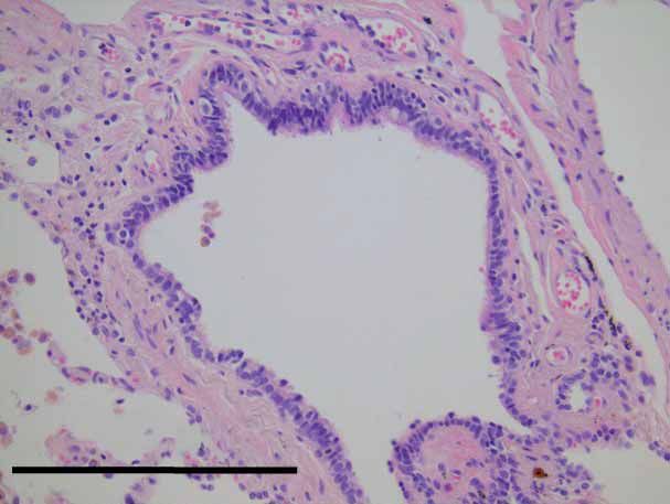
ASTHMA | W.B. VAN DEN BOSCH ET AL.

                                  come into focus. A systematic review showed that in 50–60% of people with asthma, SAD is present [5].
                                  In a large prospective study in adults (ATLANTIS study) it was shown that the small airways are involved
                                  across almost all asthma severities, and that they show proportionally more structural and functional
                                  abnormalities with increasing clinical severity of asthma [6]. To date, it is unclear whether the involvement
                                  of the small airways is related to severity of asthma or to a specific group of patients with asthma [7].
                                  Since the introduction of the term SAD by HOGG et al. [1], several hundred publications about the small
                                  airways have appeared in the medical literature, describing structure, function, diagnostic measures and
                                  therapeutic options. However, only a few studies have reviewed the potential mechanisms and associated
                                  pathophysiological changes leading to SAD in patients with asthma. Therefore, we aim to discuss the
                                  structural and functional changes of the small airways of humans in asthma in detail.
                                  For this review we conducted an extensive literature search by using PubMed. We used the following
                                  terms: “structure”’’, “function”, “remodelling”, “asthma”, “small airways”, “peripheral airways” “distal
                                  airways”, “airway smooth muscle”, “extra cellular matrix”, “epithelium”, “imaging”, “ventilation defects”,
                                  “mechanics” and “( patho)physiology” in different combinations, orders and in alternate spelling. From
                                  this literature search we retrieved approximately 1700 papers. On the basis of relevance for the aim of our
                                  review, approximately 150 papers were selected and reviewed. In the first section of the review we discuss
                                  the structural components in relation to SAD, followed by what is currently known about functional issues
                                  in relation to SAD and finally, we discuss the role that diagnostic measurements, imaging and modelling
                                  can play in our understanding of SAD. Inflammatory cells and pathways are of key importance in the
                                  pathophysiology of asthma. However, due to the extent and complexity of these cellular mechanisms and
                                  pathways, we have not included them as they are beyond the scope of this review. We suggest the in-depth
                                  review by LAMBRECHT et al. [8] for more information on inflammatory cells and pathways in asthma.

                                  Epithelium
                                  In healthy airways the epithelium lines the airways as a continuous sheet (figure 1). In the large
                                  airways the epithelium is pseudo-stratified and becomes columnar and cuboidal in the small airways.
                                  Furthermore, the epithelium consists of goblet, serous, basal, club and immune cells [9]. Compared with
                                  the large airways, the epithelium of the small airways has a greater proportion of ciliated and club cells
                                  and few or almost no goblet cells (figure 2) [10]. The epithelial cells are held together by apical tight
                                  junctions, basal adherens junctions and desmosomes [11].
                                  The predominant function of the epithelium was long thought to be a physical barrier to pathogens. The
                                  production of mucus, the mucociliary escalator, cell-to-cell adhesion proteins and inflammatory mediators
                                  are all components of this barrier function. Mucin protects the airway epithelium from inhaled pathogens by
                                  trapping them and preventing them from penetrating the mucus into the epithelium. Moreover, mucins play
                                  a vital role in clearance of pathogens, pollutant particles and inflammatory stimuli from the airways [12].
                                  MUC5B and MUC5AC are the main gel-forming mucins in human airways. MUC5B is secreted throughout
                                  the airways in healthy small and large airways. In healthy lungs almost no MUC5AC is produced in the
                                  small airways [13]. In addition, mucins in combination with the periciliary layer and ciliated epithelium
                                  form the mucociliary escalator [14]. Besides this physical barrier function, the epithelium and its
                                  components play an important role in the secretion of a wide array of cytokines, chemokines, growth factors
                                  and other regulatory mediators when exposed to allergens, pathogens and other stimuli [9, 12]. It has also
                                  been proposed that the epithelium can play a role in smooth muscle contraction by the release of soluble
                                  mediators released from damaged epithelium [15]. Overall, the epithelium plays an important role in
                                  maintaining airway patency throughout the airway tree.
                                  Shedding and desquamation of epithelium has often been described in the medical literature. It is
                                  important to note that many of these observations were in endobronchial biopsies and post mortem
                                  biopsies of large airways in patients who had died from asthma [16–18]. In more recent studies it was
                                  suggested that the observed shedding and desquamation could be a result of sampling artefacts, as no
                                  differences in biopsies between epithelial shedding or desquamation were observed comparing people with
                                  asthma with controls [19]. Moreover, epithelial desquamation increased with decreasing biopsy size [20].
                                  In small airways, epithelial shedding has not been reported [21, 22]. However, increased numbers of
                                  columnar epithelial cells in induced sputum from patients with severe asthma have been observed,
                                  suggesting increased epithelial cell turnover or shedding in these patients [23]. In general, the epithelium
                                  in small airways of people with asthma is more heterogeneous and more varied in thickness [24].
                                  Epithelial cell to cell adhesion, either through tight junctions, adherens junctions or desmosomes, is
                                  compromised in the large airways of people with asthma [25, 26]. To our knowledge, no specific studies
                                  focussing on the cell-to-cell adhesion in the epithelium of small airways have been conducted. The
                                  information provided below is derived from studies that studied epithelial cell-to-cell adhesion in large

https://doi.org/10.1183/16000617.0186-2020                                                                                                   2
ASTHMA | W.B. VAN DEN BOSCH ET AL.
a)
Outer airway wall area
Mucus
Inner airway wall area
Epithelium
ASM
Parenchyma
Basement membrane
Extracellular matrix
Thickening of ASM layer:
b)
hyperplasia/hypertrophy
Epithelium:
Variation in thickness
Altered cell to cell adhesion
Goblet cell hyper/metaplasia
Abnormal alveolar attachment
Basement membrane thickening
Changes in ECM composition
Increased mucus production
FIGURE 1 a) Schematic drawing of a cross section of a normal small airway embedded in the lung parenchyma. The different compartments and
components of the airway wall are shown. b) Schematic drawing of a cross section of a constricted small airway embedded in the parenchyma of
a patient with asthma. The structural alterations frequently observed in small airways in asthma are shown. These alterations are located in the
epithelium, extracellular matrix (ECM), airway smooth muscle (ASM) and parenchyma. Note that, the ECM is only partly depicted. In vivo the ECM
forms an extensive network throughout the airway wall.
https://doi.org/10.1183/16000617.0186-2020 3ASTHMA | W.B. VAN DEN BOSCH ET AL.
a) b)
FIGURE 2 Photomicrograph (haematoxylin & eosin stain, magnification ×20) of a) a small airway of a healthy
subject and b) a patient with asthma. The small airway of the asthmatic subject shows clear structural
alterations to the airway, such as goblet cell metaplasia, reticular basement membrane thickening, airway
smooth muscle hyperplasia and eosinophilia. Scale bars=500 µm. Images courtesy of J.H. von der
Thüsen (Dept of Pathology, Erasmus MC, University Medical Center Rotterdam, Rotterdam, The Netherlands).
airway biopsy samples. Inflammation is thought to play an important role in the loss of epithelial integrity.
Several studies have shown that the expression of proteins involved in cell-to-cell adhesion was decreased
when epithelium was exposed to tumour necrosis factor-α [27] or other cytokines [28, 29]. Therefore,
exposure to cytokines and other inflammatory mediators might result in structural defects to the
epithelium. However, it has been suggested that the fragility of the epithelium of patients with asthma is a
fundamental feature of asthma [26] characterised by abnormal repair responses to injury [30].
Normal cell-to-cell adhesion properties of the epithelial cells may also be altered in a process described as
epithelial–mesenchymal transition. In this process epithelial cells transition to fibroblast-like mesenchymal
cells. This process has been suggested as a means by which abnormal epithelial cells may contribute to the
airway wall (including smooth muscle) remodelling observed in asthma [31]. As a result, epithelial cells
lose their characteristics and functionality, such as cell-to-cell adhesion [11, 31]. Transforming growth
factor (TGF)-β has been shown to play an important role in inducing epithelial to mesenchymal
transition. In-depth information about cell-to-cell adhesion dysfunction has been discussed in reviews by
LAMBRECHT et al. [11] and HACKETT et al. [31].
In healthy lungs, goblet cells are only sparsely present in the small airways (figure 2). However, goblet cell
hyperplasia (increased number of cells), metaplasia (change in cell phenotype) and mucus accumulation
have been frequently described in the small airways of patients with fatal asthma [21, 32–36], severe
asthma [37, 38] and in mouse models [39] (table 1). MUC5AC is not seen in healthy small airways but is
upregulated in the small airways of patients with asthma [41]. Alterations in expression of MUC5AC may
contribute to mucus accumulation and mucus plugging eventually leading to airway obstruction in the
small airways of patients with asthma [42]. It has been suggested that tethering of MUC5AC to epithelial
mucous cells may further contribute to luminal mucus plugging by impairing mucociliary transport [41].
The increased amount of goblet cells in small airways of people with asthma is most likely a result of
goblet cell metaplasia (figure 2) instead of goblet cell hyperplasia [43]. Several possible mechanisms
behind goblet cell metaplasia have been suggested. First, trans-differentiation of club and ciliated cells
into goblet cells is the main mechanism for goblet cell metaplasia, rather than proliferation of pre-existing
goblet cells [11]. Secondly, it is suggested that inflammatory mediators play an important role in the
initiation of goblet cell metaplasia [43]. Thirdly, epithelial compression due to bronchoconstriction in
asthmatic patients is thought to contribute to goblet cell hyperplasia and metaplasia and subsequent
mucus over-production [44].
Many of the functional properties of the epithelium are compromised in patients with asthma. Alterations
to the barrier function may lower the protective effect of the epithelium and make the lung more
susceptible for inhaled pathogens, pollutants and bronchoconstrictor agents [26]. The impaired mucociliary
function and fragile epithelium, in combination with inflammatory cells and increased mucus production,
may contribute to luminal narrowing or occlusion by forming mucus plugs, a phenomenon frequently
described in small airways of cases of fatal asthma [16, 33, 34, 40]. Although mucus accumulation and
plugging has also been observed in small airways of patients with milder and nonfatal asthma [22, 37, 38],
we should be cautious to draw general conclusions based on observations in samples of fatal asthma
patients. Fatal asthma represents an acute terminal event, associated with inflammatory infiltrates,
vascular leakage, severe bronchoconstriction and increased mucus production and accumulation [36, 40].
https://doi.org/10.1183/16000617.0186-2020 4ASTHMA | W.B. VAN DEN BOSCH ET AL.
TABLE 1 Studies of structural epithelial alterations in mild, moderate, severe and fatal asthma
Methods Population Findings [Ref.]
Post mortem specimen: large and small Fatal asthma (n=20) Occluding (mucus) plugs were seen in all [16]
airways (not defined) small bronchi
Epithelial cell shedding was observed in
medium-sized bronchi#
Post mortem specimen: segmental and Fatal asthma (n=11) Greater mucus gland area in airways [21]
lobar bronchi (Pbm 10>18 mm); Nonfatal asthma (n=13) (4–10 mm) in fatal asthma compared to
subsegmental bronchi (4–10 mm); Control (n=11) controls
small and large membranous No difference in percentage of basement
bronchioles (Pbm 1 mm from patients with [22]
post mortem specimen: large and Paediatric asthma in remission (n=2) asthma in remission mucus plugging,
small airways (not defined) goblet cell hyperplasia and basement
membrane thickening was observed;
detachment of the epithelial lining was not
observed
In airwaysASTHMA | W.B. VAN DEN BOSCH ET AL.
TABLE 1 Continued
Methods Population Findings [Ref.]
Lung biopsy specimen obtained through Severe asthma clinically, with diagnosis of Papillary epithelial hyperplasia, basal cell [37]
VATS: large and small airways (not asthmatic granulomatosis (n=10) after hyperplasia and goblet cell metaplasia was
defined) histologic evaluation observed in all cases
Mucus plugging was observed in eight out
of 10 cases
Subepithelial basement membrane
thickening was seen in all cases
Lung biopsy specimen obtained through Severe asthma (n=29) subdivided into four Mucus plugging and goblet cell metaplasia [38]
VATS: small airways (airways ⩽2 mm groups: 1) severe asthma; 2) severe asthma was observed in 20 cases, in cases of
in size and no presence of cartilage) with autoimmune disease; 3) severe asthma severe asthma with asthmatic
with asthmatic granulomatosis; and 4) granulomatosis and autoimmune disease
severe asthma with asthmatic this was observed less frequently than in
granulomatosis and autoimmune disease the other subgroups
Post mortem specimen: small airways Fatal asthma (n=6) Degree of lumen occlusion observed in all [40]
(ASTHMA | W.B. VAN DEN BOSCH ET AL.
release of periostin which stimulates TGF-β production [11]. TGF-β is the most important protein
stimulating the production of ECM components by fibroblasts [53]. The ECM has several functions. First,
providing structure and rigidity to the airway wall. Secondly, ECM serves as a reservoir function for a diverse
array of inflammatory mediators and growth factors [50]. The ECM, therefore, plays an important role in
inflammatory and remodelling processes that occur in asthma [48]. Thirdly, the ECM serves a role in
intercellular communication, facilitating cell migration and mechanical force transduction through cell-to-cell
or cell-to-matrix adhesion [48, 56, 57]. Although knowledge regarding the ECM has improved during the
past years, there are still many questions to be answered, especially concerning the function of ECM.
Compared with controls, both increased and decreased levels of ECM components have been observed in
the small airways of patients with asthma and changes in ECM composition are observed within different
compartments of the airway wall [58–62] including the inner airway wall area, within the ASM and in the
outer airway wall area (table 2). Thickening of the basement membrane is one of the most commonly
described structural alteration in the airways of people with asthma (figure 2) [66]. The structural alterations
of the ECM are thought to be the result of increased, as well as reduced, production of ECM components
by fibroblasts [53, 63] and myofibroblasts [65]. Altered production may be mediated by epithelial cells [67],
ASM cells [68] and inflammatory cells [69]. TGF-β is the most important inducer of ECM production and
levels of TGF-β have been shown to be increased in asthma patients [70]. More recently, the extracellular
protein, fibulin 1c has been suggested as an important regulator of remodelling and inflammation [71].
Alterations in the composition or structure of the ECM in the inner and outer airway wall area [38, 64]
may change the biomechanical properties of the airways [51]. Increased collagen content in the ECM may
result in stiffening of the airways [52, 72]. Stiffening of the airway wall creates a greater load opposing
shortening of the ASM and might have a protective effect against airway narrowing by ASM [73, 74].
Remodelling of the ECM could, therefore, be a mechanism of the airway to counteract increased ASM
force. However, remodelling and stiffening might not only oppose smooth muscle shortening. A number
of studies have shown that a deep inspiration in patients with asthma does not have the same
bronchodilation or bronchoprotective effect as it does in people without asthma [75, 76]. Although the
mechanism behind the bronchoprotective and bronchodilating effects of a deep inspiration in people
without asthma is not clear, it is thought to depend on the refractoriness of the ASM, relative to the
surrounding lung parenchyma after it is lengthened. This lengthening or stretching upon inhalation
subsequently relates to the compliance of the airway. In other words, a change in transpulmonary pressure,
due to lung inflation and deflation, causes ASM to lengthen or to shorten. Increased stiffness of the airway
wall could result in less transduction of force to the ASM, impaired bronchodilation during a deep
inspiration or tidal breathing and increased susceptibility to bronchoconstriction. A study using bronchial
biopsy samples showed an association between deep inspiration-induced bronchodilation and certain
components of the ECM [77]. Moreover, a recent study showed that increased stiffness of the ECM by
itself has been shown to increase force generation by ASM cells and change ASM cell interconnectivity
[52]. In fatal asthma, an association between decreased elastic fibre content and the number of abnormal
alveolar attachments has been observed [59]. The parenchyma exerts outward tethering forces on the
airway wall of the airways through airway–alveolar attachments. These outward tethering forces contribute
to the patency of the airway. Airway–alveolar uncoupling as a result of structural changes in the outer
airway wall will result in diminished tethering forces and contribute to increased airway narrowing by
ASM shortening [59, 71, 78]. Uncoupling could also result in less force transduction to the ASM, resulting
in less strain and subsequent relaxation of the ASM.
Increased inner airway wall area due to thickening of the reticular basement membrane and the
submucosa will contribute to excessive airway narrowing as the surrounding ASM shortens due to
encroachment of the airway wall on the airway lumen [79]. In a study using endobronchial biopsies,
relationships between structural alterations of the ECM within the ASM, especially collagen I, and clinical
indicators of bronchodilation and bronchoconstriction have been observed [80]. However, to our
knowledge no studies have been reported linking structural alterations of the ECM in small airways of
people with asthma to spirometry indices of peripheral obstruction.
ASM
Asthma is characterised by excessive and reversible airway narrowing (or AHR). AHR is demonstrable in
the lung function laboratory by administering inhaled drugs, allergens or stimulants that result in ASM
contraction and can be reversed in the laboratory or clinically by drugs that relax ASM. Not surprisingly
therefore, the ASM is probably the most discussed and investigated constituent of the large and small
airways in asthma. Despite extensive research there are still a lot of unanswered questions regarding the
structure and function of ASM in small airways of patients with asthma. In the trachea, the transverse
ASM layer is limited to the membranous posterior wall. After bifurcation of the trachea the smooth
https://doi.org/10.1183/16000617.0186-2020 7ASTHMA | W.B. VAN DEN BOSCH ET AL. TABLE 2 Studies of extracellular matrix composition in mild, moderate and severe asthma Methods Population Findings [Ref.] Post mortem specimen: large airways (Pi Fatal asthma (n=18) Inner airway wall area: proteoglycans (versican [58] >6 mm) and small airways (Pi
ASTHMA | W.B. VAN DEN BOSCH ET AL.

                                  airways, its thickness in relation to total airway wall volume increases [82]. The amount of ASM in the
                                  airway wall diminishes in the terminal bronchioles although contractile elements persist even in the lung
                                  parenchyma, mostly at the alveolar openings. It should be noted that the smooth muscle layer consists not
                                  only of smooth muscle but also of ECM, blood vessels, resident mast cells and (few) inflammatory cells [83].
                                  In people with asthma the structure of the ASM layer is altered. This is most clear in the large airways but
                                  may also be seen in the small airways (figures 1 and 2) [7, 21, 35, 44, 60, 71, 79, 84, 85]. Although many
                                  studies have observed increased thickness of the ASM layer in the small airways, based on the sampling to
                                  date, it appears that this occurs in
ASTHMA | W.B. VAN DEN BOSCH ET AL.
about the normal physiology of ASM but also about the origins of the increased ASM seen in patients
with asthma.
The characteristic symptom cluster in asthma is variable cough, shortness of breath, chest tightness and/or
wheeze. Physiologically this manifests as AHR (excessive and reversible airway narrowing) in response to
contractile stimuli that activates ASM. Over the years, many studies have been conducted to examine the
role of ASM in causing excessive airway narrowing. The majority of these studies were conducted in vitro
using samples of large human airways or with animal airways. It has been shown that ASM of humans is
in certain ways comparable to that of animals [109] and therefore the result of these studies can be used to
draw conclusions regarding ASM in humans. There are only a few studies focussing on the mechanical
properties of ASM in small airways of patients with asthma, mainly due to the difficulty of obtaining lung
tissue samples from patients with asthma and the technical challenges of studying the ASM of the small
airways in isolation in vivo due to their size and difficulty of access. However, we identified some studies
that focussed on the ASM of the small airways in asthma which we will discuss below.
Increased contractility
An early hypothesis of the mechanism of the AHR observed in patients with asthma, was that the ASM
intrinsically generates higher contractile force and therefore could narrow the airways more easily. In the
studies of the larger airways of people with asthma there is conflicting evidence regarding the difference in
maximal contractile force between patients with and without asthma [109–114]. The majority of
these studies showed no difference between groups with and without asthma. In small airways the majority
of studies showed no difference in contractile force either [115–117]. However, in one study the contractile
response of ASM from a single patient with asthma was significantly higher compared with ASM from
people without asthma [118].
Adaptation
A process that might be related to ASM-induced airway narrowing is called length adaptation or ASM
plasticity. This is the ability of ASM to generate and maintain optimal force by adapting its contractile
function to different lengths [119]. In people without asthma, length adaptation of the ASM is
continuously perturbed by the dynamic movement of the lung during normal breathing. In patients with
asthma it is thought that there are fewer length fluctuations that perturb the force-generating capacity due
to stiffening or ( partial) airway–alveolar uncoupling, as mentioned earlier in the ECM section [119].
Force adaptation is another mechanism through which the contractile force can be increased. This
characteristic of ASM was first described by BOSSE et al. [120]. The authors showed that in segments of
sheep trachea increased ASM basal tone, induced by exposure to acetylcholine, led to a time-dependent
increase in total contraction force of ASM [120]. In a subsequent modelling study, they showed that force
adaptation may lead to increased airway narrowing in any generation [121], and that this force adaptation
is reversible after removing the contractile agonist. It is suggested that inflammation-derived spasmogens
in people with asthma result in increased basal tone of ASM. This increased basal tone could subsequently
lead to force adaptation and eventually excessive bronchoconstriction.
Increased maximal shortening or velocity of shortening
Besides alterations in contraction force, other properties of the ASM in asthma might play a role in
excessive airway narrowing. It was suggested that increased maximal shortening of ASM might be such a
feature. MITCHELL et al. [122] observed that in passively sensitised bronchial rings of peripheral airways,
maximal ASM shortening was significantly greater in patients with asthma compared with individuals
without asthma. In ASM obtained through endobronchial biopsies of patients with asthma [123] and an
asthma model using sheep trachea [124] it was observed that there was increased maximal shortening of
the ASM. However, in another study in large airways in patients with asthma no difference in maximal
shortening was observed [109].
The study of MITCHELL et al. [122] also found that the shortening velocity of ASM was significantly
increased in peripheral airways. In an equine model of asthma [125] it was shown that maximal
shortening velocity of peripheral ASM was significantly greater compared with controls and compared to
that of their tracheal ASM. The majority of studies of large airways have shown no differences in
shortening velocity between people with and without asthma [110, 115]. However, in another study it was
shown that shortening velocity of bronchial smooth muscle cells from the large airways of patients with
asthma was significantly increased [123]. SOLWAY et al. [126] postulated that possible alterations
in shortening velocity might affect the ASM relaxation by deep inspiration. They reasoned that increased
shortening velocity of the ASM in patients with asthma allows the ASM to return to its pre-stretched state
more quickly and thus shorten more in response to a contractile stimulus [126].
https://doi.org/10.1183/16000617.0186-2020 10ASTHMA | W.B. VAN DEN BOSCH ET AL.
As discussed in the ECM section the bronchodilation or bronchoprotective effect of a deep inspiration is
impaired in patients with asthma [75, 76]. A study of bovine lungs showed that small airways are two
times more compliant than larger airways when subjected to transmural pressure oscillation [127]. In this
study, increased compliance of the small airways led to a greater strain of the ASM and subsequently a
greater recovery after induced bronchoconstriction [127]. The impaired ability to impose a strain on the
ASM in patients with asthma could be the result of stiffening or ( partial) airway–alveolar uncoupling. The
strain imposed on the ASM in order to stretch and relax the ASM is not sufficient to fully reverse induced
bronchoconstriction [128]. Therefore, these results suggest that in addition to abnormalities to the ASM
other factors might be of importance in the mechanism that leads to excessive bronchoconstriction
observed in patients with asthma.
ASM mass
The increase in ASM mass in patients with asthma [16, 21, 38, 40, 84, 85, 87, 97, 129] and its related force
generation is potentially the most important contributing factor to the excessive airway narrowing observed
in asthma. The increased maximal airway narrowing in patients with asthma is induced by stimuli that
contract ASM directly or indirectly and are reversed by stimuli that relax ASM [130]. It has been shown in a
computer model that ASM contraction contributed the most to increased resistance after induced
bronchoconstriction [131]. Ex vivo studies of airway segments from patients with and without asthma have
shown that the volume of ASM within the airway wall was related to maximal airway narrowing [132]
demonstrating that the remodelled ASM retains its contractile properties and is capable of overcoming
potentially increased loads opposing shortening in a remodelled airway. In addition, narrowing of the
airway lumen during even normal shortening of the ASM will be exaggerated by the space-occupying effects
of the remodelled airway wall (including the ASM) [86]. This effect was estimated to result in a 37-fold
increase, compared with controls, in the resistance of airwaysASTHMA | W.B. VAN DEN BOSCH ET AL.
to derive and predict the behaviour of the small airways in vivo. This approach has led to new and
important insights regarding the small airways.
Static imaging
In its early days, chest computed tomography (CT) was not able to visualise small airways because their
size was below the resolution of the CT scanners. Thanks to the development of high-resolution CT
scanners and advanced reconstruction techniques it is now possible to visualise airways in the range of
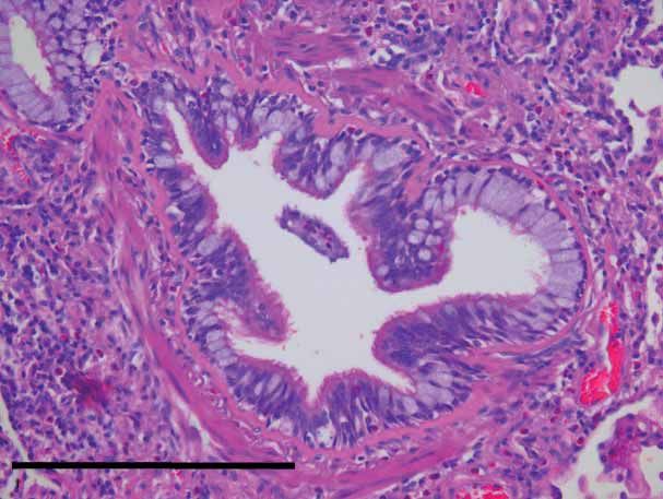
1 mm in diameter using low-dose protocols. CT images acquired during a breath hold at full inspiration
can provide detailed information on airway generation and airway wall dimensions (figure 3) [138]. In
operated patients with COPD it was shown that airway size measured by pre-operative CT correlated to
airway size as measured histologically [139]. In asthma it was shown that the airway walls, as measured on
CT, were thicker compared to healthy subjects. Furthermore, it was shown that increased airway wall
thickness observed on CT was related to lower forced expiratory volume in 1 s and higher risk of
exacerbations [138, 140]. Static CT images acquired during expiration can show regions of low attenuation
that are thought to reflect SAD (figure 2). These low-attenuation regions depict areas of gas trapping and/
or hypoperfusion, due to premature closure of small airways during expiration and reduced ventilation
leading to hypoperfusion. In clinical studies in patients with asthma, low-attenuation regions have been
associated with disease duration, asthma exacerbations, airflow obstruction and inflammation [141].
Hence, low-attenuation regions can be used in clinical studies as an outcome measure to evaluate efficacy
of treatment targeting the small airways [142]. Well-standardised volume-controlled CT protocols and the
development of sensitive automated image-analysis systems to measure airway dimensions and
low-attenuation regions are needed to further facilitate the use of chest CT-related outcome measures in
clinical studies. A limitation of chest CTs is that they expose the patient to ionising radiation, which
restricts the number of CTs that can be made for monitoring airway dimensions and low-attenuation
regions in response to therapies and furthermore for studying the lungs dynamically.
Dynamic imaging
Magnetic resonance imaging (MRI) is a radiation-free alternative to CT that is able to provide us with
information on lung structure and function. Ventilation defects or ventilation heterogeneity can be
visualised with MRI using hyperpolarised gases [143] or after expiration to residual volume level. Other
imaging modalities, such as single-photon emission CT and positron emission tomography, can also be
used in ventilation imaging. Ventilation defects are thought to represent regions of heterogeneous and
abnormal gas or air distribution. It is hypothesised that ventilation heterogeneity or defects correspond to
regions in the lung that are not well-ventilated [144]. It has been shown that the presence of ventilation
defects, frequently observed in patients with asthma, is due to heterogeneous airway closure or narrowing
of small airways [145]. In several studies it was shown that ventilation defects are not seen randomly but
they might be predefined [146] or a result of remodelling [147]. Furthermore, it has been shown that
ventilation defects persist over time [146, 148, 149]. Ventilation defects in patients with asthma could
resolve after induced bronchoconstriction most likely as a result of redistribution of airflow, resulting in
poorly ventilated regions receiving more airflow and vice versa [148]. It was suggested that if ventilation
defects persist over long periods, one would expect that atelectasis would occur in these regions [149].
a) b)
FIGURE 3 Spirometer-guided chest computed tomography (CT) in a patient with severe asthma. a) Inspiratory
chest CT showing clear airway wall thickening. b) Expiratory chest CT in the same patient showing extensive
regions of low attenuation.
https://doi.org/10.1183/16000617.0186-2020 12ASTHMA | W.B. VAN DEN BOSCH ET AL.
TABLE 3 Summary of possible structural and functional changes in the small airways of asthmatics
Structural changes Functional changes
Epithelium Variation in thickness Impaired barrier function
Altered epithelial cell to cell adhesion Increased mucus production
Goblet cell hyper/metaplasia Epithelial–mesenchymal transition
Mucus accumulation/plugging
Alterations in expression of MUC5AC
ECM
Inner airway wall area Changes in ECM composition Altered ECM production
Basement membrane thickening Stiffening of the airways
Increased ASM opposing force
Within ASM Changes in ECM composition Altered ECM production
Stiffening of the airways
Outer airway wall area Changes in ECM composition Altered ECM production
Outer airway wall area thickening Stiffening of the airways
Increased ASM opposing force
Decreased force transduction
ASM Thickening of ASM layer Increased contractile force
ASM hyperplasia/hypertrophy Length adaptation
Force adaptation
Increased maximal shortening
Increased shortening velocity
Impaired bronchodilating and protective effect after a deep inspiration
Parenchyma Abnormal airway alveolar attachments Decreased force transduction
Decreased parenchymal tethering force
ECM: extracellular matrix; ASM: airway smooth muscle.
Therefore, some sort of partial or intermittent closure must be present in the patients with persisting
ventilation defects or these areas are ventilated through collateral ventilation [150]. Ventilation defects are
associated with disease severity [144, 147, 151, 152], lung clearance index [152] and spirometry [144, 151].
An in-depth review by KING et al. [140], reported important findings on CT and three-dimensional
ventilation imaging in the large airways, small airways and the parenchyma of patients with asthma. In
that review, KING et al. [140] further elaborate on the current and possible future use of different imaging
modalities to improve our understanding of the role of small airways in asthma pathophysiology and on
what further research is needed.
Modelling
Over a number of years efforts have been made to explain the emergent behaviour of airways leading to
ventilation defects in asthma. Based on a single airway model by ANAFI et al. [153], VENEGAS et al. [145]
extended this model and discovered that behaviour of an individual airway affects the whole airway tree
and vice versa. A further extension of this model by WINKLER et al. [154] proposed a mechanism for the
emergence of ventilation defects. The model describes a vicious circle based on interdependence of
airways. In short, they show that if bronchoconstriction reaches a critical point in which airways become
unstable, then any change in airflow causing a reduction in airflow in the daughter airway leads to a
smaller tidal volume. Subsequently, reduced expansion of the adjacent lung parenchyma increases airway
constriction and airway resistance, which in turn leads to a greater reduction in airflow through the airway
relative to parallel airways. This leads to a vicious circle in which bronchoconstriction further increases.
This model is a step forward in understanding the complex behaviour of airways mechanics in asthma
exacerbations. Therefore, it is possible that localised areas of remodelling that result in excessive airway
narrowing will alter behaviour of airways in other parts of the airway tree. These complexities, including
the heterogeneity of airway remodelling need to be considered when observing behaviour of the lung in
asthma. Future modelling studies, combining morphologically, clinically and imaging-derived parameters
on structure and function with computer simulation will improve our understanding of airway mechanical
behaviour.
Conclusion
The European Respiratory Society International Congress in 2019 in Madrid, Spain, once again
highlighted the importance of the need to increase knowledge on the small airways in asthma. The high
https://doi.org/10.1183/16000617.0186-2020 13ASTHMA | W.B. VAN DEN BOSCH ET AL.
prevalence of small airways involvement in people with asthma observed in the ATLANTIS study
accentuates this need [6]. This review article provides us with an overview of the structural and functional
alterations observed in the small airways of patients with asthma (table 3). Asthma is a heterogeneous
condition and therefore, it is unlikely that there is one overarching mechanism that will do justice to the
complexity of its pathophysiology. Since Hippocrates first used the term asthma our ideas and
understanding on pathophysiology, diagnostics and treatment of asthma have changed, revealing a number
of possible phenotypes covered by the term asthma. SAD in asthma could be such a phenotype which
deserves further study.
Conflict of interest: W.B. van den Bosch reports grants from Vectura Group Plc., during the conduct of the study.
A.L. James has nothing to disclose. H.A.W.M. Tiddens reports grants from Vectura Group Plc., during the conduct of
the study; other from Roche and Novartis, grants from CFF, Vertex, Gilead and Chiesi, outside the submitted work. In
addition, H.A.W.M. Tiddens has a patent Vectura licensed, and a patent PRAGMA-CF scoring system issued and is
heading the Erasmus MC-Sophia Children’s Hospital Core Laboratory Lung Analysis.
Support statement: Funding was received from Vectura Group plc. (unconditional grant for PhD research programme).
Funding information for this article has been deposited with the Crossref Funder Registry.
References
1 Hogg JC, Macklem PT, Thurlbeck WM. Site and nature of airway obstruction in chronic obstructive lung disease.
N Engl J Med 1968; 278: 1355–1360.
2 Macklem PT, Mead J. Resistance of central and peripheral airways measured by a retrograde catheter. J Appl
Physiol 1967; 22: 395–401.
3 Weibel ER, Gomez DM. Architecture of the human lung. Use of quantitative methods establishes fundamental
relations between size and number of lung structures. Science 1962; 137: 577–585.
4 Su ZQ, Guan WJ, Li SY, et al. Evaluation of the normal airway morphology using optical coherence tomography.
Chest 2019; 156: 915–925.
5 Usmani OS, Singh D, Spinola M, et al. The prevalence of small airways disease in adult asthma: a systematic
literature review. Respir Med 2016; 116: 19–27.
6 Postma DS, Brightling C, Baldi S, et al. Exploring the relevance and extent of small airways dysfunction in
asthma (ATLANTIS): baseline data from a prospective cohort study. Lancet Respir Med 2019; 7: 402–416.
7 Elliot JG, Jones RL, Abramson MJ, et al. Distribution of airway smooth muscle remodelling in asthma: relation to
airway inflammation. Respirology 2015; 20: 66–72.
8 Lambrecht BN, Hammad H, Fahy JV. The cytokines of asthma. Immunity 2019; 50: 975–991.
9 Knight DA, Holgate ST. The airway epithelium: structural and functional properties in health and disease.
Respirology 2003; 8: 432–446.
10 Crystal RG, Randell SH, Engelhardt JF, et al. Airway epithelial cells: current concepts and challenges. Proc Am
Thorac Soc 2008; 5: 772–777.
11 Lambrecht BN, Hammad H. The airway epithelium in asthma. Nat Med 2012; 18: 684–692.
12 Tam A, Wadsworth S, Dorscheid D, et al. The airway epithelium: more than just a structural barrier. Ther Adv
Respir Dis 2011; 5: 255–273.
13 Okuda K, Chen G, Subramani DB, et al. Localization of secretory mucins MUC5AC and MUC5B in normal/
healthy human airways. Am J Respir Crit Care Med 2019; 199: 715–727.
14 Kirch J, Guenther M, Doshi N, et al. Mucociliary clearance of micro- and nanoparticles is independent of size,
shape and charge– an ex vivo and in silico approach. J Control Release 2012; 159: 128–134.
15 Zhou J, Alvarez-Elizondo MB, Botvinick E, et al. Local small airway epithelial injury induces global smooth
muscle contraction and airway constriction. J Appl Physiol 2012; 112: 627–637.
16 Dunnill MS. The pathology of asthma, with special reference to changes in the bronchial mucosa. J Clin Pathol
1960; 13: 27–33.
17 Montefort S, Roberts JA, Beasley R, et al. The site of disruption of the bronchial epithelium in asthmatic and
non-asthmatic subjects. Thorax 1992; 47: 499–503.
18 Laitinen LA, Heino M, Laitinen A, et al. Damage of the airway epithelium and bronchial reactivity in patients
with asthma. Am Rev Respir Dis 1985; 131: 599–606.
19 Ordonez C, Ferrando R, Hyde DM, et al. Epithelial desquamation in asthma: artifact or pathology? Am J Respir
Crit Care Med 2000; 162: 2324–2329.
20 Fehrenbach H, Wagner C, Wegmann M. Airway remodeling in asthma: what really matters. Cell Tissue Res 2017;
367: 551–569.
21 Carroll N, Elliot J, Morton A, et al. The structure of large and small airways in nonfatal and fatal asthma. Am
Rev Respir Dis 1993; 147: 405–410.
22 Cutz E, Levison H, Cooper DM. Ultrastructure of airways in children with asthma. Histopathology 2002; 41:
22–36.
23 Qin L, Gibson PG, Simpson JL, et al. Dysregulation of sputum columnar epithelial cells and products in distinct
asthma phenotypes. Clin Exp Allergy 2019; 49: 1418–1428.
24 Mostaco-Guidolin L, Hajimohammadi S, Vasilescu DM, et al. Application of Euclidean distance mapping for
assessment of basement membrane thickness distribution in asthma. J Appl Physiol 2017; 123: 473–481.
25 de Boer WI, Sharma HS, Baelemans SM, et al. Altered expression of epithelial junctional proteins in atopic
asthma: possible role in inflammation. Can J Physiol Pharmacol 2008; 86: 105–112.
26 Xiao C, Puddicombe SM, Field S, et al. Defective epithelial barrier function in asthma. J Allergy Clin Immunol
2011; 128: 549–556.
https://doi.org/10.1183/16000617.0186-2020 14ASTHMA | W.B. VAN DEN BOSCH ET AL.
27 Carayol N, Campbell A, Vachier I, et al. Modulation of cadherin and catenins expression by tumor necrosis
factor-alpha and dexamethasone in human bronchial epithelial cells. Am J Respir Cell Mol Biol 2002; 26:
341–347.
28 Coyne CB, Vanhook MK, Gambling TM, et al. Regulation of airway tight junctions by proinflammatory
cytokines. Mol Biol Cell 2002; 13: 3218–3234.
29 Saatian B, Rezaee F, Desando S, et al. Interleukin-4 and interleukin-13 cause barrier dysfunction in human
airway epithelial cells. Tissue Barriers 2013; 1: e24333.
30 Canas JA, Sastre B, Rodrigo-Munoz JM, et al. Eosinophil-derived exosomes contribute to asthma remodelling by
activating structural lung cells. Clin Exp Allergy 2018; 48: 1173–1185.
31 Hackett T-L. Epithelial–mesenchymal transition in the pathophysiology of airway remodelling in asthma. Curr
Opin Allergy Clin Immunol 2012; 12: 53–59.
32 Shimura S, Andoh Y, Haraguchi M, et al. Continuity of airway goblet cells and intraluminal mucus in the
airways of patients with bronchial asthma. Eur Respir J 1996; 9: 1395–1401.
33 Kuyper LM, Pare PD, Hogg JC, et al. Characterization of airway plugging in fatal asthma. Am J Med 2003; 115:
6–11.
34 Malmstrom K, Lohi J, Sajantila A, et al. Immunohistology and remodeling in fatal pediatric and adolescent
asthma. Respir Res 2017; 18: 94.
35 Elliot JG, Noble PB, Mauad T, et al. Inflammation-dependent and independent airway remodelling in asthma.
Respirology 2018; 23: 1138–1145.
36 Aikawa T, Shimura S, Sasaki H, et al. Marked goblet cell hyperplasia with mucus accumulation in the airways of
patients who died of severe acute asthma attack. Chest 1992; 101: 916–921.
37 Wenzel SE, Vitari CA, Shende M, et al. Asthmatic granulomatosis: a novel disease with asthmatic and
granulomatous features. Am J Respir Crit Care Med 2012; 186: 501–507.
38 Trejo Bittar HE, Doberer D, Mehrad M, et al. Histologic findings of severe/therapy-resistant asthma from
video-assisted thoracoscopic surgery biopsies. Am J Surg Pathol 2017; 41: 182–188.
39 Blyth DI, Pedrick MS, Savage TJ, et al. Lung inflammation and epithelial changes in a murine model of atopic
asthma. Am J Respir Cell Mol Biol 1996; 14: 425–438.
40 Saetta M, Di Stefano A, Rosina C, et al. Quantitative structural analysis of peripheral airways and arteries in
sudden fatal asthma. Am Rev Respir Dis 1991; 143: 138–143.
41 Bonser LR, Erle DJ. Airway mucus and asthma: the role of MUC5AC and MUC5B. J Clin Med 2017; 6: 112.
42 Evans CM, Raclawska DS, Ttofali F, et al. The polymeric mucin Muc5ac is required for allergic airway
hyperreactivity. Nat Commun 2015; 6: 6281.
43 Curran DR, Cohn L. Advances in mucous cell metaplasia: a plug for mucus as a therapeutic focus in chronic
airway disease. Am J Respir Cell Mol Biol 2010; 42: 268–275.
44 Grainge CL, Lau LC, Ward JA, et al. Effect of bronchoconstriction on airway remodeling in asthma. N Engl J
Med 2011; 364: 2006–2015.
45 Dunican EM, Watchorn DC, Fahy JV. Autopsy and imaging studies of mucus in asthma. lessons learned about
disease mechanisms and the role of mucus in airflow obstruction. Ann Am Thorac Soc 2018; 15: Suppl 3,
S184–SS91.
46 Wan H, Kaestner KH, Ang SL, et al. Foxa2 regulates alveolarization and goblet cell hyperplasia. Development
2004; 131: 953–964.
47 Wagers S, Lundblad LK, Ekman M, et al. The allergic mouse model of asthma: normal smooth muscle in an
abnormal lung? J Appl Physiol 2004; 96: 2019–2027.
48 Sorokin L. The impact of the extracellular matrix on inflammation. Nat Rev Immunol 2010; 10: 712–723.
49 Saglani S, Molyneux C, Gong H, et al. Ultrastructure of the reticular basement membrane in asthmatic adults,
children and infants. Eur Respir J 2006; 28: 505–512.
50 Hynes RO, Naba A. Overview of the matrisome – an inventory of extracellular matrix constituents and functions.
Cold Spring Harb Perspect Biol 2012; 4: a004903.
51 Suki B, Bates JH. Extracellular matrix mechanics in lung parenchymal diseases. Respir Physiol Neurobiol 2008;
163: 33–43.
52 Polio SR, Stasiak SE, Jamieson RR, et al. Extracellular matrix stiffness regulates human airway smooth muscle
contraction by altering the cell-cell coupling. Sci Rep 2019; 9: 9564.
53 Malmstrom J, Larsen K, Malmstrom L, et al. Proteome annotations and identifications of the human pulmonary
fibroblast. J Proteome Res 2004; 3: 525–537.
54 Elshaw SR, Henderson N, Knox AJ, et al. Matrix metalloproteinase expression and activity in human airway
smooth muscle cells. Br J Pharmacol 2004; 142: 1318–1324.
55 Johnson PR, Burgess JK, Underwood PA, et al. Extracellular matrix proteins modulate asthmatic airway smooth
muscle cell proliferation via an autocrine mechanism. J Allergy Clin Immunol 2004; 113: 690–696.
56 Hendrix AY, Kheradmand F. The role of matrix metalloproteinases in development, repair, and destruction of
the lungs. Prog Mol Biol Transl Sci 2017; 148: 1–29.
57 Couchman JR. Transmembrane signaling proteoglycans. Annu Rev Cell Dev Biol 2010; 26: 89–114.
58 de Medeiros Matsushita M, da Silva LF, dos Santos MA, et al. Airway proteoglycans are differentially altered in
fatal asthma. J Pathol 2005; 207: 102–110.
59 Mauad T, Silva LF, Santos MA, et al. Abnormal alveolar attachments with decreased elastic fiber content in distal
lung in fatal asthma. Am J Respir Crit Care Med 2004; 170: 857–862.
60 Araujo BB, Dolhnikoff M, Silva LF, et al. Extracellular matrix components and regulators in the airway smooth
muscle in asthma. Eur Respir J 2008; 32: 61–69.
61 Mauad T, Xavier AC, Saldiva PH, et al. Elastosis and fragmentation of fibers of the elastic system in fatal asthma.
Am J Respir Crit Care Med 1999; 160: 968–975.
62 Dolhnikoff M, da Silva LF, de Araujo BB, et al. The outer wall of small airways is a major site of remodeling in
fatal asthma. J Allergy Clin Immunol 2009; 123: 1090–1097.
63 Nihlberg K, Andersson-Sjoland A, Tufvesson E, et al. Altered matrix production in the distal airways of
individuals with asthma. Thorax 2010; 65: 670–676.
https://doi.org/10.1183/16000617.0186-2020 15ASTHMA | W.B. VAN DEN BOSCH ET AL.
64 Bai TR, Cooper J, Koelmeyer T, et al. The effect of age and duration of disease on airway structure in fatal
asthma. Am J Respir Crit Care Med 2000; 162: 663–669.
65 Weitoft M, Andersson C, Andersson-Sjoland A, et al. Controlled and uncontrolled asthma display distinct
alveolar tissue matrix compositions. Respir Res 2014; 15: 67.
66 Keglowich LF, Borger P. The three A’s in asthma - airway smooth muscle, airway remodeling & angiogenesis.
Open Respir Med J 2015; 9: 70–80.
67 Holgate ST. Epithelium dysfunction in asthma. J Allergy Clin Immunol 2007; 120: 1233–1244; quiz 45-6.
68 Johnson PR, Black JL, Carlin S, et al. The production of extracellular matrix proteins by human passively
sensitized airway smooth-muscle cells in culture: the effect of beclomethasone. Am J Respir Crit Care Med 2000;
162: 2145–2151.
69 Doucet C, Brouty-Boye D, Pottin-Clemenceau C, et al. Interleukin (IL) 4 and IL-13 act on human lung
fibroblasts. Implication in asthma. J Clin Invest 1998; 101: 2129–2139.
70 Al-Alawi M, Hassan T, Chotirmall SH. Transforming growth factor β and severe asthma: a perfect storm. Respir
Med 2014; 108: 1409–1423.
71 Liu G, Cooley MA, Nair PM, et al. Airway remodelling and inflammation in asthma are dependent on the
extracellular matrix protein fibulin-1c. J Pathol 2017; 243: 510–523.
72 Wilson JW, Li X, Pain MC. The lack of distensibility of asthmatic airways. Am Rev Respir Dis 1993; 148:
806–809.
73 Niimi A, Matsumoto H, Takemura M, et al. Relationship of airway wall thickness to airway sensitivity and airway
reactivity in asthma. Am J Respir Crit Care Med 2003; 168: 983–988.
74 Lambert RK, Codd SL, Alley MR, et al. Physical determinants of bronchial mucosal folding. J Appl Physiol 1994;
77: 1206–1216.
75 Jensen A, Atileh H, Suki B, et al. Selected contribution: airway caliber in healthy and asthmatic subjects: effects
of bronchial challenge and deep inspirations. J Appl Physiol 2001; 91: 506–515.
76 Brown RH, Scichilone N, Mudge B, et al. High-resolution computed tomographic evaluation of airway
distensibility and the effects of lung inflation on airway caliber in healthy subjects and individuals with asthma.
Am J Respir Crit Care Med 2001; 163: 994–1001.
77 Slats AM, Janssen K, van Schadewijk A, et al. Expression of smooth muscle and extracellular matrix proteins in
relation to airway function in asthma. J Allergy Clin Immunol 2008; 121: 1196–1202.
78 Khan MA, Ellis R, Inman MD, et al. Influence of airway wall stiffness and parenchymal tethering on the
dynamics of bronchoconstriction. Am J Physiol Lung Cell Mol Physiol 2010; 299: L98–L108.
79 Pascoe CD, Seow CY, Hackett TL, et al. Heterogeneity of airway wall dimensions in humans: a critical
determinant of lung function in asthmatics and nonasthmatics. Am J Physiol Lung Cell Mol Physiol 2017; 312:
L425–L431.
80 Yick CY, Ferreira DS, Annoni R, et al. Extracellular matrix in airway smooth muscle is associated with dynamics
of airway function in asthma. Allergy 2012; 67: 552–559.
81 James A, Carroll N. Airway smooth muscle in health and disease; methods of measurement and relation to
function. Eur Respir J 2000; 15: 782–789.
82 Ebina M, Yaegashi H, Takahashi T, et al. Distribution of smooth muscles along the bronchial tree. A
morphometric study of ordinary autopsy lungs. Am Rev Respir Dis 1990; 141: 1322–1326.
83 Jones RL, Elliot JG, James AL. Estimating airway smooth muscle cell volume and number in airway sections.
Sources of variability. Am J Respir Cell Mol Biol 2014; 50: 246–252.
84 Kuwano K, Bosken CH, Pare PD, et al. Small airways dimensions in asthma and in chronic obstructive
pulmonary disease. Am Rev Respir Dis 1993; 148: 1220–1225.
85 Ebina M, Takahashi T, Chiba T, et al. Cellular hypertrophy and hyperplasia of airway smooth muscles
underlying bronchial asthma. A 3-D morphometric study. Am Rev Respir Dis 1993; 148: 720–726.
86 James AL, Pare PD, Hogg JC. The mechanics of airway narrowing in asthma. Am Rev Respir Dis 1989; 139:
242–246.
87 James AL, Elliot JG, Jones RL, et al. Airway smooth muscle hypertrophy and hyperplasia in asthma. Am J Respir
Crit Care Med 2012; 185: 1058–1064.
88 Prescott GW, Gregory MD, Ronald EF, et al. Hyperplasia of smooth muscle in mild to moderate asthma without
changes in cell size or gene expression. Am J Respir Crit Care Med 2004; 169: 1001–1006.
89 Leclere M, Lavoie-Lamoureux A, Gelinas-Lymburner E, et al. Effect of antigenic exposure on airway smooth
muscle remodeling in an equine model of chronic asthma. Am J Respir Cell Mol Biol 2011; 45: 181–187.
90 Johnson PR, Roth M, Tamm M, et al. Airway smooth muscle cell proliferation is increased in asthma. Am J
Respir Crit Care Med 2001; 164: 474–477.
91 James AL, Noble PB, Drew SA, et al. Airway smooth muscle proliferation and inflammation in asthma. J Appl
Physiol 2018; 125: 1090–1096.
92 Ward JE, Harris T, Bamford T, et al. Proliferation is not increased in airway myofibroblasts isolated from
asthmatics. Eur Respir J 2008; 32: 362–371.
93 Ijpma G, Panariti A, Lauzon AM, et al. Directional preference of airway smooth muscle mass increase in human
asthmatic airways. Am J Physiol Lung Cell Mol Physiol 2017; 312: L845–L854.
94 Hassan M, Jo T, Risse PA, et al. Airway smooth muscle remodeling is a dynamic process in severe long-standing
asthma. J Allergy Clin Immunol 2010; 125: 1037–1045.
95 Salter B, Pray C, Radford K, et al. Regulation of human airway smooth muscle cell migration and relevance to
asthma. Respir Res 2017; 18: 156.
96 Wang KCW, Le Cras TD, Larcombe AN, et al. Independent and combined effects of airway remodelling and
allergy on airway responsiveness. Clin Sci 2018; 132: 327–338.
97 James AL, Bai TR, Mauad T, et al. Airway smooth muscle thickness in asthma is related to severity but not
duration of asthma. Eur Respir J 2009; 34: 1040–1045.
98 O’Reilly R, Ullmann N, Irving S, et al. Increased airway smooth muscle in preschool wheezers who have asthma
at school age. J Allergy Clin Immunol 2013; 131: 1024–1032.
99 Owens L, Laing IA, Zhang G, et al. Infant lung function predicts asthma persistence and remission in young
adults. Respirology 2017; 22: 289–294.
https://doi.org/10.1183/16000617.0186-2020 16ASTHMA | W.B. VAN DEN BOSCH ET AL.
100 Mead J. Point: airway smooth muscle is useful. J Appl Physiol 2007; 102: 1708–1709.
101 Fredberg JJ. Counterpoint: airway smooth muscle is not useful. J Appl Physiol 2007; 102: 1709–1710.
102 Seow C, Mitzner W, Irvin C, et al. Point:counterpoint comments. J Appl Physiol 2007; 102: 1712.
103 Ford LE. Comment on point:counterpoint: “airway smooth muscle is/is not useful”. J Appl Physiol 2007; 102:
2407.
104 Seow CY, Fredberg JJ. Historical perspective on airway smooth muscle: the saga of a frustrated cell. J Appl Physiol
2001; 91: 938–952.
105 Mitzner W. Airway smooth muscle: the appendix of the lung. Am J Respir Crit Care Med 2004; 169: 787–790.
106 Schittny JC, Miserocchi G, Sparrow MP. Spontaneous peristaltic airway contractions propel lung liquid through
the bronchial tree of intact and fetal lung explants. Am J Respir Cell Mol Biol 2000; 23: 11–18.
107 Cieri RL. Pulmonary smooth muscle in vertebrates: a comparative review of structure and function. Integr Comp
Biol 2019; 59: 10–28.
108 Howarth PH, Knox AJ, Amrani Y, et al. Synthetic responses in airway smooth muscle. J Allergy Clin Immunol
2004; 114: Suppl. 2, S32–S50.
109 Chin LY, Bosse Y, Pascoe C, et al. Mechanical properties of asthmatic airway smooth muscle. Eur Respir J 2012;
40: 45–54.
110 Cerrina J, Labat C, Haye-Legrande I, et al. Human isolated bronchial muscle preparations from asthmatic
patients: effects of indomethacin and contractile agonists. Prostaglandins 1989; 37: 457–469.
111 Bai TR. Abnormalities in airway smooth muscle in fatal asthma. A comparison between trachea and bronchus.
Am Rev Respir Dis 1991; 143: 441–443.
112 Bramley AM, Thomson RJ, Roberts CR, et al. Hypothesis: excessive bronchoconstriction in asthma is due to
decreased airway elastance. Eur Respir J 1994; 7: 337–341.
113 Thomson RJ, Bramley AM, Schellenberg RR. Airway muscle stereology: implications for increased shortening in
asthma. Am J Respir Crit Care Med 1996; 154: 749–757.
114 Ijpma G, Kachmar L, Matusovsky OS, et al. Human trachealis and main bronchi smooth muscle are
normoresponsive in asthma. Am J Respir Crit Care Med 2015; 191: 884–893.
115 Whicker SD, Armour CL, Black JL. Responsiveness of bronchial smooth muscle from asthmatic patients to
relaxant and contractile agonists. Pulm Pharmacol 1988; 1: 25–31.
116 Bjorck T, Gustafsson LE, Dahlen SE. Isolated bronchi from asthmatics are hyperresponsive to adenosine, which
apparently acts indirectly by liberation of leukotrienes and histamine. Am Rev Respir Dis 1992; 145: 1087–1091.
117 Goldie RG, Spina D, Henry PJ, et al. In vitro responsiveness of human asthmatic bronchus to carbachol,
histamine, beta-adrenoceptor agonists and theophylline. Br J Clin Pharmacol 1986; 22: 669–676.
118 de Jongste JC, Mons H, Bonta IL, et al. In vitro responses of airways from an asthmatic patient. Eur J Respir Dis
1987; 71: 23–29.
119 Bosse Y, Sobieszek A, Pare PD, et al. Length adaptation of airway smooth muscle. Proc Am Thorac Soc 2008; 5:
62–67.
120 Bosse Y, Chin LY, Pare PD, et al. Adaptation of airway smooth muscle to basal tone: relevance to airway
hyperresponsiveness. Am J Respir Cell Mol Biol 2009; 40: 13–18.
121 Bosse Y, Chapman DG, Pare PD, et al. A ’good’ muscle in a ’bad’ environment: the importance of airway smooth
muscle force adaptation to airway hyperresponsiveness. Respir Physiol Neurobiol 2011; 179: 269–275.
122 Mitchell RW, Ruhlmann E, Magnussen H, et al. Passive sensitization of human bronchi augments smooth
muscle shortening velocity and capacity. Am J Physiol 1994; 267: L218–L222.
123 Ma X, Cheng Z, Kong H, et al. Changes in biophysical and biochemical properties of single bronchial smooth
muscle cells from asthmatic subjects. Am J Physiol Lung Cell Mol Physiol 2002; 283: L1181–L1189.
124 Oliver MN, Fabry B, Marinkovic A, et al. Airway hyperresponsiveness, remodeling, and smooth muscle mass:
right answer, wrong reason? Am J Respir Cell Mol Biol 2007; 37: 264–272.
125 Matusovsky OS, Kachmar L, Ijpma G, et al. Peripheral airway smooth muscle, but not the trachealis, is
hypercontractile in an equine model of asthma. Am J Respir Cell Mol Biol 2016; 54: 718–727.
126 Solway J, Fredberg JJ. Perhaps airway smooth muscle dysfunction contributes to asthmatic bronchial
hyperresponsiveness after all. Am J Respir Cell Mol Biol 1997; 17: 144–146.
127 Harvey BC, Parameswaran H, Lutchen KR. Can breathing-like pressure oscillations reverse or prevent narrowing
of small intact airways? J Appl Physiol 2015; 119: 47–54.
128 Robinson PD, Latzin P, Verbanck S, et al. Consensus statement for inert gas washout measurement using
multiple- and single- breath tests. Eur Respir J 2013; 41: 507.
129 Huber HL, Koessler KK. The pathology of bronchial asthma. Arch Int Med 1922; 30: 689–760.
130 Woolcock AJ, Salome CM, Yan K. The shape of the dose-response curve to histamine in asthmatic and normal
subjects. Am Rev Respir Dis 1984; 130: 71–75.
131 Lambert RK, Wiggs BR, Kuwano K, et al. Functional significance of increased airway smooth muscle in asthma
and COPD. J Appl Physiol 1993; 74: 2771–2781.
132 Noble PB, Jones RL, Needi ET, et al. Responsiveness of the human airway in vitro during deep inspiration and
tidal oscillation. J Appl Physiol 2011; 110: 1510–1518.
133 Cockcroft DW, Davis BE, Boulet LP, et al. The links between allergen skin test sensitivity, airway responsiveness
and airway response to allergen. Allergy 2005; 60: 56–59.
134 Barnig C, Purohit A, Casset A, et al. Nonallergic airway hyperresponsiveness and allergen-specific IgE levels are
the main determinants of the early and late asthmatic response to allergen. J Investig Allergol Clin Immunol 2013;
23: 267–274.
135 Illi S, von Mutius E, Lau S, et al. Perennial allergen sensitisation early in life and chronic asthma in children: a
birth cohort study. Lancet 2006; 368: 763–770.
136 Konstantinos Katsoulis K, Kostikas K, Kontakiotis T. Techniques for assessing small airways function: possible
applications in asthma and COPD. Respir Med 2016; 119: e2–e9.
137 McNulty W, Usmani OS. Techniques of assessing small airways dysfunction. Eur Clin Respir J 2014; 1.
138 Patyk M, Obojski A, Sokolowska-Dabek D, et al. Airway wall thickness and airflow limitations in asthma
assessed in quantitative computed tomography. Ther Adv Respir Dis 2020; 14: 1753466619898598.
https://doi.org/10.1183/16000617.0186-2020 17You can also read