Relapse of Acute Myeloid Leukemia after Allogeneic Stem Cell Transplantation: Prevention, Detection, and Treatment - MDPI
←
→
Page content transcription
If your browser does not render page correctly, please read the page content below
International Journal of
           Molecular Sciences

Review
Relapse of Acute Myeloid Leukemia after Allogeneic
Stem Cell Transplantation: Prevention, Detection,
and Treatment
Christina Rautenberg, Ulrich Germing, Rainer Haas, Guido Kobbe and Thomas Schroeder *
 Department of Hematology, Oncology and Clinical Immunology, University of Duesseldorf, Medical Faculty,
 40225 Duesseldorf, Germany; Christina.Rautenberg@med.uni-duesseldorf.de (C.R.);
 Germing@med.uni-duesseldorf.de (U.G.); Haas@med.uni-duesseldorf.de (R.H.);
 Kobbe@med.uni-duesseldorf.de (G.K.)
 * Correspondence: thomas.schroeder@med.uni-duesseldorf.de; Tel.: +49-211-81-17720; Fax: +49-211-81-18853

 Received: 28 November 2018; Accepted: 3 January 2019; Published: 8 January 2019

 Abstract: Acute myeloid leukemia (AML) is a phenotypically and prognostically heterogeneous
 hematopoietic stem cell disease that may be cured in eligible patients with intensive chemotherapy
 and/or allogeneic stem cell transplantation (allo-SCT). Tremendous advances in sequencing
 technologies have revealed a large amount of molecular information which has markedly improved
 our understanding of the underlying pathophysiology and enables a better classification and risk
 estimation. Furthermore, with the approval of the FMS-like tyrosine kinase 3 (FLT3) inhibitor
 Midostaurin a first targeted therapy has been introduced into the first-line therapy of younger
 patients with FLT3-mutated AML and several other small molecules targeting molecular alterations
 such as isocitrate dehydrogenase (IDH) mutations or the anti-apoptotic b-cell lymphoma 2 (BCL-2)
 protein are currently under investigation. Despite these advances, many patients will have to undergo
 allo-SCT during the course of disease and depending on disease and risk status up to half of them will
 finally relapse after transplant. Here we review the current knowledge about the molecular landscape
 of AML and how this can be employed to prevent, detect and treat relapse of AML after allo-SCT.

 Keywords: acute myeloid leukemia; allogeneic transplantation; relapse; maintenance; minimal
 residual disease; salvage therapy

1. Introduction
     Allogeneic stem cell transplantation (allo-SCT) is besides the use of conventional chemotherapy
the second backbone of therapy for patients with acute myeloid leukemia (AML) who are eligible
for intensive therapy. Allo-SCT offers the highest potential for long-term survival as postremission
therapy in those with an intermediate or high-risk disease and as salvage therapy in those with relapsed
or resistant disease, regardless of prognostic biological characteristics [1]. Still, despite substantial
improvements regarding the reduction of non-relapse mortality during the last decades [2] relapse
represents the major cause of treatment failure and up 50% of AML patients finally relapse after
allo-SCT depending on disease status and characteristics [3]. Their prognosis is generally dismal,
since many of them, in particular those with early relapse, can either not tolerate or are refractory
to low-dose or intensive chemotherapy that are usually applied in this situation. Furthermore, even
with cellular therapies such as donor lymphocyte infusions [4] and second transplantation in selected
cases or the use of investigational agents only a minority of patients can be rescued in the long run.
As a consequence, 2-year survival rates of patients with AML who relapsed after allo-HCT are below
20% independent from the choice of salvage therapy [5–8]. This poor prognosis and the limited success
of salvage therapies imply the need for novel strategies to prevent, detect and to treat relapse.

Int. J. Mol. Sci. 2019, 20, 228; doi:10.3390/ijms20010228                          www.mdpi.com/journal/ijms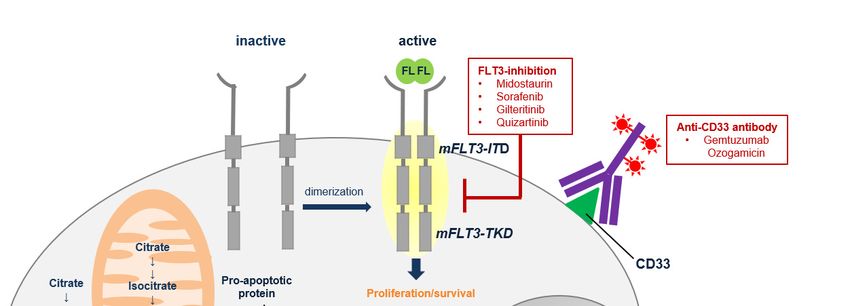
Int.Int.
J. Mol. Sci.Sci.
J. Mol. 2019, 20,19,
2018, 228x FOR PEER REVIEW 2 of220
of 20
poor prognosis and the limited success of salvage therapies imply the need for novel strategies to
During
prevent, the last
detect andtwo decades
to treat extensive advances in molecular techniques, in particular massive
relapse.
parallelDuring
or nextthegeneration sequencing,
last two decades extensive haveadvances
facilitated inthe discovery
molecular of a largeinnumber
techniques, of massive
particular molecular
aberrations
parallel orin patients
next with sequencing,
generation AML. Thesehave enabled a comprehensive
facilitated the discoveryview on the
of a large molecular
number landscape
of molecular
and have substantially moved forward our understanding of the pathophysiology
aberrations in patients with AML. These enabled a comprehensive view on the molecular landscape of AML [9].
Furthermore, this knowledge now allows us to group patients into
and have substantially moved forward our understanding of the pathophysiology of AML [9].distinct molecular subgroups,
to Furthermore,
perform riskthis estimation
knowledge andnowtoallows
adjustusour post-remission
to group patients intotreatment strategy, subgroups,
distinct molecular e.g., high-dose
to
AraC-based
perform riskconsolidation
estimation and or allo-SCT
to adjust [1,10,11]. In addition,
our post-remission we are using
treatment more
strategy, e.g.,and more molecular
high-dose AraC-
based consolidation
information identified or allo-SCT
at the time of[1,10,11].
diagnosis In to
addition,
track the wedisease
are using more
at the and level
lowest moreasmolecular
technically
information
possible identified
(=minimal at the time disease,
or measurable of diagnosis
MRD) to track
during theand
disease
afterat the lowest
therapy level as technically
by molecular techniques
in possible
order to (=minimal
adapt therapy or measurable
if neededdisease,
[12–15]MRD)
(Figure during and after
1). Finally, new therapy
therapiesby molecular
targeting techniques
in particular
in order to
molecular adapt therapy
aberrations (e.g.,ifFMS-like
needed [12–15] (Figure
tyrosine 1). 3Finally,
kinase (FLT3)new
andtherapies
isocitratetargeting in particular
dehydrogenase (IDH)
inhibitors) [16–18], anti-apoptotic pathways (e.g., b-cell lymphoma 2 (BCL) inhibitors) [19–21],(IDH)
molecular aberrations (e.g., FMS-like tyrosine kinase 3 (FLT3) and isocitrate dehydrogenase but also
inhibitors)
surface [16–18],
antigens anti-apoptotic
on leukemic pathways
cells (e.g., (e.g., b-cell
gemtuzumab lymphoma [22,23]
ozogamicin) 2 (BCL)andinhibitors) [19–21], but
new pharmacokinetic
also surface antigens on leukemic cells (e.g., gemtuzumab ozogamicin) [22,23] and new
compositions of classical cytostatic drugs (e.g., CPX-351) [24] have entered the therapeutic arena. Here,
pharmacokinetic compositions of classical cytostatic drugs (e.g., CPX-351) [24] have entered the
we aim to give an overview how this knowledge about the molecular landscape can be integrated into
therapeutic arena. Here, we aim to give an overview how this knowledge about the molecular
the care of patients with AML in order to prevent, detect and treat relapse after allo-SCT. Giving the
landscape can be integrated into the care of patients with AML in order to prevent, detect and treat
rapidly evolving evidence in this field we are aware that we may have not been able to cover all
relapse after allo-SCT. Giving the rapidly evolving evidence in this field we are aware that we may
relevant work. Indeed, we have selected relevant articles mainly based on the intention of current
have not been able to cover all relevant work. Indeed, we have selected relevant articles mainly based
applicability or availability
on the intention of currentin the near future.
applicability or availability in the near future.
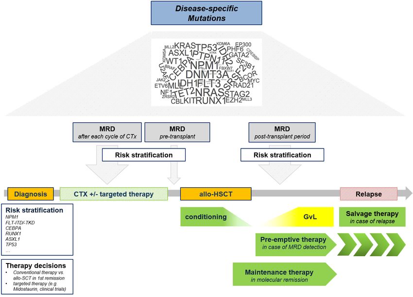
Figure1.1. Clinical
Figure Clinicaluse of molecular
use information
of molecular to prevent,
information detect, and
to prevent, treat relapse
detect, afterrelapse
and treat allogeneic
after
stem cellstem
allogeneic transplantation (allo-SCT).(allo-SCT).
cell transplantation MRD (minimal or measurable
MRD (minimal residual disease);
or measurable residual NPM1
disease);
(Nucleophosmin);
NPM1 FLT3-ITD
(Nucleophosmin); (FMS-like(FMS-like
FLT3-ITD tyrosine kinase 3-internal
tyrosine kinase tandem duplication);
3-internal tandem FLT3-TKD
duplication);
(FMS-like tyrosine kinase 3-tyrosine kinase domain); CEBPA (CCAT/enhaner-binding
FLT3-TKD (FMS-like tyrosine kinase 3-tyrosine kinase domain); CEBPA (CCAT/enhaner-binding protein alpha);
RUNX1 (Runt-related transcription factor 1); ASXL1 (additional sex comb-like 1);
protein alpha); RUNX1 (Runt-related transcription factor 1); ASXL1 (additional sex comb-like 1); TP53TP53 (Tumor
Protein
(Tumor 53); allo-SCT
Protein (allogeneic
53); allo-SCT stem cell
(allogeneic stemtransplantation); GvL (Graft-versus-Leukemia);
cell transplantation); GvL (Graft-versus-Leukemia);CTx
(Chemotherapy).
CTx (Chemotherapy).Int. J. Mol. Sci. 2019, 20, 228 3 of 20
2. Mutational Landscape and Pathophysiology of AML
Large, comprehensive genomic discovery studies and advances in stem cell biology have
tremendously improved our understanding of the pathogenesis and the molecular heterogeneity
of AML. Even though not completely understood, AML is believed to originate from a hematopoietic
stem or progenitor cell (HSPC) that acquires genomic and molecular alterations. As a consequence
of this, HPSC gains stem-cell like properties of unrestricted self-renewal thereby becoming
capable to maintain the malignant clone [25,26]. It seems to be the case that specific mutations
involved in epigenetic regulation, such as mutations in DNA-methyltransferase 3a (DNMT3A),
Tet methylcytosine dioxygenase 2 (TET2) and Additional sex comb-like 1 (ASXL1) genes, occur
early during leukemogenesis as founder events prior to leukemogenic alterations (such as NPM1 or
other mutations in signaling molecules) [27–29]. Such early mutations in pre-leukemic HSPC may
even be present in patients decades before leukemia evolves or can be found in approximately 10% of
healthy individuals older than 65 years without evidence for a hematological malignancy. The latter
state has been termed “clonal hematopoiesis of indeterminate potential” (CHIP) acknowledging the
aspect that those patients have, besides an additional association with atherosclerotic cardiovascular
disease, an elevated risk to develop a myeloid or other hematological malignancy. Notably, the risk to
develop overt hematological malignancy from CHIP state is estimated to be between 0.5 to 1.0% per
year and thereby comparable with other pre-malignant states such as progression from monoclonal
gammopathy of undetermined significance (MGUS) to multiple myeloma [30,31].
Overall, the number of mutations per AML genome seems to be lower than for most other cancers
with an average of 13 coding mutations (single nucleotide variants and insertion/deletions) per person.
These recurrent mutations occur in commonly deregulated pathways such as DNA methylation
associated genes, spliceosome-complex genes, cohesion-complex genes, chromatin-modifying genes,
signaling genes and 96% patients of the AML patients have at least one driver mutation in one
of these genes [9,29,32]. This genomic and combinatorial diversity represents the background that
drives the heterogeneity in phenotype and prognosis of patients with AML. Together with the results
from conventional cytogenetics the information about these mutations is the essential component to
diagnose AML according to the WHO 2016 classification and to estimate the prognosis according to
the ELN Genetic Risk stratification [32,33].
To further refine the classification and prognostication Papaemannuil and colleagues recently
performed a comprehensive sequencing analysis of 111 genes in 1540 patients. Hereby, they were able to
define in addition to 8 established AML subsets three new subtypes which included AML with spliceosome
mutations, AML with Tumor Protein 53 (TP53) mutations and AML with IDH2R172 mutations [32].
Besides a better disease classification, risk stratification and tailoring of therapies with regard to
the decision between consolidation with conventional chemotherapy and allo-SCT this knowledge
about the underlying molecular biology enables high-resolution tracking of the disease after transplant
by quantitative assessment of molecular markers and targeted therapeutic interventions.
3. Methods for Relapse Detection—Monitoring of Minimal Residual Disease (MRD)
While AML patients, who relapse after transplantation, generally have a dismal prognosis,
recent studies clearly showed that treatment is more effective if started at molecular relapse with
low disease burden rather than at hematological relapse [8,34,35]. Thus, successful therapeutic
intervention requires an early detection of imminent relapse ideally at a molecular level, which can
be accomplished by regular monitoring of MRD. The aim is to detect leukemic cells in a background
of normal hematopoiesis down to a level from 1:102 to 1:106 cells depending on the method applied.
The evaluation of MRD is validated to identify patients at risk for relapse who are in morphological
remission prior transplant [36]. In addition, it can also serve as a trigger for pre-emptive therapeutic
interventions after transplant in order to prevent overt hematological relapse. In the following part we
give an overview about methodical approaches for MRD detection consisting of molecular techniques,
chimerism analyses and multi-parameter flow cytometry (Table 1).Int. J. Mol. Sci. 2019, 20, 228 4 of 20
Table 1. Methods to detect Minimal Residual Disease (MRD) in Patients with AML after allo-SCT.
Molecular Genetics (Fusion
Multiparametric
Transcripts, Point Mutations, Chimerism
Flow Cytometry
Gene Overexpression)
qPCR // digital droplet PCR qPCR // Indel-PCR //
Methods/Approaches LAIP/DfN
(ddPCR) // NGS STR-based // XY-FISH
10−2 –10−3
Sensitivity 10−3 –10−4 10−3 –10−6
10−4 –10−5
applicable in all patients
Advantages broad applicability high sensitivity and specificity
after allo-SCT
low sensitivity and
mostly restricted to a small
specificity
part of patients
Disadvantages/ need for not directly detecting
need for standardization MRD
Perspectives standardization leukemic cells CD34+
monitoring based on
sorted chimerism offers
mutations not yet established
increased sensitivity
DfN (different from normal); ELN (European LeukemiaNet); FISH (fluoreszenz in-situ hybridization);
Indel (insertion and deletions); LAIP (Leukemia-associated immunephenotype); NGS (next generation sequencing);
pB (peripheral blood); qPCR (quantitative polymerase chain reaction); STR /short-tandem-repeats).
4. Molecular MRD Assessment
The genetic heterogeneity of AML as previously described provides several disease-specific
markers for MRD detection after allo-SCT. Molecular assessment of MRD can be performed by
monitoring (1) mutated genes, (2) fusion gene transcripts and (3) overexpressed genes. PCR-based
techniques to quantitatively measure these markers currently represent the standard of care with the
highest sensitivity (down to 1:106 cells) and specificity. This will be further improved in the near future
by the introduction of sensitive next-generation sequencing (NGS) assays and digital-droplet based
PCR as routine techniques for molecular MRD monitoring [15,37].
5. Detection of Gene Mutations for MRD Assessment
About 30% of patients with normal karyotype AML expose a mutation in NPM1 (Nucleophosmin 1)
gene, which is assumed to be relatively stable during disease course hereby fulfilling a major
requirement for a suitable MRD marker [11,38,39]. Several groups already demonstrated the
negative prognostic impact of increase in peripheral blood (PB)/bone marrow (BM) samples after
conventional chemotherapy on relapse incidence and overall survival in NPM1-mutated AML,
as recently reviewed [40–44]. Furthermore is was recently shown that MRD-positivity documented
either by qRT-PCR or by NGS also has a negative prognostic impact on relapse-free and overall survival
after allo-SCT [42,45–47]. Molecular testing for FLT3 mutations is part of the diagnostic setting at
the time of first diagnosis and offers important prognostic information [1,48]. However, it is not
routinely used to monitor MRD after allo-SCT due to its relative instability during disease course
and a lack of a broadly available quantitative assay [49–51]. Generally, almost all mutations in the
commonly mutated genes in AML may theoretically be measurable for MRD detection but have not
been integrated into routine care yet. Indeed, due to their relatively low frequency being only present
in small patient subgroups these markers lack a broad applicability and have therefore not been
studied in larger cohorts. So far, only one report addressed the value of monitoring known DNMT3A
and/or IDH1/2 mutations by digital droplet PCR in a small group of patients [49]. Representing an
additional limitation, some of the above-mentioned so-called pre-leukemogenic mutations such as
TET2, DNMT3A and ASXL1 may be retained at remission after chemotherapy and their prognostic
impact is uncertain [52–57]. Furthermore, the investigation of AML-specific mutations by NGS after
transplant may confront us with the phenomenon of donor-derived CHIP, a state with unknown
impact on the risk for the development of donor-cell leukemia [58].Int. J. Mol. Sci. 2019, 20, 228 5 of 20
6. Detection of Fusion Gene Transcripts for MRD Assessment
In about 20% of patients with AML, excluding acute promyelocytic leukemia (APL), distinct
fusion genes are approachable for MRD monitoring with most of them represented by core-binding
factor (CBF) AML (RUNX1-RUNX1T1 and CBFB-MYH11 fusion genes) [1,12,13,59]. Several reports
already demonstrated that MRD-positivity detected by quantitative PCR is predictive for imminent
relapse in patients with CBF leukemias after conventional therapy [60–63]. Due to the low frequency of
patients who receive allo-SCT in this good-risk AML category the evidence for MRD monitoring of CBF
fusion genes in the post-transplant period is restricted to a small number of reports. As an example,
Wang et al. reported a significantly higher cumulative incidence of relapse and shorter leukemia
free survival for pts with MRD persistence after allo-SCT depicted by RUNX1-RUN1T1-positivity
in PCR [64]. Besides CBF leukemias there are several other genetic rearrangements that are
also accessible for post-transplant MRD monitoring such as t(9;11)(p21.3;q23.3)/MLLT3-KMT2A,
t(6;9)(p22;q34)/DEK-NUP and inv(3)(q21q26)/RPN1-MECOM, but again their use is restricted to
a low number of individuals with these molecular alterations [12,13,65].
7. Quantification of Gene Expression for MRD Assessment
In contrast to the limited frequency of individual gene mutations and fusion-genes mentioned
above overexpression of Wilms Tumor 1 (WT1) mRNA is present in about 90% of patients with AML
and 50% of patients with MDS. Thus, monitoring of WT1 is broadly applicable in a large proportion
of AML and MDS patients [66,67]. Furthermore, WT1 expression is measurable in peripheral blood
with an even higher sensitivity and specificity than in bone marrow thereby facilitating high patient
comfort in contrast to other methods for molecular MRD monitoring that require BM biopsy to
gain a comparable sensitivity. As an additional advantage, WT1 expression can be performed using
a standardized, European LeukemiaNet (ELN) certified assay that offers a validated and reproducible
cut-off level and comparability of results among different laboratories [66]. Several studies including
one from our group recently demonstrated that longitudinal monitoring of PB WT1 expression offers
high sensitivity and specificity concerning detection of imminent relapse and appeared favorable
compared to other methods for MRD monitoring such as cytogenetics, NGS-based molecular testing
or chimerism analyses [68–70]. As a consequence, measurement of WT1 is valuable option for MRD
detection in patients with AML, at least in those cases where mutations or fusion genes are not
accessible for sensitive PCR-based approaches [15,37].
8. Chimerism Analyses for MRD Assessment
Donor/recipient chimerism analysis is the standard practice to monitor donor cell engraftment
and can be performed in all patients after allo-SCT. Analysis of chimerism also complementary
augments MRD measurement and relapse prediction after transplant, even though it reflects not a direct
proof of malignant cells by a leukemia-specific marker. Chimerism analysis detects host-derived
hematopoiesis on the basis of genomic differences at highly variable gene loci between the recipient
and the donor and this cannot directly be equated with relapse of the leukemic clone in all cases.
However, in malignant disorders such as AML decrease of donor chimerism is often associated with
disease recurrence [15,37]. Apart from this method-inherent limitation chimerism analysis has further
restrictions. The conventional and the most widely adopted method using fragment analysis of short
tandem repeats (STR) offers a sensitivity of 1 × 10⁻² to 1 × 10⁻³ only [71–73]. This also applies for
XY-FISH analysis in sex-mismatched donor/recipient constellations which provides a similar low
sensitivity of only about 1 × 10⁻² to 1 × 10⁻³ [74]. By employing variant-allele-specific quantitative
PCR-based approaches to detect small DNA insertions or deletion sensitivity can be increased to
a level with 1 × 10⁻⁴ to 1 × 10⁻⁵ cells [75,76]. Sensitivity and specificity of chimerism analysis can
also be improved in patients with AML and MDS by evaluating the CD34+ cell subset [72,77]. Overall,Int. J. Mol. Sci. 2019, 20, 228 6 of 20
chimerism analysis should be routinely performed after allo-SCT in conjunction with other more
sensitive methods in order to identify patients at risk for relapse and to guide preventive interventions.
9. MRD Assessment by Multiparameter Flow Cytometry (MFC)
MFC is a standard MRD method to directly identify residual leukemic cells and can be performed
in >90% with AML [37]. Two separate MFC approaches are capable to detect AML cells: the leukemia
associated immunophenotype (LAIP) method defines a disease-specific expression pattern at diagnosis
and facilitates subsequent tracking of this phenotype during follow-up period. If information about
the immunophenotype at diagnosis is not available or if the occurrence of new or the disappearance
of primary alterations are suspected, the different from normal (DfN) approach can be exerted [15].
These two approaches facilitate MRD assessment reaching a sensitivity of 10−3 to 10−4 [15]. To achieve
optimal results, sensitivity and specificity an international expert panel recently recommended to
use BM as primary material for examination, to use a minimum of 8 colors and to analyze the first
BM pull to avoid hemodilution [15]. Several mostly retrospective reports have demonstrated the
prognostic impact of MRD detected by MFC in patients with myeloid neoplasms after allo-SCT
showing a significantly higher relapse risk for the patients with MRD-positivity compared to those
without evidence for MRD by MFC [47,78–81]. Despite the main advantages of broad applicability and
high sensitivity there still remain relevant limitations of this method in terms of a lack of comparability
and reproducibility among different laboratories, the use of different instruments, fluorophores,
and operating procedures that require further standardization [15,37].
10. Prevention and Treatment of Relapse after Allo-SCT
Treatment of AML relapse after transplant is challenging due to a high rate of patients who either
cannot tolerate intensive therapies as a consequence of toxicity of the previous transplant or do not
achieve durable remissions by any treatment [3,6,8]. In general, any treatment for relapse of AML
after transplant aims to deliver direct antileukemic activity and/or to enhance the immunological
graft-versus-leukemia (GvL) effect. On the one hand, those therapeutic approaches consist of
cellular-based therapies such as donor lymphocyte infusions [4] and second transplant in individual
patients, which have been recently reviewed elsewhere [82,83]. On the other hand, pharmacological
treatments represent the second backbone of relapse therapy and are often administered before any
cellular therapy in order to reduce the leukemic burden. Traditionally, pharmacological approaches
have mainly consisted of low-dose or intensive chemotherapy. However, as exemplified by a large
registry-based analysis, the response to chemotherapy is limited (CR rate mild chemotherapy 17%,
intensive chemotherapy 27%) and not all patients relapsing after transplantation can tolerate another
round of intensive chemotherapy. Remissions after chemotherapy are generally not long-lasting if given
alone. Those patients achieving CR after chemotherapy substantially benefit from donor cell-based
consolidation. Indeed, 2-year OS was 55% in CR patients receiving donor lymphocyte infusion (DLI)
or second transplantation compared to 20% in those without donor-cell treatment [8]. During the last
years, hypomethylating agents, in particular Azacitidine (Aza) have proven to be a well-tolerated and
efficient treatment alternative in this setting [35,84,85]. Furthermore, the decipherment of the molecular
landscape in AML has facilitated the still ongoing development of targeted therapies. Some of them
have already been licensed like the FLT3 inhibitor Midostaurin (EMA- and FDA-approved) and
Ivosidenib and Enasidenib, inhibitors of IDH1 and IDH2, respectively [16–18], while others like the
BCL2 inhibitor Venetoclax are still under investigation [19–21,86] (Figure 2). Based on the time point of
intervention they can be divided into prophylactic approaches started when no evidence for leukemia
is present, preemptive approaches which are initiated at the time of MRD detection to avoid frank
hematological relapse and those approaches administered at the stage of hematological relapse.Int. J. Mol.
Int. J. Sci.
Mol.2019, 20, 228
Sci. 2018, 19, x FOR PEER REVIEW 7 of 20 7 of 20
Figure
Figure 2. Potentialtargets
2. Potential targets for
for prophylactic
prophylacticand therapeutic
and interventions
therapeutic after allogeneic
interventions stem cell stem
after allogeneic
transplantation (allo-SCT) in patients with acute myeloid leukemia (AML). mFLT3-ITD
cell transplantation (allo-SCT) in patients with acute myeloid leukemia (AML). mFLT3-ITD (mutant FMS-
like tyrosine kinase 3-internal tandem duplication); mFLT3-TKD (mutant FMS-like
(mutant FMS-like tyrosine kinase 3-internal tandem duplication); mFLT3-TKD (mutant FMS-like tyrosine kinase 3-
tyrosine kinase domain); FL (FLT3 ligand); bcl2 (b-cell lymphoma 2); IDH1 (isocitrate dehydrogenase
tyrosine kinase 3-tyrosine kinase domain); FL (FLT3 ligand); bcl2 (b-cell lymphoma 2); IDH1
1); IDH2 (isocitrate dehydrogenase 2); αKG (alpha ketoglutarate); mIDH1 (mutant isocitrate
(isocitrate dehydrogenase 1); IDH2 (isocitrate dehydrogenase 2); αKG (alpha ketoglutarate);
dehydrogenase 1); mIDH2 (mutant isocitrate dehydrogenase 2); 2HG (2-hydroxyglutarate)
mIDH1 (mutant isocitrate dehydrogenase 1); mIDH2 (mutant isocitrate dehydrogenase 2); 2HG
(2-hydroxyglutarate)
11. FLT3 Inhibitors
Mutations in the gene encoding for the FMS-like tyrosine kinase 3 (FLT3) are present in about
11. FLT3 Inhibitors
30% of adults with newly-diagnosed AML. Almost three quarters of these patients have so called
Mutations in theduplications
internal tandem gene encoding
(ITD),for the FMS-like
which are mostlytyrosine
located inkinase 3 (FLT3) are present
the juxtra-membrane domainin of
about
the 30%
of adults withand
receptor newly-diagnosed AML.
result in a duplication ofAlmost
betweenthree
3 andquarters
more thanof100 these
aminopatients have
acids. The so called25%
remaining internal
tandem duplications
of patients have a (ITD), which are
point mutation mostly
in the located
tyrosine kinaseindomain
the juxtra-membrane
(TKD) of the receptordomain of the
[32,87]. receptor
While
and result in a mutations,
FLT3-ITD duplicationinofparticular
betweenthose
3 andwithmore thanmutant-to-wildtype
a high 100 amino acids. The remaining
allelic 25%confer
ratio (≥0.51), of patients
an adverse prognosis, the prognostic impact of TKD mutations is uncertain
have a point mutation in the tyrosine kinase domain (TKD) of the receptor [32,87]. While FLT3-ITD[14,32,87]. Both mutations
lead toinaparticular
mutations, constitutive, ligand-independent
those activation of the allelic
with a high mutant-to-wildtype FLT3 ratio
receptor whichconfer
(≥0.51), mediates a
an adverse
continuous proliferative stimulus on leukemic cells. Giving this pivotal
prognosis, the prognostic impact of TKD mutations is uncertain [14,32,87]. Both mutations leadrole for the pathogenesis of
AML and the relatively high frequency of FLT3 mutations small-molecule FLT3 inhibitors have been
to a constitutive, ligand-independent activation of the FLT3 receptor which mediates a continuous
developed. The first-generation of these molecules such as Midostaurin, Sorafenib, and Lestaurtinib
proliferative stimulus on leukemic cells. Giving this pivotal role for the pathogenesis of AML and
were rather multi-kinase inhibitors than specific FLT3 inhibitors. This property to additionally inhibit
the relatively highkinases
several other frequency of FLT3the
may explain mutations small-molecule
limited efficacy FLT3 inhibitors
when administered as singlehave been
agent anddeveloped.
some
The first-generation
of the off-target effects of these substances, which may either be undesirable in terms of sidewere
of these molecules such as Midostaurin, Sorafenib, and Lestaurtinib effectsrather
multi-kinase
but also ofinhibitors
interest withthan specific
regard FLT3 efficacy
to potential inhibitors. This
in FLT3 property
wildtype AML to[88,89].
additionally inhibit
In a large, placebo-several
othercontrolled
kinases may explainphase-III
randomized the limited efficacy when
trial Midostaurin wasadministered
the first agent toasshow
single agentwith
benefit andregard
sometoof the
overall
off-target survival
effects which
of these resulted inwhich
substances, the approval
may eitherof Midostaurin
be undesirable for first-line
in termstreatment (induction,
of side effects but also of
consolidation
interest with regard andtomaintenance therapy)
potential efficacy inof patients
FLT3 with FLT3-mutated
wildtype AML [88,89].AML. In combination
In a large, with
placebo-controlled
conventional chemotherapy Midostaurin led to a 22% risk reduction for death
randomized phase-III trial Midostaurin was the first agent to show benefit with regard to overall and in particular those
patients transplanted in first complete remission (CR1) seemed to benefit. Unfortunately, the role of
survival which resulted in the approval of Midostaurin for first-line treatment (induction, consolidation
Midostaurin as maintenance therapy after allo-SCT was not addressed in this trial and therefore
and maintenance therapy) of patients with FLT3-mutated AML. In combination with conventional
chemotherapy Midostaurin led to a 22% risk reduction for death and in particular those patients
transplanted in first complete remission (CR1) seemed to benefit. Unfortunately, the role of Midostaurin
as maintenance therapy after allo-SCT was not addressed in this trial and therefore Midostaurin is onlyInt. J. Mol. Sci. 2019, 20, 228 8 of 20
licensed as maintenance therapy after conventional chemotherapy [18]. Meanwhile, second generation
molecules such as Gilteritinib and Quizartinib, which inhibit FLT3 more specifically, have been
developed and appear to be more effective even when used as single agents [88,90]. Updated results
from the latter randomized phase-III trial in relapsed/refractory AML patients with FLT3-ITD mutation
have recently been updated and show prolonged overall survival 0.76 (95% CI, 0.58–0.98; p = 0.0177)
and a higher composite complete remission (CR) rate (48% vs. 27%) for Quizartinib in comparison to
salvage chemotherapy. Both of these trials included patients who were treated for relapsed AML after
allo-SCT but did not report the details of these subgroups separately. Moreover, gilterinib is currently
tested as maintenance therapy after allo-SCT in an ongoing, randomized, placebo-controlled phase III
trial, where patients are envisaged to receive maintenance therapy with 120 mg Gilteritinib per day
for 24 months (EudraCT #2016-001061-83). Patients are planned to start maintenance therapy early
after allo-SCT between day 30 and day 90, but as a particular strength they are already registered
prior transplant. The latter enables estimation of drop-out rate prior to the start of maintenance
therapy and thereby the interpretation how many patients will finally be able to start and tolerate
maintenance with an FLT3 inhibitor after allo-SCT. Furthermore, Crenolanib is another highly selective
and potent FLT3 tyrosine kinase inhibitor (TKI) with activity against ITD mutants and FLT3/D835 point
mutants [91]. Crenolanib is currently under investigation as salvage therapy in relapsed/refractory
patients [92]. Still, a randomized phase-III trial comparing Crenolanib with standard salvage therapies
(NCT02298166) has been stopped this year due to strategical considerations of the company. Despite
this, investigation of Crenolanib as maintenance therapy after allo-SCT (NCT02400255), but also in
addition to first-line chemotherapy in a head-to-head comparison to midostaurin (NCT03258931) is
ongoing. Another interesting option regarding the use of crenolanib in AML patients may be the
combination with CAR T-cells, since preclinical analyses revealed a synergistical effect of FLT3 targeting
CAR T-cells and crenolanib [93]. A synergistical effect of crenolanib has also been demonstrated for
the combination with azacitidine. In this context, azacitidine renders FLT3+ AML cells more sensitive
to crenolanib by effectively abrogating stromal protection [94]. As salvage therapy for relapsed
FLT3-mutated AML most evidence comes from the use of Sorafenib. The knowledge about Sorafenib
is again based on retrospective case series and reports, but not controlled prospective trials [95–97].
The use of Sorafenib in FLT3-mutated AML after allo-SCT was performed as off-label use on an
individual basis facilitated by the availability of Sorafenib as licensed drug for the treatment of renal
and liver cancer. Nevertheless, in these reports monotherapy with Sorafenib seems to induce durable
remissions and long-term survival in a subgroup of patients and the anti-leukemic effect of Sorafenib
seems to synergize with allo-immune effects. As a mechanistic explanation, Mathew et al. recently
discovered that Sorafenib increases IL-15 production by FLT3-ITD+ leukemia cells in mice and human
thereby inducing leukemia-reactive T cells [98]. To take advantage of these properties Sorafenib has
also been tested as maintenance therapy after allo-SCT in a randomized phase II trial (EUDRACT
2010-018539-16). Data from this well-designed, randomized trial have recently presented at the
annual meeting of the American Society of Hematology and suggest a relevant benefit of Sorafenib
maintenance therapy in terms of overall and relapse-free survival [99]. Even though not licensed
in this indication, this may have practical implications when considering maintenance therapy in
FLT3-mutated patients.
12. Isocitrate Dehydrogenase (IDH) Inhibitors
Another interesting molecular target also in the context of allo-SCT are mutations of the cytosolic
IDH1 (R132) and the mitochondrial IDH2 (R140 and R172) enzyme which can each be found in about
5–15% of patients with AML. Usually, these enzymes catalyze the transformation from isocitrate to
α-ketoglutarate. Mutations in these enzymes found in patients with AML result in a neomorphic
enzyme activity which now produce 2-hydroxyglutarate. This oncometabolite impairs cellular
differentiation in hematopoietic stem and progenitor cells and thereby substantially contributes to the
pathogenesis of AML [16,100,101]. Taking this into account IDH inhibitors have been developed inInt. J. Mol. Sci. 2019, 20, 228 9 of 20
order to target these mutations selectively. The orally available inhibitors (IDH1, ivosidenib; IDH2,
enasidenib) have initially been tested in patients with relapsed/refractory patients with AML and
demonstrated encouraging rates of complete remissions (CR, CR + CR incomplete: ivosidenib 30.4%;
enasidenib 26.1%) [16,17]. Based upon these results, both drugs were approved by the FDA for the
treatment of relapsed/refractory AML and are now tested in the front-line setting either in combination
with intensive chemotherapy or with hypomethylating agents. Their unique mode of action conferring
antileukemic activity by the promotion of differentiation instead of a primary cytotoxic effect resulted
in a limited and class-specific toxicity profile [16,17,101,102].
Both, the acceptable toxicity and the mode of action imply that these substances may also be an
interesting option for patients with IDH-mutated AML who have relapsed after allo-SCT. In detail,
by inducing cellular differentiation the IDH inhibitors may promote an allo-immunologic reaction by
antigen upregulation on leukemic cells thereby enhancing their immunogenicity. Although both trials
in patients with relapsed/refractory AML also included a limited number of patients with relapse after
allo-SCT, their specific outcome has not been reported in detail so far.
To explore the role of IDH inhibition either as maintenance and or as salvage therapy also after
allo-SCT more in detail, there are now several prospective studies including one from our group on
the way.
13. Hypomethylating Agents and HDAC Inhibitors
Unfortunately, apart from those with IDH and FLT3 mutations many AML patients have no
molecular alteration that can currently be therapeutically addressed by a targeted approach prior to
transplant as well as to prevent or treat relapse after transplant. In addition, even if present before
transplant, a specific molecular target, such as FLT3 mutations, may have been lost after transplant
as a process of clonal evolution [103,104]. Conversely, new molecular lesions may occur at relapse,
leading to the need to perform extensive molecular examinations in cases of AML recurrence. Thus,
a preventive or therapeutic approach after transplant may rather be a more general option for a relevant
proportion of patients if it is effective independent from any molecular structure or biological features.
The hypomethylating agents Azacitidine (Aza) and Decitabine (DAC) seem to fulfill many of
these criteria and have intensively been investigated during the last years. They mediate a direct
anti-leukemic effect in patients with AML and MDS independent from a distinct molecular phenotype,
seem to beneficially influence the balance between GvL effect and GvHD and are not associated with
an excess of toxicity [90,105–108]. In detail, by upregulating epigenetically silenced leukemia antigens
such as cancer-testis antigens [109] and the re-expression of endogenous retroviral elements and
dsRNA causing an antiviral interferon response they increase the immunological visibility of AML and
MDS cells [110]. Furthemore, HMA seem to favorably modulate the activity of T- and NK cells towards
an enhanced GvL effect, which is not counterbalanced by an increased risk of GvHD [111–113].
To take advantage of these properties the HMA Aza (n = 4) or DAC (n = 2) have been tested
as maintenance therapy to avoid relapse after allo-SCT in 5 prospective single-arm studies and one
retrospective case series covering a total of 148 patients [111,114–118]. Those early-phase trial generally
demonstrated feasibility and identified the optimal dosage and schedule for future trials, which was
lower than the one recommended for the primary indication was generally administered in these
trials. Indeed, Aza (32 mg/m2 per day, day 1 to day 5) is currently tested for relapse prevention after
allo-SCT in an ongoing, randomized placebo-controlled trial in patients with high-risk AML or MDS
(NCT00887068). Results from this trial are awaited next year.
Another interesting option in this context is the use of an oral formulation of Aza (CC-486).
This compound is currently tested in a large company-driven randomized, placebo-controlled trial as
maintenance therapy in elderly AML after induction chemotherapy (NCT01757535) and has also been
recently tested as maintenance therapy after allo-SCT in a single-arm phase I/II dose finding study
including 31 patients with AML or MDS [119]. Advantages of this formulation may be a better patientInt. J. Mol. Sci. 2019, 20, 228 10 of 20
compliance and convenience, due to outpatient administration as well as its pharmacokinetic and
pharmacodynamic properties facilitating prolonged exposure and sustained DNA hypomethylation.
Aza and DAC have also been tested in several retrospective analyses and in 4 small-sized
prospective studies as salvage therapy either alone or in combination with DLI in patients with relapse
of AML or MDS after allo-SCT. As recently reviewed elsewhere [85,120,121], results from these analyses
clearly demonstrated that this well-tolerated combination of a HMA and donor cells can induce durable
remissions (CR rates ranging from 10% to 75%) and long-term survival in a relevant proportion of
patients (2-year survival rates ranging from 12% to 80%). In particular patients with a diagnosis of MDS
and those treated at the stage of molecular relapse instead of hematological relapse seems to benefit
from this combination highlighting the need for stringent MRD monitoring [35,122]. This concept of
pre-emptive therapy at the time of molecular relapse after allo-SCT has been tested by Platzbecker and
colleagues in two prospective trials. In an initial proof-of concept study (RELAZA-1) 20 patients with
MDS or AML were treated with up to 4 cycles Aza as soon as the CD34+ donor chimerism dropped
in peripheral blood below a threshold of 80%, while patients were still in hematological remission.
Despite an improvement of chimerism (>80%) in half of the patients, this early intervention was able
to induce durable remissions only in 3 (30%) of the responders and did not avoid progression towards
hematological relapse in the majority of patients [122]. They expanded this analysis in a second trial
(RELAZA-2) covering 53 patients (24 after allo-SCT, 29 after conventional chemotherapy), who were
monitored by CD34+ donor chimerism or molecular markers such as NPM1 and RUNX1-RUNX1T1.
In case of MRD positivity patients could receive up to 24 cycles Aza. The study met its primary
endpoint with 31 patients (58%) free of relapse after 6 months and 19 of 53 patients achieved a major
response (36%). However, similar to their first trial treatment with Aza, even though with an expanded
number of cycles, could only delay relapse in many patients and 26 patients (49%) finally relapsed [123].
In those patients after allo-SCT this is probably related to the fact that DLI were not part of the
protocols of both trials. Although especially the combination of Aza and DLI has been established
as valuable treatment alternative besides chemotherapy and second transplantation, the responses
rates in particular in patients treated at the stage of hematological relapse indicate there is still space
for improvement [35]. Here, the combination of Aza with a targeted approach may be an option to
improve outcome in patients with relapse after allo-SCT. In this context, DiNardo and colleagues
recently reported encouraging response and survival data in treatment-naïve elderly AML patients
treated with HMA in combination with Venetoclax, a potent, selective oral inhibitor of the antiapoptotic
b-cell lymphoma 2 (BCL-2) protein. Response rates and overall survival seemed to be higher than
observed after monotherapy with HMA making this combination an attractive option also for patients,
including subjects post allo-SCT [19]. This also applies to the combination of HMA and FLT3 inhibitors
such as Sorafenib, which has also been demonstrated to induce long-term remissions in individual
patients [124–126].
Besides HMA also the HDAC inhibitor Panobinostat has demonstrated feasibility as maintenance
therapy after allo-SCT in 42 patients with AML or high-risk MDS in an open-label, multi-center phase
I/II trial. Consequently, Panobinostat is currently investigated in a large randomized trial.
14. Cellular Therapies
14.1. Donor Lymphocyte Infusions
Besides pharmacological anti-leukemic approaches (e.g., chemotherapy and targeted therapies)
cellular interventions represent the second backbone to prevent and to treat relapse of AML after
allo-SCT. Donor lymphocyte infusions (DLI) are a cellular product of mononuclear cells containing
a defined number of donor-derived CD3+ T cells. DLI can be obtained either as aliquots from the initial
G-CSF-mobilized PB stem cell product or by an unstimulated leukapheresis of the original donor [127].
Following an initial description in 3 patients with chronic myeloid leukemia [128] the application of DLI
has also been extensively employed in patients with AML [127]. Likewise in CML the aim is to enhanceInt. J. Mol. Sci. 2019, 20, 228 11 of 20
the GvL effect in order to either prevent relapse in case of remission after transplant (e.g., prophylactic
DLI) or in case of disease recurrence to treat relapse (e.g., therapeutic DLI). Several retrospective
analyses and a limited number of prospective studies have reported on the use of DLI as prophylactic
approach. In T-cell depleted (TCD) setting the beneficial effect of prophylactic DLI on progression-free
and overall survival is well established [129–131]. In the non-TCD setting prophylactic DLI also
seemed to improve the outcome of high-risk AML patients after sequential conditioning using the
FLAMSA-RIC regimen when compared to historical controls [132,133]. Prophylactic DLI were given
to 46 patients with high-risk AML starting from day +120 after allo-SCT if patients were in still
in remission, off immunosuppression for 30 days and free of GvHD. After a median follow-up of
7.2 years OS was 67% at 7 years in those patients receiving prophylactic DLI compared to 31% in
the control group [133]. These data have been supported by a recent matched-pair analysis from the
EBMT registry [134]. Nevertheless, the retrospective character of most analyses and the relatively late
timepoint of DLI application suggest a relevant selection bias. To overcome the potentially higher
GvHD risk associated with earlier application Wang et al. administered G-CSF mobilized DLI by day
+40–60 followed by short-term GvHD prophylaxis [135]. The lack of randomized prospective trials and
the heterogeneity of the retrospective data in different settings do not facilitate a clear recommendation
on indication, timing, and dosage of prophylactic DLI. Data regarding therapeutic DLI are also mainly
based on retrospective analyses but also suggest a limited efficacy, as indicated by a 2-year OS rate of
21% in relapsed patients receiving DLI compared 9% in those not receiving DLI [7,136]. The success of
therapeutic DLI strongly correlated with disease burden at relapse and remission state at the time of
DLI application [7,136]. This implies the need for regular MRD monitoring for early relapse detection
but also highlights the need for efficient salvage therapies to induce remission prior to DLI.
14.2. Second Transplantation
Apart from DLI, another form of cellular therapy is a second allogeneic transplantation after
anew conditioning followed by immunosuppression. In a large retrospective analysis of 179 patients
with acute leukemia (AML n = 132, acute lymphoblastic leukemia n = 46) the 2-year OS rate after
second allo-SCT was 25% in the entire cohort [5]. Second transplantation in CR, an interval of >6
months between first allo-SCT and relapse and a matched related donor at first transplantation were
associated with a better outcome in multivariate analysis. Assuming that the GvL effect mediated
by the first donor was insufficient to control leukemia a donor change at second transplant is worth
considering, although in the analysis by Christopeit et al. no significant impact of donor change
could be demonstrated in multivariate analysis. Given the advances in the field of haploidentical
transplantation a second allo-HSCT using a haploidentical donor may also be an option that is currently
employed in individual patients [137]. However, in this context a comprehensive analysis of the HLA
constellation of the recipient and donors is required to guide donor selection, as loss of HLA may
have driven relapse after allo-SCT 1 [138–140]. Finally, it needs to be taken into account that patients
included in these retrospective analyses represent a positively selected cohort that survived until
intervention. Furthermore, as exemplified by a median age of 38.5 years at allo-SCT2 in the report of
Christopeit et al. [5], patients receiving a second transplant are mostly younger than the majority of
AML patients relapsing after allo-SCT1 and thereby fit enough to tolerate such an intensive therapy.
Based on this, the decision on a second transplantation can only be made individually by carefully
weighting the risks and chances of this procedure.
14.3. New Cellular Therapies
Alloreactivity of DLI and second transplantation is mainly mediated by recognition of disparities
in minor histocompatibility antigens in the matched donor setting and of HLA disparities in case of
mismatched donors. Current investigations aim to improve the efficacy and toxicity profile of cellular
therapies by selection of specific immune cells (e.g., NK cells, γ/δ T cells etc.) or by manipulation of
the cellular product (e.g., external cytokine stimulation or genetic modification) [127]. In this context,Int. J. Mol. Sci. 2019, 20, 228 12 of 20
chimeric-antigen-receptor (CAR)-modified T cells are also under investigation for patients with AML
and initial data seem promising [93]. However, in contrast to patients with B cell malignancies the
transfer of CAR T cells from bench to bedside in patients with AML is slower. This is mainly related
to the difficulty to identify AML-specific target epitopes, which are expressed as few as possible on
normal tissues, but homogenous on AML cells of a larger proportion of patients and exert an essential
function for AML biology. Among others, current targets of interests consist of CD123, CD33, folate
receptor β, FLT3, CLL-1 and CD44v6 [141].
15. Conclusions
During the last decade, our understanding of the molecular heterogeneity and pathophysiology of
AML has been greatly advanced by the use of high-throughput sequencing techniques. The discovery
of several recurrently mutated genes and chromosomal aberrations has markedly improved the
classification and risk stratification. Furthermore, first steps towards a more targeted therapy for
patients with AML have been made by the introduction of Midostaurin and several other compounds
will probably follow in the near future. This knowledge about molecular alterations and the
opportunity to monitor MRD has, similar to the pre-transplant phase, already been adopted into
routine care after transplant. While randomized studies investigating maintenance strategies with
HMA or targeted approaches such as FLT3 inhibitors are currently ongoing, we are already using such
compounds to treat patients with relapse after transplant. Until those studies will have demonstrated
a benefit of maintenance therapy, we suggest to employ the molecular information in individuals
patients for stringent MRD monitoring and to perform preemptive therapeutic interventions guided
by disease phenotype and genetics.
Author Contributions: C.R. and T.S. wrote the manuscript. U.G., R.H. and G.K. read and critically revised the
manuscript. C.R. and T.S. prepared the illustrations.
Funding: This research received no external funding.
Conflicts of Interest: T.S. had a consulting role for Celgene Corporation, Germany and received financial travel
support and lecture fees from Celgene Corporation, Germany. T.S. also received financial travel support and lecture
fees from Janssen-Cilag GmbH Germany, Novartis GmbH Germany, Pfizer GmbH Germany and Teva GmbH
Germany. C.R. received financial travel support from Celgene Corporation, Germany and Jazz Pharmaceutical
GmbH Germany. R.H. and U.G. have nothing to declare. G.K. received financial travel support, research funding
and lecture fees from Celgene Corporation, Germany.
References
1. Döhner, H.; Estey, E.; Grimwade, D.; Amadori, S.; Appelbaum, F.R.; Büchner, T.; Dombret, H.; Ebert, B.L.;
Fenaux, P.; Larson, R.A.; et al. Diagnosis and management of AML in adults: 2017 ELN recommendations
from an international expert panel. Blood 2017, 129, 424–447. [CrossRef] [PubMed]
2. Gooley, T.A.; Chien, J.W.; Pergam, S.A.; Hingorani, S.; Sorror, M.L.; Boeckh, M. Reduced mortality after
allogeneic hematopoietic cell transplantation. N. Engl. J. Med. 2010, 363, 2091–2101. [CrossRef] [PubMed]
3. De Lima, M.; Porter, D.L.; Battiwalla, M.; Bishop, M.R.; Giralt, S.A.; Hardy, N.M. Proceedings from the
National Cancer Institute’s Second International Workshop on the Biology, Prevention, and Treatment of
Relapse after Hematopoietic Stem Cell Transplantation: Part III. Prevention and Treatment of Relapse after
Allogeneic Transplantation. Biol. Blood Marrow Transplant. 2014, 20, 4–13. [PubMed]
4. Bhagat, T.D.; Chen, S.; Bartenstein, M.; Barlowe, A.T.; Von Ahrens, D.; Choudhary, G.S.
Epigenetically Aberrant Stroma In MDS Propagates Disease Via Wnt/β-Catenin Activation. Cancer Res.
2017, 77, 4846–4857. [CrossRef] [PubMed]
5. Christopeit, M.; Kuss, O.; Finke, J.; Bacher, U.; Beelen, D.W.; Bornhäuser, M. Second Allograft for Hematologic
Relapse of Acute Leukemia After First Allogeneic Stem-Cell Transplantation From Related and Unrelated
Donors: The Role of Donor Change. J. Clin. Oncol. 2013, 31, 3259–3271. [CrossRef] [PubMed]
6. Schmid, C.; de Wreede, L.C.; van Biezen, A.; Finke, J.; Ehninger, G.; Ganser, A. Outcome after relapse
of myelodysplastic syndrome and secondary acute myeloid leukemia following allogeneic stem cellInt. J. Mol. Sci. 2019, 20, 228 13 of 20
transplantation: A retrospective registry analysis on 698 patients by the Chronic Malignancies Working Party
of the European Society of Blood and Marrow Transplantation. Haematologica 2018, 103, 237–245. [PubMed]
7. Schmid, C.; Labopin, M.; Nagler, A.; Bornhäuser, M.; Finke, J.; Fassas, A. Donor lymphocyte infusion in
the treatment of first hematological relapse after allogeneic stem-cell transplantation in adults with acute
myeloid leukemia: A retrospective risk factors analysis and comparison with other strategies by the EBMT
Acute Leukemia Working Party. J. Clin. Oncol. 2007, 25, 4938–4945. [PubMed]
8. Schmid, C.; Labopin, M.; Nagler, A.; Niederwieser, D.; Castagna, L.; Tabrizi, R. Treatment, risk factors,
and outcome of adults with relapsed AML after reduced intensity conditioning for allogeneic stem cell
transplantation. Blood 2012, 119, 1599–1606. [CrossRef]
9. Bullinger, L.; Döhner, K.; Döhner, H. Genomics of Acute Myeloid Leukemia Diagnosis and Pathways.
J. Clin. Oncol. 2017, 35, 934–946. [CrossRef]
10. Koreth, J.; Schlenk, R.; Kopecky, K.J.; Honda, S.; Sierra, J.; Djulbegovic, B.J. Allogeneic stem cell
transplantation for acute myeloid leukemia in first complete remission: Systematic review and meta-analysis
of prospective clinical trials. JAMA 2009, 301, 2349–2361. [CrossRef]
11. Schlenk, R.F.; Döhner, K.; Krauter, J.; Fröhling, S.; Corbacioglu, A.; Bullinger, L. Mutations and treatment
outcome in cytogenetically normal acute myeloid leukemia. N. Engl. J. Med. 2008, 358, 1909–1918. [CrossRef]
[PubMed]
12. Grimwade, D.; Freeman, S.D. Defining minimal residual disease in acute myeloid leukemia: Which platforms
are ready for “prime time”? Blood 2014, 124, 3345–3355. [CrossRef]
13. Grimwade, D.; Ivey, A.; Huntly, B.J.P. Molecular landscape of acute myeloid leukemia in younger adults and
its clinical relevance. Blood 2016, 127, 29–41. [CrossRef] [PubMed]
14. Kayser, S.; Schlenk, R.F.; Grimwade, D.; Yosuico, V.E.D.; Walter, R.B. Minimal residual disease–directed
therapy in acute myeloid leukemia. Blood 2015, 125, 2331–2335. [CrossRef] [PubMed]
15. Schuurhuis, G.J.; Heuser, M.; Freeman, S.; Béné, M.-C.; Buccisano, F.; Cloos, J. Minimal/measurable residual
disease in AML: A consensus document from the European LeukemiaNet MRD Working Party. Blood 2018,
131, 1275–1291. [CrossRef]
16. DiNardo, C.D.; Stein, E.M.; de Botton, S.; Roboz, G.J.; Altman, J.K.; Mims, A.S. Durable Remissions with
Ivosidenib in IDH1-Mutated Relapsed or Refractory AML. N. Engl. J. Med. 2018, 378, 2386–2398. [CrossRef]
17. Stein, E.M.; DiNardo, C.D.; Pollyea, D.A.; Fathi, A.T.; Roboz, G.J.; Altman, J.K. Enasidenib in mutant IDH2
relapsed or refractory acute myeloid leukemia. Blood 2017, 130, 722–731. [CrossRef]
18. Stone, R.M.; Mandrekar, S.J.; Sanford, B.L.; Laumann, K.; Geyer, S.; Bloomfield, C.D. Midostaurin plus
Chemotherapy for Acute Myeloid Leukemia with a FLT3 Mutation. N. Engl. J. Med. 2017, 377, 454–464.
[CrossRef]
19. DiNardo, C.D.; Pratz, K.; Pullarkat, V.; Jonas, B.A.; Arellano, M.; Becker, P.S.; Frankfurt, O.; Konopleva, M.;
Wei, A.H.; Kantarjian, H.M.; et al. Venetoclax combined with decitabine or azacitidine in treatment-naive,
elderly patients with acute myeloid leukemia. Blood 2019, 33, 7–17. [CrossRef]
20. DiNardo, C.D.; Rausch, C.R.; Benton, C.; Kadia, T.; Jain, N.; Pemmaraju, N. Clinical experience with the
BCL2-inhibitor venetoclax in combination therapy for relapsed and refractory acute myeloid leukemia and
related myeloid malignancies. Am. J. Hematol. 2018, 93, 401–407. [CrossRef]
21. Konopleva, M.; Pollyea, D.A.; Potluri, J.; Chyla, B.; Hogdal, L.; Busman, T.; McKeegan, E.; Salem, A.H.;
Zhu, M.; Ricker, J.L.; et al. Efficacy and Biological Correlates of Response in a Phase II Study of Venetoclax
Monotherapy in Patients with Acute Myelogenous Leukemia. Cancer Discov. 2016, 6, 1106–1117. [CrossRef]
22. Hills, R.K.; Castaigne, S.; Appelbaum, F.R.; Delaunay, J.; Petersdorf, S.; Othus, M. Addition of gemtuzumab
ozogamicin to induction chemotherapy in adult patients with acute myeloid leukaemia: A meta-analysis of
individual patient data from randomised controlled trials. Lancet Oncol. 2014, 15, 986–996. [CrossRef]
23. Lambert, J.; Pautas, C.; Terré, C.; Raffoux, E.; Turlure, P.; Caillot, D.; Legrand, O.; Thomas, X.; Gardin, C.;
Gogat-Marchant, K.; et al. Gemtuzumab ozogamicin for de novo acute myeloid leukemia: Final efficacy and
safety updates from the open-label, phase 3 ALFA-0701 trial. Haematologica 2019, 104, 113–119. [CrossRef]
24. Lancet, J.E.; Uy, G.L.; Cortes, J.E.; Newell, L.F.; Lin, T.L.; Ritchie, E.K. CPX-351 (cytarabine and daunorubicin)
Liposome for Injection Versus Conventional Cytarabine Plus Daunorubicin in Older Patients With Newly
Diagnosed Secondary Acute Myeloid Leukemia. J. Clin. Oncol. 2018, 36, 2684–2692. [CrossRef] [PubMed]
25. Dick, J.E.; Lapidot, T. Biology of normal and acute myeloid leukemia stem cells. Int. J. Hematol. 2005, 82,
389–396. [CrossRef] [PubMed]You can also read