Heat Shock Protein 70 (HSP70) Induction: Chaperonotherapy for Neuroprotection after Brain Injury - MDPI
←
→
Page content transcription
If your browser does not render page correctly, please read the page content below
cells
Review
Heat Shock Protein 70 (HSP70) Induction:
Chaperonotherapy for Neuroprotection after
Brain Injury
Jong Youl Kim 1 , Sumit Barua 1 , Mei Ying Huang 1,2 , Joohyun Park 1,2 , Midori A. Yenari 3, * and
Jong Eun Lee 1,2, *
 1   Department of Anatomy, Yonsei University College of Medicine, Seoul 03722, Korea;
     jongyoulkim@yuhs.ac (J.Y.K.); sumit@yuhs.ac (S.B.); mayhuang@yuhs.ac (M.Y.H.); jhpark922@yuhs.ac (J.P.)
 2   BK21 Plus Project for Medical Science and Brain Research Institute, Yonsei University College of Medicine,
     50-1 Yonsei-ro, Seodaemun-gu, Seoul 03722, Korea
 3   Department of Neurology, University of California, San Francisco & the San Francisco Veterans Affairs
     Medical Center, Neurology (127) VAMC 4150 Clement St., San Francisco, CA 94121, USA
 *   Correspondence: yenari@alum.mit.edu (M.A.Y.); jelee@yuhs.ac (J.E.L.); Tel.: +1-415-750-2011 (M.A.Y.);
     +82-2-2228-1646 (ext. 1659) (J.E.L.); Fax: +1-415-750-2273 (M.A.Y.); +82-2-365-0700 (J.E.L.)

 Received: 17 July 2020; Accepted: 26 August 2020; Published: 2 September 2020

 Abstract: The 70 kDa heat shock protein (HSP70) is a stress-inducible protein that has been shown
 to protect the brain from various nervous system injuries. It allows cells to withstand potentially
 lethal insults through its chaperone functions. Its chaperone properties can assist in protein folding
 and prevent protein aggregation following several of these insults. Although its neuroprotective
 properties have been largely attributed to its chaperone functions, HSP70 may interact directly with
 proteins involved in cell death and inflammatory pathways following injury. Through the use of
 mutant animal models, gene transfer, or heat stress, a number of studies have now reported positive
 outcomes of HSP70 induction. However, these approaches are not practical for clinical translation.
 Thus, pharmaceutical compounds that can induce HSP70, mostly by inhibiting HSP90, have been
 investigated as potential therapies to mitigate neurological disease and lead to neuroprotection.
 This review summarizes the neuroprotective mechanisms of HSP70 and discusses potential ways in
 which this endogenous therapeutic molecule could be practically induced by pharmacological means
 to ultimately improve neurological outcomes in acute neurological disease.

 Keywords: heat shock protein 70; brain injury; chaperone neuroprotection; pharmacological induction

1. Introduction
     After various insults to the brain, a coordinated stress response which seems to protect it from
further injury occurs. Heat shock proteins (HSPs) are the most exhaustively studied stress proteins.
They were originally noted when sublethal heat stress was applied to cells. Postmortem studies have
also documented induction of HSPs in the human brain following different types of thermal stress, such
as hyperthermia or fire-related fatalities [1]. When core body temperature exceeded 40 ◦ C, increased
transcripts of HSPs were detected in postmortem brain specimens. The study of autopsied brain
specimens of patients who had suffered from hyperthermia also led to the conclusion that HSP70
induction could be a brain biomarker of death [2]. HSPs are chaperones that typically act within
the cytosolic space, engaged in assisting with protein folding, degradation, complex assembly, and
translocation. They have demonstrated the ability to inhibit the accumulation of damaged proteins as
well as to facilitate the construction of polypeptides of newly synthesized proteins. The diverse roles
by which HSP70 and HSP90 regulate aggregated proteins appear to be involved in neuroprotection as

Cells 2020, 9, 2020; doi:10.3390/cells9092020                                        www.mdpi.com/journal/cells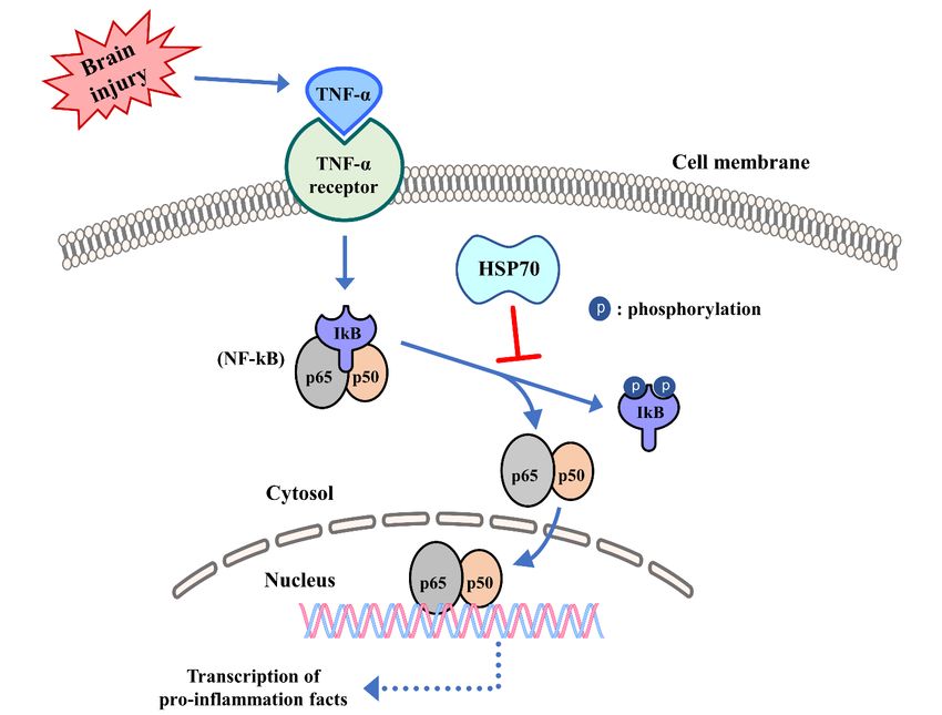
Cells 2020, 9, 2020                                                                                  2 of 17

demonstrated by models of brain injury. HSP70 induction also represents an endogenous protective
mechanism that occurs in the penumbra of the hippocampus, but not of other core areas, in the
ischemic stroke model [3,4]. More than two decades of research involving such models have shown
that HSP70 has the ability to protect against multiple types of cell death, including apoptosis and
necrosis. Specifically, HSP70 interferes with multiple cell death pathways [5,6]. HSP70 also modulates
inflammatory pathways and, thus, appears to improve neurological outcomes through interrupting both
cell death and immune responses [7]. It should be noted, however, that these studies possessed limited
translational utility because they relied upon either genetic mutant models or gene transfer models, and
upon heat stress to induce HSP70 overexpression. In the HSP70 research trajectory, multiple disciplines
have studied geldanamycin (GA) and 17-allyamino-demethoxygeldamycin (17-AAG), which block
HSP90 leading to the induction of HSP70 [8,9]. The possible clinical applications of HSP70-inducing
pharmacological compounds in neuroprotective therapies for ischemic stroke and associated conditions
warrant further research [8]. Here, we discuss the mechanisms of HSP70 neuroprotection in brain
injury (ischemic stroke and traumatic brain injury (TBI)), along with pharmacological HSP70 inducers
and their possible applications at the clinical level.

2. Classification and Functional Role of Heat Shock Protein 70
     At the onset of brain injury (for instance, of ischemic stroke or TBI), the synthesis of most cellular
proteins is downregulated. However, HSPs belong to a small class of proteins that are, instead,
upregulated, and have been collectively referred to as stress proteins. HSPs are classified in accordance
to their molecular mass. Constitutive HSPs, such as HSP90, HSP40, and HSP70, have housekeeping
functions within the cell [10]. HSP70 and HSP90 are two highly conserved ATP-dependent HSPs that
modulate unfolded proteins.
     HSP90 is an ATP-dependent chaperone associated with protein homeostasis [11]. It is required for
the homeostasis of a number of key cellular proteins and protein complexes. HSP90 client proteins
belong to distinct functional classes, such as transcription factors (e.g., HIF1α, ATF3, and p53), steroid
hormone receptors (e.g., estrogen receptor, glucocorticoid receptor, and progesterone receptor), and
kinases (e.g., EGFR, B-raf, and SRC). HSP90 and cochaperones bind to client proteins in an ordered
pathway that involves sequential ATP-dependent interactions of the client protein with HSP70 and
HSP90 [12]. Cochaperones are fundamental in regulating the ATP enzymatic activity of HSP90 in
the cytoplasm and in mediating interactions between HSP90 and substrate [12,13]. They regulate the
function of HSP90 by either inhibiting or activating the ATPase of HSP90 and by recruiting specific
client proteins in different ways [14,15]. HSP90 directs the folding and activation of a wide variety of
substrate proteins, most of which are kinases and transcription factors involved in signal transduction
and regulatory processes [16,17]. Furthermore, many diverse pathological conditions such as cancer,
neurodegenerative diseases, and infectious diseases involve HSPs. Of note, HSP90 functions in tandem
with many additional chaperones, including HSP70 and HSP40, and also with cochaperones, including
those containing tetratricopeptide repeat (TPR), to refold many denatured proteins [18].
     HSP70 exists in several forms, which are either constitutive or inducible. Heat stress is an HSP70
catalyst for brain cells; it produces a marked expression of induced HSP70 family chaperones [19].
The neuroprotective properties associated with HSP70 have been the most comprehensively studied.
HSP70 family chaperones exist in a multitude of different forms, including cytosolic, HSP73 (also
Heat shock cognate [HSC]70); inducible cytosolic, HSP72; mitochondrial, HSP75/mortalin; and ER,
HSP78/BIP. Other variants of HSP70 include HSP72 and HSP70i. HSP70 reacts to hydrophobic peptide
segments, and these reactions are ATP-dependent. In HSP70, a C-terminal substrate-binding domain
works to identify unstructured polypeptide segments, while a N-terminal ATPase domain is involved in
HSP70’s protein folding functions. For the protein folding role, HSP70 fluctuates between ATP-bound
open and closed states with low and high substrate affinity, respectively [20].
     The chaperone functions of HSP70 and HSP90 work in a coordinated manner to improve denatured
protein stability [21]. In contrast to the refolding function of these proteins, when HSP90 is blocked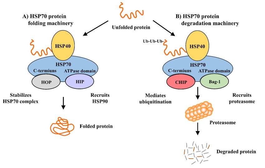
Cells 2020, 9, 2020                                                                                 3 of 17

by pharmacological substances such as GA or 17-AAG, it rapidly degrades client proteins through
the ubiquitin–proteasome pathway [22]. Moreover, HSP70 and its cochaperone HSP40 assist in the
degradation of various proteins [21].

3. Mechanism of Heat Shock Protein 70 Induction and Its Interactions with Cochaperones and
Client Proteins
      HSP transcription is regulated by transcription factor-heat shock factors (HSFs), which are
translocated into the nucleus where they interact with conserved heat shock elements (HSEs) to
upregulate genes that code for the induced HSPs. HSEs are generally located in the upstream
untranslated region of HSFs target genes. HSF activation is accomplished at the level of protein–protein
interaction and posttranslational modification [23]. Conversely, some HSFs expressed through
HSPA1A/B/L (HSP70), HSPA1A (HSP72), and HSPC1 (HSP90), directly interrupt HSFs through binding
to its trimerization domain. In addition, a cytoplasmic histone deacetylase and a valosin-containing
protein lead to the suppression of the HSF–HSP complex [24].
      Ischemia and other conditions, which are associated with unfolded proteins, lead to the dissociation
of HSFs from HSPs. Dissociated HSFs migrate to the nucleus of the stressed cell, where they become
phosphorylated, often by a protein such as kinase C. This process forms activated trimers that bind to
the HSE, which is a highly conserved regulatory sequence located on the heat shock gene. After binding
to HSE, HSFs then bind to the HSP70 gene’s promoter region, which leads to more HSP70 generation [8].
HSP70 production is influenced by HSP90, because the latter binds to HSF1. HSF1 can be freed to bind
to HSEs in instances where HSP90 dissociates from it, thus facilitating further HSP70 induction [25].
      The ATPs, HSP40 and HSP90 induce HSP70 synthesis and act as the molecular chaperones of
damaged cells by repairing their denatured proteins. This role is completed through refolding and
trafficking of the denatured proteins in the damaged cells, ultimately resulting in a multiple iteration
of protein refolding. HSP40 binds to HSP70 with high affinity and is responsible for the ATPase
activity of HSP70 and for maintaining it in an ADP-bound state. HSP40, by itself, cannot stabilize
HSP70’s ADP state. Due to this, and because of newly minted HSP70-substrate complexes, early
dissociation occurs [26]. Thus, there are additional cochaperones involved. HSP70 interacting protein
(HIP) binds to the HSP70 ATPase domain, thus maintaining HSP70’s ADP-bound state, and protects
the chaperone–substrate complex from dissociating [26]. Another cofactor, HSP70/HSP90-organization
protein (HOP), binds to the HSP70 C-terminus, allowing HOP to recruit HSP90 to the HSP70 substrate
complex [27–29]. As HIP stabilizes the ADP/ATP exchange of HSP70, HOP’s interaction with HSP70
and HSP90 releases HSP70 chaperone components (Figure 1A). HSP70 utilizes its cochaperones, CHIP
(C-terminus of HSP70/HSC70 interacting protein) and Bcl-2–associated athanogene (Bag)-1, indirectly
for its role in protein degeneration (Figure 1B). CHIP, an ubiquitin ligase, interacts with HSP70 by
rounding unfolded proteins and then ubiquitinating them, thus facilitating the degradation of seized
proteins [30,31]. Under stress conditions, HSP70 induction is improved by CHIP function. Moreover,
CHIP regulates HSP70’s development in the stress recovery process; it probably achieves protein
homeostasis by regulating chaperone levels while in stress and recovery processes [32]. Bag-1, an HSP70
cochaperone, is also associated with the proteasomal degradation of abnormal proteins. Bag-1 and
CHIP’s interaction and co-expression directly enhance the model chaperone substrate degradation [33].
They work together to update the chaperone systems from a protein folding role to a degradation
function. HSP90 is interlinked with HSP70 regarding protein degradation. Both chaperones transitorily
associate with shared cochaperones. HSP90 transfers client proteins to HSP70 via a transient interaction.
CHIP, that is associated to HSP70, ubiquitinates and pushes client proteins to proteasome mechanisms
(Figure 1B) [34–36]. Thus, HSP90 is an inhibitor to substrate ubiquitination and degradation. In contrast,
HSP70 is a promoter of ubiquitination and degradation [37,38].
Cells 2020, 9, 2020 4 of 17
Cells 2020, 9, x FOR PEER REVIEW 4 of 17
Figure1.1.HSP70
Figure HSP70chaperone
chaperonemachinery.
machinery.HOP
HOPandandCHIP
CHIPcompete
competefor forHSP70’s
HSP70’sC-terminus
C-terminusduring
during
protein folding and degradation, while HIP and Bag-1 compete for the ATPase domain.
protein folding and degradation, while HIP and Bag-1 compete for the ATPase domain. Depending Depending
ononthe
thesite
siteofofHIP
HIPororBag-1
Bag-1binding,
binding,HSP70
HSP70may
maylead
leadtotoprotein
proteinfolding
folding(HIP/HOP
(HIP/HOPpathway;
pathway;(A)
(A)orortoto
protein degradation through the proteasome (CHIP/Bag-1 pathway; (B). Ub =
protein degradation through the proteasome (CHIP/Bag-1 pathway; (B). Ub = ubiquitin. ubiquitin.
4. Role of Heat Shock Protein 70 in Brain Injury
4. Role of Heat Shock Protein 70 in Brain Injury
Injury to the brain either from loss of blood flow or direct trauma to the brain evokes a complex
Injury to the brain either from loss of blood flow or direct trauma to the brain evokes a complex
multicellular pathophysiological state. Such injuries lead to different types of cellular damage, ranging
multicellular pathophysiological state. Such injuries lead to different types of cellular damage,
from acute excitotoxic stress to instances of delayed programmed cell death. Inducible HSP70 levels
ranging from acute excitotoxic stress to instances of delayed programmed cell death. Inducible HSP70
are low while in homeostasis; however, injury drastically raises its expression. Therefore, HSP70 is
levels are low while in homeostasis; however, injury drastically raises its expression. Therefore,
demonstrated to be a marker of stress in cells. HSP70 induction was originally studied in a stroke
HSP70 is demonstrated to be a marker of stress in cells. HSP70 induction was originally studied in a
model; within this model, HSP70 was initially observed in neurons of the brain regions that appeared
stroke model; within this model, HSP70 was initially observed in neurons of the brain regions that
relatively resistant to injury [39,40]. It was also detected in brain regions surrounding the infarct
appeared relatively resistant to injury [39,40]. It was also detected in brain regions surrounding the
(penumbra) [41]. Later, HSP70 was observed in endothelial cells and glia, such as astrocytes and
infarct (penumbra) [41]. Later, HSP70 was observed in endothelial cells and glia, such as astrocytes
microglia [41]. In particular, HSP70 was detected in the brain regions surrounding the area of stroke
and microglia [41]. In particular, HSP70 was detected in the brain regions surrounding the area of
(penumbra), but was absent in the areas most affected by the infarct (core) [42]. However, HSP70
stroke (penumbra), but was absent in the areas most affected by the infarct (core) [42]. However,
mRNA was still detected within this core [41]. Thus, it was concluded that HSP70 was mainly detected
HSP70 mRNA was still detected within this core [41]. Thus, it was concluded that HSP70 was mainly
in cells that survived the ischemic insult, while brain areas that failed to translate HSP70 remained
detected in cells that survived the ischemic insult, while brain areas that failed to translate HSP70
vulnerable to ischemic injury. Similarly, in a model of global cerebral ischemia, which attempts to
remained vulnerable to ischemic injury. Similarly, in a model of global cerebral ischemia, which
recreate ischemic brain injury due to cardiac arrest, HSP70 was mainly observed in the cornu amonis
attempts to recreate ischemic brain injury due to cardiac arrest, HSP70 was mainly observed in the
(CA) 3 and the dentate granules of the hippocampus that tend to survive the ischemic episode, whereas
cornu amonis (CA) 3 and the dentate granules of the hippocampus that tend to survive the ischemic
HSP70 was not induced in CA1 neurons [43]. Equally to that observed in stroke models, HSP70 mRNA
episode, whereas HSP70 was not induced in CA1 neurons [43]. Equally to that observed in stroke
was detected in all ischemic hippocampal regions, including CA1 neurons. However, CA1 neurons,
models, HSP70 mRNA was detected in all ischemic hippocampal regions, including CA1 neurons.
which tend to be more vulnerable to ischemic insults compared to neurons of other hippocampal
However, CA1 neurons, which tend to be more vulnerable to ischemic insults compared to neurons
regions, failed to express the protein [44]. These observations generated debate as to whether HSP70
of other hippocampal regions, failed to express the protein [44]. These observations generated debate
expression was an epiphenomenon of the injury or facilitated cell survival.
as to whether HSP70 expression was an epiphenomenon of the injury or facilitated cell survival.
Experimentation demonstrating that HSP70 overexpression using viral vectors improved survival
Experimentation demonstrating that HSP70 overexpression using viral vectors improved
of neurons and astrocytes in stroke models finally provided direct evidence of a neuroprotective role
survival of neurons and astrocytes in stroke models finally provided direct evidence of a
of HSP70 [45,46]. Similarly, transgenic mice that overexpress HSP70 have smaller lesion sizes and
neuroprotective role of HSP70 [45,46]. Similarly, transgenic mice that overexpress HSP70 have
better neurological outcomes, whereas a deficiency exacerbates lesions and poor outcomes [47,48].
smaller lesion sizes and better neurological outcomes, whereas a deficiency exacerbates lesions and
poor outcomes [47,48]. Although less studied compared to brain ischemia, HSP70 was found to
exhibit similar patterns in experimental models of TBI [7]. Many mechanisms leading to thisCells 2020, 9, 2020 5 of 17
Although
Cells 2020, 9, x less studied
FOR PEER compared
REVIEW to brain ischemia, HSP70 was found to exhibit similar patterns
5 of 17
in experimental models of TBI [7]. Many mechanisms leading to this protective effect have since
protective effect have in
been demonstrated since beenand
stroke demonstrated in stroke
related models, and related
ranging models, ranging
from prevention fromaggregates
of protein preventionto
ofmodulation
protein aggregates to modulation
of cell death of cell death pathways [20].
pathways [20].
5.5.The
TheNeuroprotective
NeuroprotectiveEffect
EffectofofHeat
HeatShock
ShockProtein
Protein70
70via
viaCell
CellDeath
DeathPathways
Pathways
HSP70induction
HSP70 inductioncancanlower
lowerprotein
proteinaggregates
aggregatesand andintracellular
intracellularinclusions
inclusions[49],
[49],but
butspecific
specific
chaperone interactions seem to underlie other ways in which HSP70 may lead to
chaperone interactions seem to underlie other ways in which HSP70 may lead to neuroprotection. neuroprotection.
Afterbrain
After braininjury,
injury,apoptosis
apoptosis occurs
occurs either
either viavia
thethe intrinsic
intrinsic or mitochondrial
or mitochondrial pathway
pathway [50]via
[50] or orthe
via
the extrinsic
extrinsic or surface
or surface receptor-mediated
receptor-mediated pathway
pathway [51].
[51]. Recent
Recent research
research demonstratedthat
demonstrated thatHSP70
HSP70
impedesapoptosis
impedes apoptosisviaviadirect
directand
andindirect
indirectmeans
means(Figure
(Figure2).
2).
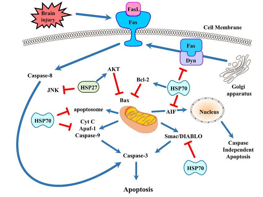
Braininjury
Figure2.2.Brain
Figure injuryinduces
inducesapoptotic
apoptoticcell
celldeath
deathbybydistinct
distinctpathways.
pathways.TheTheintrinsic
intrinsicpathway
pathwayisis
centered around signals emanating from the mitochondria, whereas the extrinsic
centered around signals emanating from the mitochondria, whereas the extrinsic or receptor- or receptor-mediated
pathwaypathway
mediated begins when
beginsdeath
whenreceptors bind tobind
death receptors theirtoligands. The prototypical
their ligands. deathdeath
The prototypical receptor Fas is
receptor
shown here. HSP70 and HSP27 have been shown to interfere with apoptosis as indicated
Fas is shown here. HSP70 and HSP27 have been shown to interfere with apoptosis as indicated in the in the figure.
See text
figure. Seefortext
more
fordetails. (FasL =(FasL
more details. Fas ligand; AIF = apoptosis
= Fas ligand; inducing
AIF = apoptosis factor; Akt
inducing = protein
factor; Akt = kinase
proteinB;
Apaf-1B;= Apaf-1
kinase apoptosis protease activating
= apoptosis protease factor-1;
activatingCyt C = cytochrome
factor-1; Cyt C = c; JNK = c-Jun
cytochrome c; N-terminal
JNK = c-Junkinase;
N-
Dyn = dynamin).
terminal kinase; Dyn = dynamin).
The intrinsic apoptotic pathway initiates with the release of various factors from the cell’s
The intrinsic apoptotic pathway initiates with the release of various factors from the cell’s
mitochondria. In response to brain injury (and to the resultant oxidative stress), the mitochondria
mitochondria. In response to brain injury (and to the resultant oxidative stress), the mitochondria
develop a permeability transition pore which leads to the discharge of cytochrome c to the cytosol, where
develop a permeability transition pore which leads to the discharge of cytochrome c to the cytosol,
a number of pro-apoptotic molecules ultimately causes the activation of effector caspases. Among these
where a number of pro-apoptotic molecules ultimately causes the activation of effector caspases.
molecules are the Bcl-2 family members, some of which are pro-apoptotic. These molecules are
Among these molecules are the Bcl-2 family members, some of which are pro-apoptotic. These
primary regulators of the mitochondrial membrane. There are three subgroups classified by their
molecules are primary regulators of the mitochondrial membrane. There are three subgroups
structural homology. The first group includes the anti-apoptotic proteins Bcl-2, Bcl-XL, and Bcl-w.
classified by their structural homology. The first group includes the anti-apoptotic proteins Bcl-2, Bcl-
XL, and Bcl-w. The second group includes the pro-apoptotic proteins Bax and Bak. The third group
includes the BH3-only proteins Bad, Bid, Bim, Noxa, and PUMA. Bcl-2 family proteins conduct
multiple functions during brain injury. BH3-only proteins, via interactions with Bcl-2 familyCells 2020, 9, 2020 6 of 17
The second group includes the pro-apoptotic proteins Bax and Bak. The third group includes
the BH3-only proteins Bad, Bid, Bim, Noxa, and PUMA. Bcl-2 family proteins conduct multiple
functions during brain injury. BH3-only proteins, via interactions with Bcl-2 family members,
assist with neuronal cell death after ischemic stroke [52]. Mitochondria-based apoptosis is related
to the apoptosome. This occurs when procaspase-9 interacts with apoptosis protease activating
factor-1 (Apaf-1) in the cytosol and leads to activation when cytochrome c is translocated to the
cytosol from the mitochondria. Bcl-2, an antiapoptotic protein, interrupts cytochrome c release.
Furthermore, caspase-9 activation induces the activation of a multitude of effector caspases such as
caspase-3. In neuronal stem cells, induction of HSP70 by recombination plasmid pEGFP-C2-HSP70
significantly blocks caspase-3 and reduces neural cytotoxicity, including neuron loss and synapsis
damnification, in cocultured cells [53]. After an inflammatory stimulus, oligodendrocyte precursor
cells and mature oligodendrocytes of HSP70 deficient mice enter apoptosis caused by caspase-3
activation [54]. Caspase-independent pathways have been described to cause apoptosis via apoptosis
inducing factor (AIF) [55]. AIF translocates into the nucleus from the mitochondria. Once in the nucleus,
AIF can trigger apoptosis in the absence of caspases. Like cytochrome c, AIF release can also be prevented
by Bcl-2 [56]. Second mitochondria-derived activator of caspases (Smac)/direct inhibitor-of-apoptosis
protein (IAP) binding protein with low pi (Smac/DIABLO) is also a regulator of apoptosis. These are also
discharged from the mitochondria and impede apoptosomal inhibition by IAPs. HSP70 seems to affect
multiple aspects of the intrinsic apoptotic pathway. HSP70 interacts with components of the apoptotic
machinery both upstream [57,58] and downstream of mitochondrial events [59]. In experimental stroke
models, HSP70 interfered with cytochrome c release [43,60] and inhibited AIF translocation to the
nucleus [61] while reducing ischemic brain injury. It lowers the recruitment of procaspase-9 into the
apoptosome, as observed in HSP70 overexpressing transgenic mice, and sequesters AIF [62]. HSP70
also prevents the release of proapoptotic protein Smac/DIABLO from myocyte mitochondria [63].
Mitochondrial HSP70/HSP75/mortalin assistants act to maintain mitochondrial membrane potential,
which could be beneficial to mitochondrial function and mitochondrial protein import [64]. Astrocytes
with HSP induction showed reduced cell death following in vitro stroke models with preserved
ATP [65]. The above outcomes are connected to lowered reactive oxygen species (ROS) creation, and
maintained mitochondrial membrane potentials [66] and glutathione levels [67]. Overexpression of
HSP70 has been demonstrated to elevate mitochondrial antioxidant enzyme activity in myocardial
cells [68]. Bcl-2 is a critical component in hindering the onset of apoptosis due its ability to block
the release of cytochrome c and AIF, and viral vector-mediated HSP70 overexpression was linked
to elevated Bcl-2 protein levels in hippocampal neurons [69]. HSP70 has also been shown to reduce
heat-induced apoptosis by preventing the migration of the pro-apoptotic Bcl-2 family member Bax,
and thus blocking the mitochondrial discharge of pro-apoptotic factors [58]. Previous studies of HSP27
and its anti-apoptotic activity have established that stress-induced Bax oligomerization and migration
to the mitochondria can be suppressed indirectly by HSP27 [70]. HSP27 has also been shown to
phosphorylate the survival kinase Akt/protein kinase B (PKB) [71] and to inactivate prodeath c-Jun
N-terminal kinase (JNK) [72]. HSP70 also interferes with the activity of Apaf-1, a critical component
for apoptosome formation and activation of caspase-9 [62], although others have failed to show such
an interaction [57].
The extrinsic or cell surface-mediated pathway of apoptosis involves interactions with death receptors
found on the plasma membrane. This pathway is also known as the “death receptor pathway”. Death
receptor ligation activates caspase-8 and caspase-10. This has the potential to activate effector caspase-3 [73].
The TNF family of ligands, including FasL, TNF, CD40L, and TRAIL, promotes the activation of many
death receptors. Many of these ligands are released extracellularly as part of the ischemic immune
response [74]. One of the first death receptors identified was Fas, which activates when it binds to its
ligand FasL. When FasL binds to Fas, the cytoplasmic adaptor protein Fas-associated death domain
protein (FADD) is recruited to this complex. FADD possesses a “death effector domain” at the N-terminus
that can bind to procaspase-8 [75]. This is known as the death-inducing signaling complex (DISC).Cells 2020, 9, 2020 7 of 17
The proteolytic cleavage, which causes transactivation of procaspase-8 to caspase-8, is catalyzed by the
DIAC [75]. Activated caspase-8 ultimately leads to the activation of caspases-3 and -10 [76].
HSP70 has been demonstrated to engage with extrinsic death receptor signaling pathways. HSP70
is known to bind to cell surface receptors TRAILR1 and TRAILR2, and to death receptors 4 (DR4) and
5 (DR5), where they bind to a cytokine called TRAIL to induce TNF-related apoptosis. Therefore, the
TRAIL-induced assembly and activity of the DISC becomes inhibited [77,78]. HSP70 can neutralize
Bid activation and the following apoptosis once DISC formation has occurred and caspase-8 has
been activated [79]. HSP70 has been demonstrated to interact with the Fas pathway. Dynamin, a
molecule typically associated with synaptic transmission, is also known to traffic Fas to the cell’s surface
from the Golgi apparatus [80]. Fas binds to FasL when it translocates to the cell surface, activating
caspase-8 and, therefore, leading to cell death. We recently showed that HSP70 inhibits Fas trafficking
to the cell surface via its interaction with dynamin [48]. Thus, HSP70 also prevents the extrinsic or
receptor-mediated apoptosis with specific chaperone interactions.
Several studies have shown that microRNAs (miRNAs), which interact with multiple target
messenger RNAs (mRNAs), coordinate the regulation of target genes. Several miRNAs such
as miRNA-1, miRNA-21, and miRNA-24 may contribute to an increased expression of several
cytoprotective proteins, although no targets have been validated. Some studies showed that
muscle-specific miRNA-1 levels can change in the condition of ischemic myocardium [81–83], and
that two of miRNA-1’s targets are HSP60 and HSP70 [84]. Recently, Ouyang et al. showed that
miRNA-181a, which is expressed at high levels in the brain, regulates HSP70 family chaperones and
the outcome of ischemic stroke [85]. Mutual expression of miRNA-181a and HSP78/BIP was found
both in the core and the penumbra in an ischemic stroke model. miRNA-181a mimic reduces and
its inhibitor/antagomir enhances HSP78/BIP expression [85]. miRNA-181a also targets Bcl2, which is
an antiapoptotic protein [86] and has been shown to be upregulated by HSP70 overexpression [69].
miRNA-181 can also target HSP78/BIP and potentially target the 30 UTRs of HSP72 and HSP75/mortalin.
Following ischemic stroke, miRNAs could conceivably target multiple chaperones and efficiently
modulate cell death.
6. Inflammation Regulation of Heat Shock Protein 70
Inflammation of the central nervous system (CNS) is a feature of many acute neurological injuries,
including brain trauma, stroke, and other cerebral hypoxic-ischemic injuries [87]. Brain injury elicits
an inflammatory reaction beginning with the activation of endogenous microglia and with peripheral
leukocyte influx into the cerebral parenchyma [88,89]. Upon inflammatory cell activation, cytotoxic
agents such as some cytokines, which are increasingly viewed as key contributors to ischemic cell
death [90], are discharged. Some studies indicate that nonimmunologic brain cells, such as astrocytes
and even neurons, can elaborate these same inflammatory molecules. These inflammatory responses
are thought to exacerbate brain damage, thus presenting a major opportunity for potential treatments.
HSP70 has been demonstrated to possess a modulating role in regulating immune responses in cases
of brain injury. HSP70 has been demonstrated to control inflammation both inside and outside the
cell. In the intracellular setting, HSP70 appears to inhibit pro-immune responses; whereas in the
extracellular setting, it seems to do the opposite and potentiate such responses.
HSP70 is capable of interacting with transcription factors, including those which trigger
inflammations, such as the nuclear factor-kappaB (NF-kB, which consists of the heterodimers p65
and p50). One study in astrocytes showed that HSP70 was capable of reducing NF-kB activation [91].
HSP70 can also interact with immune molecules themselves, such as matrix metalloproteinases (MMPs)
and ROS; it seems to limit inflammatory responses in such cases. Increased HSP70 within the cell
has been shown to decrease the production of nitric oxide and inducible nitric oxide synthase (iNOS)
in inflammatory cells. Heat stress is also associated with the reduced secretion of tumor necrosis
factor-alpha (TNF-α) as well as with diminished generation of ROS. HSP70 has been shown to inhibit
the cellular responses to inflammatory cytokines including TNF-α and interleukin (IL)-1 [92,93], andCells 2020, 9, 2020 8 of 17
increased HSP70 in macrophages prevents lipopolysaccharide (LPS)-induced increases in TNF, IL-1,
IL-10, and IL-12 [93,94]. In a model of intracerebral hemorrhage, HSP70 also reduced TNF-α while
attenuating blood brain barrier (BBB) disruption and brain edema, and enhanced neurological function
as well [95].
Induction of HSP70 in phagocytes through heat shock decreased nicotinamide adenine dinucleotide
phosphate hydrogen (NADPH) oxidase activity in neutrophils and increased superoxide dismutase, a
superoxide scavenger [96]. Recently, we demonstrated that HSP70 induction by heat stress halts IkB,
JNK, and p38 phosphorylation. HSP70 also seems to inhibit the binding to DNA of several transcription
factors, including NF-kB, activator protein-1 (AP-1), and signal transducer and activator of transcription
factor 1 (STAT-1), with subsequent inhibition of their pro-inflammatory transgenes [97]. Other studies
have also demonstrated that prior-heat stress, due, in part, to HSP70 overexpression and prevention of
nuclear NF-kB translocation, interferes with these inflammatory responses [98,99]. When ischemic
stroke induces generation of cytokines, such as TNF-α that can activate NF-kB–dependent gene
transcription through the NF-kB pathway, the inhibitor of kB (IkB) acts as another target of HSP70 and,
when bound, can interrupt NF-kB activation by preventing phosphorylation of IkB (Figure 3) [91,92].
Some studies have also shown that HSP70 interacts with NF-kB and/or its regulatory proteins [100,101],
but the mechanisms may depend on the nature of the stimulus. In a cell death model induced by
TNF-α, HSP70 directly inhibited IkB kinase (IKK) activity [102], while in a stroke model, HSP70
seemed to interact with both NF-kB and IkB, ultimately leading to reduced IkB phosphorylation by
IKK [100]. NF-kB inhibition by HSP70 further prevented transcription of immune genes and led to a
neuroprotective effect.
NF-kB regulates many pro-inflammatory genes, including matrix metalloproteinase (MMP)-9.
Our group showed that MMP-9 mRNA was reduced when HSP70 was overexpressed in astrocytes
later exposed to ischemia-like injury [46]. Furthermore, HSP70 in astrocytes suppresses MMP-9 protein
expression [46]. MMP-2, while not regulated by NF-kB, was also decreased in an HSP70 overexpression
context, which suggests that HSP70 inhibits other transcription factors as well. In fact, studies in heat
stressed alveolar macrophages [18] and primary astrocytes [97], showed that HSP70 interfered with
STAT-1, which is known to regulate MMP-2 expression [103]. Additionally, HSP70 seems to inhibit the
generation of activated MMPs, as evidenced by a reduction in cleaved forms of several MMPs. In a
model of TBI, HSP70 overexpression inhibited, while HSP70 deficiency exacerbated, brain hemorrhage,
and this was correlated with changes in MMP levels and activity [7]. Thus, there are several ways in
which HSP70 inhibits the post injury immune response.
Extracellular HSP70 also has the capacity to modulate immune responses [104]. HSP70 has
been shown to accomplish opposite roles depending on the type of insult and the immune response
that follows. Extracellular HSP70 appears to potentiate adaptive immune responses, and has been
widely studied. Complexes of HSP70 and peptides trigger CD8+T-cell responses [105]. Injecting mice
with these HSP70–peptide complexes leads to similar responses, and suggests that HSP70 acts as an
adjuvant [106]. In the extracellular environment, HSP70 can also potentiate antigen presentation, and
reacts with macrophages and dendritic cells via CD40, CD91, and LOX-1 receptors [107]. Extracellular
HSP70 has also been shown to elicit innate immune responses by interacting with Toll-like receptors
(TLRs), causing subsequent NF-kB activation and target gene upregulation, including iNOS and
pro-inflammatory cytokines [108]. HSP60 and HSP90 appear to interact with TLR-2 and -4 [109];
however, these results have been controversial, as some preparations of recombinant HSPs may have
contained endotoxin, the classic ligand for TLR-4 [110].preventing phosphorylation of IkB (Figure 3) [91,92]. Some studies have also shown that HSP70
interacts with NF-kB and/or its regulatory proteins [100,101], but the mechanisms may depend on
the nature of the stimulus. In a cell death model induced by TNF-α, HSP70 directly inhibited IkB
kinase (IKK) activity [102], while in a stroke model, HSP70 seemed to interact with both NF-kB and
IkB,2020,
Cells ultimately
9, 2020 leading to reduced IkB phosphorylation by IKK [100]. NF-kB inhibition by HSP70
9 of 17
further prevented transcription of immune genes and led to a neuroprotective effect.
Figure 3. Influence of of HSPs
HSPs inininnate
innateimmunity.
immunity.Following
Followingischemic
ischemicstroke,
stroke, HSPs
HSPs have
have been
been shown
shown to
inhibit the activation of the transcription factor NF-kB and to prevent its nuclear translocation.
to inhibit the activation of the transcription factor NF-kB and to prevent its nuclear translocation. Acute
brain insults
Acute brain (brain
insultsinjury)
(braintrigger
injury)activation of NF-kB by
trigger activation of causing
NF-kB by thecausing
phosphorylation and degradation
the phosphorylation and
of its inhibitorofprotein
degradation IkB, which
its inhibitor normally
protein IkB, keeps
whichNF-kB (which
normally consists
keeps NF-kBof the heterodimers
(which consistsp65
of and
the
p50) tethered p65
heterodimers to the
andcytosol. Once NF-kB
p50) tethered is able to
to the cytosol. translocate
Once NF-kB istoable
thetonucleus, it binds
translocate tonucleus,
to the promoter it
regions of several pro-inflammatory genes and leads to an inflammatory response.
binds to promoter regions of several pro-inflammatory genes and leads to an inflammatory response.
7. Heat Shock Protein 70 as a (Pharmacological) Therapeutic Target for Brain Injury
NF-kB regulates many pro-inflammatory genes, including matrix metalloproteinase (MMP)-9.
Our group showed that
Understanding MMP-9 mRNA
the mechanisms was reduced
by which when HSP70
HSPs function wascell
within the overexpressed in astrocytes
under conditions of stress
and injury may reveal potential therapeutic targets. A few studies have attempted a direct delivery of
HSP70 to the brain, while others have employed the cell’s endogenous capacity to upregulate HSP70
through its coordinated actions with HSP90. After brain injury, treatments to increase HSP70 in the cell
might be a worthwhile approach to treat neurological conditions associated with inflammation and
cell death. A few pharmacological approaches to delivering HSP70 have been explored. In one study,
exogenous HSP70 was conjugated to the HIV TAT protein in order to improve brain delivery following
intravenous administration. HSP70-TAT reduced infarct volumes, improved neurological outcomes,
and led to higher survival of neural progenitors in a stroke model [111]. Similar observations were
found when recombinant HSP70 was administered in a rodent stroke model [112]. In this latter study,
HSP70 was conjugated with Fv, which is a single chain fragment of the anti-DNA antibody mAb
3E10 that allows for penetration into the cell, and the Fv fragment was used as a delivery vehicle.
Interestingly, systemic Fv-HSP70 treatment seemed to enter the ischemic side of the brain but not the
contralateral nonischemic side, which suggests that it may bind to a still unknown target within the
injured brain.
Since HSP90 sequesters HSF1 in the cytosol and prevents transcription of target HSPs, including
HSP70, the administration of pharmacological inhibitors of HSP90 is another strategy to endogenously
increase HSP70 expression. A few studies have now explored the efficacy of pharmacological inductionCells 2020, 9, 2020 10 of 17
of HSP70 via HSP90 inhibitors in brain injury models. This was recently reviewed by us, and we refer
the reader to the review for more details on this topic [8]. Such inducers have shown salutary effects
against both local and global experimental cerebral ischemia [113,114]. Due to the HSP70 abilities to
reduce apoptosis and inflammation after ischemic stroke, there is an obvious interest in the HSP70
induction capacity of HSP90 inhibitor agents. One such HSP90-antagonist, GA, has been studied in
stroke models with salutary results [113], but has failed at the clinical level due to poor solubility in
water [115] and to unacceptable renal and hepatic toxicity [116,117]. The GA analogue 17-AAG is
less toxic and has since advanced to phase 3 clinical trials for cancer therapy [116,118]. Consistent
with the findings seen with GA, 17-AAG treatment improved outcomes in experimental TBI, with a
reduction in brain hemorrhage [119]. 17-(2-dimethylaminoethyl) amino-17-demethoxygeldanamycin
(17-DMAG), with better solubility, was developed, and was found to decrease microglial activation
and to inhibit phosphorylation of IκB resulting in reduced nuclear translocation of NF-kB (p65) in a
stroke model [120].
There are other HSP70-inducing groups, such as the purine-based compounds and the resorcinols.
Purine-based compounds were designed to resemble ansamycins; hence, they use ADP to bind to
HSP90’s ATP binding site [121]. BIIB021 (also named CNF-2024) is also an HSP70-inducer and an
HSP90-inhibitor. It is being studied in a phase 2 clinical trial using an oral form [118,122]. Another
study in lymphoma cells demonstrated BIIB021 to be a potent anti-inflammatory compound that
can suppress NF-kB [123]. What is still unclear is if these purine-based compounds accomplish the
same roles in other types of cells, and whether they can penetrate the BBB. The resorcinol group
includes radicicol based compounds, which bind to HSP90’s ATP-binding pocket [124,125]. Despite
their HSP90-inhibiting properties, drug development of the resorcinol groups is slow because they
have been found to easily degrade in vivo [118]. Some variants such as NVP-AUY922 and AT-13387
were designed to avoid this limitation, and have also been studied as potential anti-inflammatory
compounds for cancer treatment. Resorcinol has yet to be studied in brain injury or inflammation [118].
There is, however, some work published about another HSP70 inducer, geranylgeranylacetone (GGA),
known for its antiulcer properties. In a stroke model, GGA led to not only a neuroprotective effect
but also to a reduced post stroke inflammation via HSP70 upregulation and protein kinase C (PKC)
activation [126,127]. Others found that the administration of GGA also led to a neuroprotective effect
in a TBI model by inhibiting microglial activation and reducing apoptosis of neurons [128]. Thus,
promising preclinical studies of HSP90 inhibitors together with clinical experience in cancer patients
may stimulate appropriate trials in stroke and TBI patients.
Other pharmacological inducers of HSPs have been studied in models of neurodegeneration
and cancer, but they have yet to be tested in brain injury models. Myricetin, a flavonoid, was shown
to enhance intracellular levels of HSF-1 and HSP70, and to suppress abnormal protein aggregation
and remove various toxic neurodegenerative disease-associated proteins [129]. Celastrol, a quinone
methide family member isolated from the Thunder God Vine, has been shown to upregulate HSF-1,
which, in turn, leads to HSP induction; this has been shown in human neuronal cells [130]. Celastrol
has been shown to protect motor neurons from kainic acid induced excitotoxicity while upregulating
HSPs [131]. Celastrol has also been shown to improve outcome in a stroke model, but this study did not
explore whether this was related to HSP induction [132]. In a metastatic cancer model, a recent study
showed that electrochemotherapy with betulinic acid or with cisplatin increased HSP27 and HSP70,
and they proposed that electric field stress combined with drug administration led to the induction of
HSPs [133]. Thus, promising observations in related nervous system disease models suggest that these
approaches should be tested in acute brain injury models.
8. Conclusions
The role of the HSP family as potential neuroprotectants for the treatment of acute brain insults has
been increasingly recognized, along with a more detailed elucidation of the mechanisms underlying
this beneficial effect. Several studies have now reported a neuroprotective effect, particularly afterCells 2020, 9, 2020 11 of 17
HSP70 induction or administration through pharmacological manipulations. HSP70 also appears
to be accountable for multiple protective mechanisms as a molecular chaperone. The multifaceted
mechanisms of protection by HSPs suggest that these may be an effective therapeutic target. While some
HSP90 inhibiting compounds have already been investigated in cancer clinical trials, there are no
studies in patients with neurological conditions. With the development of several pharmacological
HSP70 inducers and of methods to deliver the protein itself, it seems that we may be ready for
clinical trials.
Author Contributions: J.E.L. and M.A.Y. provided concept, design and overall supervision of this study. J.Y.K.
contributed in the writing and drawing. S.B., M.Y.H. and J.P. participated in the discussion and revision. All authors
approved and agreed to be accountable for all aspects of the work. All authors have read and agreed to the
published version of the manuscript.
Funding: This research was supported by grants from the National Institutes of Health (RO1 NS106441)
and Department of Defense and the Veteran’s Merit Award (I01 BX000589) to M.A.Y. Grants to M.A.Y. were
administered by the Northern California Institute for Research and Education, and supported by resources of the
Veterans Affairs Medical Center, San Francisco, California. Basic Science Research Program through the National
Research. Foundation of Korea (NRF) funded by the Ministry of Education (NRF-2020R1I1A1A01064803) to
J.Y.K., and the National Research Foundation of Korea (NRF) grant funded by the Korea Government (MSIP;
NRF-2017R1A2B2005350) to J.E.L.
Conflicts of Interest: The authors declare that they have no competing interests.
References
1. Doberentz, E.; Genneper, L.; Wagner, R.; Madea, B. Expression times for hsp27 and hsp70 as an indicator of
thermal stress during death due to fire. Int. J. Legal Med. 2017, 131, 1707–1718. [CrossRef]
2. Mash, D.C.; Duque, L.; Pablo, J.; Qin, Y.; Adi, N.; Hearn, W.L.; Hyma, B.A.; Karch, S.B.; Druid, H.; Wetli, C.V.
Brain biomarkers for identifying excited delirium as a cause of sudden death. Forensic Sci. Int. 2009, 190,
e13–e19. [CrossRef] [PubMed]
3. Weinstein, P.R.; Hong, S.; Sharp, F.R. Molecular identification of the ischemic penumbra. Stroke 2004, 35,
2666–2670. [CrossRef] [PubMed]
4. States, B.A.; Honkaniemi, J.; Weinstein, P.R.; Sharp, F.R. DNA fragmentation and hsp70 protein induction in
hippocampus and cortex occurs in separate neurons following permanent middle cerebral artery occlusions.
J. Cereb. Blood Flow Metab. 1996, 16, 1165–1175. [CrossRef] [PubMed]
5. Frebel, K.; Wiese, S. Signalling molecules essential for neuronal survival and differentiation. Biochem.
Soc. Trans. 2006, 34, 1287–1290. [CrossRef] [PubMed]
6. De Maio, A. Extracellular hsp70: Export and function. Curr. Protein Pept. Sci. 2014, 15, 225–231. [CrossRef]
7. Kim, J.Y.; Kim, N.; Zheng, Z.; Lee, J.E.; Yenari, M.A. The 70 kda heat shock protein protects against
experimental traumatic brain injury. Neurobiol. Dis. 2013, 58, 289–295. [CrossRef]
8. Kim, N.; Kim, J.Y.; Yenari, M.A. Anti-inflammatory properties and pharmacological induction of hsp70 after
brain injury. Inflammopharmacology 2012, 20, 177–185. [CrossRef]
9. Kacimi, R.; Yenari, M.A. Pharmacologic heat shock protein 70 induction confers cytoprotection against
inflammation in gliovascular cells. Glia 2015, 63, 1200–1212. [CrossRef]
10. Saibil, H. Chaperone machines for protein folding, unfolding and disaggregation. Nat. Rev. Mol. Cell Biol.
2013, 14, 630–642. [CrossRef]
11. Schopf, F.H.; Biebl, M.M.; Buchner, J. The hsp90 chaperone machinery. Nat. Rev. Mol. Cell Biol. 2017, 18,
345–360. [CrossRef] [PubMed]
12. Zuehlke, A.; Johnson, J.L. Hsp90 and co-chaperones twist the functions of diverse client proteins. Biopolymers
2010, 93, 211–217. [CrossRef]
13. Johnson, J.L.; Brown, C. Plasticity of the hsp90 chaperone machine in divergent eukaryotic organisms.
Cell Stress Chaperones 2009, 14, 83–94. [CrossRef] [PubMed]
14. Taipale, M.; Jarosz, D.F.; Lindquist, S. Hsp90 at the hub of protein homeostasis: Emerging mechanistic
insights. Nat. Rev. Mol. Cell Biol. 2010, 11, 515–528. [CrossRef]
15. Li, J.; Soroka, J.; Buchner, J. The hsp90 chaperone machinery: Conformational dynamics and regulation by
co-chaperones. Biochim. Biophys. Acta 2012, 1823, 624–635. [CrossRef]Cells 2020, 9, 2020 12 of 17
16. Xu, Z.S.; Li, Z.Y.; Chen, Y.; Chen, M.; Li, L.C.; Ma, Y.Z. Heat shock protein 90 in plants: Molecular mechanisms
and roles in stress responses. Int. J. Mol. Sci. 2012, 13, 15706–15723. [CrossRef] [PubMed]
17. Krukenberg, K.A.; Street, T.O.; Lavery, L.A.; Agard, D.A. Conformational dynamics of the molecular
chaperone hsp90. Q. Rev. Biophys. 2011, 44, 229–255. [CrossRef] [PubMed]
18. Howard, M.; Roux, J.; Lee, H.; Miyazawa, B.; Lee, J.W.; Gartland, B.; Howard, A.J.; Matthay, M.A.; Carles, M.;
Pittet, J.F. Activation of the stress protein response inhibits the stat1 signalling pathway and inos function in
alveolar macrophages: Role of hsp90 and hsp70. Thorax 2010, 65, 346–353. [CrossRef] [PubMed]
19. Brown, I.R. Heat shock proteins and protection of the nervous system. Ann. N. Y. Acad. Sci. 2007, 1113,
147–158. [CrossRef]
20. Giffard, R.G.; Yenari, M.A. Many mechanisms for hsp70 protection from cerebral ischemia. J. Neurosurg.
Anesthesiol. 2004, 16, 53–61. [CrossRef]
21. Sherman, M.Y.; Goldberg, A.L. Cellular defenses against unfolded proteins: A cell biologist thinks about
neurodegenerative diseases. Neuronal 2001, 29, 15–32. [CrossRef]
22. Isaacs, J.S.; Xu, W.; Neckers, L. Heat shock protein 90 as a molecular target for cancer therapeutics. Cancer Cell
2003, 3, 213–217. [CrossRef]
23. Akerfelt, M.; Morimoto, R.I.; Sistonen, L. Heat shock factors: Integrators of cell stress, development and
lifespan. Nat. Rev. Mol. Cell Biol. 2010, 11, 545–555. [CrossRef]
24. Pernet, L.; Faure, V.; Gilquin, B.; Dufour-Guerin, S.; Khochbin, S.; Vourc’h, C. Hdac6-ubiquitin interaction
controls the duration of hsf1 activation after heat shock. Mol. Biol. Cell 2014, 25, 4187–4194. [CrossRef]
25. Zhao, H.; Michaelis, M.L.; Blagg, B.S. Hsp90 modulation for the treatment of alzheimer’s disease.
Adv. Pharmacol. 2012, 64, 1–25. [CrossRef]
26. Hohfeld, J.; Minami, Y.; Hartl, F.U. Hip, a novel cochaperone involved in the eukaryotic hsc70/hsp40 reaction
cycle. Cell 1995, 83, 589–598. [CrossRef]
27. Frydman, J.; Hohfeld, J. Chaperones get in touch: The hip-hop connection. Trends Biochem. Sci. 1997, 22,
87–92. [CrossRef]
28. Chen, S.; Smith, D.F. Hop as an adaptor in the heat shock protein 70 (hsp70) and hsp90 chaperone machinery.
J. Biol. Chem. 1998, 273, 35194–35200. [CrossRef]
29. Hernandez, M.P.; Sullivan, W.P.; Toft, D.O. The assembly and intermolecular properties of the
hsp70-hop-hsp90 molecular chaperone complex. J. Biol. Chem. 2002, 277, 38294–38304. [CrossRef]
30. Connell, P.; Ballinger, C.A.; Jiang, J.; Wu, Y.; Thompson, L.J.; Hohfeld, J.; Patterson, C. The co-chaperone chip
regulates protein triage decisions mediated by heat-shock proteins. Nat. Cell Biol. 2001, 3, 93–96. [CrossRef]
31. Murata, S.; Minami, Y.; Minami, M.; Chiba, T.; Tanaka, K. Chip is a chaperone-dependent e3 ligase that
ubiquitylates unfolded protein. EMBO Rep. 2001, 2, 1133–1138. [CrossRef] [PubMed]
32. Qian, S.B.; McDonough, H.; Boellmann, F.; Cyr, D.M.; Patterson, C. Chip-mediated stress recovery by
sequential ubiquitination of substrates and hsp70. Nature 2006, 440, 551–555. [CrossRef]
33. Demand, J.; Alberti, S.; Patterson, C.; Hohfeld, J. Cooperation of a ubiquitin domain protein and an e3
ubiquitin ligase during chaperone/proteasome coupling. Curr. Biol. 2001, 11, 1569–1577. [CrossRef]
34. Meacham, G.C.; Patterson, C.; Zhang, W.; Younger, J.M.; Cyr, D.M. The hsc70 co-chaperone chip targets
immature cftr for proteasomal degradation. Nat. Cell Biol. 2001, 3, 100–105. [CrossRef] [PubMed]
35. Esser, C.; Scheffner, M.; Hohfeld, J. The chaperone-associated ubiquitin ligase chip is able to target p53 for
proteasomal degradation. J. Biol. Chem. 2005, 280, 27443–27448. [CrossRef]
36. Luo, W.; Zhong, J.; Chang, R.; Hu, H.; Pandey, A.; Semenza, G.L. Hsp70 and chip selectively mediate
ubiquitination and degradation of hypoxia-inducible factor (hif)-1alpha but not hif-2alpha. J. Biol. Chem.
2010, 285, 3651–3663. [CrossRef]
37. Peng, H.M.; Morishima, Y.; Clapp, K.M.; Lau, M.; Pratt, W.B.; Osawa, Y. Dynamic cycling with hsp90 stabilizes
neuronal nitric oxide synthase through calmodulin-dependent inhibition of ubiquitination. Biochemistry
2009, 48, 8483–8490. [CrossRef]
38. Peng, H.M.; Morishima, Y.; Pratt, W.B.; Osawa, Y. Modulation of heme/substrate binding cleft of neuronal
nitric-oxide synthase (nnos) regulates binding of hsp90 and hsp70 proteins and nnos ubiquitination.
J. Biol. Chem. 2012, 287, 1556–1565. [CrossRef]
39. Nowak, T.S., Jr.; Bond, U.; Schlesinger, M.J. Heat shock rna levels in brain and other tissues after hyperthermia
and transient ischemia. J. Neurochem. 1990, 54, 451–458. [CrossRef]Cells 2020, 9, 2020 13 of 17
40. Abe, K.; Tanzi, R.E.; Kogure, K. Induction of hsp70 mrna after transient ischemia in gerbil brain. Neurosci. Lett.
1991, 125, 166–168. [CrossRef]
41. Sharp, F.R.; Lu, A.; Tang, Y.; Millhorn, D.E. Multiple molecular penumbras after focal cerebral ischemia.
J. Cereb. Blood Flow. Metab. 2000, 20, 1011–1032. [CrossRef] [PubMed]
42. Beretta, S.; Cuccione, E.; Versace, A.; Carone, D.; Riva, M.; Padovano, G.; Dell’Era, V.; Cai, R.; Monza, L.;
Presotto, L.; et al. Cerebral collateral flow defines topography and evolution of molecular penumbra in
experimental ischemic stroke. Neurobiol. Dis. 2015, 74, 305–313. [CrossRef] [PubMed]
43. Tsuchiya, D.; Hong, S.; Matsumori, Y.; Kayama, T.; Swanson, R.A.; Dillman, W.H.; Liu, J.; Panter, S.S.;
Weinstein, P.R. Overexpression of rat heat shock protein 70 reduces neuronal injury after transient focal
ischemia, transient global ischemia, or kainic acid-induced seizures. Neurosurgery 2003, 53, 1179–1187.
[CrossRef] [PubMed]
44. DeGracia, D.J.; Jamison, J.T.; Szymanski, J.J.; Lewis, M.K. Translation arrest and ribonomics in post-ischemic
brain: Layers and layers of players. J. Neurochem. 2008, 106, 2288–2301. [CrossRef] [PubMed]
45. Batulan, Z.; Taylor, D.M.; Aarons, R.J.; Minotti, S.; Doroudchi, M.M.; Nalbantoglu, J.; Durham, H.D. Induction
of multiple heat shock proteins and neuroprotection in a primary culture model of familial amyotrophic
lateral sclerosis. Neurobiol. Dis. 2006, 24, 213–225. [CrossRef]
46. Lee, J.E.; Kim, Y.J.; Kim, J.Y.; Lee, W.T.; Yenari, M.A.; Giffard, R.G. The 70 kda heat shock protein suppresses
matrix metalloproteinases in astrocytes. Neuroreport 2004, 15, 499–502. [CrossRef]
47. Lee, S.H.; Kim, M.; Yoon, B.W.; Kim, Y.J.; Ma, S.J.; Roh, J.K.; Lee, J.S.; Seo, J.S. Targeted hsp70.1 disruption
increases infarction volume after focal cerebral ischemia in mice. Stroke 2001, 32, 2905–2912. [CrossRef]
48. Kim, J.Y.; Kim, N.; Zheng, Z.; Lee, J.E.; Yenari, M.A. 70-kda heat shock protein downregulates dynamin in
experimental stroke: A new therapeutic target? Stroke 2016, 47, 2103–2111. [CrossRef]
49. Hartl, F.U.; Hayer-Hartl, M. Molecular chaperones in the cytosol: From nascent chain to folded protein.
Science 2002, 295, 1852–1858. [CrossRef]
50. Green, D.R.; Reed, J.C. Mitochondria and apoptosis. Science 1998, 281, 1309–1312. [CrossRef]
51. Ashkenazi, A.; Dixit, V.M. Death receptors: Signaling and modulation. Science 1998, 281, 1305–1308.
[CrossRef] [PubMed]
52. Okuno, S.; Saito, A.; Hayashi, T.; Chan, P.H. The c-jun n-terminal protein kinase signaling pathway mediates
bax activation and subsequent neuronal apoptosis through interaction with bim after transient focal cerebral
ischemia. J. Neurosci. 2004, 24, 7879–7887. [CrossRef] [PubMed]
53. Lu, D.; Xu, A.; Mai, H.; Zhao, J.; Zhang, C.; Qi, R.; Wang, H.; Lu, D.; Zhu, L. The synergistic effects of heat
shock protein 70 and ginsenoside rg1 against tert-butyl hydroperoxide damage model in vitro. Oxid. Med.
Cell Longev. 2015, 2015, 437127. [CrossRef] [PubMed]
54. Mansilla, M.J.; Costa, C.; Eixarch, H.; Tepavcevic, V.; Castillo, M.; Martin, R.; Lubetzki, C.; Aigrot, M.S.;
Montalban, X.; Espejo, C. Hsp70 regulates immune response in experimental autoimmune encephalomyelitis.
PLoS ONE 2014, 9, e105737. [CrossRef] [PubMed]
55. Susin, S.A.; Lorenzo, H.K.; Zamzami, N.; Marzo, I.; Snow, B.E.; Brothers, G.M.; Mangion, J.; Jacotot, E.;
Costantini, P.; Loeffler, M.; et al. Molecular characterization of mitochondrial apoptosis-inducing factor.
Nature 1999, 397, 441–446. [CrossRef] [PubMed]
56. Zhao, H.; Yenari, M.A.; Cheng, D.; Barreto-Chang, O.L.; Sapolsky, R.M.; Steinberg, G.K. Bcl-2 transfection via
herpes simplex virus blocks apoptosis-inducing factor translocation after focal ischemia in the rat. J. Cereb.
Blood Flow Metab. 2004, 24, 681–692. [CrossRef]
57. Steel, R.; Doherty, J.P.; Buzzard, K.; Clemons, N.; Hawkins, C.J.; Anderson, R.L. Hsp72 inhibits apoptosis
upstream of the mitochondria and not through interactions with apaf-1. J. Biol. Chem. 2004, 279, 51490–51499.
[CrossRef]
58. Stankiewicz, A.R.; Lachapelle, G.; Foo, C.P.; Radicioni, S.M.; Mosser, D.D. Hsp70 inhibits heat-induced
apoptosis upstream of mitochondria by preventing bax translocation. J. Biol. Chem. 2005, 280, 38729–38739.
[CrossRef]
59. Ravagnan, L.; Gurbuxani, S.; Susin, S.A.; Maisse, C.; Daugas, E.; Zamzami, N.; Mak, T.; Jaattela, M.;
Penninger, J.M.; Garrido, C.; et al. Heat-shock protein 70 antagonizes apoptosis-inducing factor. Nat. Cell Biol.
2001, 3, 839–843. [CrossRef]
60. Lee, S.H.; Kwon, H.M.; Kim, Y.J.; Lee, K.M.; Kim, M.; Yoon, B.W. Effects of hsp70.1 gene knockout on the
mitochondrial apoptotic pathway after focal cerebral ischemia. Stroke 2004, 35, 2195–2199. [CrossRef]You can also read