Relative Contribution of Proprioceptive and Vestibular Sensory Systems to Locomotion: Opportunities for Discovery in the Age of Molecular Science ...
←
→
Page content transcription
If your browser does not render page correctly, please read the page content below
International Journal of
               Molecular Sciences

Review
Relative Contribution of Proprioceptive and Vestibular Sensory
Systems to Locomotion: Opportunities for Discovery in the Age
of Molecular Science
Turgay Akay 1, * and Andrew J. Murray 2, *

                                          1   Atlantic Mobility Action Project, Brain Repair Centre, Department of Medical Neuroscience, Life Science
                                              Research Institute, Dalhousie University, Halifax, NS B3H 4R2, Canada
                                          2   Sainsbury Wellcome Centre for Neural Circuits and Behaviour, University College London,
                                              London W1T 4JG, UK
                                          *   Correspondence: turgay.akay@dal.ca (T.A.); a.murray@ucl.ac.uk (A.J.M.); Tel.: +1-902-494-2647 (T.A.);
                                              +44-20-3108-8016 (A.J.M.)

                                          Abstract: Locomotion is a fundamental animal behavior required for survival and has been the
                                          subject of neuroscience research for centuries. In terrestrial mammals, the rhythmic and coordinated
                                          leg movements during locomotion are controlled by a combination of interconnected neurons in the
                                          spinal cord, referred as to the central pattern generator, and sensory feedback from the segmental
                                          somatosensory system and supraspinal centers such as the vestibular system. How segmental
                                          somatosensory and the vestibular systems work in parallel to enable terrestrial mammals to locomote
                                          in a natural environment is still relatively obscure. In this review, we first briefly describe what is
                                          known about how the two sensory systems control locomotion and use this information to formulate
                                a hypothesis that the weight of the role of segmental feedback is less important at slower speeds

                                          but increases at higher speeds, whereas the weight of the role of vestibular system has the opposite
Citation: Akay, T.; Murray, A.J.          relation. The new avenues presented by the latest developments in molecular sciences using the
Relative Contribution of                  mouse as the model system allow the direct testing of the hypothesis.
Proprioceptive and Vestibular
Sensory Systems to Locomotion:            Keywords: somatosensory feedback; vestibular feedback; locomotion; speed; molecular sciences
Opportunities for Discovery in the
Age of Molecular Science. Int. J. Mol.
Sci. 2021, 22, 1467. https://doi.org/
10.3390/ijms22031467
                                          1. Introduction
Academic Editor: Cynthia L. Jordan             Locomotion is a fundamental animal behavior that is necessary for survival and,
Received: 16 December 2020                consequently, has been a strong focus for research in life sciences. In most terrestrial animals,
Accepted: 26 January 2021                 from insects to mammals, locomotion is achieved by moving multiple multi-segmented
Published: 2 February 2021                appendages, the legs, in a rhythmic and coordinated fashion [1,2]. Current advances in
                                          molecular sciences have presented unprecedented opportunities to investigate the neuronal
Publisher’s Note: MDPI stays neutral      mechanisms underlying this intra-leg and the inter-leg coordination. In particular the
with regard to jurisdictional claims in   mouse model, with its combination of relatively complex behavior and neural circuit access
published maps and institutional affil-   via molecular–genetic methods, has emerged as a key tool in our quest to understand
iations.                                  terrestrial locomotion [3–6].
                                               Though central neuronal circuits, and even the isolated spinal cord alone, can generate
                                          the basic locomotor rhythm, sensory feedback plays a crucial role in the production of
                                          coordinated and goal-directed locomotion. In this review, we will briefly outline our current
Copyright: © 2021 by the authors.         understanding on the neuronal control of locomotion with a specific focus on the role of
Licensee MDPI, Basel, Switzerland.        sensory feedback from two sources. First, that from the legs (segmental somatosensory
This article is an open access article    feedback) signaling touch, movement or position of the leg or the force. Second, sensory
distributed under the terms and           feedback from the inner ear (vestibular feedback) that signals the rotation and acceleration
conditions of the Creative Commons        of the head. We will outline why we believe the respective roles of proprioceptive and
Attribution (CC BY) license (https://     vestibular feedback are dependent on locomotor speed, vestibular feedback being critical at
creativecommons.org/licenses/by/          lower speeds and somatosensory feedback is necessary at higher velocities. Finally, we will
4.0/).

Int. J. Mol. Sci. 2021, 22, 1467. https://doi.org/10.3390/ijms22031467                                           https://www.mdpi.com/journal/ijms
Int. J. Mol. Sci. 2021, 22, 1467                                                                                            2 of 18

                                   discuss how modern molecular–genetic techniques provide exceptional possibilities to
                                   further understand the locomotor circuitry.

                                   1.1. Intra-Leg Coordination during Locomotion
                                         Intra-leg coordination, i.e., coordination of the movement of a single limb, involves
                                   movement in multiple joints. In mammals, the coordinated movement of the three main
                                   joints (hip, knee and ankle) of a leg is achieved by the temporally constrained contractions
                                   of several dozen muscles [1,4]. These coordinated muscle contractions are controlled by
                                   pools of individual muscle-dedicated motor neurons located in the cervical or lumbar
                                   enlargements of the spinal cord for the fore or the hind limbs, respectively [7]. The activity
                                   of these motor neuron pools is driven by a complex network of premotor interneurons that
                                   make up the central pattern generator (CPG) [4,7]. The CPG works in collaboration with
                                   sensory feedback from the leg (segmental somatosensory) [2,8] or the supraspinal centers
                                   (e.g., visual or the vestibular) [9,10] to generate a locomotor pattern that is flexible enough
                                   to deal with obstacles or unpredictable changes in the terrain.
                                         When we consider the rhythmic movement of an individual leg, we recognize two
                                   stages that make up one step: the stance and the swing phase. The foot is on the ground
                                   during the stance phase and moves in the opposite direction of locomotion with respect to
                                   the body and provides body support and propulsion. When the leg is extended to a certain
                                   degree, the foot lifts off the terrain and moves in the direction of locomotion to be placed
                                   back on the ground and start the next stance phase [1,2]. This overall structure of the step
                                   is similar at all speeds of walking, but the relative duration of the stance phase as a portion
                                   of a step cycle (the duty cycle) is modulated as the speed changes during locomotion [1,11].

                                   1.2. Inter-Leg Coordination during Locomotion
                                         Inter-leg coordination involves organization of the movement of multiple legs. Spinal
                                   commissural pathways are in place to maintain the coordination of legs on the left and
                                   right sides, and propriospinal pathways coordinate legs of different segments [12–14].
                                   The main goal here is for animals with multiple legs to maintain an area of body support
                                   that surrounds the extrapolated center of the body mass or places the feet in front of the
                                   extrapolated center of mass during running gaits to avoid destabilization and falls [15,16].
                                   This is achieved by diverse interlimb coordination patterns such as walking–trotting–
                                   galloping–bounding in quadrupeds [17], walking–running in bipedal humans [18] or
                                   tripod and tetrapod pattern in insects [19,20].
                                         In quadrupedal mammals, when locomoting at slow speeds, walking is the preferred
                                   gait, where the swing phases of the left and right legs alternate with each other (they move
                                   in antiphase). Moreover, during walking, the hind and front legs of the same side swing
                                   temporally closer to each other but do not overlap. The overlap of the swing movements
                                   (in phase) of homolateral legs occurs in a relatively uncommon gait called pacing [21],
                                   which will not be discussed here. During trotting, which occurs at slightly faster locomotion
                                   speeds, not only are the left and right legs in antiphase, but the hind and front legs also
                                   swing in antiphase, causing the diagonal legs to swing in synchrony. At higher speeds,
                                   the left and right leg swing movements start to overlap (in phase), leading to the galloping
                                   gait. Finally, at the fastest locomotion speed, the swing movements of the left and hind
                                   legs are synchronous, a gait that is called bounding [17]. In bipedal humans, the gait for
                                   the slowest locomotor speed is walking, when at least one foot is on the ground at all
                                   times. At faster speeds, the locomotor gait changes to running, where there are periods
                                   with both feet in the air. Faster running may also be classified as sprinting, though it is
                                   unclear whether this is a distinct gait. Mechanically, the main difference between walking
                                   and running is the way animals use their kinetic and potential energy in the most efficient
                                   way to reduce the work needed to accelerate and maintain the desired speed [22–24].
                                   In hexapod locomotion in insects, the animals have a quadrupedal coordination pattern at
                                   their slowest speed, which transitions gradually into a tripodal coordination pattern as their
                                   speed increases [19,20]. Research in animal models indicate that segmental somatosensory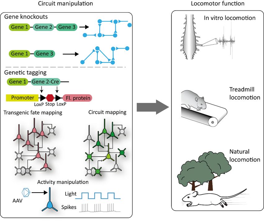
Int. J. Mol. Sci. 2021, 22, 1467 3 of 18
feedback [25,26] and feedback from the vestibular system [27] might underlie the correct
coordination pattern during locomotion, but the details of how these two feedback patterns
play a role at different speeds is not understood.
In this review, we sought to summarize the research investigating the role of segmental
somatosensory feedback and the feedback from the vestibular system during locomotion
at different speeds in mammals. This review will lead to the hypothesis that the weight of
the role of segmental feedback is less important at slower speeds but increases at higher
speeds, whereas the weight of the role of the vestibular system has the opposite relation.
2. Role of Segmental Somatosensory Sensory Feedback in Locomotion
2.1. Overview on Segmental Sensory Feedback on Locomotion
A major source of the sensory feedback during locomotion comes from the segmental
afferents that signal the current position, movement and force of the body, collectively
called proprioception and signals originating from the external world, referred to as
exteroception [28]. Proprioceptive information is mainly transmitted by myelinated Group
Ia and Group II afferents from the muscle spindles and the Group Ib afferents from the Golgi
tendon organs, respectively [29,30]. The signals provided by the Group Ia/II afferents from
the muscle spindles are related to muscle stretch and are therefore an indirect measurement
of the angular displacement of individual joints. On the other hand, the signals conveyed
by the Group Ib afferents relate to tension in the tendons and therefore measure the force
or load. Experiments on human subjects suggest that touch and stretch-sensitive cutaneous
afferent feedback also contributes to the sensation of joint movements [31–34]. The stimuli
that are related to proprioception therefore originate from one’s own body posture or
movement. Exteroceptive information is conveyed by a large array of cutaneous afferents
coming from the skin cutaneous receptors that signal skin deformation due to touch, stretch,
vibration, pressure (mechanoreception), temperature (thermoception) or stimuli perceived
as painful (nociception) [35,36]. The common aspect of these exteroceptors is that the
stimuli originate from outside of the body. Different aspects of locomotor movements are
influenced by either of these feedback modalities; we outline how below.
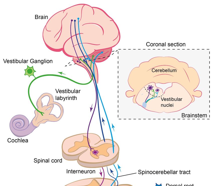
The cell bodies of segmental sensory afferent neurons are located in the dorsal root
ganglia adjacent to the spinal cord. From this cell body, a single neuronal process emerges
that further diverges into a peripheral and a central process (giving these sensory neurons
a monopolar neuron structure) (Figure 1). The peripheral branch of the proprioceptive
afferent neurons project out to the muscles to innervate either the muscle spindles, as for
the Group Ia and II afferents, or the Golgi tendon organs, as for the Group Ib afferents,
whereas the peripheral processes of the cutaneous afferents innervate the skin [37–39].
The central branch enters the spinal cord through the dorsal root entry zone and forms
synapses with diverse inter- or motor neurons located in the grey matter of the spinal
cord [40–42]. While some of these interneurons are involved in information processing
within the spinal cord, others carry the sensory information further to the brain through
specific pathways, such as the dorsal and ventral spinocerebellar pathways [43,44] and the
spinothalamic tracts [39,45]. In addition to these second-order afferents, a branch of the
primary afferents, conveying proprioceptive information, projects up to the supraspinal
centers through the dorsal column of the white mater [39]. Of all the afferent fibers entering
the spinal cord, only the Group Ia afferents’ central projections makes direct synaptic
contact with the motor neurons [46,47]. A number of these pathways are summarized in
Figure 1.Int. J. Mol. Sci. 2021, 22, 1467 4 of 18
Int. J. Mol. Sci. 2021, 22, x FOR PEER REVIEW 4 of 19
Figure 1. Summary of the somatosensory and vestibular sensory pathways and their integration into the brain and
Figure 1. Summary of the somatosensory and vestibular sensory pathways and their integration into the brain and spi‐
spinal cord.
nal cord.
2.2. Exteroceptive Sensory Feedback and Locomotion
Research conducted in early 20th century demonstrated that the removal of cutaneous
feedback in an otherwise intact cat does not cause significant changes in its walking behav-
ior, suggesting that exteroceptive sensory feedback is not necessary for locomotion [48].
These observations were confirmed in later studies showing that the removal of cutaneousInt. J. Mol. Sci. 2021, 22, 1467 5 of 18
feedback caused only minor, transient changes in locomotion on a flat terrain, and these
effects diminished in a matter of weeks [49]. However, significant changes were observed
when descending information from the brain was also missing in addition to the absence
of the cutaneous feedback [50]. In more recent studies, it has been shown that cutaneous
feedback is important in maintaining lateral stability during walking on a split-belt tread-
mill [51]. The role of cutaneous feedback becomes more apparent when cutaneous afferent
fibers are experimentally stimulated during ongoing walking. For example, the stimulation
of cutaneous afferents from the plantar surface of the foot during walking causes either an
increase or initiation of the extensor muscle activity, depending on the phase of the step
cycle during stimulation [52]. Interestingly, a stimulation of cutaneous afferents from the
dorsal surface of the foot either augments the extensor activity if the stimulation occurs
during a stance or causes flexor muscle activity if the stimulation occurs during a swing
phase [53,54]. The latter flexor response to dorsal foot stimulation is called the “stumbling
corrective reaction”, as it is a reflex response that initiates a higher swing movement to
clear obstacles hitting the dorsal side of the food while walking [55]. Overall, it appears
that cutaneous feedback is essential for the fine control of locomotion, though its loss can
be at least partially compensated for by the supraspinal centers.
2.3. Segmental Sensory Feedback and Posture
The general physical rules dictate that, for a stable posture in terrestrial animals,
the center of mass must be kept within the base of support during standing [15]. Due to
the dynamic conditions of locomotion—that is, the forward and lateral movements of
the body—a modification has been proposed that incorporates the velocity of locomotion
and length of the limb into the center of mass position [16]. However, does segmental
somatosensory feedback play a role in maintaining this stable posture during movement?
Past research in cats suggests that cutaneous feedback is important to maintaining stabil-
ity during locomotion, especially in the presence of external perturbations [26,51,56–58].
A critical role of cutaneous feedback in the regulation of the center of mass has also been
suggested in human experiments [59]. Furthermore, muscle spindles have been shown
to provide information regarding the direction and velocity of perturbations, which is
critical for maintaining stability in dynamic environments [60]. In accordance with this,
proprioceptive feedback has been shown to be important to maintaining stability in hu-
mans [61–63]. These results suggest that segmental sensory feedback is necessary for a
stable posture during standing and walking.
Ensuring proper posture, with the center of mass maintained within the base of sup-
port, in an irregular environment requires sensory feedback to coordinate the position and
movement of multiple legs. A very successful historical overview of these reflexes was pre-
sented by [64]. The most intensively investigated interlimb reflex are the crossed reflexes,
where the stimulation of somatosensory afferents of one leg causes a motor response in
the contralateral legs. This reflex has been described in cats [48,65–67], rodents [68–70] and
humans [71,72]. Moreover, using animal models, commissural interneurons have been
identified that are involved in transmitting somatosensory information to the contralateral
side of the spinal cord [40,73]. Besides the crossed reflexes, sensory influences that coor-
dinate the activity of the hind and fore legs have also been demonstrated in cats [26] and
rats [12]. Significant progress is underway in order to understand the neuronal circuitry
that coordinates the activity between the legs and, therefore, maintains a stable posture
during standing and walking, despite perturbations.
2.4. Proprioceptive Sensory Feedback and Locomotion
In contrast to exteroceptive feedback, proprioceptive feedback is required for normal
stepping behavior with a loss of proprioception due to diseases in humans or experimental
animals having a detrimental effect on locomotion [74–76]. The loss of proprioceptive
feedback in humans with a rare form of large fiber neuropathy causes severe deficits
in movement and locomotion, unless these patients learn to compensate for the loss ofInt. J. Mol. Sci. 2021, 22, 1467 6 of 18
proprioceptive feedback with vision [76]. It has also been shown using animal models
that the removal of proprioceptive feedback, either along with all other types of afferent
feedback using surgical methods [77–79] or selectively using genetics [74,75] or chemical
ablation [80], has a detrimental effect on the generation of the locomotor pattern. However,
the effect on quadrupedal animals seems to be less severe than in bipeds. The reason for
the milder effect in animals such as mice is presumably due to the more stable postures of
quadrupeds vs. bipeds due to the lower center of mass and increased base of support [81].
Moreover, if proprioceptive feedback from the muscle spindles is missing but the feedback
from the Golgi tendon organs remains intact, the effect is less severe and more prominent in
the swing phase of the step cycle [74]. These observations suggest that normal locomotion
in a natural environment requires proprioceptive feedback.
Is the proprioceptive influence on the locomotor pattern similar at all speeds, or is
this type of feedback more predominant during certain velocities? The H-, or Hoffmann,
reflex provides a potential means of examining the strength of proprioceptive feedback
during behaviors [82,83]. Here, a stimulation of the peripheral nerve is propagated to
the spinal cord, where the synaptic actions on motor neurons can be read out as EMG
recordings from the muscles. The strength of this reflex can be modified by changes in the
central circuitry, such as alpha-motor neuron excitability or the presynaptic inhibition of
proprioceptive sensory terminals. The H-reflex decreases in gain during running compared
to walking in humans [84], potentially suggesting a reduced proprioceptive feedback
at faster speeds. However, animal investigations have shown that, in the absence of
proprioceptive sensory feedback from the muscle spindles, mice do not locomote at faster
speeds, suggesting proprioceptive feedback is required at higher velocities [11]. We see two
possibilities that could reconcile these apparently contradictory results. First, the human
studies compared H-reflex across two distinct gaits, running and walking, whereas the
mice maintained a trotting gait across a variety of speeds [11,84]. The second explanation
concerns the route of proprioceptive feedback through the nervous system. The H-reflex
measures excitability mainly at the proprioceptive-motor neuron synapse. The presynaptic
inhibition of sensory afferents permits the nervous system to reduce an activity at a specific
branch, while not affecting the other outputs of the same neuron [85]. Indeed, the H-
reflex gain is reduced during behaviors where proprioceptive feedback should be critically
important, such as in the absence of vision or when standing on an unstable surface [86].
It has been suggested that this downregulation of H-reflex gain occurs via presynaptic
inhibition of the sensory neuron to motor neuron synapse and serves to attenuate the spinal
stretch reflexes that could hinder balance [87]. The observation that presynaptic inhibition
can attenuate local reflex responses and ascending information flow independently [85]
suggests that individuals are protected against the loss of balance while preserving the
awareness of limb positions. A similar mechanism could be at play during locomotion at
different speeds.
Nevertheless, these observations do not mean that proprioceptive feedback is not
required for slow locomotion, as many studies have demonstrated that the normal lo-
comotor pattern is eroded in the absence of proprioceptive feedback [74,75]. A possible
explanation is that an alternative mechanism might be able to compensate for the loss
of proprioceptive feedback at slower speeds but not at faster speeds. We posit that this
alternative mechanism is vestibular feedback, as it has been demonstrated that animals,
including humans, with vestibulopathy avoid walking slower speeds [88,89], suggest-
ing that vestibular feedback has a more significant role during slow walking than faster
locomotor speeds.
3. Role of Vestibular Sensory Feedback in Locomotion
3.1. Vestibular Sensory Feedback
As the head moves through space, both rotation and linear acceleration are detected by
organs in the vestibular labyrinth. Rotation is perceived by three bilateral, orthogonal semi-
circular canals. These canals contain a viscous fluid, the endolymph, whose movementInt. J. Mol. Sci. 2021, 22, 1467 7 of 18
deflects hair cells, altering their activity [90]. Linear acceleration is detected in two planes
(horizontal and vertical) by the otolith organs, where small grains known as otoconia
move in response to acceleration and again deflect hair cells. In turn, sensory signals
are transferred to the brain via vestibular sensory neurons that project to the brainstem
and cerebellum.
How the central nervous system uses vestibular sensory information largely depends
on two factors. (i) The type of sensory afferent that conveys vestibular signals and (ii) where
in the nervous system those sensory afferents project. There are two types of vestibular
sensory afferents, classified according to their discharge patterns in the absence of stimula-
tion. These are regular and irregular afferents, with the two types also having differences
in their anatomical, as well as physiological, properties [91]. Both types of afferent have a
resting discharge that permits a bidirectional response to stimulations, i.e., a decrease in
firing with hair cell deflection in one direction and an increase when deflected in the other.
Regular, or tonic firing, afferents encode the angular head velocity (from the canals) and
linear acceleration with respect to gravity (from the otoliths). Irregular, or phasic, afferents
encode both the changes in head velocity and acceleration [92,93]. These sensory neurons
are bimodal, with their cell bodies in the vestibular, or Scarpa’s, ganglion. The central
branch of the vestibular nerve mostly terminates with the various nuclei of the vestibular
nuclear complex. However, primary afferents also innervate the floccular–nodular lobe of
the cerebellum, and there are reports that some fibers innervate non-vestibular nuclei of
the brainstem, such as the cuneate and lateral reticular nucleus (Figure 1) [94].
In considering the vestibular contributions to locomotion, it is important to note that
there are rarely “pure” vestibular signals found in the brain. As the vestibular end organs
are located in the head and the head can be positioned on multiple planes on the body, the
correct interpretation of vestibular signals also requires the immediate integration of propri-
oceptive information. Proprioception from the neck allows the nervous system to infer the
position of the head on the body and then the direction of rotation or acceleration detected
by the vestibular organs. Indeed, many second-order vestibular neurons (i.e., those that
receive input from primary vestibular afferents) are concurrently innervated by propriocep-
tive afferents [95]. In turn, this combined sensory signal can influence multiple descending
pathways that have access to spinal motor circuits. For example, multiple reticulospinal
populations receive second-order vestibular sensory information [96]. Vestibulospinal pop-
ulations also innervate the cervical and lumbar cord and may themselves play an important
contribution in locomotion, though their close connection with the cerebellum means that
it is challenging to infer how much of their output is mediated by vestibular afferents vs.
higher order pathways [10,97].
The initial neural circuitry of the vestibular system is complex, with different afferent
types encoding different angles and velocities of the head movement, which, in turn,
is relayed to multiple regions in the brainstem and cerebellum. There are therefore multiple
potential ways in which vestibular signaling could influence locomotion. Below, we outline
some of the potential roles.
3.2. Maintaining Vision during Locomotion
A key behavioral process that requires the vestibular system is not directly related to
the locomotor pattern itself but does facilitate the behavior. During locomotion, both the
head and body are deflected in the vertical plain. The head can be stabilized on the body
via the actions of the vestibulocollic reflex (VCR), generated by vestibular feedback. This re-
flex stabilizes the head on the body in response to locomotion and other movements [98].
Stabilization of the gaze itself is achieved by a complimentary reflex, the vestibular ocular
reflex (VOR). The VOR uses vestibular afferent information, routed through the vestibular
nucleus and floculus of the cerebellum, to directly control ocular motor neurons [99]. Dur-
ing locomotion, the actions of the VOR mean that visual acuity can be maintained to similar
levels during walking and running as observed by standing in place [100]. Though most
studies point to the role of the vestibular organs in the VOR, this reflex may be influencedInt. J. Mol. Sci. 2021, 22, 1467 8 of 18
by ascending circuits associated with the spinal central pattern generator [101]. Interest-
ingly, the VOR may be the most effective at slow locomotor speeds, with a feedforward
approach preferred at higher velocities [102].
3.3. Maintaining Stability and Balance
The most well-known function of the vestibular system is the maintenance of balance
and stable posture. The vestibular system is required for maintaining balance during stand-
ing, particularly in humans and other bipeds [103]. Further, postural reflexes that respond
to unexpected perturbations require a functioning vestibular system [104]. The vestibular
system can therefore be considered as “stabilizing”, acting to counteract the effects of body
movement, gravity and other external forces. This may seem counterintuitive to a role in
locomotion; if the vestibular system wants to keep us in place, why would it be required
for locomotion, which is inherently unstable? This problem was initially postulated by
von Holst and Mittelstaedt [105]. Interestingly, vestibular reflexes have been shown to
be downregulated when humans transition from standing to walking [106,107]—that is,
its use is state-dependent. Initially, vestibular pathways are downregulated to allow gait
initiation but are then utilized again during the double-support phase of walking [107].
This study points to a phase-specific role for vestibular pathways during locomotor behav-
iors, with the vestibular sensory information most predominant during the double-support
phase of bipedal stepping. Whether similar mechanisms are found in more stable bipeds is
not clear.
3.4. Vestibular Damage and Gait
Patients with damage to the vestibular system suffer from postural instability and an
inability to appropriately respond to unexpected perturbations [108]. A central problem
in ascribing a functional role to the vestibular system in locomotion is an inability to
disambiguate the vestibular system’s role in maintaining balance and upright posture
and the potential role in the generation of the locomotor pattern. That is, as we locomote,
we must maintain our balance. So, is the vestibular system simply coordinating with other
motor pathways to ensure that we maintain an upright posture during locomotion or does
it have a fundamental role in generating the locomotor pattern itself? Though this is an
interesting question from a circuit perspective, from the point of view of animal behavior,
it may be a moot point. Given that it is impossible to generate natural locomotion in the
absence of an upright posture, does the nervous system even consider these as two separate
control problems?
Nevertheless, we can infer some functions of the vestibular system during locomotion
by examining people and animals with either damage to the vestibular organs or by electri-
cal stimulation of these organs. Damage or disruption to the vestibular system in humans
can result from disorders such as vestibular neuritis or Meniere’s disease. The locomotor
pattern in patents with peripheral vestibular damage is severely altered. Patients show an
increased trunk sway, reduced step length, increased base of support, prolonged double-
stance phase and increased variability [109]. At first glance, this phenotype would seem
to indicate that the vestibular system plays multiple roles in the generation of the normal
locomotor pattern. However, many of these can be considered as a secondary consequence
of a loss of balance. Similarly, in quadrupedal animals with vestibular lesions, the main
phenotypes are also associated with disruptions to the balance system. Animals generally
maintain a lower center of mass, reduced cadence, shorter swing and variability in foot
placement [110,111].
In humans, the primary vestibular afferents can be stimulated by galvanic vestibular
stimulation, the application of an electrical current through the mastoid process, resulting
in an increase in vestibular afferent activity on the side of the cathode, and a decrease on the
side on the anode. During walking, galvanic stimulation results in deviations of the heading
direction towards the side of the anode [27]. Similarly, unilateral damage to the vestibular
apparatus results in heading deviations towards the side of the lesion, particularly duringInt. J. Mol. Sci. 2021, 22, 1467 9 of 18
slow walking [88,112]. Galvanic vestibular stimulation also provides the opportunity to
alter vestibular afferent firing during particular phases of the step cycle. The effects on
the gait are largest when stimulation is initiated at heel contact and minimized during the
swing phase [113], and could contribute to a role of the vestibular system in the planning
of future foot placement for forward progression [113,114], with the current swing phase
being coordinated by local spinal circuits. Interestingly, this phase-dependent role of
the vestibular system was only present in the limbs, whereas control of the upper body
was independent of the step cycle, perhaps indicating separate control systems for the
maintenance of posture and locomotion. Interestingly, vestibular stimulation appears to
have less effect on gait direction and variability when running compared to walking [115].
Vestibular damage also results in gait variability [89], a potential phenotype that may
not be directly related to an inability to balance or poor postural control. This could indicate
that vestibular feedback can have a role in foot placement, perhaps due to the requirement
of the vestibular system for understanding the position of the body in space [116]. Temporal
gait variability is associated with damage to both the vestibular system and the cerebellum.
Interestingly, variability associated with cerebellar damage manifests at both slow and fast
walking speeds, whereas the variability found in vestibular patients is only observed at
slow speeds, with a normal variance found at higher gait speeds [89]. This indicates that
the role of the vestibular system in locomotion may be speed-dependent, which is further
discussed below.
3.5. Locomotor Speed and Vestibular Influence
In general, vestibular damage results in a slower gait speed [117], at least partly due to
patients taking longer, slower steps when walking [118]. As discussed above, this slow gait
is highly variable both in the temporal and spatial domains [119]. Intuitively, there could
be two potential reasons for this. First, the vestibular system is believed to be involved in
setting the desired pace of locomotion, and vestibular stimulation can result in an increase
in gait speed [120]. Therefore, vestibular damage may result in a distorted perception of
locomotor speed. Second, a general feeling of disequilibrium or instability could simply
result in the nervous system, reducing locomotor speed to protect the body from falls.
The reduced locomotor speeds would increase the duty cycle; therefore, there is a higher
number of legs providing support. This, in turn, could mean a higher rate of somatosensory
feedback due to longer ground contact (cutaneous feedback) or load signals (group Ib
feedback from the Golgi tendon organs).
However, several studies have noted that gait variability resulting from vestibular
damage is reduced when the locomotor speed is increased. As mentioned, gait variability
is reduced in patients with bilateral vestibular failure during fast walking but not in
patients suffering from cerebellar ataxia [89]. Animals with vestibular damage that are
unable to walk in straight lines when walking are capable of maintaining a constant
heading direction when running [88]. Similarly, humans with vestibular neuritis are able to
maintain a constant heading when running slowly but not when walking [88]. This same
study suggested that these differences could be explained in the differences between spinal
vs. the descending control of locomotion [88], with the largely spinal high-speed locomotion
prompting an inhibition of the descending pathways carrying vestibular information [115].
The potential speed dependence of vestibular signaling provides some important
clues as to the relative roles of vestibular and proprioceptive feedback during locomotion.
We discuss these below.
4. Perspective
Locomotor behavior is controlled by interactions between the CPG, sensory feedback
from the segmental somatosensory system, as well as from supraspinal sensory input,
which includes the vestibular system. The control of normal locomotion requires the inter-
actions of multiple sensory systems. Indeed, both vestibular and somatosensory signals
can be found in similar brain regions, particularly cortical regions [121], and vestibularInt. J. Mol. Sci. 2021, 22, 1467 10 of 18
processing can be influenced by somatosensory signals [122]. However, the role and way
that segmental somatosensory feedback and the vestibular system affect the function of the
CPG is very likely to be distinct in three ways. First, while somatosensory feedback has a
patterning effect for each locomotor cycle, vestibular feedback seems to have a more subtle,
indirect influence on locomotion, unless the locomotion is perturbed. Second, the avail-
ability of vestibular influence on locomotion appears to be state-dependent, whereas
somatosensory feedback is available throughout the locomotion. Though it is worth noting
that some specific reflex pathways are reduced starting just before the initiation of move-
ment or even reversed in sign during locomotion compared to the rest [123,124]. Third,
the influence of segmental somatosensory feedback and vestibular feedback on locomotor
behavior seems to be speed-dependent, such that segmental feedback is necessary at higher
speeds, whereas vestibular feedback is required for slower speeds [11,88]. In the following,
these three aspects will be discussed separately.
4.1. Effect of Segmental Somatosensory and Vestibular Feedback on the Generation of Locomotion
It has been established that the locomotor pattern driving well-coordinated locomotor
behavior is generated by the interactive function of the CPG and sensory feedback [2].
We discussed above that segmental somatosensory feedback does influence very specific
aspects of locomotor movements transiently on a cycle-to-cycle basis, likely through the
direct and specific influence of the CPG network. This influence seems to be important
during unperturbed locomotion [2,8,74,125], as well as to compensate for mechanical
perturbations [8,55,126]. In contrast, vestibular pathways seem to play several accessory
roles in locomotion; most of these can be explained by the need to maintain an upright
posture when walking. Potentially, the vestibular system does not directly influence the
CPG but, rather, has a more general influence on the overall function of the CPG when
the animal is performing a smooth, undisturbed locomotion. However, when locomotion
is perturbed, such as a sudden lateral movement of the terrain, supraspinal pathways
influenced by the vestibular system can step in to provide the necessary motor program
that enables the animal to perform corrective movements [127]. From this, it seems that
both segmental somatosensory feedback, as well as vestibular feedback, are important for
the generation of a functional locomotor pattern, but the way these feedbacks are utilized
is different.
4.2. State-Dependent Modulation of Segmental Somatosensory and Vestibular Feedback
Segmental somatosensory feedback is modulated in state and phase-dependent man-
ners [128], but the feedback is always available to the nervous system so that ongoing
locomotor behavior can be modified in different terrains. This is different from vestibu-
lar feedback. Vestibular feedback is the key to maintaining posture and avoiding mov-
ing/swaying during standing. It is counterintuitive at first glance for the vestibular system,
functioning as a stabilizer to keep us in place, to have a function during locomotion,
which is defined as moving from one place to another and is inherently unstable. However,
it was shown that the vestibular feedback is downregulated at the transition to locomo-
tion and has different effects during different parts of the step cycle [107,113], at least in
humans. This suggests that vestibular feedback is available during locomotion and can be
transitorily called upon at different points of the step cycle to ensure that locomotion does
not disrupt the upright posture. This ability likely requires the nervous system to integrate
both current vestibular sensory feedback, as well make feedforward predictions of how
locomotor actions will impact the postural stability. This integration of sensory feedback
and feedforward predictions likely underlies natural locomotion and could involve other
supraspinal structures, such as the cerebellum [129].
4.3. Influence of Segmental Somatosensory and Vestibular Feedback at Different Locomotor Speeds
Even though segmental sensory feedback is important for the generation of a nor-
mal locomotor behavior, its relative necessity seems to be speed-dependent. It has beenInt. J. Mol. Sci. 2021, 22, 1467 11 of 18
shown that animals, including humans, can perform locomotion if segmental feedback is
partially removed, even though specific changes occur in the timing and amplitude of the
motor activity [74,125]. However, if the segmental proprioceptive feedback is completely
removed, the movements become maladaptive [74]. Moreover, in animal models with
proprioceptive sensory feedback removed from the muscle spindles, locomotion remains at
slower speeds, with higher locomotor speeds avoided [11]. Interestingly, there seems to be
a reverse relationship with the vestibular feedback and locomotor speeds. That is, humans
and animals with vestibular damage prefer a higher locomotor speed [88]. These observa-
tions indicate that, whereas segmental somatosensory feedback, especially proprioceptive
feedback, is required for higher speeds, vestibular feedback is necessary for lower speeds.
However, these observations do not address the question why this would be the case,
nor what the underlying circuit mechanism are. It is conceivable that higher speeds are
prevented due to issues related to biomechanics or spinal circuit functions. Addressing
this will require further investigation.
4.4. Current Working Hypothesis
Based on the above, we hypothesize: “the role of segmental and vestibular feedback
during locomotion depends on speed, vestibular feedback is required at lower speeds
while the somatosensory feedback is necessary at higher locomotor speeds.” The rational
for this hypothesis is the following:
(i). Slower locomotor speeds are more variable and unstable than higher speeds. As slower
walking speeds are more likely to be associated with exploratory activities, they re-
quire frequent changes in the heading direction and body position. Here, the nervous
system must consider how each movement or variation in foot placement could affect
the equilibrium. Therefore, a direct link to the head movement, and overall position
of the body, is imperative.
(ii). Due to the slower speed of movement, there is ample time for the brain to influence
the spinal networks to control foot placement and posture. Multiple descending
pathways influence locomotion, with the vestibular system “in charge” of making
sure that these movements do not cause the animal to lose balance.
(iii). During walking, the features of the step cycle, such as overall cycle duration or length
of the support phase, are variable at different walking speeds, whereas the timing of
these phases during running is consistent across speeds [130]. High-speed locomotion
could therefore be more stereotyped and perhaps dominated by local spinal networks
where segmental somatosensory feedback is the main source of sensory feedback,
allowing minor adaptations to the musculature as locomotion continues.
(iv). The segmental proprioceptive feedback seems to be important for all speeds, it is nec-
essary for higher speeds, as, without them, animals do not locomote at higher speeds.
5. Future Perspective in the Age of Molecular Sciences
Clearly, locomotion is a complex behavior requiring multiple modalities of sensory
feedback, as well as feedforward predictions from the brain. How are we to untangle this
complexity of different neural circuits? Traditionally, the identification of the neural cir-
cuitry that facilitates locomotion has relied on the electrophysiological mapping of neurons
in the spinal cord, sensory pathways and brain [131,132]. Furthermore, many of the studies
discussed above have been based during observations of human patients, precluding a de-
tailed analysis of the underlying circuitry. As we mentioned in the introduction, the mouse
presents an important opportunity to dissect the neural circuitry underlying locomotion.
Mouse genetics can be used in two broad ways to target neural circuitry. First, knockout
strategies can be used to remove individual genes in neurons. The resultant phenotype can
be observed, and some conclusions can be drawn around the role of the underlying circuit
changes. Genetic knockouts can be either global, i.e., the simple removal of a gene from
the entire organism, or conditional, where the gene is removed only from select tissues
or cell types. Further information on the use of gene knockouts in neuroscience studieslocomotion. Mouse genetics can be used in two broad ways to target neural circuitry. First,
knockout strategies can be used to remove individual genes in neurons. The resultant
phenotype can be observed, and some conclusions can be drawn around the role of the
underlying circuit changes. Genetic knockouts can be either global, i.e., the simple re‐
Int. J. Mol. Sci. 2021, 22, 1467 moval of a gene from the entire organism, or conditional, where the gene is removed 12 of 18 only
from select tissues or cell types. Further information on the use of gene knockouts in neu‐
roscience studies can be found elsewhere [133]. As well as traditional gene knockouts, in
the future, more contemporary technologies that use gene editing, such as CRISPR–Cas
can be found elsewhere [133]. As well as traditional gene knockouts, in the future, more
systems, willtechnologies
contemporary also be highly important
that use for understanding
gene editing, the roles
such as CRISPR–Cas of particular
systems, will also becircuits in
motor behaviors [134].
highly important for understanding the roles of particular circuits in motor behaviors [134].
AnAn important
important example
example usingusing traditional
traditional geneticgenetic
knockoutsknockouts can be
can be found found
in the manip-in the ma‐
nipulation
ulation of axonofguidance
axon guidance
moleculesmolecules that
that guide guide
axons axons
to their to their
targets targets
during during develop‐
development,
such
ment,as the
suchgenetic
as thedeletion
genetic of the EPhA4
deletion receptor
of the EPhA4and resultant
receptor andphenotype
resultantof the syn- of the
phenotype
chronous left–right
synchronous “hopping”
left–right gait [135,136].
“hopping” This genetic
gait [135,136]. strategy
This hasstrategy
genetic led to an has
increased
led to an in‐
understanding of the role of commissural interneurons in the coordination of
creased understanding of the role of commissural interneurons in the coordination of left–left–right
alternations [137] (Figure
right alternations [137]2).
(Figure 2).
Figure
Figure 2. 2. Summary
Summary of the
of the molecular
molecular genetic
genetic strategies
strategies that
that can becan
usedbetoused to the
dissect dissect the locomotor
locomotor
circuitry.
circuitry. Gene
Gene knockouts
knockouts (top(top
line)line) can to
can lead lead to circuit
circuit rearrangements
rearrangements that canthat can be combined
be combined with with
several
several techniques
techniques to analyze
to analyze the function
the function ofcircuit
of that that circuit
(right).(right). Similarly,
Similarly, gene expression
gene expression can be can be
used
used to to
tagtag populations
populations of neurons
of neurons and probe
and probe their functions
their functions via manipulations
via manipulations of their
of their activity activity or
or by
by tracing their synaptic inputs. Fl. protein = fluorescent
tracing their synaptic inputs. Fl. protein = fluorescent protein. protein.
Complimentary strategies utilize gene expression patterns in specific subsets of neu-
rons, but rather than probe the function of that gene directly, they use the expression as
a “marker” for that subtype and target genetic or viral tools to probe the circuit function.
In the spinal CPG, the most common strategy has been to utilize the wealth of knowledge
we have regarding transcription factor expression during development and the consequent
sorting of spinal interneurons into four cardinal domains (V0–V4) [5,138]. These classes can
be further subdivided into more genetically and anatomically neuronal classes [139,140].
A common strategy is to “fate map” developing spinal interneurons and express proteins
in adult animals that can either alter their function or remove them from the circuit com-
pletely (Figure 2). Fate mapping generally involves the use of two separate mouse lines
bred together to produce progeny where select tissues or cell types permanently express
a transgene (for example, a fluorescent protein). One mouse line contains a site-specific
recombinase (such as cre) inserted downstream of the gene of interest, and the expressionInt. J. Mol. Sci. 2021, 22, 1467 13 of 18
of the recombinase will therefore be tied to that gene’s appearance. The second mouse line
contains an exogenous gene, such as a fluorescent protein, inserted into the genome under
the control of a ubiquitous promoter. Importantly, this gene will be downstream from a
transcriptional stop flanked by LoxP sites (in the case of cre recombinase). Under wildtype
conditions, the fluorescent protein is not expressed. When both cre and this reporter con-
struct are in the same cell, the transcriptional stop is permanently removed. As this removal
is not reversible, the fluorescent protein will continue to be expressed even after the gene
of interest has been downregulated. This makes fate mapping an important strategy to
study developmentally regulated genes, where exogenous genes can be expressed in the
adult dependent on their developmental gene expression profile. Further information on
the use of fate mapping can be found in a different review [141].
These strategies have yielded important information regarding the function of these
broad classes of neurons, such as V1 interneuron involvement in locomotor speed [142].
Finally, through the introduction of genetic recombinases, gene expressions in adult spinal
neurons can be exploited to target genetic actuators of distinct cells types. These strategies
can be used to manipulate the function of spinal neurons—for example, with chemo- or
optogenetics—or reveal neuronal inputs with transsynaptic tracers, such as the rabies
virus (Figure 2). An important example of this was the use of Chx10 cre lines to probe the
function of V3-derived neurons in both the brain and spinal cord [143–145].
Despite the common use of genetic strategies to dissect the CPG circuitry, the use of
the same tools to dissect sensory contributions to locomotion such as the vestibular and
proprioceptive systems has been relatively lacking. These pathways, though, are amenable
to the same types of mapping and manipulation strategies. For example, rabies virus
tracing can be utilized to dissect the neuronal outputs of vestibular and proprioceptive
sensory neurons [146]. Genetic knockout strategies have also been used to pinpoint the role
of proprioceptive neurons in locomotion, such as the use of Egr3-mutant mice to investigate
the role of proprioceptive sensory feedback in locomotion [74], while a combination of
genetic markers and viral manipulations have been used to remove proprioceptive feedback
from select muscle groups [11].
Compared to other sensory pathways, such as the sensory neurons involved pain or
touch [35,36] or the auditory system [147], our understanding of the genetic subclasses
of the proprioceptive and vestibular pathways is relatively limited. However, important
progress is being made in these pathways. The subtypes of proprioceptive neurons can
be subdivided based on their expression of the ETS transcription factor [148], and the
combinatorial expression pattern of various markers can be used to target phenotypically
distinct proprioceptive neurons [149]. Similarly, in the vestibular system, the developmen-
tal pathways that delineate both vestibular sensory neurons and vestibulospinal neurons
are known [150,151], although there remains much work to do. A continuing push towards
a genetic understanding of the neuronal subtypes in the proprioceptive and vestibular cir-
cuitry, combined with modern molecular genetic strategies, will result in a more complete
understanding of these sensory pathways roles in movement.
Funding: The work in the authors’ laboratories was supported by the Gatsby Charitable Founda-tion
(GAT336) and Wellcome (090843/F/09/Z) (A.J.M.) and Canadian Institutes of Health Research
(162357), Natural Sciences and Engineering Research Council (RGPIN-2015-03871), and National
Institute of Health (R01NS115900-01) (T.A.).
Acknowledgments: We thank Boris Prilutsky (Georgia Institute of Technology) for comments on the
manuscript and Miranda Mathews (Sainsbury Wellcome Centre) for the work on Figure 1.
Conflicts of Interest: The authors declare no conflict of interest.Int. J. Mol. Sci. 2021, 22, 1467 14 of 18
References
1. Grillner, S. Control of locomotion in bipeds, tetrapods, and fish. In Handbook of Physiology: The Nervous System, 2, Motor Control;
Brooks, V., Ed.; Wiley: New York, NY, USA, 1981; pp. 1176–1236.
2. Akay, T. Sensory Feedback Control of Locomotor Pattern Generation in Cats and Mice. Neuroscience 2020, 450, 161–167. [CrossRef]
[PubMed]
3. Grillner, S.; Jessell, T.M. Measured motion: Searching for simplicity in spinal locomotor networks. Curr. Opin. Neurobiol. 2009, 19,
572–586. [CrossRef] [PubMed]
4. Grillner, S.; El Manira, A. Current Principles of Motor Control, with Special Reference to Vertebrate Locomotion. Physiol. Rev.
2020, 100, 271–320. [CrossRef] [PubMed]
5. Goulding, M. Circuits controlling vertebrate locomotion: Moving in a new direction. Nat. Rev. Neurosci. 2009, 10, 507–518.
[CrossRef]
6. Kiehn, O. Decoding the organization of spinal circuits that control locomotion. Nat. Rev. Neurosci. 2016, 17, 24–238. [CrossRef]
7. Guertin, P.A. Central Pattern Generator for Locomotion: Anatomical, Physiological, and Pathophysiological Considerations.
Front. Neurol. 2013, 3, 1–15. [CrossRef]
8. Santuz, A.; Akay, T.; Mayer, W.P.; Wells, T.L.; Schroll, A.; Arampatzis, A. Modular organization of murine locomotor pattern in
the presence and absence of sensory feedback from muscle spindles. J. Physiol. 2019, 597, 3147–3165. [CrossRef]
9. McVea, D.A.; Pearson, K.G. Object Avoidance During Locomotion. Adv. Exp. Med. Biol. 2009, 629, 293–315. [CrossRef]
10. Witts, E.C.; Murray, A.J. Vestibulospinal contributions to mammalian locomotion. Curr. Opin. Physiol. 2019, 8, 56–62. [CrossRef]
11. Mayer, W.P.; Murray, A.J.; Brenner-Morton, S.; Jessell, T.M.; Tourtellotte, W.G.; Akay, T. Role of muscle spindle feedback in
regulating muscle activity strength during walking at different speed in mice. J. Neurophysiol. 2018, 120, 2484–2497. [CrossRef]
12. Juvin, L.; Simmers, J.; Morin, D. Propriospinal circuitry underlying interlimb coordination in mammalian quadrupedal locomotion.
J. Neurosci. 2005, 25, 6025–6035. [CrossRef] [PubMed]
13. Akay, T.; McVea, D.A.; Tachibana, A.; Pearson, K.G. Coordination of fore and hind leg stepping in cats on a transversely-split
treadmill. Exp. Brain Res. 2006, 175. [CrossRef] [PubMed]
14. Frigon, A. The neural control of interlimb coordination during mammalian locomotion. J. Neurophysiol. 2017, 117, 2224–2241.
[CrossRef] [PubMed]
15. Winter, D.A. A.B.C (Anatomy, Biomechanics and Control) of Balance During Standing and Walking; Waterloo Biomechanics: Waterloo,
CA, USA, 1995.
16. Hof, A.L.; Gazendam, M.G.J.; Sinke, W.E. The condition for dynamic stability. J. Biomech. 2005, 38, 1–8. [CrossRef]
17. Lemieux, M.; Josset, N.; Roussel, M.; Couraud, S.; Bretzner, F. Speed-dependent modulation of the locomotor behavior in adult
mice reveals attractor and transitional gaits. Front. Neurosci. 2016, 10, 42. [CrossRef]
18. Novacheck, T.F. The biomechanics of running. Gait Posture 1998, 7, 77–95. [CrossRef]
19. Mendes, C.S.; Bartos, I.; Akay, T.; Márka, S.; Mann, R.S. Quantification of gait parameters in freely walking wild type and sensory
deprived Drosophila melanogaster. eLife 2013, 2013, 1–11. [CrossRef]
20. Wosnitza, A.; Bockemühl, T.; Dübbert, M.; Scholz, H.; Büschges, A. Inter-leg coordination in the control of walking speed in
Drosophila. J. Exp. Biol. 2013, 216, 480–491. [CrossRef]
21. Hildebrand, M. The Quadrupedal Gaits of Vertebrates. Bioscience 1989, 39, 766–775. [CrossRef]
22. Cavagna, G.A.; Thys, H.; Zamboni, A. The sources of external work in level walking and running. J. Physiol. 1976, 262, 639–657.
[CrossRef]
23. Cavagna, G.A.; Heglund, N.C.; Taylor, C.R. Mechanical work basic mechanisms in terrestrial locomotion: Two for minimizing
energy expenditure. Am. J. Physiol. 1977, 233, R243–R261. [CrossRef] [PubMed]
24. Lee, C.R.; Farley, C.T. Determinants of the center of mass trajectory in human walking and running. J. Exp. Biol. 1998, 201,
2935–2944.
25. Bidaye, S.S.; Bockemühl, X.T.; Büschges, A. Six-legged walking in insects: How CPGs, peripheral feedback, and descending
signals generate coordinated and adaptive motor rhythms. J. Neurophysiol. 2018, 119, 459–475. [CrossRef] [PubMed]
26. Hurteau, M.-F.; Thibaudier, Y.; Dambreville, C.; Danner, S.M.; Rybak, I.A.; Frigon, A. Intralimb and Interlimb Cutaneous Reflexes
during Locomotion in the Intact Cat. J. Neurosci. 2018, 38, 4104–4122. [CrossRef] [PubMed]
27. Bent, L.R.; McFadyen, B.J.; Merkley, V.F.; Kennedy, P.M.; Inglis, J.T. Magnitude effects of galvanic vestibular stimulation on the
trajectory of human gait, Neurosci. Lett. Lett. 2000, 279, 157–160.
28. Bigley, G.K. Sensation. In Clinical Methods History Physical Laboratory Examinations, 3rd ed.; Walker, H., Hall, W., Hurst, J., Eds.;
Butterworths: Boston, UK, 1990; pp. 343–350.
29. Jack, J.J.B. Some methods for selective activation of muscle afferent fibres. In Studies in Neurophysiology: Presented to A. K. McIntryre;
Porter, R., Ed.; Cambridge University Press: London, UK; Melbourne: New York, NY, USA, 1978; pp. 155–176.
30. Bewick, G.S.; Banks, R.W. Mechanotransduction in the muscle spindle. Eur. J. Physiol. 2015, 467, 175–190. [CrossRef]
31. Collins, D.F.; Refshauge, K.M.; Todd, G.; Gandevia, S.C. Cutaneous Receptors Contribute to Kinesthesia at the Index Finger,
Elbow, and Knee. J. Neurophysiol. 2005, 94, 1699–1706. [CrossRef]
32. Collins, D.F.; Prochazka, A. Movement illusions evoked by ensemble cutaneous input from the dorsum of the human hand.
J. Physiol. 1996, 496, 857–871. [CrossRef]You can also read