Regulation Is in the Air: The Relationship between Hypoxia and Epigenetics in Cancer - MDPI
←
→
Page content transcription
If your browser does not render page correctly, please read the page content below
cells
Review
Regulation Is in the Air: The Relationship between
Hypoxia and Epigenetics in Cancer
Diego Camuzi 1 , Ísis Salviano Soares de Amorim 2 , Luis Felipe Ribeiro Pinto 1 ,
Leonardo Oliveira Trivilin 3 , André Luiz Mencalha 2 and Sheila Coelho Soares Lima 1, *
 1    Programa de Carcinogênese Molecular, Instituto Nacional de Câncer, Rio de Janeiro CEP 20231-050, Brazil;
      drdcamuzi@gmail.com (D.C.); lfrpinto@inca.gov.br (L.F.R.P.)
 2    Laboratório de Biologia do Câncer (LABICAN), Departamento de Biofisica e Biometria (DBB), Instituto de
      Biologia Roberto Alcântara Gomes (IBRAG), Universidade do Estado do Rio de Janeiro (UERJ),
      Rio de Janeiro CEP 20511-010, Brazil; isisssoares@yahoo.com (Í.S.S.d.A.);
      almencalha@yahoo.com.br (A.L.M.)
 3    Programa de Pós-Graduação em Ciências Veterinárias, Universidade Federal do Espírito Santo (UFES),
      Espírito Santo CEP 29500-000, Brazil; leotrivilin@gmail.com
 *    Correspondence: sheilacoelho@gmail.com; Tel.: +55-21-32076520

 Received: 23 February 2019; Accepted: 26 March 2019; Published: 1 April 2019

 Abstract: Hypoxia is an inherent condition of tumors and contributes to cancer development and
 progression. Hypoxia-inducible factors (HIFs) are the major transcription factors involved in response to
 low O2 levels, orchestrating the expression of hundreds of genes involved in cancer hallmarks’ acquisition
 and modulation of epigenetic mechanisms. Epigenetics refers to inheritable mechanisms responsible
 for regulating gene expression, including genes involved in the hypoxia response, without altering the
 sequence of DNA bases. The main epigenetic mechanisms are DNA methylation, non-coding RNAs,
 and histone modifications. These mechanisms are highly influenced by cell microenvironment, such
 as O2 levels. The balance and interaction between these pathways is essential for homeostasis and is
 directly linked to cellular metabolism. Some of the major players in the regulation of HIFs, such as prolyl
 hydroxylases, DNA methylation regulators, and histone modifiers require oxygen as a substrate, or have
 metabolic intermediates as cofactors, whose levels are altered during hypoxia. Furthermore, during
 pathological hypoxia, HIFs’ targets as well as alterations in epigenetic patterns impact several pathways
 linked to tumorigenesis, such as proliferation and apoptosis, among other hallmarks. Therefore, this
 review aims to elucidate the intricate relationship between hypoxia and epigenetic mechanisms, and its
 crucial impact on the acquisition of cancer hallmarks.

 Keywords: hypoxia; hypoxia-inducible factors; epigenetics; DNA methylation; histones
 modifications; cancer hallmarks

1. Microenvironment in Solid Tumors: Hypoxia
     Hypoxia is a common feature of solid tumors, associated with tumor progression, resistance to
treatment, and poor prognosis. The microenvironment of low oxygen levels arises as a consequence of the
tumor cells high proliferation rate, which is often not accompanied by an efficient angiogenesis [1–4].
     Cell response to hypoxia is mainly regulated by hypoxia-inducible factors (HIFs), a family of
transcription factors involved in coordinating the expression of many genes that allow adaptation
of the tumor cell to this hostile environment [2,5,6]. Hypoxia-inducible factors are heterodimeric
transcription factors, which can be constituted by one of the three α-subunits that are regulated
by oxygen (HIF-1α, HIF-2α and HIF-3α), and a constitutive subunit HIF-1β (also known as aryl
hydrocarbon receptor nuclear translocator, ARNT) [7–9].

Cells 2019, 8, 300; doi:10.3390/cells8040300                                       www.mdpi.com/journal/cells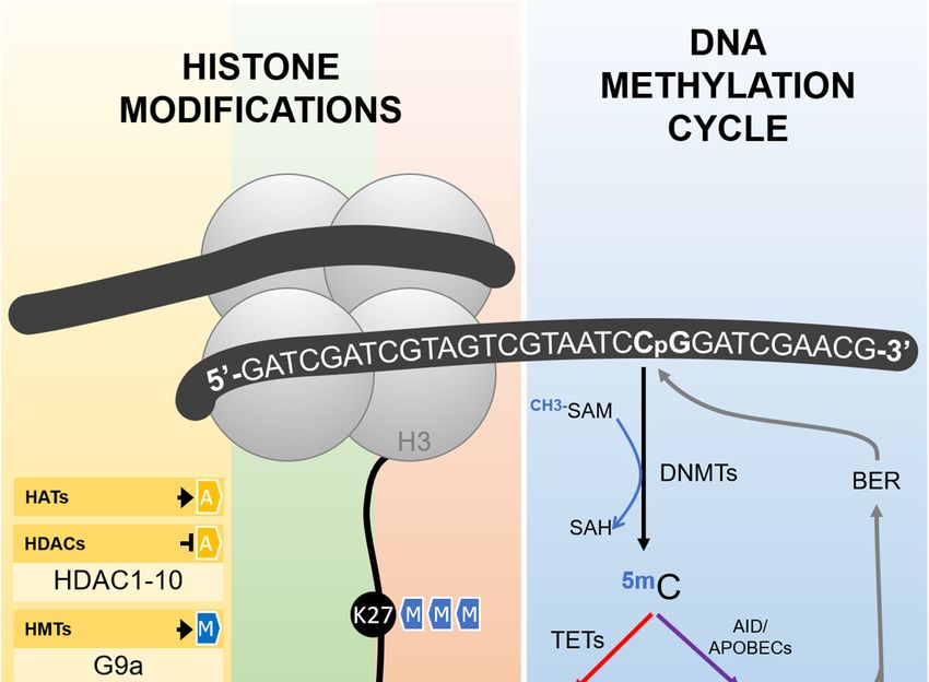
Cells 2019, 8, 300                                                                                  2 of 22

      The two types of HIF subunits are classified as basic helix-loop-helix (HLH)-PER-ARNT-SIM
(bHLH-PAS) proteins [10]. Alpha subunits are constituted by a DNA-binding domain, which is
the basic HLH domain; and two PAS domains (PAS-A and PAS-B), which mediate the interaction
with the subunit HIF-1β. In addition, the N-terminal transactivation domain (N-TAD), found in
the oxygen-dependent degradation domain (ODD), where hydroxylation occurs, together with
the c-terminal transactivation domain (C-TAD), regulate hypoxia-inducible factors transcriptional
activity [11–13]. HIF-1α and HIF-2α have both N-terminal and C-terminal TADs, while HIF-3α has
only the N-terminal TAD [14].
      HIF-1α and HIF-2α are the most commonly studied subunits, whereas the HIF-1α is the
first subunit to respond to hypoxia, and HIF-2α is stabilized after longer hypoxia periods [15].
Although these two subunits share redundant functions, they also exhibit unique and even opposing
activities [6]. For example, by altering the activity of transcription factors such as p53 and MYC, HIF-1α
and HIF-2α exert different effects on cell proliferation and apoptosis, with HIF-1α being known to
inhibit proliferation and induce apoptosis, whereas HIF- 2α has the opposite effect [6].
      On the other hand, HIF-3α has been described with distinct roles in hypoxia, generally thought
to impact negatively by inhibiting HIF-1α and HIF2α. However, it has been suggested that HIF-3α
variants play different roles in regulating gene expression, with stimulating or inhibiting effects
depending on stimulus, organism and cellular source [16].
      Under normoxic conditions, the hypoxia pathway is inactivated by post-translational
modifications. So, in the presence of normal oxygen levels, α subunits proline residues 402 and
564 (HIF-1α), 405 and 531 (HIF-2α), and 490 (HIF-3α) are hydroxylated by the prolyl hydroxylase
enzymes (PHDs). Proline modifications are then recognized by Von Hippel–Lindau (VHL) proteins
that are substrates for the E3 ubiquitination complex. Thus, α subunits are rapidly degraded by the
proteolytic pathway [17,18].
      In hypoxia, hydroxylation rates are reduced, and α subunits are stabilized. They translocate to the
nucleus, dimerize with HIF-1β/ARNT, recruit co-activators such as P300/CBP, and bind to hypoxia
response elements (HRE) in the DNA consensus sequence [A/G]CGTG localized in the promoter
region of target genes [19,20].
      Hypoxia-inducible factors’ target genes allow tumor cell survival in the hypoxic
microenvironment and are involved in regulating pH, angiogenesis, metabolic reprogramming,
proliferation, autophagy, apoptosis, redox homeostasis, inflammation, tumor stem cell maintenance,
invasion, metastasis and treatment resistance [6,21–23]. For instance, HIF-1α and/or HIF-2α induce
the expression of genes such as TGFA and CCND1 to stimulate cell cycle progression and proliferation;
BNIP3 to induce apoptosis and autophagy [24]; GLUT1, HK1, HK2, PFK, ALDA, PGK1, and LDHA to
depict glycolysis; ADM1 and VEGF to promote angiogenesis [25]; MDR1 [26] and ABCG2 [27] to confer
treatment resistance; and TWIST [28] and MMP2 [29] to stimulate invasion and metastasis.
      Apart from their direct effect on gene expression, hypoxia inducible factors and hypoxia
microenvironment are also capable of regulating epigenetic mediators. The term “Epigenetics” states
for inheritable molecular alterations that affect gene expression without changing the bases in the DNA
sequence. However, a broader definition, proposed in the 1940s by Conrad Waddington, emphasizes
the importance of the interaction between environment and genes through epigenetic mechanisms
to define phenotype [30]. This concept is of major importance in the interplay between hypoxia and
epigenetic alterations. In fact, epigenetic mechanisms could be considered as sensors of cell exposure
and as effectors of cell fate. Therefore, hypoxia consequences could be sensed by epigenetic mediators
and, depending on how these responses are orchestrated, could push cells towards a transformed
state. So, cancer hallmarks may be acquired by hypoxia-associated epigenetic alterations, and this
association will be the focus of this review, particularly DNA methylation and histone modifications
(Table 1).
Cells 2019, 8, 300 3 of 22
Table 1. Summary of epigenetics and hypoxia interplay.
Hypoxia and DNA Methylation
Epigenetic
Hypoxic or
Modifier/Modification HIF Involved Cancer/Cell Type Functional Impact Reference
Hypoxic-Like Condition
Involved
Eleven human and murine cell lines from different
Decreased 5hmC levels following hypoxia. In MCF7, 5hmC
normal tissues and tumor types (HepG2, HT-1080,
0.5% O2 for 24 h 5hmC ND was decreased near transcription start sites of NSD1, [31]
MCF10A, H358, MCF7, Hep3B, LLC, mESC WT, N2a,
FOXA1 and CDKN2A
mES Tet1-/- and A549)
MAT2A induction; decreased SAM levels; genomic DNA
HIF-1α Human hepatoma cells (Hep3B) [32]
hypomethylation
1% O2 for 24 h DNA methylation
Human hepatoblastoma cells (HepG2) Increased SAM levels [33]
ND
Human cervix adenocarcinoma cells (HeLa) Decreased SAM levels [34]
In vivo (rats) cerebral
Decreased SAM production; higher global methylation
hypoperfusion (ischemia) DNMT3A ND Brain [35]
levels; higher DNMT3A expression levels
for 90 days
Decreased DNMTs expression, which may contribute to the
In vitro ischemia for 24 h DNMTs ND Human colorectal carcinoma cells (HCT116) [36]
low DNA methylation observed in colorectal tumors
1% O2 for 24 h DNMT1 and DNMT3A ND Human hepatoma cells (Hep3B) Increased DNMTs expression [32]
Healthy human fetal lung fibroblasts (HFL1) and lung Increased DNMT1 expression; HIF-2α hypermethylation
2% O2 for up to 96 h HIF-2α [37]
cancer and decreased expression
DNMT1
DNMT1 recruitment to SPRY2 promoter and its consequent
3% O2 for 24 h HIF-1α and HIF-2α Human hepatoma cells (HuH7 and Hep3B) [38]
decreased expression
1% O2 for up to 8 days DNMT1 and DNMT3B Human primary cardiac fibroblasts (HCF) Increased DNMTs expression [39]
Increased TET1 expression; accumulation of
1% O2 for 48 h TET1 Human neuroblastoma cells (SK-N-BE) [40]
5-hydroxymethylcitosine in hypoxia-responsive genes
1% O2 for 24 h TETs Human hepatoma cells (HepG2) Induced expression of TET enzymes [41]
HIF-1α
Human breast cancer cell lines (MCF7 and Global hydroxymethylation; TNFα overexpression and
1% O2 for 24 h TET1 and TET3 [42]
MDA-MB-231) and primary breast cancer cells activation of the TNFα-p38-MAPk signaling axis
Human metastatic melanoma cells (WM9) and human
Normoxia TET2 Reduced TET2 expression [43]
glioblastoma cells (T98G)
Human hypopharynx carcinoma cells (FaDu) and Increased TET1 expression; regulation of gene expression in
1% O2 for 18 h TET1 HIF-1α and HIF-2α human non-small cell lung cancer cells (derived from response to hypoxia; INSIG1 induced expression; [44]
lymph node metastasis, H1299) promotion of epithelial-mesenchymal transition
VHL deficiency + c-MYC HIF-2α stabilization and PLA2R1 repression by promoter
DNA methylation HIF-2α Human kidney cancer cells (ACHN, RCC10 and 786-O) [45]
amplification hypermethylationCells 2019, 8, 300 4 of 22
Table 1. Cont.
Hypoxia and Histone Modifications
Epigenetic
Hypoxic or
Modifier/Modification HIF Involved Cancer/Cell Type Functional Impact Reference
Hypoxic-Like Condition
Involved
Human fibrosarcoma cells (HT1080), human colon In normoxia, SIRT1 deacetylates HIF-1α, blocking
In vitro 1% O2 for up to
HIF-1α cancer cells (HCT116), human embryonic kidney cells p300 recruitment, and represses HIF-1α targets. This is [46]
24 h; in vivo (mice) 8% O2
(HEK293 and HEK293T) and mice reversed in hypoxia, when SIRT1 levels decrease
SIRT1
In vitro 1% O2 ; in vivo
HIF-2α Human hepatoma cells (Hep3B) and mice Stimulation of HIF-2α activity [47]
(Sirt1+/– mice) 6% O2
Increased affinity of HIF-1α by PHD2, after deacetylation
1% O2 for 16 h SIRT2 Human cervix adenocarcinoma cells (HeLa) [48]
by SIRT2; HIF-1α degradation by proteasome
In vitro 1% O2 for up to
30 h; in vivo (mice) 10% SET7/9 and LSD1 Human cervix adenocarcinoma cells (HeLa) and mice Regulation of HIF-1α stability [49]
O2 for up to 14 days
Human embryonic kidney cells (HEK293T), human
lung adenosquamous carcinoma cells (NCI-H596),
1% O2 for up to 8 h Increased HIF-1α stability; glycolysis upregulation [50]
human colon adenocarcinoma cells (Colo-205) and
LSD1
human clear cell renal cell carcinoma cells (RCC4)
Human lung adenosquamous carcinoma cells Decreased RFK expression, reduced FAD+ levels; HIF-1α
1% O2 for up to 24 h [50].
(NCI-H596) degradation.
Increased JMJD expression and proliferation induction;
1% O2 for 24 h JMJD2B Human colorectal cancer cells (SW480 and HCT116) [51]
reduced H3K9me3 levels in ELF3 and IFI6
HIF-1α Human clear cell renal cell carcinoma cells (RCC4) and Increased JMJD expression and regulation of
0.5% O2 for 16 h [52]
human colon cancer cells (HCT116) hypoxia-inducible genes
In vitro 0.5% O2 or JMJD1A
chemical hypoxia for up Human embryonic kidney cells (HEK293); brain, heart,
Increased JMJD1A expression [53]
to 18 h; in vivo (rats) 8% kidney and liver
O2 for up to 12 h
Human prostate cancer cells (LNCaP), human cervix
adenocarcinoma cells (HeLa) and human renal [54]
Increased JMJD expression
adenocarcinoma cells (786–0 RCC)
0.5% O2 for 16 h JMJD1A and JMJD2B
Human osteosarcoma cells (U2OS), human breast
cancer cells (MCF-7), human cervix adenocarcinoma
[55]
cells (HeLa), human neuroblastoma cells (IMR32) and
human promyelocytic leukaemia cells (HL60)
Human Burkitt’s lymphoma cells (P493-6), mouse PDK1 activation; blockage of PDH activity; reduced
0.1–0.5% O2 for up to 72 h;
HATs? TETs? embryonic fibroblasts (MEF) and mouse hepatoma cells acetyl-CoA synthesis; widespread repression of RNA and [56,57]
0.2% O2 for 48 h
(Hepa 1–6) mRNA synthesisCells 2019, 8, 300 5 of 22
Table 1. Cont.
Hypoxia and Histone Modifications
Epigenetic
Hypoxic or
Modifier/Modification HIF Involved Cancer/Cell Type Functional Impact Reference
Hypoxic-Like Condition
Involved
Human glioblastoma cells (SF188), human embryonic
kidney cells (HEK293T), mouse embryonic fibroblasts
Increased 2-hydroxyglutarate (2HG) production; KDM4C
0.5% O2 for up to 48 h KDM4C ND (MEF), human neuroblastoma cells (SH-SY5Y), mouse [58]
inhibition, increased H3K9me3 levels
bone marrow cells (32D) and mice fetal liver cells
(FL5.12)
JMJD3 activity is reduced, resulting in increased
JMJD3, JARID1A andCells 2019, 8, 300 6 of 22
2. Hypoxia and DNA Methylation
Cytosine DNA methylation is probably the most commonly studied epigenetic mechanism,
usually associated with gene silencing. It consists of the addition of a methyl group to the carbon
five of cytosines followed by guanines (CpG sites), reaction catalyzed by DNA methyl-transferases
(DNMTs), which depend on the universal methyl donor S-adenosylmethionine (SAM) (Figure 1) [67].
CpG sites are spread along the whole genome, but in some specific regions, such as gene promoters
and repetitive elements, a high frequency of these dinucleotides is found, characterizing the CpG
islands. In general, DNA methylation in these regions is associated with transcription silencing, either
by the recruitment of other proteins, as methyl-binding domain proteins (MBDs) and histone modifiers,
or by the inhibition of transcription factors binding to DNA [68,69].
However, DNA methylation is a dynamic process and methyl marks can be removed by both
passive and active demethylation pathways. In the first case, DNA methylation is lost during replication
because of an ineffective copy of methyl marks into the newly synthesized strand. The active pathway
(Figure 1), by comparison, is dependent on an orchestrated sequence of oxidation or deamination enzymatic
reactions. Ten-Eleven Translocation (TETs) enzymes, dependent on alpha-ketoglutarate and oxygen,
are the major players in the oxidative pathway and catalyze the conversion of 5-methylcytosine to
5-hydroxymethylcytosine, which can be further oxidized, forming 5-formylcytosine and 5-carboxylcytosine.
The last two bases are not recognized as normal by the base excision repair (BER) machinery, are removed
from the DNA strand, and finally replaced by a non-methylated cytosine [67,70].
The deamination pathway is controlled by the AID/APOBEC (Activity Induced
Deaminase/Apolipoprotein B mRNA Editing Catalytic Polypeptide-like) family of enzymes that can
deaminate 5-methylcytosine and 5-hydroxymethylcytosine to form thymine and 5-hydroxymethyluracil,
respectively, generating a mispairing between these bases and guanine. This is recognized by the BER
machinery, which removes the mispaired base to bring back a non-methylated cytosine [67,70].
Whereas in normal cells promoter-associated CpG islands are usually demethylated and repetitive
regions are hypermethylated (and consequently silenced), the inverse profile is observed in tumor cells,
with global genomic hypomethylation and hypermethylation of tumor suppressors, which contributes
to both carcinogenesis and tumor progression. In solid tumors, hypoxia may be able to promote this
aberrant DNA methylation profile [31,71]. Thienpont et al. [31] showed that oxygen shortage leads to a
reduced TET activity (without decrease in TET expression), resulting in tumor suppressor gene promoter
hypermethylation. Furthermore, the hypermethylated genes were functionally grouped into cell cycle
arrest, DNA repair and apoptosis, as well as suppressor genes of glycolysis, angiogenesis and metastasis.
Hypoxia-inducible factors can also act directly on the regulation of DNA methylation mechanism.
Liu and colleagues [32] showed that hypoxia reduces the level of the universal methyl donor,
S-adenosylmethionine (SAM), causing hypomethylation of genomic DNA in hepatoma cells.
Although the mechanisms are not clear, the authors observed that hypoxia induces MAT2A (methionine
adenosyltransferase) expression through HIF-1α, which results in the increase of the enzyme activity
and a decrease in SAM production, thus inducing genomic DNA demethylation. This is intriguing
since MAT2A is one of the enzymes responsible for SAM synthesis and, therefore, the induction of
its expression was expected to result in higher SAM levels. Based on this, the authors hypothesize
that SAM could be consumed for polyamine biosynthesis or the different kinetic of MAT isoforms
could play a role. Furthermore, SAM inhibits MAT2A activity [72]. Lower SAM levels have also been
observed in the brain of rats after chronic cerebral hypoperfusion, although higher global methylation
levels were detected [35]. In contrast, Hermes et al. [33] observed the opposite—increased SAM levels
following hypoxia in HepG2 hepatoma lineage. In 2005, the same group showed that hypoxic HeLa
cells present decreased SAM levels [34]. Taking these data into consideration, new studies should be
performed to elucidate the effects of hypoxia on SAM levels in hepatic and other tumor cell types,
since it might be cell-dependent. Notwithstanding, if it is proved that the hypoxic microenvironment
can affect the levels of the methyl donor, its effects on the transcriptome will gain another layer of
complexity, not only based on HIFs activity, but also on global DNA and histone methylation.Cells 2019, 8, 300 7 of 22
Cells 2019, 8, x 8 of 24
Figure 1. Epigenetic mediators involved in the establishment and erasure of histone post-translational
Figure 1. Epigenetic mediators involved in the establishment and erasure of histone
modifications and DNA methylation, as described in the text. Histone acetyltransferases (HATs) are
post-translational modifications and DNA methylation, as described in the text. Histone
responsible for the transfer of an acetyl group to different histone amino acid residues, including lysines
acetyltransferases (HATs) are responsible for the transfer of an acetyl group to different histone
4 andamino
9 (K4 acid
and residues,
K9). Histone deacetylases
including lysines 4 (HDACs)
and 9 (K4 andcatalyze the removal
K9). Histone of this group.
deacetylases (HDACs) Methyl
catalyzegroups
can also be added to different amino acids in the tails of the histones by histone methyltransferases
the removal of this group. Methyl groups can also be added to different amino acids in the tails of the
(HMTs) and removed
histones by histone by methyltransferases
histone demethylases (HDMs).
(HMTs) Histone acetylation
and removed always results
by histone demethylases in an active
(HDMs).
chromatin
Histonestatus, while the
acetylation effects
always of histone
results in anmethylation depends
active chromatin on the
status, number
while of groups
the effects added and
of histone
methylation depends on the number of groups added and the amino acid residue
the amino acid residue involved. As exemplified in the figure, H3K27me3 and H3K9me3 are repressive involved. As
exemplified in the figure, H3K27me3 and H3K9me3 are repressive marks,
marks, while H3K4me3 is an active mark. DNA methylation takes place more frequently in cytosineswhile H3K4me3 is an
active mark. DNA methylation takes place more frequently in cytosines followed
followed by guanines in the so-called CpG sites, reaction catalyzed by DNA-methyltransferases by guanines in the
so-called CpG sites, reaction catalyzed by DNA-methyltransferases (DNMTs) using
(DNMTs) using S-adenosylmethionine (SAM) as methyl donor. The erasure of DNA methylation can
S-adenosylmethionine (SAM) as methyl donor. The erasure of DNA methylation can be driven by an
be driven by an active pathway, either by sequential oxidations or deamination. In the oxidation
active pathway, either by sequential oxidations or deamination. In the oxidation pathway (depicted
pathway (depicted in red), Ten-eleven translocation enzymes (TETs) catalyze the conversion of
in red), Ten-eleven translocation enzymes (TETs) catalyze the conversion of 5-methylcytosine (5mC)
5-methylcytosine (5mC) in 5-hydroxymethylcytosine (5hmC), which is further oxidized to form
in 5-hydroxymethylcytosine (5hmC), which is further oxidized to form 5-formylcytosine (5fC) and
5-formylcytosine (5fC) and 5-carboxylcytosine
5-carboxylcytosine (5caC). Both 5fC and 5caC (5caC). Both 5fC by
are recognized andthe
5caC
baseare recognized
excision repairby the base
(BER)
excision repair (BER) machinery and removed from the DNA strand, resulting in
machinery and removed from the DNA strand, resulting in the reincorporation of a non-methylated the reincorporation
of a non-methylated cytosine. Inpathway
cytosine. In the deamination the deamination
(depicted in pathway
purple), (depicted
5mC and 5hmCin purple),
can be5mC and 5hmC
deaminated by can
be deaminated
AID/APOBEC by AID/APOBEC
(Activity Induced (Activity Induced Deaminase/Apolipoprotein
Deaminase/Apolipoprotein B mRNA Editing B mRNA Editing
Catalytic
Polypeptide-like)
Catalytic family
Polypeptide-like) of enzymes,
family generating
of enzymes, generatingthymine and and
thymine 5-hydroxymethyluracil
5-hydroxymethyluracil (5hmU),(5hmU),
respectively.
respectively. TheThemispairing
mispairing ofof these
thesebases
bases with guanine
with in the
guanine inopposite strand activates
the opposite the BER the
strand activates
BER machinery and results in the reincorporation of a non-methylated cytosine. G9a: histone
methyltransferase G9a; SET7/9: histone-lysine N-methyltransferase SETD7; JARID: Jumonji/ARID
Domain-Containing Protein; JMJD: Jumonji Domain-Containing Protein; LSD1: Lysine-specific
demethylase 1; SAH: S -adenosylhomocysteine.Cells 2019, 8, 300 8 of 22
In addition, Skowronski et al. [36] observed that hypoxia and hypoglycemia cause DNMTs
downregulation in human colorectal carcinoma cells, which may contribute to the pattern of low DNA
methylation observed in colorectal tumors. However, Liu et al. [32] observed that hypoxia induced the
expression of DNMT1 and DNMT3A in Hep3B hepatoma cells.
In human primary cardiac fibroblasts, Watson et al. [39] showed that the increased activity of
DNMT1 and DNMT3B promoters in hypoxia is regulated by HIF-1α. Likewise, hypoxia-induced
HIF-2α induced DNMT1 expression, which promoted HIF-2α promoter hypermethylation and
decreased its mRNA expression, functioning as a negative feedback mechanism for HIF-2α regulation
in healthy human fetal lung fibroblasts [37].
Apart from HIFs’ effects on DNMTs expression, HIFs can also recruit these enzymes to specific
gene regions and affect the transcription of important genes for tumorigenesis. The protein Sprouty
2 (SPRY2) is known to attenuate tyrosine kinase receptor signaling, and acts as a tumor suppressor.
In addition, SPRY2 protein levels are often reduced in several cancers, such as liver, lung, prostate
and breast, which is associated with poor prognosis and shorter survival [73–76]. To elucidate the
mechanisms involved in SPRY2 downregulation, Gao et al. [38] employed the Hep3B hepatoma cells.
This study showed that HIF-1α and HIF-2α facilitate DNA methylation mediated by DNMT1 in the
regulatory region of SPRY2, leading to its decreased expression [38].
Mariani et al. [40] showed an increase in TET1 expression, mediated by HIF-1α, resulting mainly
in the accumulation of 5-hydroxymethylcitosine in hypoxia-responsive genes, and the induction of
the hypoxia response transcriptional program in neuroblastoma. These data indicated an interaction
between epigenetic mechanisms and hypoxia-inducible factors in the regulation of gene expression
related to hypoxia. Similarly, Lin et al. [41] indicated that hypoxia regulates DNA methylation by
increasing HIF-1α-mediated TET expression in hepatocellular carcinoma cell, HepG2.
In addition, Wu et al. [42] indicated that hypoxia, through HIF-1α, enhanced the expression of
TET1 and TET3 enzymes, leading to increased global hydroxymethylation. In this context, the authors
also observed TNFα overexpression and activation of the TNFα-p38-MAPk signaling axis, which
contributed to the acquisition of breast tumor-initiating cell characteristics.
However, Fischer and Miles, [43] have observed that TET2 expression was reduced by the action of
HIF-1α on WM9 human metastatic melanoma cells and T98G glioblastoma cells. The same study showed
that HIF-1α silencing reverted TET2 downregulation and increased ascorbic acid-induced TET2-dependent
5-hydroxymethylation. They further suggested that the combined use of HIF-1 inhibitor and ascorbic acid
may promote the re-expression of methylated tumor suppressors and may be useful in antitumor therapy.
Finally, Tsai et al. [44] suggested that hypoxia, through HIF-2α, upregulates TET1 expression,
which acts as a co-activator of HIF-1α and HIF-2α, contributing to the regulation of gene expression in
response to hypoxia, including the promotion of epithelial-mesenchymal transition. Furthermore, they
demonstrated that TET1, acting as a coactivator, increased the expression of the main regulator of
cholesterol biosynthesis, INSIG1 (insulin induced gene 1), mediated by HIF-2α. Knockdown of TET1
and INSIG1 reduced hypoxia-induced epithelial-mesenchymal transition (EMT). These data showed
a new role of TET1 enzyme as a cofactor of HIF-1α and HIF-2α and an association between HIF-2α,
TET1, INSIG1 and hypoxia induced-EMT.
Based on these data, we may conclude that the hypoxic phenotype is at least partially mediated
by DNA methylation alterations, depending on both the modulation of SAM’s availability and the
regulation of enzymes involved in DNA methylation and demethylation by HIFs. In the context of
cancer, this link contributes to the establishment of a recurrent tumor epigenotype, involving both
global DNA hypomethylation and tumor suppressor hypermethylation. Therefore, DNA methylation
alterations induced by hypoxia may play a pivotal role in tumorigenesis.
3. Hypoxia and Histone Modifications
Histones are conserved proteins involved in DNA packing and play a central role in regulating
accessibility to genomic information through chromatin remodeling. This process is coordinated byCells 2019, 8, 300 9 of 22
a long list of post-translational alterations, which includes acetylation, methylation, sumoylation,
phosphorylation, among others, and it is crucial for the regulation of gene expression and DNA
repair [77]. Different enzymes are responsible for writing and erasing these post-translational marks
(Figure 1), but they can also modify non-histone proteins, such as HIFs, altering their stability.
In addition, hypoxia regulates both the activity and expression of these enzymes, which could have a
global impact on gene expression profiles.
Sirtuins (SIRTs) are a family of NAD+ -dependent deacetylases that regulate transcription by
inducing heterochromatin formation following the deacetylation of histone tails, but have also been
shown to play a central role in the regulation of key transcription factors, functioning both as oncogenes
and tumor suppressor genes [78]. For instance, the deacetylation of p53 at lysine residue 382 by
SIRT1 leads to a reduction of its transcriptional activity [79]. Complementary, SIRT1 may also act as a
tumor suppressor by deacetylating HIF-1α lysine 674, blocking p300 recruitment and repressing HIF-1α
targets [46]. The same deacetylase has been shown to stimulate HIF-2α activity [47]. Other SIRTs have
been shown to regulate HIF-1α stability. SIRT2-mediated deacetylation of HIF-1α increases its affinity
for prolyl hydroxylase 2, which hydroxylates HIF, inducing its degradation by the proteasome [48].
However, the association between SIRTs and hypoxia is not a one-way street.
SIRT activity is directly dependent on NAD+ levels; therefore, these enzymes are considered
sensors of the cellular redox state. Severe hypoxia increases NADH/NAD+ ratios [80], possibly as
a consequence of oxidative phosphorylation (OXPHOS) inhibition. In this context, hypoxia would
reduce SIRTs activity, leading to a positive feedback by increasing HIF-1α stability and activity.
Thus, these deacetylases could represent one of the links between hypoxia and cancer. In agreement
with this hypothesis, decreased SIRT1 levels have been observed in BRCA1-mutated breast cancers [81].
In addition, SIRT2 downregulation was reported in glioma, hepatocarcinoma, esophageal and gastric
adenocarcinomas [82,83], while small cell lung carcinoma [84], breast cancer and leukemia [85] present
lower levels of SIRT4. Other SIRTs are also dysregulated in cancer (for a review, access [86]), but it
is not clear what comes first during the carcinogenesis process, the inhibition of these enzymes
by the hypoxia-associated reduction of NAD+ levels, the increased HIF-1α stability and activity
resulting from SIRT downregulation, or even if other mechanisms are involved. For example,
a hypoxic-like phenotype is induced during ageing, one of the strongest risk factors associated with
cancer development, with a reduction of NAD+ levels. This leads to a reduction of SIRT1 activity, with
a consequent HIF-1α stabilization [87]. Either way, evidence points to a strong association between
sirtuins and metabolism, consequently hypoxia, and such association should be further explored in
cancer, including their potential as new tumor therapy targets.
Other histone-modifying enzymes are also directly linked to HIF-1α protein stability and involved
in the promotion of tumor growth. Kim et al. [49] have shown in an in vivo model that non-canonical
post-translational modifications in HIF-1α determine the protein’s fate. Methylation of HIF-1α by
SET7/9 (Histone-lysine N-methyltransferase SETD7) marks the protein for degradation, even in
the nucleus. The demethylation process was catalyzed by lysine-specific demethylase 1 (LSD1).
Mice carrying HIF-1A S28Y and R30Q mutations were shown to be resistant to HIF-1α methylation,
resulting in protein stabilization. In these mice, the implantation of Lewis lung carcinoma cells showed
higher tumor growth [49].
LSD1, a FAD-dependent demethylase, removes mono- and dimethyl marks from histone 3,
inducing transcriptional disturbances dependent on the affected amino acid residue (lysine 4 or 9) [88].
While H3K4me and H3K4me2 are associated with active transcription, H3K9me2 is a repressive
mark [89]. Apart from its role in chromatin regulation and HIF-1α demethylation, LSD1 is capable
of augmenting HIF-1α stability, both in normoxia and hypoxia, via an oxygen-independent pathway,
but dependent on its demethylase activity. LSD1 demethylates RACK1K271me2, which then fails
to bring the Elongin C-containing E3 ubiquitin ligase complex to HIF-1α, avoiding its proteasomal
degradation [50]. Therefore, in the early stages of normoxia and hypoxia, LSD1 plays a role in
upregulating glycolysis via HIF-1α stabilization. Consistent with this, LSD1 upregulation is observedCells 2019, 8, 300 10 of 22
in hematological and solid tumors [90]. However, during prolonged hypoxia, FAD+ levels decrease as
a consequence of the diminished expression of riboflavin kinase (RFK) and flavin adenine dinucleotide
synthetase 1 (FLAD1), leading to impaired LSD1 activity and consequent HIF-1α degradation [50].
This strong correlation between LSD1-promoted demethylation and HIF-1α levels is further reinforced
by observations in human samples. In triple-negative breast cancer, both hypoxia pathway signature
and the average expression of LSD1 target genes are associated with a poor prognosis [50,91].
Therefore, LSD1 inhibitors are currently in clinical trials involving different tumor types, such as
acute myeloid leukemia, small cell lung carcinoma, prostate cancer, among others [90].
Histone demethylases from the Jumonji Domain-Containing (JMJD) family of proteins have also
been suggested to play a role in the interplay between hypoxia and epigenetics. Wellmann et al. [53]
showed in human embryonic kidney cells (HEK-293) and human microvascular endothelial cells
(HMEC-1) that hypoxia and hypoxia mimetic agents induce JMJD1A expression, which is abrogated
after HIF-1A silencing [53]. The authors further showed that JMJD1A promoter harbors a hypoxia
responsive element, enabling its induction by HIF-1α both in vitro and in vivo. HIF-1α is also able to
bind to JMJD2B promoter, inducing its expression [54]. The regulation of JMJD proteins by hypoxia was
also evaluated in other human cancer cell lines (U2OS, MCF7, HeLa, IMR32 and HL60), confirming
the induction of JMJD1A and JMJD2B [55]. JMJD2B upregulation by HIF-1α during hypoxia has
been linked to the modulation of hypoxic gene expression, and to increased cell proliferation [51].
Furthermore, the regulation of a subset of hypoxia-inducible genes, including ADM and GDF15,
has been shown to be dependent on JMJD1A in renal cell and colon carcinoma cell lines, reinforcing
the connection between hypoxia, HIF-1α and JMJD-mediated chromatin remodeling [52].
Acetyl-CoA, the fuel of tricarboxylic acid cycle (TCA) cycle, is the universal donor for acetylation
reactions in a cell. Therefore, the activity of histone acetyl-transferases (HATs) is highly dependent
on the concentrations of this intermediate. HATs transfer an acetyl group to histone tails, especially
to lysine residues, annulling their positive charge and causing a weaker interaction with the DNA
molecule, with a consequent euchromatin formation [92]. In hypoxic conditions, the stabilization of
HIF-1 complex leads to activation of pyruvate dehydrogenase kinase 1 (PDK1), blocking pyruvate
dehydrogenase (PDH) activity. As a consequence, acetyl-CoA synthesis from pyruvate is impaired [56].
In this context, it is reasonable to hypothesize that a global reduction of histone acetylation would take
place in hypoxic microenvironments, inducing a heterochromatin state. In fact, a widespread repression
of RNA and mRNA synthesis is induced by hypoxia [57], although the reduction of HAT activity
cannot be pointed at as the sole cause. As previously mentioned, the activity of other histone modifiers
is modulated during O2 deprivation by NAD+ and FAD+ altered levels and, to add more complexity to
this scenery, the activity of some histone demethylases can be limited by α-ketoglutarate availability.
Alpha-ketoglutarate (or 2-oxoglutarate) is a rate-limiting intermediate of the TCA cycle, which
plays an important role in cellular energy metabolism, protein synthesis modulation, immune
system homeostasis, among others [93]. As previously mentioned, it is a cofactor of TET enzymes,
involved in the active demethylation process, but it is also a cofactor of JMJD. During hypoxia,
α-ketoglutarate and the following citrate production through oxidative metabolism of glucose is
reduced, but α-ketoglutarate levels are actually increased. In these conditions, α-ketoglutarate is
produced from glutamine metabolism, and it goes through reductive carboxylation to form citrate,
which supports cytosolic macromolecular synthesis, enabling cell proliferation even in a hypoxic
microenvironment [94]. Therefore, TETs and JMJDs could have enough α-ketoglutarate available to
catalyze their reactions. However, the increase in α-ketoglutarate levels during hypoxia is followed
by an increase of 2-hydroxyglutarate (2HG) production by lactate dehydrogenase A in cells with
wild-type IDH1 and IDH2. 2-hydroxyglutarate has been considered an oncometabolite, since it can
block tumor cell differentiation, maybe because of its potential inhibition of α-ketoglutarate-dependent
enzymes such as TETs and JMJDs [58,94]. Indeed, 2HG produced during hypoxia is sufficient to induce
the spreading of histone repressive marks, including H3K9me3 [58].Cells 2019, 8, 300 11 of 22
The interplay between hypoxia, HIFs and histone modifications seems to be even more complex
than that with DNA methylation. Indeed, the data gathered here show that histone modifiers are
capable of modulating HIFs stability, having a direct impact on hypoxic signaling, but HIFs are also
capable of modulating the expression of histone modifiers. Furthermore, metabolism intermediates,
highly sensible to O2 levels, are cofactors of these enzymes, and HIFs may also exert its transcriptional
activities through the interaction with chromatin remodelers. In cancer, the expression of different
histone-modifying enzymes has been shown to be dysregulated, what can be mediated at least in part
by HIFs, and has an impact on tumor phenotype and prognosis. Furthermore, some hypoxia-associated
tumor transcriptional programs seem to be acquired by the cooperation of HIFs and histone modifiers.
Although much is left to be clarified, these observations suggest a strong connection between hypoxia
and this epigenetic mechanism in the establishment of cancer hallmarks.
4. Painting the Cancer Hallmarks with Epigenetics
The hypoxia phenotype triggered by the activation of hypoxia-inducible factors due to low
O2 levels may eventually become a paradox on tissue homeostasis. Some healthy tissues keep
homeostasis during hypoxia, such as in early embryonic development [95], or in the intestine of
adult individuals [96]. However, along the carcinogenesis and tumor progression13the
Cells 2019, 8, x of 24
consequence of
low O2 levels is selection of more resistant clones [97], and stimulation of several phenotypes linked
adult individuals [96]. However, along the carcinogenesis and tumor progression the consequence
to cancer hallmarks, such as increased angiogenesis, proliferation, invasion, metastasis, genomic
of low O2 levels is selection of more resistant clones [97], and stimulation of several phenotypes
instability, andlinked
immune leakage,
to cancer hallmarks,among others [98–100].
such as increased Regardlessinvasion,
angiogenesis, proliferation, of themetastasis,
hallmark, epigenetic
genomic instability, and immune leakage, among others [98–100]. Regardless of the hallmark,
mechanisms are also altered, responding to hypoxia or interacting with hypoxia-inducible factors to
epigenetic mechanisms are also altered, responding to hypoxia or interacting with
modulate cell fate [101,102], as
hypoxia-inducible described
factors in the
to modulate next
cell fate sections
[101,102], and summarized
as described in Figure
in the next sections and 2 and Table 1.
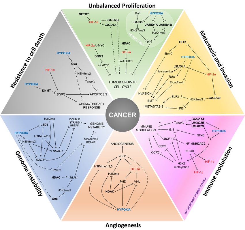
summarized in Figure 2 and Table 1.
Figure 2. Interactions betweenbetween
Figure 2. Interactions hypoxia, HIFs
hypoxia, HIFs and epigenetic
and epigenetic players
players in the establishment
in the establishment of cancer of cancer
hallmarks. The figure summarizes how the establishment of cancer hallmarks can be influenced by
hallmarks. The the
figure summarizes how the establishment of cancer hallmarks can be
cross-regulation of the expression and activity of HIFs and epigenetic modifiers during hypoxia.
influenced by the
cross-regulationText
of the expression
in blue: hypoxia due and activity
to low of HIFs
O2 levels; text in and epigenetic modifiers
red: hypoxia-inducible during
factors; text in bold:hypoxia. Text in
epigenetic players.
blue: hypoxia due to low O2 levels; text in red: hypoxia-inducible factors; text in bold: epigenetic players.
4.1. Unbalanced Proliferation
Cell cycle is controlled by cyclins and cyclin-dependent kinases (CDK), the latter being directly
inhibited by specific inhibitors (cyclin-dependent kinase, inhibitors, CDKI). The cyclin-dependent
kinase Inhibitor 2A (CDKN2AINK4A or p16) is responsible for monitoring the cell during the
progression of G1/S and through oncogenic stimuli leads to cell senescence [103]. Alterations of p16
are found in several tumors, both downregulation and upregulation [104], and p16 proteinCells 2019, 8, 300 12 of 22
4.1. Unbalanced Proliferation
Cell cycle is controlled by cyclins and cyclin-dependent kinases (CDK), the latter being directly
inhibited by specific inhibitors (cyclin-dependent kinase, inhibitors, CDKI). The cyclin-dependent
kinase Inhibitor 2A (CDKN2AINK4A or p16) is responsible for monitoring the cell during the
progression of G1/S and through oncogenic stimuli leads to cell senescence [103]. Alterations of
p16 are found in several tumors, both downregulation and upregulation [104], and p16 protein
expression is also used as a biomarker of the oncogenic Human Papillomavirus 16 (HPV16) infection,
particularly in oropharyngeal tumors [105].
In human lung fibroblasts, Raf reduces H3K27me3 repressive marks present in the p16 gene
by the activation of a p16 locus-specific demethylase, JMJD3, causing its activation. However, even
if recruited to the p16 locus, during hypoxia, the activity of the enzyme JMJD3 is reduced by O2
deprivation, its substrate, resulting in increased H3K27me3 levels [59]. However, despite the increase
in the repressive mark, the expression of p16 is not affected. Other analyses pointed to a bivalent
chromatin state where, in addition to the increase of the repressive trimethylation in H3K27, there
is also an increase in the activation trimethylation in H3K4 by the inhibition of the demethylases
JARID1A and JARID1B, also caused by the reduced O2 levels [59]. Thus, showing a role of bivalent
chromatin in the regulation of cell cycle checkpoints during hypoxia. However, this phenotype is
controversial, since p16 can be downregulated during hypoxia in mesenchymal stem cells, pointing to a
tissue-specific regulation [106]. In both cases, however, non-tumoral cell lineages were used as models,
raising the question of how p16 expression would be modulated in tumor cells in hypoxic conditions.
Proliferation pathways have been shown to be altered by DNA methylation mechanisms during
hypoxia. The c-MYC proto-oncogene is dysregulated in a series of tumors and is linked to increased
proliferation and apoptosis evasion, having hypoxia-inducible factors as partners or antagonists in
several processes [107]. So, while HIF-1α antagonizes c-MYC-induced proliferation, HIF-2α increases
it by potentiating the oncogenic effect of c-MYC in an in vitro model with VHL-deficient renal cell
carcinoma [108]. One of the oncogenic mechanisms resulting from this HIF-2α and c-MYC interaction
may be epigenetic changes in target suppressor genes. PLA2R1 (Phospholipase A2 Receptor 1) is a
potential tumor suppressor gene with a few known functions, but its increased expression has
been linked to cell cycle arrest and apoptosis induction [109]. Renal cell carcinomas with VHL
deficiency and c-MYC amplification exhibit HIF-2α stabilization and PLA2R1 repression by promoter
hypermethylation. In vitro treatment with the DNMT inhibitor and DNA demethylating agent
5-azadeoxycytidine restored PLA2R1 expression [45].
In contrast, in an in vivo model of sarcoma, HIF-1α is active, whereas HIF-2α has its expression
inhibited. When cells are treated with Vorinostat, an inhibitor of histone deacetylases class I and
II (HDAC1-10), HIF-2α expression is restored, without changes in HIF-1α levels. In these cells,
the restoration of HIF-2α plays a tumor suppressor role, inhibiting the mTORC1 pathway and
promoting a decrease in cell proliferation and tumor growth [60]. These differences suggest that
the role of hypoxia-inducible factors in the regulation of cell proliferation might be dependent on the
tumor type.
4.2. Angiogenesis
The main player of angiogenesis is the vascular endothelial growth factor (VEGF), regulating the
creation of new vessels and vascularization of new tissues, allowing nutrient and O2 supply. In tumors,
when cell growth overcomes that of healthy tissues, a hypoxia microenvironment is created, and VEGF
can be stimulated independently or directly by hypoxia-inducible factors [110,111]. Breast carcinoma
cells in hypoxia (0.01% O2 ) exhibit increased methylation in H3K4 and acetylation in H3K9, both
activation marks, in VEGF promoter region [61]. Furthermore, some studies with in vitro or in vivo
models have shown that treatment with deacetylase inhibitors can inhibit VEGF expression and thus
angiogenesis by restoring the expression of HIF-1α suppressors such as VHL and PHD [112,113].Cells 2019, 8, 300 13 of 22
4.3. Metastasis and Invasion
Hypoxia is a double-edged sword, whereas low O2 levels induce the death of several tumor
cells, the hypoxia-resistant cells exhibit a more invasive and metastatic phenotype [97]. In melanomas,
the loss of 5-hydroxymethylcytosine (5hmC), caused by decreased expression of TETs and of isocitrate
dehydrogenases (IDH1 and IDH2), the latter coding for the enzymes that produce α-ketoglutarate,
is a hallmark of this tumor, particularly to differentiate between melanomas and benign melanocytic
nevi [114,115].
HIF-1α knockdown in a metastatic melanoma cell line, is followed by an increase in TET2 gene
and protein levels. These cells, when supplemented with ascorbic acid (cofactor of TETs enzymes),
show higher 5hmC levels, which are also observed in glioblastoma cell lines [43]. TET2 overexpression
and the consequent increase in 5hmC levels decrease melanoma cell invasion in murine models [114].
Thus, targeting HIF-1α along with ascorbic acid supplementation may be a therapeutic option.
As previously mentioned, HIF-1α increases the transcription of JMJD1A and JMJD2B in malignant
cell lines, such as prostate adenocarcinoma, cervical cancer [54], and clear cell renal carcinoma [52].
Increased expression of JMJD1A is related to the induction of the expression of several genes linked
to invasion [52]. In addition, JMJD1A nuclear expression is associated with tumor stage and lymph
node metastasis in oral and oropharyngeal squamous cell carcinoma [116]. The regulation of JMJD2B
in a HIF-1α-dependent manner during hypoxia increases the invasiveness of colorectal cancer cells
by reducing the levels of the repressive mark H3K9me3 in genes like ELF3 (E74-like ETS transcription
factor 3) and IFI6 (Interferon Alpha Inducible Protein 6) [51]. ELF3 overexpression is involved in the
metastasis process in non-small cell lung cancer [117] and its expression can be a useful biomarker
of lymph node metastasis in colorectal cancer [118], while IFI6 promotes metastasis in breast cancer
cells [119].
JMJD1A is found overexpressed in hepatocellular carcinomas and is related to higher recurrence
rates [62]. During hypoxia in vitro, the expression of the epithelial marker E-cadherin is reduced,
followed by increased levels of the mesenchymal markers N-caderin and Twist. JMJD1A silencing
during hypoxia leads to a reduction of N-cadherin and Twist, along with increased E-cadherin levels [62].
4.4. Genomic Instability
DNA damage is readily sensed and usually corrected by the DNA repair machinery in a normal
cell in order to avoid the accumulation of mutations. Among the types of damage, double-strand
breaks are a major source of stress and DNA repair system impairments can result in genomic
instability, including changes in nucleic acid sequences, chromosomal rearrangements or aneuploidy.
BRCA1 and RAD51 are classical proteins involved in double-strand break repair through homologous
recombination [120] and both proteins have high clinical relevance for breast cancer [121,122].
Breast carcinoma cells submitted to hypoxia (0.01% O2 ) showed a decrease in H3K4 methylation
(H3K4me2,3), associated with increased transcriptional activity in BRCA1 and RAD51 promoter
regions, and, therefore, presented a decreased expression of these genes. In these cells, it was verified
whether this demethylation could be performed by the demethylases JARID1A, JARID1B and LSD1;
however, only LSD1 was involved. Furthermore, the suppressor marker H3K9me3 was increased in
the promoter region of BRCA1 and RAD51. There was also a decrease of H3K9 acetylation, a marker
present in transcriptionally active regions. Interestingly, for both genes, the demethylation of H3K4 was
performed independently of HIF-1α expression [61].
MLH1 and PMS2 are tumor suppressor proteins that form a heterodimer involved in DNA
mismatch repair [123]. During hypoxia, an increased expression of the histone methyltransferase G9a
(G9a) is observed, which is related to global and localized increase of the suppressor mark H3K9me2.
Under these conditions, higher H3K9me2 levels are detected in the promoter region of MLH1,
decreasing its expression [63]. Interestingly, cells treated with histone deacetylase inhibitors during
hypoxia have restored MLH1 levels. Although PMS2 mRNA expression is not altered during hypoxia,Cells 2019, 8, 300 14 of 22
there is a drop in its protein levels. These cells with MLH1 and PMS2 impairment during hypoxia
have their repair system affected, resulting in increased mutagenesis and genomic instability [64].
4.5. Immune Modulation
Cells from the immune system act as guardians actively looking for invaders and altered cells
causing the elimination of these in order to maintain body homeostasis. On the other hand, after
immune escape and tumor establishment, a subversion of the immune system occurs, altering the
expression of cytokines that will work in support of the tumor [124]. During this review, we observed
how hypoxia influences the epigenome of malignant cells and vice versa, now we will discuss how the
same phenomenon can be observed in immune cells.
During hypoxia, the expression of the demethylases JMJD1A, JMJD2B and JMJD2D tends to
increase in macrophages possibly as a mechanism to compensate for the low activity of these enzymes
due to O2 deprivation. Despite their increased gene expression, an overall increase in H3K9 and
H3K36 methylation is observed [65], with phenotypes similar to those observed in tumors [52].
Increased H3K9 methylation affects target cytokine genes, as monocyte chemoattractant protein-1 (MCP-1,
also known as CCL2), CC Motif Chemokine Receptor 1 (CCR1) and CC Motif Chemokine Receptor 5 (CCR5),
reducing their gene expression [65]. Similarly, in HeLa cells, MCP-1 gene expression is also reduced
during hypoxia [66].
MCP-1 and IL-8 possess NF-kappaB (NF-kB) responsive elements in their promoters.
However, during hypoxia, MCP-1 expression is suppressed, while IL-8 expression is induced.
Interestingly, NF-κB interacts with both genes, but, at low O2 levels, NF-κB promotes IL-8 transcription,
and in MCP-1 it acts as a repressive factor, recruiting HDAC2 to its promoter region [66]. The increase in
NF-kB expression is common in several tumors and is related to increased malignancy [125], the same
applies to its transcriptional target IL-8 [126]. NF-kB also directly regulates HIF-1α and HIF-1β gene
expression, resulting in upregulation of several hypoxia-like genes [127–129].
4.6. Resistance to Cell Death
In healthy tissues, the balance between proliferation, differentiation and cell death is critical to
maintaining the individual’s homeostasis. Evading apoptosis is a key step during carcinogenesis and
progression. The ability to bypass the mechanisms of cell death is directly related to resistance to
treatment and malignancy [130–132].
Euchromatic histone-lysine N-methyltransferase 2 (EHMT2/G9a) regulates H3K9me2 and is
found overexpressed in several tumor cell lines during hypoxia [63]. The expression of G9a confers
resistance to chemotherapy [133] and the pharmacological inhibition of G9a induces apoptosis in
tumor cells [134,135]. Nonetheless, the connection between G9a and hypoxia on tumor progression is
poorly understood.
BCL-2 interacting protein 3 (BNIP3) is an HIF target gene, involved in apoptosis and autophagy
pathways [136], and its expression is increased during hypoxia in several cell lines by direct HIF-1α
activation [137]. On the contrary, in hypoxic tumor cells, the BNIP3 promoter can be hypermethylated,
resulting in its silencing regardless of HIF-1α expression. Treatment with DNA demethylating
agents and, interestingly not with HDAC inhibitors, restores its expression [138]. The regulation
of BNIP3 during tumor hypoxia is poorly understood and further studies are needed to understand its
causal effect.
5. Final Considerations
Although different studies have tried to elucidate the impact of hypoxia on epigenetic programing,
much is left to be learned. Nevertheless, the epigenetic machinery is an important hypoxia sensor,
and copes with hypoxia-inducible factors to depict the hypoxic phenotype. However, modulation
of epigenetic mediators’ expression by these transcription factors makes evident a highly complex
regulation mechanism. Indeed, the levels of many cofactors and radical donors for reactions catalyzedCells 2019, 8, 300 15 of 22
by epigenetic regulators are affected during hypoxia and have shown to exert a global effect on
gene expression.
One of the clearest consequences of the exposure to a hypoxic environment is the metabolic switch
from oxidative phosphorylation (OXPHOS) to glycolysis, and not surprisingly, one of the proposed
cancer hallmarks is exactly this switch. In this context, epigenetic alterations can both play a role in the
establishment of this phenotype, and be affected by it, indicating a central role of epigenetics in the
acquisition of this cancer hallmark associated with hypoxia. Although epigenetic alterations have also
been associated with the acquisition of other cancer hallmarks, only more recently an association with
hypoxia has been proposed, as made evident by the information gathered here.
Furthermore, data presented in this review suggest hypoxia-epigenetics interaction might be
tissue-specific and dependent on other factors, such as oxygen deprivation duration. Although such
a highly complex interaction is observed during carcinogenesis and tumor progression, modulation
of specific players such as histone modifiers presents promising results in cancer cell control in vitro.
This highlights the potential of alterations of this regulatory axis to be used as biomarkers of tumor
aggressiveness as well as therapeutic targets.
Acknowledgments: We thank the Conselho Nacional de Desenvolvimento Científico e Tecnológico (CNPq) and
the Bridge from the Swiss Private Banking Community to International Cancer Research (Swiss Bridge) for the
financial support of our research and for covering the costs of open access publishing.
Conflicts of Interest: The authors declare no conflict of interest.
References
1. Evans, S.M.; Koch, C.J. Prognostic significance of tumor oxygenation in humans. Cancer Lett. 2003, 195, 1–16.
[CrossRef]
2. Vaupel, P.; Harrison, L. Tumor hypoxia: Causative factors, compensatory mechanisms, and cellular response.
Oncologist 2004, 9 (Suppl. 5), 4–9. [CrossRef]
3. Bertout, J.A.; Patel, S.A.; Simon, M.C. The impact of O2 availability on human cancer. Nat. Rev. Cancer 2008,
8, 967–975. [CrossRef]
4. Petrova, V.; Annicchiarico-Petruzzelli, M.; Melino, G.; Amelio, I. The hypoxic tumour microenvironment.
Oncogenesis 2018, 7, 10. [CrossRef] [PubMed]
5. Semenza, G.L. Hypoxia-inducible factor 1: Master regulator of O2 homeostasis. Curr. Opin. Genet. Dev. 1998,
8, 588–594. [CrossRef]
6. Keith, B.; Johnson, R.S.; Simon, M.C. HIF1α and HIF2α: Sibling rivalry in hypoxic tumour growth and
progression. Nat. Rev. Cancer 2011, 12, 9–22. [CrossRef] [PubMed]
7. Wang, G.L.; Jiang, B.H.; Rue, E.A.; Semenza, G.L. Hypoxia-inducible factor 1 is a basic-helix-loop-helix-PAS
heterodimer regulated by cellular O2 tension. Proc. Natl. Acad. Sci. USA 1995, 92, 5510–5514. [CrossRef]
[PubMed]
8. Tian, H.; McKnight, S.L.; Russell, D.W. Endothelial PAS domain protein 1 (EPAS1), a transcription factor
selectively expressed in endothelial cells. Genes Dev. 1997, 11, 72–82. [CrossRef]
9. Gu, Y.Z.; Moran, S.M.; Hogenesch, J.B.; Wartman, L.; Bradfield, C.A. Molecular characterization and
chromosomal localization of a third alpha-class hypoxia inducible factor subunit, HIF3alpha. Gene Expr.
1998, 7, 205–213. [PubMed]
10. Bersten, D.C.; Sullivan, A.E.; Peet, D.J.; Whitelaw, M.L. bHLH-PAS proteins in cancer. Nat. Rev. Cancer 2013,
13, 827–841. [CrossRef]
11. Jiang, B.H.; Rue, E.; Wang, G.L.; Roe, R.; Semenza, G.L. Dimerization, DNA binding, and transactivation
properties of hypoxia-inducible factor 1. J. Biol. Chem. 1996, 271, 17771–17778. [CrossRef]
12. Jiang, B.H.; Zheng, J.Z.; Leung, S.W.; Roe, R.; Semenza, G.L. Transactivation and inhibitory domains of
hypoxia-inducible factor 1alpha. Modulation of transcriptional activity by oxygen tension. J. Biol. Chem.
1997, 272, 19253–19260. [CrossRef]
13. O’Rourke, J.F.; Tian, Y.M.; Ratcliffe, P.J.; Pugh, C.W. Oxygen-regulated and transactivating domains in
endothelial PAS protein 1: Comparison with hypoxia-inducible factor-1alpha. J. Biol. Chem. 1999, 274,
2060–2071. [CrossRef]You can also read


