Evolution of limb development in cephalopod mollusks - eLife
←
→
Page content transcription
If your browser does not render page correctly, please read the page content below
RESEARCH ARTICLE

                                     Evolution of limb development in
                                     cephalopod mollusks
                                     Oscar A Tarazona1,2, Davys H Lopez1, Leslie A Slota2, Martin J Cohn1,2*
                                     1
                                      Department of Molecular Genetics and Microbiology, University of Florida,
                                     Gainesville, United States; 2Department of Biology, UF Genetics Institute, University
                                     of Florida, Gainesville, United States

                                     Abstract Cephalopod mollusks evolved numerous anatomical novelties, including arms and
                                     tentacles, but little is known about the developmental mechanisms underlying cephalopod limb
                                     evolution. Here we show that all three axes of cuttlefish limbs are patterned by the same signaling
                                     networks that act in vertebrates and arthropods, although they evolved limbs independently. In
                                     cuttlefish limb buds, Hedgehog is expressed anteriorly. Posterior transplantation of Hedgehog-
                                     expressing cells induced mirror-image limb duplications. Bmp and Wnt signals, which establish
                                     dorsoventral polarity in vertebrate and arthropod limbs, are similarly polarized in cuttlefish.
                                     Inhibition of Bmp2/4 dorsally caused ectopic expression of Notum, which marks the ventral sucker
                                     field, and ectopic sucker development. Cuttlefish also show proximodistal regionalization of Hth,
                                     Exd, Dll, Dac, Sp8/9, and Wnt expression, which delineates arm and tentacle sucker fields. These
                                     results suggest that cephalopod limbs evolved by parallel activation of a genetic program for
                                     appendage development that was present in the bilaterian common ancestor.
                                     DOI: https://doi.org/10.7554/eLife.43828.001

                                     Introduction
                                     Animal appendages have widely varying morphologies and perform a multitude of functions, includ-
                                     ing locomotion, feeding, and reproduction (Nielsen, 2012; Ruppert et al., 2004). Limbs evolved on
*For correspondence:
mjcohn@ufl.edu                       multiple occasions, and the absence of shared ontogenetic or morphological precursors of appen-
                                     dages in many animal lineages is consistent with their independent origins (Minelli, 2003;
Competing interests: The             Pueyo and Couso, 2005; Shubin et al., 1997). This has led to the view that appendages in different
authors declare that no
                                     clades of Bilateria are non-homologous morphological innovations that arose by convergent evolu-
competing interests exist.
                                     tion (Nielsen, 2012; Ruppert et al., 2004). However, despite more than 500 million years of diver-
Funding: See page 15                 gence, the independently evolved limbs of arthropods and vertebrates share developmental genetic
Received: 22 November 2018           similarities (Pueyo and Couso, 2005; Shubin et al., 1997; Tabin et al., 1999).
Accepted: 08 May 2019                     These discoveries led to debate over whether the genetic program for appendage development
Published: 18 June 2019              evolved in the common ancestor of all bilaterians in the early Cambrian, or whether arthropod and
                                     vertebrate appendages have undergone rampant convergence of developmental programs (Mine-
Reviewing editor: Nikola-
                                     lli, 2000; Minelli, 2003; Panganiban et al., 1997; Pueyo and Couso, 2005; Shubin et al., 1997;
Michael Prpic-Schäper, University
of Giessen, Germany                  Tabin et al., 1999). A major obstacle to resolving this question is that the evidence of a conserved
                                     program derives almost exclusively from Ecdysozoa and Deuterostomia (Pueyo and Couso, 2005;
   Copyright Tarazona et al. This
                                     Shubin et al., 1997), and little is known about molecular mechanisms of limb development in Spira-
article is distributed under the
                                     lia, the third major superphylum of Bilateria (Grimmel et al., 2016; Prpic, 2008; Winchell and
terms of the Creative Commons
Attribution License, which
                                     Jacobs, 2013; Winchell et al., 2010).
permits unrestricted use and              Within spiralians, the phylum Mollusca is the largest lineage, displaying a rich diversity of body
redistribution provided that the     plans (Figure 1A) dating back to the Cambrian explosion (Ruppert et al., 2004; Smith et al., 2011).
original author and source are       The evolution of arms and tentacles in cephalopod mollusks contributed to the successful adaptive
credited.                            radiation of these agile marine predators (Kröger et al., 2011; Ruppert et al., 2004). Cephalopod

Tarazona et al. eLife 2019;8:e43828. DOI: https://doi.org/10.7554/eLife.43828                                                        1 of 19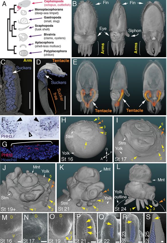
Research article                                                                   Developmental Biology Evolutionary Biology

                                   eLife digest Legs, wings, flippers and tentacles are just some examples of the diverse variety of
                                   animal limbs. Despite striking differences in form and function, all limbs develop in embryos using
                                   similar fundamental processes, like producing an outgrowth from the body and placing structures
                                   such as fingers, feathers, or suckers at appropriate positions. Animals have solved this problem
                                   multiple times during the history of life on Earth, in that limbed animals have arisen from limbless
                                   ancestors on many separate occasions. It is not clear, however, whether the same genetic
                                   instructions shape the developing limbs of all species.
                                       Species that have limbs fall under three main groups of animals: arthropods, such as insects and
                                   crustaceans; vertebrates, like amphibians, reptiles and mammals; and a specialized group of
                                   mollusks known as cephalopods, which includes squid, cuttlefish and octopuses. It has been over
                                   two decades since the discovery that the limbs of vertebrates and insects develop using a similar
                                   molecular recipe, but the mechanisms responsible for the limbs of cephalopods had not been
                                   determined.
                                       Tarazona et al. have now established that the genetic mechanisms that control how cuttlefish
                                   limbs develop are the same as those used by the limbs of vertebrates and insects. These
                                   mechanisms are also applied for similar purposes in each animal group. Notably, a signaling
                                   pathway called hedgehog, which controls the number of fingers that develop on a hand, also
                                   dictates the number of suckers on a cuttlefish arm. This may mean that an ancient system for
                                   creating limbs emerged over 500 million years ago in the earliest animals with bilateral symmetry (i.
                                   e., animals with mirror image halves), and activating this ancient genetic program resulted in the
                                   evolution of limbs in different animal lineages.
                                       The extent of the genetic similarities between cuttlefish, mammals and insects suggests that this
                                   mechanism is likely to provide instructions about where cells position themselves in the developing
                                   limb. The next step is to examine how these common systems are interpreted differently to give
                                   arms, legs, wings and other limb forms.
                                   DOI: https://doi.org/10.7554/eLife.43828.002

                                   limbs are highly muscular appendages that bear cup-shaped suckers on their ventral sides. Arms are
                                   short and have suckers along the entire ventral surface (Figure 1B and C), whereas tentacles are lon-
                                   ger, retractable appendages with suckers restricted to a distal pad (Figure 1D and E). Tentacles are
                                   thought to be specialized serial homologs of the arms (Arnold, 1965; Lemaire, 1970;
                                   Shigeno et al., 2008) and are present in decapods (squid and cuttlefish) but absent in nautilids and
                                   octopods. Limbs likely evolved de novo in cephalopods (Figure 1A), since no homologous precursor
                                   structures have been identified in any other mollusk lineages (Lee et al., 2003; Shigeno et al.,
                                   2008). To test the hypothesis that cephalopod limbs evolved by recruitment of an ancient gene reg-
                                   ulatory network for appendage development that is conserved across Bilateria, we investigated arm
                                   and tentacle development in embryos of the cuttlefish, Sepia officinalis.

                                   Results
                                   Development of arms and tentacles in the cuttlefish (Sepia officinalis)
                                   Cuttlefishes are decapod cephalopods that have eight arms and two tentacles (Figure 1B–E; Fig-
                                   ure 1—videos 1 and 2). Fertilized cuttlefish eggs undergo superficial cleavage, and scanning elec-
                                   tron microscopy and optical projection tomography show that most embryonic development is
                                   restricted to the animal pole (Figure 1H and I). The first sign of limb formation is observed at stage
                                   16, when all ten limb primordia (five on each side) can be detected as small swellings around the
                                   periphery of a flat-shaped embryo, which lies at the top of the large yolk mass (Figure 1H and M).
                                   Analysis of the mitotic marker phospho-histone H3 (PHH3) at stage 15 revealed localized clusters of
                                   PHH3-positive cells in each of the early limb primordia (Figure 1F and G), indicating that initiation of
                                   limb outgrowth is caused by localized cell proliferation. Discrete limb buds are observed from stage
                                   17 (Figure 1I and N; Figure 1—video 3). As the embryo begins to rise-up on the animal pole
                                   around stage 19, the limb buds start to elongate along the proximodistal axis (Figure 1J and O;

Tarazona et al. eLife 2019;8:e43828. DOI: https://doi.org/10.7554/eLife.43828                                                       2 of 19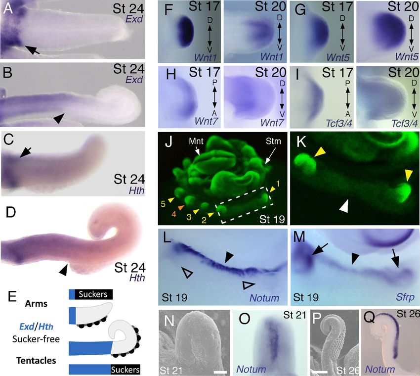
Research article                                                                            Developmental Biology Evolutionary Biology

                                   Figure 1. Development of arms and tentacles in the cuttlefish, Sepia officinalis. (A) Phylogenetic relationships of
                                   Mollusca based on phylogenomic data (Smith et al., 2011) illustrating the unique morphology of the cephalopod
                                   body plan compared to other mollusks. (B) OPT reconstruction of a cuttlefish hatchling showing positions of the
                                   limbs; only arms are visible (see also Figure 1—video 1). (C to D), SEM of the ventral side of a cuttlefish arm (C)
                                   and tentacle (D). Suckers are pseudocolored blue. Note distal restriction of suckers in tentacle relative to arm. (E)
                                   OPT reconstruction illustrating the internally retracted tentacles. Specimens are same as in (B), but here the
                                   tentacles are displayed in orange and the rest of the tissue is rendered translucent (see also Figure 1—video 2).
                                   Figure 1 continued on next page

Tarazona et al. eLife 2019;8:e43828. DOI: https://doi.org/10.7554/eLife.43828                                                                    3 of 19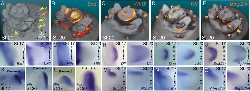
Research article                                                                            Developmental Biology Evolutionary Biology

                                   Figure 1 continued
                                   (F and G) Phospho-histone H3 (PHH3) immunostaining at stage 15 shows localized clusters of proliferating cells at
                                   the onset of limb development (black arrowheads) but little proliferation in the interlimb region (open arrowhead).
                                   (F) PHH3 detection by colorimetric reaction with DAB in a whole mount. (G) PHH3 immunofluorescence (red) on
                                   limb cryosection (white bracket). Cell nuclei (blue) are labeled by Hoechst staining. (H-L) OPT reconstructions of
                                   cuttlefish embryos at stages 16 to 24. Cuttlefishes have five bilaterally symmetric limb pairs (ten limbs; eighth arms
                                   and two tentacles). Numbered arrowheads mark all five limbs/limb buds on the left side of each embryo. The left
                                   tentacle differentiates from position number four (orange arrowhead), whereas arms form from limb buds at the
                                   other positions (yellow arrowheads). See also Figure 1—videos 3—5. A, anterior; P, posterior; D, dorsal; V,
                                   ventral; Stm, stomodeum; Mnt, mantle. (M to O) SEM during early stages of cuttlefish limb development (stages
                                   16 to 19). Morphogenesis of the limb is first observed as a slight swelling (M) that transforms into a limb bud (O) as
                                   proximodistal outgrowth progresses. D, dorsal; V, ventral. (P–S), SEM at later stages of cuttlefish limb
                                   development (stages 21 to 25) showing the formation of sucker buds on the ventral surface of a developing limb.
                                   A primordial sucker band (yellow arrows) is observed along the ventral midline of a stage 21 limb bud (P). At later
                                   stages, the band cleaves superficially from the proximal end to form the sucker buds (pseudocolored blue in Q to
                                   S). Scale bars: 0.5 mm (C and D) and 100 mm (M to S).
                                   DOI: https://doi.org/10.7554/eLife.43828.003
                                   The following video and figure supplement are available for figure 1:
                                   Figure supplement 1. Sucker morphogenesis.
                                   DOI: https://doi.org/10.7554/eLife.43828.004
                                   Figure 1—video 1. OPT 3D reconstruction showing cuttlefish hatchling morphology.
                                   DOI: https://doi.org/10.7554/eLife.43828.005
                                   Figure 1—video 2. OPT 3D reconstruction showing the internal location of the tentacles in a cuttlefish hatchling.
                                   DOI: https://doi.org/10.7554/eLife.43828.006
                                   Figure 1—video 3. OPT 3D reconstruction showing morphology of a cuttlefish embryo at stage 17.
                                   DOI: https://doi.org/10.7554/eLife.43828.007
                                   Figure 1—video 4. OPT 3D reconstruction showing morphology of a cuttlefish embryo at late stage 19.
                                   DOI: https://doi.org/10.7554/eLife.43828.008
                                   Figure 1—video 5. OPT 3D reconstruction showing the morphology of a cuttlefish embryo at stage 24.
                                   DOI: https://doi.org/10.7554/eLife.43828.009

                                   Figure 1—video 4), and by stage 24, the differential length and morphology of arms relative to ten-
                                   tacles is apparent (Figure 1L; Figure 1—video 5).
                                       Analysis of sucker development showed that a sucker field primordium initially forms as a narrow
                                   proximodistal ridge along the ventral surface of each limb (evident by stage 21; Figure 1P). At later
                                   stages, the sucker field ridge cleaves superficially, segregating sucker buds from proximal to distal
                                   (Figure 1Q). As the arms elongate, the sucker buds are laid down on the entire ventral surface of
                                   each arm (Figure 1L and R; Figure 1—figure supplement 1A and C–G), forming four parallel rows
                                   across the anteroposterior axis (Figure 1C; Figure 1—figure supplement 1A). In the tentacles, the
                                   primordial sucker band is restricted to the distal tip, where sucker buds form in eight rows along the
                                   anteroposterior axis of the tentacle sucker pads (Figure 1D; Figure 1—figure supplement 1B). The
                                   full complement of immature sucker bud rows is present on each limb at hatching, and differentia-
                                   tion of the suckers continues during post-hatch development (Figure 1—figure supplement 1H and
                                   I).

                                   Molecular analysis of cuttlefish limbs reveals conservation of
                                   proximodistal, anteroposterior, and dorsoventral patterning networks
                                   To test the hypothesis that cuttlefish limb development is regulated by the same molecular mecha-
                                   nisms that pattern arthropod and vertebrate limbs, despite their independent evolutionary origins,
                                   we cloned and characterized cuttlefish orthologs of genes that pattern the three axes of vertebrate
                                   and arthropod limbs, and then analyzed their expression patterns during cuttlefish limb develop-
                                   ment (Figure 2 and Figure 2—figure supplements 1–10).
                                      Partial sequences of cuttlefish cDNAs (Sepia officinalis and Sepia bandensis) were isolated by rt-
                                   PCR, and preliminary identities were determined by comparison with NCBI sequence databases,
                                   including the octopus genome. Molecular phylogenetic reconstructions were then made by maxi-
                                   mum likelihood phylogenetic inference using the best amino acid substitution model for each gene

Tarazona et al. eLife 2019;8:e43828. DOI: https://doi.org/10.7554/eLife.43828                                                                    4 of 19
Research article Developmental Biology Evolutionary Biology
Figure 2. Molecular regionalization of proximodistal, anteroposterior, and dorsovental axes during cephalopod limb development. (A) OPT
reconstruction of cuttlefish embryo at stage 20 showing all five limb buds on the left side of the embryo (arms, yellow arrowheads; tentacle, orange
arrowhead). (B to E) OPT reconstructions showing four representative genes with polarized expression patterns along major axes of limb buds (gene
expression indicated by orange/yellow). Proximodistally polarized expression of Exd (B) and Wnt5 (C). Anteroposteriorly polarized expression of Hh (D),
dorsoventrally polarized expression of Bmp2/4 (E). (F to O), In situ hybridizations of cuttlefish limb buds at stage 17 (left) and stage 20 (right) showing
polarized patterns of expression along the proximodistal axis for Exd (F), Hth (G), Dll (H), Dac (I) and Sp8/9a (J); the anteroposterior axis for Hh (K) and
Ptc (L); and the dorsoventral axis for Bmp2/4 (M), En (N) and Sfrp1/2/5 (O). A, anterior; P, posterior; D, dorsal; V, ventral; Di, distal; Pr, proximal.
DOI: https://doi.org/10.7554/eLife.43828.010
The following source data and figure supplements are available for figure 2:
Source data 1. Source data for molecular phylogenetic analyses.
DOI: https://doi.org/10.7554/eLife.43828.021
Figure supplement 1. Wnt phylogeny.
DOI: https://doi.org/10.7554/eLife.43828.011
Figure supplement 2. Pan/Tcf phylogeny.
DOI: https://doi.org/10.7554/eLife.43828.012
Figure supplement 3. Dac/Dach phylogeny.
DOI: https://doi.org/10.7554/eLife.43828.013
Figure supplement 4. Sp phylogeny.
DOI: https://doi.org/10.7554/eLife.43828.014
Figure supplement 5. Homeodomain transcription factor phylogeny.
DOI: https://doi.org/10.7554/eLife.43828.015
Figure supplement 6. Notum phylogeny.
DOI: https://doi.org/10.7554/eLife.43828.016
Figure supplement 7. Frizzled/Sfrp phylogeny.
DOI: https://doi.org/10.7554/eLife.43828.017
Figure supplement 8. Tgfb phylogeny.
DOI: https://doi.org/10.7554/eLife.43828.018
Figure supplement 9. Hedgehog phylogeny.
DOI: https://doi.org/10.7554/eLife.43828.019
Figure supplement 10. Patched phylogeny.
DOI: https://doi.org/10.7554/eLife.43828.020
family (see Materials and methods for details). Tree topologies with well-supported bootstrap values
showed the position of each cuttlefish gene within the targeted gene families, which included Wnt,
Tcf/Lef, Frizzled (Fzd), Dachsund (Dac/Dach), Notum, Patched (Ptc/Ptch), Hedgehog (Hh), Bone mor-
phogenetic protein (Bmp), Specificity protein (Sp), and the ANTP and TALE homeobox gene families
(trees are shown in Figure 2—figure supplements 1–10 and are described below; gene accession
Tarazona et al. eLife 2019;8:e43828. DOI: https://doi.org/10.7554/eLife.43828 5 of 19Research article Developmental Biology Evolutionary Biology
Figure 3. Expression of proximal identity genes Exd and Hth in arms and tentacles corresponds with distribution of suckers; Wnt signaling repressors
are dorsally restricted. (A and B) Compared to arms (A), tentacles (B) show a distally expanded domain of Exd expression in the proximal region of the
limb. (C and D) A similar pattern of expression is detected for Hth during arm (C) and tentacle (D) development. Distal boundary of Exd and Hth
expression marked by black arrowheads in (A to D). (E) Expanded expression of proximal identity genes correlates with the expanded sucker-free
domain seen in tentacles compared to arms. (F and H) The Wnt ligands Wnt1, Wnt5 and Wnt7 show a distally restricted expression but no dorsoventral
polarization at stages 17 and 20. (I) The Wnt signaling transcription factor Tcf3/4 is also distally restricted but shows no dorsoventral polarization at
stages 17 and 20. (J and K) Fluorescent nuclear stain SYBR Safe highlights limb buds (yellow arrowheads). Boxed region in (J) is enlarged in (K); white
arrowhead marks interlimb region. (L and M) The Wnt ligand repressors Notum and Sfrp1/2/5 are expressed in the dorsal interlimb region (black
arrowhead in L and M; compare with K). Sfrp1/2/5 expression expands into the dorsal limb bud (black arrows in M) in stage 19 embryos, whereas
Notum stays dorsal but proximally restricted (open arrowheads mark the limb buds in L). (N and O) The earliest sign of sucker formation can be
detected by SEM as a slight swelling (N) and by Notum expression (O) on the ventral side of stage 21 limb buds. (P and Q) Expression of Notum is
maintained through later stages of sucker morphogenesis, as seen in stage 26 tentacles (lateral views).
DOI: https://doi.org/10.7554/eLife.43828.022
The following source data and figure supplements are available for figure 3:
Source data 1. Sequence similarities of Sepia bandensis clones.
DOI: https://doi.org/10.7554/eLife.43828.025
Figure supplement 1. Expression of developmental control genes in cuttlefish limb buds.
DOI: https://doi.org/10.7554/eLife.43828.023
Figure supplement 2. Analysis of gene expression in the arms and tentacles of Sepia bandensis embryos.
DOI: https://doi.org/10.7554/eLife.43828.024
ID numbers and the data set used in the phylogenetic analyses is provided in Figure 2—source data
1).
Within the Wnt family of cell signaling proteins, we isolated cuttlefish orthologs of Wnt1, Wnt2,
Wnt5, and Wnt7 (Figure 2—figure supplement 1). Phylogenetic analysis of cuttlefish transcription
Tarazona et al. eLife 2019;8:e43828. DOI: https://doi.org/10.7554/eLife.43828 6 of 19Research article Developmental Biology Evolutionary Biology
factors identified Tcf3/4, an ortholog of arthropod Pangolin and a pro-ortholog of vertebrate Tcf3
and Tcf4 (Figure 2—figure supplement 2), Dac, a pro-ortholog of vertebrate Dach1 and Dach2 (Fig-
ure 2—figure supplement 3), and Sp8/9, a pro-ortholog of vertebrate Sp8 and Sp9 (Figure 2—fig-
ure supplement 4). We also identified numerous homeobox genes, which phylogenetic analyses
confirmed to be Dll, a pro-ortholog of vertebrate Dlx genes, Exd, a pro-ortholog of vertebrate Pbx
genes, Hth, a pro-ortholog of vertebrate Meis1 and Meis2, and Engrailed, a pro-ortholog of verte-
brate En1 and En2 (Figure 2—figure supplement 5).
In addition, we cloned the Wnt extracellular inhibitors Notum and Sfrp-1/2/5, and the Wnt co-
receptor Fzd9/10 (Figure 2—figure supplements 6 and 7). Cuttlefish possess a Bmp-2/4 gene that
is an ortholog of arthropod Dpp and a pro-ortholog of vertebrate Bmp2 and Bmp4 (Figure 2—fig-
ure supplement 8), a Hh gene (Grimaldi et al., 2008) that we show to be a pro-ortholog of the ver-
tebrate hedgehog family (Figure 2—figure supplement 9), and a gene encoding the Hh receptor
Patched, a pro-ortholog of vertebrate Ptch1 and Ptch2 (Figure 2—figure supplement 10). The cut-
tlefish Sfrp ortholog that we identified as Sfrp1/2/5 was annotated incorrectly in the octopus
genome as Frizzled1 (Figure 2—source data 1). We also found two Sp8/9 genes in the octopus
genome (Figure 2—source data 1), and the cuttlefish Sp8/9 gene shows clear orthology to only one
of the two octopus genes (Figure 2—figure supplement 4), suggesting that the Sp8/9 gene under-
went a duplication in cephalopod mollusks. Therefore, we designate the octopus Sp8/9 paralogs as
Sp8/9a and Sp8/9b, and the cuttlefish Sp8/9 gene that we isolated is the ortholog of Sp8/9a.
We next investigated the spatial and temporal expression patterns of these genes during cuttle-
fish limb development. Genes that pattern the proximodistal axis of arthropod and vertebrate limbs
(Lecuit and Cohen, 1997; Mercader et al., 1999; Panganiban et al., 1997; Pueyo and Couso,
2005) showed similarly polarized patterns of expression along the proximodistal axis of cuttlefish
limb buds, with Exd and Hth restricted proximally (Figure 2B,F and G; Figure 3A–E; Figure 3—fig-
ure supplement 1A and B; and Figure 3—figure supplement 2A,I and J) and Dll, Dac, Sp8/9a,
Wnt1, Wnt5, and Wnt7 restricted distally (Figure 2C,H–J; Figure 3F–I; Figure 3—figure supple-
ment 1C–E and L–N; and Figure 3—figure supplement 2B,C,G,I and J). At stages 20–21, the distal
expression boundaries of Exd and Hth and the proximal expression boundaries of Dll and Sp8/9a
appear to mark the morphological boundary between the proximal sucker-free and the distal
sucker-forming regions (compare right panels in Figure 2F–H and J with Figure 1P). Indeed, at
stages when arms and tentacles begin to develop their distinctive morphologies – tentacles are lon-
ger and have an extensive proximal sucker-free domain – the Exd/Hth expression domains were
found to extend further distally in tentacles (Figure 3B,D) compared to arms (Figure 3A and C).
This distal expansion of the Exd/Hth expression domain matches the expanded sucker-free region
and the distal restriction of suckers in tentacles (Figure 3E).
Our finding that the proximodistal axis of cuttlefish limbs shares patterns of molecular regionaliza-
tion with arthropod and vertebrate limbs led us to examine whether anteroposterior and dorsoven-
tral axis development are also conserved. Posteriorly polarized activation of Hedgehog signaling in
arthropod and vertebrate limbs is essential for proper patterning of the anteroposterior axis, and
ectopic activation of the Hedgehog pathway induces anterior duplication of posterior structures
(Basler and Struhl, 1994; Kojima et al., 1994; Riddle et al., 1993). We analyzed Hh expression dur-
ing cuttlefish limb development at stages 16 to 20 and found that Hh expression is also polarized to
one side of cuttlefish limb buds. In cuttlefishes, however, Hh expression is restricted to the anterior
margin of the limb bud, whereas in arthropods and vertebrates, Hh/Shh is expressed posteriorly
(Figure 2D and K; and Figure 3—figure supplement 2D). Consistent with the anterior localization
of Hh, we detected expression of Patched, which serves as a readout of Hedgehog signal transduc-
tion, in an anterior-to-posterior gradient (Figure 2L). Thus, anteroposteriorly restricted activation of
the Hedgehog pathway is a conserved feature of cephalopod, arthropod, and vertebrate limb devel-
opment, but the polarity of the signaling center is reversed in cephalopod limbs. By stage 21, the
anteriorly restricted Hh domain has diminished and a new, central expression domain appears in the
location of the brachial nerve primordia (Figure 3—figure supplement 1F,K).
We then examined the dorsoventral axis, which is controlled by the antagonistic actions of wg/
Wnt and dpp/Bmp signaling in arthropods and vertebrates (Brook and Cohen, 1996; Cygan et al.,
1997; Diaz-Benjumea et al., 1994; Jiang and Struhl, 1996; Parr and McMahon, 1995). In arthro-
pods, the Wnt ligand wg is expressed ventrally, whereas the Bmp2/4 ortholog dpp is expressed dor-
sally (Basler and Struhl, 1994; Diaz-Benjumea et al., 1994). Expression and function of the Wnt-
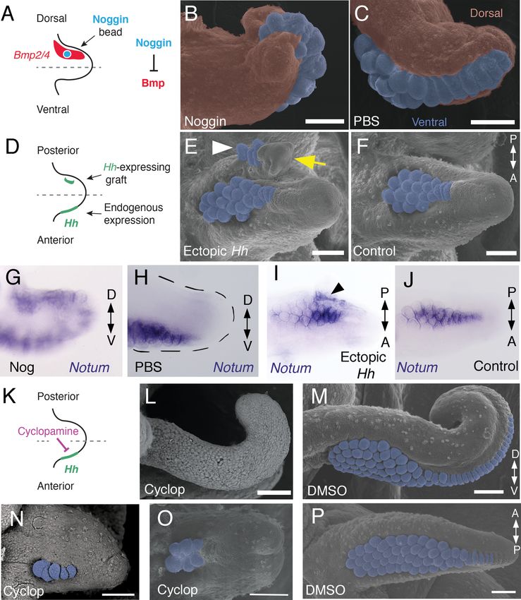
Tarazona et al. eLife 2019;8:e43828. DOI: https://doi.org/10.7554/eLife.43828 7 of 19Research article Developmental Biology Evolutionary Biology Figure 4. Bmp signaling controls dorsoventral patterning and Hh signaling regulates anteroposterior patterning of cuttlefish limbs. (A to C) Implantation of carrier beads loaded with the Bmp inhibitor Noggin (A) results in formation of ectopic sucker buds (n = 8/12) on the dorsal surface of the limb (B), whereas PBS control beads (n = 15/15) result in normal development of the dorsal limb (pseudocolored red) restriction of suckers (pseudocolored blue) to the vental side of the limb (C). (D-F), Hh-expressing cells (taken from the funnel of a stage 24 donor embryo) grafted to the Figure 4 continued on next page Tarazona et al. eLife 2019;8:e43828. DOI: https://doi.org/10.7554/eLife.43828 8 of 19
Research article                                                                            Developmental Biology Evolutionary Biology

Figure 4 continued
posterior side of a stage 17 cuttlefish limb bud (D) generates a posterior mirror-image limb duplication (n = 7/12; yellow arrow in E), whereas no
duplication (n = 8/8) results when control (Hh-negative) cells are grafted to the same position (F). Sucker buds are pseudocolored blue in (E and F);
sucker buds in duplicated limb marked with a white arrowhead. (G and H) Noggin beads induce ectopic expression of Notum on the dorsal side of the
limb (G). Limbs receiving control PBS beads show normal expression of Notum ventrally (H). (I and J) Graft of Hh-expressing tissue to the posterior side
of the limb induces ectopic domain of Notum prior to duplication of the limb (I; black arrowhead). Note the two separate domains of Notum
expression in I compared to a single Notum expression domain in the limb with the Hh-negative control graft (J). (K to P) Transitory repression of Hh
signaling by cyclopamine (K) during early stages of limb development disrupts the anteroposterior distribution of sucker bud rows (L, N, O).
Cyclopamine-treated limbs showing complete loss of suckers in tentacles (L) and reduction in the number of sucker bud rows in arms (N and O).
Control embryos treated with vehicle only (DMSO) develop the normal number of sucker bud rows in tentacles (M) and arms (P). Sucker buds are
pseudocolored blue in (B, E, F, and M- P). Scale bars 100 mm.
DOI: https://doi.org/10.7554/eLife.43828.026
The following figure supplement is available for figure 4:
Figure supplement 1. Sucker development after manipulations of Bmp and Hh signaling pathways.
DOI: https://doi.org/10.7554/eLife.43828.027

                                     Bmp network is conserved, albeit with inverted polarity, in vertebrate limbs; Wnt7a is expressed dor-
                                     sally (Parr and McMahon, 1995) and Bmp signaling activates Engrailed1 (En1) ventrally (Ahn et al.,
                                     2001), and these interactions regulate development of dorsal and ventral limb structures
                                     (Cygan et al., 1997; Parr and McMahon, 1995). During cuttlefish limb development, Bmp2/4 and
                                     En show dorsally polarized expression (Figure 2E,M and N; and Figure 3—figure supplement 2E).
                                     Genes encoding Wnt ligands (Wnt1, Wnt5 and Wnt7) and cellular components of canonical Wnt sig-
                                     naling cascade (Tcf3/4 and Frz9/10) are expressed broadly throughout the dorsoventral axis of cut-
                                     tlefish limb buds (Figure 3F–I and Figure 3—figure supplement 1L–R; and Figure 3—figure
                                     supplement 2G,I and J); however, the secreted Wnt antagonists Notum and Sfrp1/2/5 are
                                     expressed dorsally in the limb and interlimb regions (Figure 3J–M), with the Sfrp1/2/5 domain
                                     extending deeper into the dorsal limb buds (Figure 2O; Figure 3M). This dorsal expression of Wnt
                                     antagonists suggests a mechanism for restriction of Wnt signaling to the ventral side of the cephalo-
                                     pod limb buds. Taken together, these results suggest that the genetic pathways active along the
                                     proximodistal, anteroposterior, and dorsoventral axes of cephalopod limbs are homologous (specifi-
                                     cally, orthologous) to the networks that regulate limb development in arthropods and vertebrates.
                                         In order to further test this hypothesis, we next performed a series of functional experiments to
                                     determine whether polarized expression of these signaling molecules is involved in patterning the
                                     anteroposterior and dorsoventral axes of cuttlefish limbs (described below). We developed a
                                     method for ex-ovo culture of cuttlefish embryos (see Material and methods) to allow in vivo manipu-
                                     lations of genetic pathways in early limb buds.

                                     Bmp signaling controls dorsoventral patterning of cuttlefish limbs
                                     A hallmark of dorsoventral polarity is the restriction of sucker buds to the ventral surface of the limb
                                     (Figure 1C,D and S), and this is preceded by ventral expression of Notum in the sucker-forming
                                     region at stage 21 (Figure 3N–Q). We asked whether polarized expression of Bmp2/4 on the dorsal
                                     side of cuttlefish limb buds is required for the specification of dorsal identity. To repress dorsal Bmp
                                     activity, we implanted carrier beads loaded with Noggin (Nog), a secreted Bmp inhibitor protein, on
                                     the dorsal side of stage 17 limb buds (Figure 4A). Implantation of Nog beads on the dorsal side of
                                     cuttlefish limb buds resulted in ectopic, dorsal expansion of the Notum mRNA domain (n = 3/3; con-
                                     trol PBS [phosphate buffered saline] beads had no effect on Notum expression [n = 3/3])
                                     (Figure 4G,H). To determine whether inhibition of dorsal Bmp signaling respecifies dorsal cells to
                                     form ventral structures, we repeated the experiment and allowed embryos to develop to stage 26–
                                     27. Analysis of limb morphology by scanning electron microscopy revealed the presence of ectopic
                                     sucker buds on the dorsal surface of Nog-treated limbs (n = 8/12; Figure 4B; Figure 4—figure sup-
                                     plement 1A and B). The ectopic dorsal suckers extended around the distal tip of the limb and joined
                                     the ventral sucker field. By contrast, in limbs that received control PBS beads dorsally, sucker buds
                                     were restricted to ventral surface and terminated at the normal dorsal-ventral boundary at the tip of
                                     the limb (n = 15/15; Figure 4C). Our finding that antagonism of Bmp signaling results in

Tarazona et al. eLife 2019;8:e43828. DOI: https://doi.org/10.7554/eLife.43828                                                                    9 of 19
Research article Developmental Biology Evolutionary Biology Figure 5. Molecular patterning of the anteroposterior, proximodistal, and dorsoventral axes of developing limbs in vertebrates, arthropods, and cephalopods. For each lineage, the top row shows schematic representations of a generalized limb bud and an adult limb in two different orientations. Axes are indicated to the left of each limb bud (A, anterior; Po, posterior; Pr, proximal; Di, distal; Do, dorsal; Ve, ventral) Bottom rows show limb buds with gene expression domains pink color). Vertebrate gene expression based on mouse limb development. Arthropod gene expression is a compound reconstruction from chelicerate, myriapod, and hexapod limb development in order to consolidate a complete set of pro-orthologous genes comparable to vertebrate and cephalopod lineages. Cephalopod gene expression is based on findings in this study from the cuttlefish Sepia officinalis and Sepia bandensis. The figure illustrates the conserved and divergent expression patterns of homologous (orthologous) genes, some of which share equivalent roles in patterning the limb axes. The proximodistal axis displays conserved expression of transcription factors at opposite ends; Hth (pro- ortholog of vertebrate Meis genes) and Exd (pro-ortholog of vertebrate Pbx genes) are restricted proximally, whereas Dll (pro-ortholog of vertebrate Dlx genes), Wnt5 (pro-ortholog of Wnt5a) and Sp8/9 (pro-ortholog of vertebrate Sp8 and Sp9 genes, known as Sp6-9 in some arthropods) show distally restricted expression. The typical expression pattern of Dac seen in arthropods (between proximally and distally restricted genes) is not strictly conserved in vertebrates (Dac is the pro-ortholog of vertebrate Dach genes) or cephalopods. However, Dac expression in non-locomotory arthropod appendages (e.g., mandibles) is distally restricted, resembling cephalopod Dac expression (Donoughe and Extavour, 2016). Expression patterns of the diverse family of Wnt genes shows interesting variation. Although, some members of the family show variation in their expression pattern (Wnt1 and Wnt7), there is a general pattern of distal restriction of Wnt expression (represented here by Wnt5, but also seen in many other Wnt ligands) in the three lineages. At the level of individual Wnt members, Wg (pro-ortholog of vertebrate Wnt1) is restricted ventrally in arthropods but not in vertebrates or cephalopods, and Wnt7a (arthropod and cephalopod Wnt7 genes are pro-orthologs of vertebrate Wnt7a) is restricted dorsally in vertebrates but not in arthropods or cephalopods. Restricted expression of Wnt ligands either dorsally or ventrally has not been reported in cephalopods, but the dorsally restricted expression of the Wnt repressor Sfrp1/2/5 suggests a role of polarized Wnt pathway activation in the control of the dorsoventral axis of cephalopod limbs, similar to vertebrates (by dorsal Wnt7a) and arthropods (by ventral Wg). There is a clear restriction of at least one Bmp ligand (vertebrate Bmp7 and cephalopod Bmp2/4; pro-orthologs of arthropod Dpp) and the transcription factor En along the dorsoventral axis in these three lineages. Finally, polarized expression of Hh is conserved in the three lineages (posterior in vertebrates and arthropods, but anterior in cephalopods), which, together with the functional manipulations, indicates conservation of Hh signaling in patterning the anteroposterior limb axis in the three lineages. The asterisk (*) in arthropod Dac indicates that some mouth appendages show a distal expression domain (Donoughe and Extavour, 2016) (Angelini and Kaufman, 2005) more similar to cephalopod Dac limb expression than to Dac expression in arthropod legs. Two asterisks (**) indicate that Wnt5 expression shows variation in arthropods, with a sub-distal expression in chelicerates (Damen, 2002) but distal in hexapods (i.e. flour beetle) (Bolognesi et al., 2008). Three asterisks (***) indicate that Dpp shows variation in its expression domain in arthropods, with some hexapods and Figure 5 continued on next page Tarazona et al. eLife 2019;8:e43828. DOI: https://doi.org/10.7554/eLife.43828 10 of 19
Research article                                                                            Developmental Biology Evolutionary Biology

Figure 5 continued
chelicerates showing a distal expression domain, whereas in Myriapods and other hexapods it is dorsally restricted, as depicted here (Angelini and
Kaufman, 2005). Schematized gene expression domains for vertebrates and arthropods are from the following sources. Mouse gene expression: Meis1
(González-Lázaro et al., 2014), Pbx1 (Capellini et al., 2006), Sp8 (Kawakami et al., 2004), Dlx5 (Vieux-Rochas et al., 2013), Dach1 (Salsi et al.,
2008), Wnt1, Wnt5a, Wnt7a, Sfrp2 (Witte et al., 2009), Shh (Riddle et al., 1993), Bmp7 (Choi et al., 2012) and En1 (Loomis et al., 1998). Arthropod
expression based on: Chelicerates, Hth, Exd, Dll, Dac (Prpic et al., 2003), Sp8/9 (Königsmann et al., 2017), Wg (Damen, 2002), En (Damen, 2002)
and Sfrp1/2/5 (Hogvall et al., 2018); Myriapods, Hth, Exd, Dll, Dac (Prpic and Tautz, 2003), Sp8/9 (Setton and Sharma, 2018), Wg, Dpp
(Prpic, 2004), Wnt5, Wnt7, Hh, En (Janssen et al., 2004), Sfrp1/2/5 (Hogvall et al., 2018); Hexapods, flour beetle, Hth, Exd (Prpic et al., 2003), Sp8/9
(Schaeper et al., 2010), Dll (Beermann et al., 2001), Dac (Prpic et al., 2001), Wg, Wnt5 (Bolognesi et al., 2008), Dpp (Sanchez-Salazar et al., 1996),
En (Brown et al., 1994); Hexapods, cricket, Hth, Exd, Dll, Dac, Wg, Hh, Dpp, En (Donoughe and Extavour, 2016).
DOI: https://doi.org/10.7554/eLife.43828.028
The following figure supplement is available for figure 5:
Figure supplement 1. The developing funnel/siphon organ shows limb-like expression patterns of the proximodistal patterning genes Exd and Wnt5.
DOI: https://doi.org/10.7554/eLife.43828.029

                                     development of ventral structures (sucker buds) on the dorsal side of the limb indicates that dorsal
                                     Bmp2/4 activity is required for the early specification of dorsal identity in cephalopod limb
                                     development.

                                     Hedgehog signaling at the anterior margin of cuttlefish limb buds
                                     controls anteroposterior patterning of the sucker field
                                     We then investigated whether the mechanism of anteroposterior patterning is conserved between
                                     cephalopod and vertebrate/arthropod limbs. To determine whether the anterior expression of Hh in
                                     cuttlefish limb buds controls anteroposterior patterning, we grafted Hh-expressing cells from the
                                     thickened funnel epithelium (Tarazona et al., 2016) to the posterior side of stage 17 limb buds,
                                     which created an ectopic source of Hh opposite the endogenous Hh expression domain (Figure 4D).
                                     We used Hh-expressing cells from the funnel, rather than the anterior side of the limb bud, to
                                     exclude the possibility of grafted limb cells undergoing self-differentiation. Transplantation of Hh-
                                     expressing cells to the posterior side of cuttlefish limb buds resulted in posterior limb duplications
                                     (n = 7/12; Figure 4E and Figure 4—figure supplement 1C,D). Analysis of morphology and gene
                                     expression in host limbs approximately 10 days after receiving the graft revealed that the posterior
                                     duplications even contained sucker buds, which were marked by Notum expression (Figure 4I and
                                     J). By contrast, limbs that received control grafts of stage 24 funnel epithelium that lacks Hh expres-
                                     sion (Tarazona et al., 2016) developed normally (n = 8/8; Figure 4F).
                                         Although these results suggest that Hh is sufficient to re-specify anteroposterior polarity in cuttle-
                                     fish limbs, we wanted to exclude the possibility that posterior identity was induced by other factors
                                     that could be present in the graft. Therefore, we tested whether Hh signaling is necessary for antero-
                                     posterior patterning of cephalopod limbs by specifically repressing endogenous Hh signaling. A
                                     notable morphological feature of cephalopod limbs is the anteroposterior arrangement of parallel
                                     sucker rows on the ventral surface (Figure 1C,D and S). Based on the results of the transplantation
                                     experiments, we reasoned that Hh signaling could regulate the number of sucker rows along the
                                     anteroposterior axis of cephalopod limbs, similar to the manner in which Hh specifies digit number
                                     along the anteroposterior axis of vertebrate limbs (Lewis et al., 2001; Scherz et al., 2007;
                                     Zhu et al., 2008).
                                         Transitory treatment (2 days) of cuttlefish embryos at stage 16, when Hh is first expressed on the
                                     anterior side of the early limb bud, with the small molecule cyclopamine, an inhibitor of Smoothened
                                     that represses Hh signaling (Figure 4K), disrupted the anteroposterior distribution of sucker rows in
                                     arms and tentacles. Severity of this phenotype ranged from arms with a reduced number of suckers
                                     and sucker rows (n = 10/10; Figure 4N and O) to completely sucker-free tentacles (n = 8/10;
                                     Figure 4L). Control treatments with vehicle only (DMSO) did not alter the normal anteroposterior
                                     pattern of sucker rows (n = 8/8; Figure 4M and P). Finally, to confirm that the phenotype of cyclop-
                                     amine-treated embryos was not due to failure in brachial nerve differentiation, we examined acety-
                                     lated tubulin immunofluorescence, which shows that the brachial nerve cords develop in both
                                     cyclopamine and DMSO treated embryos (Figure 4—figure supplement 1E,F). These results show

Tarazona et al. eLife 2019;8:e43828. DOI: https://doi.org/10.7554/eLife.43828                                                                  11 of 19
Research article Developmental Biology Evolutionary Biology
that Hh signaling is necessary for proper patterning of the anteroposterior axis in cephalopod limb
development.
Discussion
Our finding that the proximodistal, dorsoventral, and anteroposterior axes of cuttlefish limb buds
are patterned by the same pathways that regulate arthropod and vertebrate limb development sug-
gests that the independent evolution of limbs in cephalopod mollusks involved recruitment of an
ancient genetic program for appendage development. Discovery of this appendage developmental
circuit within Spiralia demonstrates its deep conservation across all three branches of Bilateria (i.e.,
Deuterostomia, Ecdysozoa, and Spiralia), suggesting its presence in the common ancestor of all
bilaterians (Figure 5). Parallel recruitment of this ancient developmental genetic program may have
played a role in the independent evolution of a wide diversity of appendages in many bilaterian line-
ages (Moczek and Nagy, 2005; Shubin et al., 2009).
The discovery that cephalopod, arthropod, and vertebrate appendages develop using conserved
developmental mechanisms does not exclude the possibility that other types of appendages evolved
by recruiting a different set of developmental tools (or by utilizing the same tools but in different
patterns). Examination of gene expression in lateral parapodial appendages of the polychaete worm
Neanthes, also a spiralian, led to the suggestion that the molecular mechanisms of polychaete
appendage development might not be conserved with ecdysozoans and deuterostomes
(Winchell and Jacobs, 2013; Winchell et al., 2010). However, given that relatively few genes were
examined in Neanthes parapodia, it is difficult to conclude whether the reported differences
between parapodia and arthropod/vertebrate/cephalopod limbs reflect the unique nature of para-
podia or lineage-specific divergences that occurred after recruitment of the core developmental pro-
gram. A study of a different polychaete, Platynereis dumerilii, showed that gene expression is
generally conserved in appendages that form during regeneration of caudal trunk segments,
although some divergent patterns were observed and these were suggested to reflect taxon-specific
differences in appendage morphology (Grimmel et al., 2016). How parapodia fit into the picture of
animal appendage evolution will require additional studies of spiralian appendages to increase the
diversity of species, types of appendages, and number of genes/pathways interrogated. Nonethe-
less, our discovery that cephalopod arms and tentacles evolved by parallel recruitment of the same
genetic program that orchestrates appendage formation in arthropods and vertebrates suggests
that this program was present in the bilaterian common ancestor.
Activation of this ancient developmental program could also underlie the origin of other morpho-
logical innovations, including non-locomotory appendages such as beetle horns (Moczek and Nagy,
2005; Moczek et al., 2006) and external genital organs of amniote vertebrates (Cohn, 2011;
Gredler et al., 2014). We propose that the genetic program for appendage formation was stabi-
lized in Bilateria, including those lineages that lack limbs, for development of appendage-like struc-
tures. This hypothesis implies that the ancestral appendage developmental program was not a
latent developmental feature that was redeployed each time that limbs evolved, but rather it might
have been a continuously activated network that controlled formation of outgrowths in general.
One of our observations raises the possibility that the gene network that controls appendage for-
mation could be conserved in non-cephalopod mollusks, despite the absence of arms and tentacles
in those lineages. During cuttlefish funnel/siphon development, we found asymmetric expression of
Hh (Tarazona et al., 2016) and proximodistally polarized expression of Wnt5 and Exd, which par-
tially mirror their expression patterns during arm and tentacle development (Figure 5—figure sup-
plement 1). If this gene network is found to be active in the developing funnel/siphon of non-
cephalopod mollusks, then the funnel/siphon would represent a more primitive site of expression in
mollusks, given that evolution of the molluscan funnel/siphon predates the origin of cephalopod
limbs (Nielsen, 2012; Ruppert et al., 2004). Further studies of gene expression and function during
funnel/siphon development in mollusks will be needed to determine if this clade shows conservation
of the appendage development program beyond cephalopod arm and tentacle development.
Although the bilaterian common ancestor may have used this genetic program to control devel-
opment of rudimentary outgrowths (e.g., appendages, funnel/siphon, genitalia), it is also possible
that it predates the evolution of locomotory and non- locomotory appendages. Studies of cephalic
neuroectoderm showed that gene expression patterns controlling the anteroposterior axis of the
Tarazona et al. eLife 2019;8:e43828. DOI: https://doi.org/10.7554/eLife.43828 12 of 19Research article Developmental Biology Evolutionary Biology
neuroectoderm mirror the organization of gene expression territories along the proximodistal axis of
locomotory appendages, including polarized expression of Sp8, Dll, Dac and Hth (Lemons et al.,
2010). Similarly, Minelli has suggested that the appendage patterning program could reflect co-
option of a more ancient (pre-bilaterian) program for patterning the main body axis and, therefore,
bilaterian appendages are simply secondary body axes (Minelli, 2000; Minelli, 2003).
Cephalopod arms and tentacles have no direct structural homologs in non-cephalopod mollusks;
however, they likely formed from the ventral embryonic foot, a morphological and embryological
hallmark of the molluscan bodyplan (Nödl et al., 2016). Therefore, cephalopod arms and tentacles
may be considered evolutionary novelties that are derived from a structure that is conserved across
Mollusca. This raises the question of whether other foot-derived outgrowths/appendages (e.g., in
sea slugs) evolved by co-option of the same developmental program that cephalopods, arthropods,
and vertebrates use to build appendages.
Although the results presented here suggest that an ancient and conserved developmental
genetic program facilitated the origin of cephalopod limbs, they also indicate that fine-scale regula-
tory changes may have played a role in the diversification of cephalopod limb morphologies. For
example, evolution of specialized tentacles from serially homologous arms may have resulted from a
distal shift in the expression of proximal identity genes, such as Exd and Hth, which could have
extended the proximal sucker-free domain and restricted suckers to a distal pad (see Figure 3A–E).
Likewise, the results of functional manipulations of Hh signaling in cuttlefish limbs suggests that the
diversity in the number of sucker rows in cephalopod limbs (i.e. four rows in squids and cuttlefishes,
two in octopus, and one in vampire squid and glass octopus) could be explained by modulation of
Hh signaling, in the same way that gradual changes to Shh regulation has led to variation in digit
number in tetrapod vertebrates (Scherz et al., 2007; Shapiro et al., 2003; Zhu et al., 2008).
Finally, we note that while the data presented here point to the existence of a deeply conserved
genetic program for appendage development across Bilateria, this does not imply that the limbs of
cephalopods, arthropods, and vertebrates are homologous structures, or that limbs were present in
the common ancestor. Rather, these results show that homologous developmental mechanisms
underlie the multiple parallel origins of limbs in bilaterians.
Materials and methods
No statistical methods were used to predetermine sample size. Embryos were randomized in each
experiment. The investigators were not blinded to allocation during experiments and outcome
assessment.
Embryo collection and preparation
Sepia officinalis and Sepia bandensis eggs were purchased from commercial suppliers, incubated
until they reached the required stages (Lemaire, 1970), and prepared for in situ hybridization (ISH)
and immunohistochemistry as described (Tarazona et al., 2016).
Optical projection tomography (OPT)
Three-dimensional reconstructions of gene expression in cuttlefish embryos were performed as pre-
viously described (Tarazona et al., 2016).
Scanning electron microscopy
Cuttlefish embryos were fixed in 4% paraformaldehyde in phosphate buffered saline (PBS) overnight
at 4˚C and were washed with PBS the next day. Embryos were fixed in 1% osmium tetroxide solution
in PBS for 30 min and then washed three times in PBS, dehydrated through a graded ethanol series,
critical point dried, and sputter coated with gold. Embryonic samples were scanned using a Hitachi
SU5000 and Hitachi TM3000.
Gene cloning and molecular phylogenetic analysis
RNA extraction from Sepia officinalis and Sepia bandensis embryos at stages 15–26 was performed
using TRIzol reagent (Ambion) following the manufacturer’s instructions. cDNA synthesis was per-
formed by an AMV reverse transcriptase (New England Biolabs) following the manufacturer’s
Tarazona et al. eLife 2019;8:e43828. DOI: https://doi.org/10.7554/eLife.43828 13 of 19Research article Developmental Biology Evolutionary Biology
instructions. PCR amplification was carried out on Sepia cDNA pools, amplicons were cloned into TA
vectors and sequenced. We then performed multiple sequence alignments (MSA) with ClustalW
(PMID: 7984417) using the predicted amino acid sequence of our cuttlefish cDNA fragments, and
putative metazoan orthologous genes downloaded from NCBI RefSeq protein databases (Figure 2—
source data 1). We performed nine MSA for Wnt, Tcf, Sfrp, Notum, Patch, Hh, Bmp, Sp and Homeo-
domain families. Each of the nine MSA was analyzed by ProtTest (PMID: 15647292), in order to
determine the best combination of amino acid substitution model and other free parameters (amino
acid site frequency, site heterogeneity and invariant sites), using Akaike information criterion (Fig-
ure 2—source data 1). We applied the best model in RaXML (PMID: 18853362) for each MSA and
performed maximum likelihood phylogenetic inference, estimating branch support by bootstrap,
and then majority consensus of the trees from all bootstrap partitions was performed to compute
the final tree topology. All sequences have been deposited in Genbank under accession numbers
MK756067-MK756082 (complete list of entries is provided in Figure 2—source data 1).
In situ hybridization (ISH) and immunohistochemistry
Whole-mount ISH was performed using digoxigenin- and fluorescein-labeled antisense (or sense
control) RNA probes according to protocols described previously (Tarazona et al., 2016). Due to
limited availability of embryonic material at relevant early developmental stages, only a limited num-
ber of S. bandensis embryos were used for ISH. Thus, the majority of ISH were performed in S. offici-
nalis embryos using S. officinalis antisense RNA probes, however, some ISH were performed in S.
officinalis embryos using S. bandensis antisense RNA probes. We validated the specificity of S. ban-
densis probes in S. officinalis embryos by comparing the gene expression domains marked by these
probes in embryonic material from both species at stages 20 and 21. This comparison shows that
gene expression territories identified by these probes at these stages were indistinguishable
between the two species (Figure 3—figure supplement 2), consistent with their high level of
sequence similarity (Figure 3—source data 1). Excluding the S. bandensis ISH mentioned above, all
the experiments described in this work were carried out with S. officinalis embryos. Proliferating cells
were detected by immunolocalization of Histone H3 Serine 10 phosphorylation using an antibody
against H3S10p/PHH3 (06–570, EMD Millipore) and brachial nerve tissue was detected using an anti-
body against acetylated alpha tubulin (ab24610, Abcam).
Cuttlefish ex-ovo embryo culture and embryo manipulations
A protocol for ex-ovo cuttlefish embryo culture was established for this study, as a modified version
of previous descriptions of ex-ovo embryo culture in squid (Arnold, 1990). Briefly, to minimize the
problem of bacterial and fungal contamination we started the protocol by taking 10 cuttlefish eggs
at the appropriate stage, placing them in a 50 ml tube, and washing them with 0.22 mm filtered arti-
ficial sea water (FASW) five times. Eggs were then cleaned with a freshly prepared 5% bleach solu-
tion (0.25% sodium hypochlorite in FASW) for 5 s and immediately washed with FASW five times.
The bleaching and washing steps were repeated two to three times. Five additional washes with
FASW were carried out before incubating the eggs in 2X antibiotic/antimycotic solution (A5955,
Sigma) in FASW for 2 hr at ambient temperature.
Each cuttlefish egg was then transferred to a 50 mm diameter petri dish that was coated with
a ~ 5 mm layer of 0.5% low melting point agarose (16520050, ThermoFisher), and filled with culture
medium (components described below). The agarose layer had a hemispherical depression in the
center of the dish made with a sterile 10 mm acrylic necklace bead before gel solidification. The 10
mm hemispherical depression is essential to maintain the normal shape of the yolk mass once the
embryos are outside their egg case. Embryos were then extracted from their egg cases (S. officinalis
are housed individually, one embryo per egg case) very slowly and with extreme care to avoid rup-
turing the yolk mass at the vegetal pole of the egg and were carefully placed in the hemispherical
depression in the agarose. To extract the embryo, a single 5 mm diameter hole was created in the
egg case, which generates a burst of the vitelline liquid and part of the embryo out from the egg
case. With the hole kept open, the spontaneous shrinkage of the egg case aided in the expelling of
the large cuttlefish embryo. Of every ten eggs prepared this way, between two and five embryos
were damaged and had to be discarded. Embryos were cultured at 17˚C.
Tarazona et al. eLife 2019;8:e43828. DOI: https://doi.org/10.7554/eLife.43828 14 of 19Research article Developmental Biology Evolutionary Biology
Protein carrier beads and tissue grafting
For protein carrier bead implantation, 150 mm diameter Affi-Gel Blue Gel beads (153–7301, Biorad)
were selected and transferred to 1 mg/ml recombinant human Noggin protein (6057 NG, R and D
Systems) in PBS and incubated for 30 min to 1 hr at ambient temperature before being implanted in
embryos. Control beads were incubated in PBS only.
Grafts of Hh-expressing tissue were performed using stage 24 donor embryos and carefully dis-
secting the funnel side of the mantle-funnel locking system, which carries the Hh-expressing thick-
ened funnel epithelium (Tarazona et al., 2016). The dissected tissue was transferred to 10 mg/ml
Dispase II (D4693, Sigma) in cuttlefish culture medium and incubated for 40 min or until the thick-
ened epithelium was easily detaching from the underlying mesenchyme with the aid of forceps. Tis-
sue was then transferred to cuttlefish culture medium without Dispase II, where they were washed
and then grafted into limb buds of stage 17 host embryos. Control grafts were performed using the
non-Hh expressing epithelium of the funnel.
After bead implantation or tissue grafts, embryos were incubated at 17˚C until control embryos
reached stage 26, at which point all embryos were collected and prepared for SEM or ISH.
Cuttlefish culture medium
We used a modified version of a cell culture medium for squid neuron, glia and muscle cells that was
previously described (Rice et al., 1990). Cuttlefish culture medium had no glucose, was buffered
with 20 mM HEPES and adjusted the pH to 7.6. The medium contained: 430 mM NaCl, 10 mM KCl,
10 mM CaCl2, 50 mM MgCl2, 1X MEM Non-Essential Amino Acids Solution (11140–076, Life Tech-
nologies), 1X MEM Amino Acids Solution (11130–051, Life Technologies), 1X MEM Vitamin Solution
(11120–052, Life Technologies), 2 mM L-Glutamine (25030–081, Life Technologies). The medium was
supplemented with 20% heat inactivated fetal bovine serum (16000044, ThermoFisher) and 1X anti-
biotic/antimycotic solution (A5955, Sigma).
Treatments with small-molecule inhibitors
Cyclopamine treatments were performed as described previously (Tarazona et al., 2016) with the
following modifications; stage 16 embryos were treated with 10 mM cyclopamine (C988400, Toronto
Research Chemicals) for 2 days, then washed thoroughly ten times with FASW. Embryos were then
washed five more times every hour and one time every day before collecting the embryos for SEM.
Control embryos were treated with 0.1% DMSO and then washed as described above.
Acknowledgements
We thank Emily Merton for technical support, Karen L Kelley and Kimberly L Backer-Kelley (UF ICBR)
for assistance with electron microscopy, and members of our laboratory for helpful comments and
discussions. OAT was supported by a Howard Hughes Medical Institute International Student
Research Fellowship, DHL by a Society for Developmental Biology ‘Choose Development!’ fellow-
ship, and LAS by an EDEN Undergraduate Internship.
Additional information
Funding
Funder Grant reference number Author
Howard Hughes Medical Insti- Oscar A Tarazona
tute Davys H Lopez
Leslie A Slota
Martin J Cohn
Howard Hughes Medical Insti- International Student Oscar A Tarazona
tute Research Fellowship
Society for Developmental ’Choose Development!’ Davys H Lopez
Biology fellowship
National Science Foundation EDEN Undergraduate Leslie A Slota
Internship
Tarazona et al. eLife 2019;8:e43828. DOI: https://doi.org/10.7554/eLife.43828 15 of 19You can also read