Lung transcriptome of a COVID 19 patient and systems biology predictions suggest impaired surfactant production which may be druggable by ...
←
→
Page content transcription
If your browser does not render page correctly, please read the page content below
www.nature.com/scientificreports
OPEN Lung transcriptome of a COVID‑19
patient and systems biology
predictions suggest impaired
surfactant production which may
be druggable by surfactant therapy
Abul Bashar Mir Md. Khademul Islam1* & Md. Abdullah‑Al‑Kamran Khan2
An incomplete understanding of the molecular mechanisms behind impairment of lung pathobiology
by COVID-19 complicates its clinical management. In this study, we analyzed the gene expression
pattern of cells obtained from biopsies of COVID-19-affected patient and compared to the effects
observed in typical SARS-CoV-2 and SARS-CoV-infected cell-lines. We then compared gene expression
patterns of COVID-19-affected lung tissues and SARS-CoV-2-infected cell-lines and mapped those to
known lung-related molecular networks, including hypoxia induced responses, lung development,
respiratory processes, cholesterol biosynthesis and surfactant metabolism; all of which are
suspected to be downregulated following SARS-CoV-2 infection based on the observed symptomatic
impairments. Network analyses suggest that SARS-CoV-2 infection might lead to acute lung injury
in COVID-19 by affecting surfactant proteins and their regulators SPD, SPC, and TTF1 through
NSP5 and NSP12; thrombosis regulators PLAT, and EGR1 by ORF8 and NSP12; and mitochondrial
NDUFA10, NDUFAF5, and SAMM50 through NSP12. Furthermore, hypoxia response through HIF-1
signaling might also be targeted by SARS-CoV-2 proteins. Drug enrichment analysis of dysregulated
genes has allowed us to propose novel therapies, including lung surfactants, respiratory stimulants,
sargramostim, and oseltamivir. Our study presents a distinct mechanism of probable virus induced
lung damage apart from cytokine storm.
The recent Coronavirus Disease (COVID-19) pandemic has affected approximately 30 million people across 212
countries, and territories and the number of active cases is still on the rise (on 18 September, 2020)1. At the time
of this article, approximately 4% of the infected population has suffered d eath1, and the fatality rate is continu-
ously increasing due to the lack of detailed knowledge of the molecular mechanism of Severe Acute Respiratory
Syndrome Coronavirus 2 (SARS-CoV-2) infection and proper-targeted therapeutic approaches against it.
SARS-CoV-2 is an enveloped RNA virus, which contains single-stranded positive-sense RNA and belongs to
the betacoronavirus genus of c oronavirus2. It has 11 protein coding genes encompassing its ~ 29.9 Kb g enome3.
About 90% genomic identity was observed between SARS-CoV-2 and bat-derived SARS-like coronavirus, while
SARS-CoV-2 genome is ~ 79% and ~ 50% identical with that of Severe Acute Respiratory Syndrome Coronavirus
(SARS-CoV) and Middle East Respiratory Syndrome related Coronavirus (MERS-CoV), respectively2,4,5. Lu
et al.2 showed the considerable differences between SARS-CoV-2 and SARS-CoV genomes, including 380 amino
acids substitution, ORF8a deletion, ORF8b elongation, and ORF3b truncation2. Despite their identical genomic
features, SARS-CoV-2 presents unique clinical and pathophysiological features, such as prolonged incubation
period6, and latency inside the h ost7; which complicate its clinical management.
Based on the clinical exhibitions of COVID-19, most of the mild to critically affected patients show respira-
tory complications including moderate to severe pneumonia, which can further progress into acute respiratory
distress syndrome (ARDS), sepsis, and multiple organ dysfunction (MOD) in severely ill patients8. Most of
these clinical symptoms are associated with respiratory system, specifically the lungs9 resulting in the depleted
1
Department of Genetic Engineering and Biotechnology, University of Dhaka, Dhaka 1000,
Bangladesh. 2Department of Mathematics and Natural Sciences, BRAC University, Dhaka, Bangladesh. *email:
khademul@du.ac.bd
Scientific Reports | (2020) 10:19395 | https://doi.org/10.1038/s41598-020-76404-8 1
Vol.:(0123456789)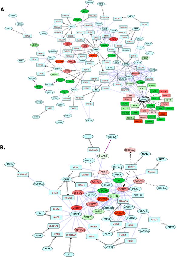
www.nature.com/scientificreports/
lung functionality. Complications in other systems such as the cardiovascular and nervous systems were also
reported10,11. Recently, cases of pulmonary embolism in the lungs of COVID-19 patients have been reported12.
In SARS-CoV and MERS-CoV infections, increased level of pro-inflammatory cytokines were evident13,
which in turn increased the activation and recruitment of inflammatory cells into the lung tissues, facilitat-
ing acute lung injury14. Similarly, increased levels of many pro-inflammatory cytokines were also detected in
moderate-to-critically affected COVID-19 p atients15, leading to respiratory failure from ARDS. However, the
complex interplays between pro-inflammatory and anti-inflammatory cytokines have not been completely illus-
trated. Apart from the cytokine storm, other factors such as host innate immunity, autoimmunity against the
pulmonary epithelial and endothelial cells, and host genetic and epigenetic factors also play important roles in
the pathogenesis of SARS-CoV infection16,17. Moreover, the multifaceted host-virus interactions are also found
to be a key player in the pathogenesis reported for other coronavirus i nfections18.
Previously, transcriptional responses in COVID-19 were experimentally recorded using various in vitro and
in vivo approaches such as cell lines, animal models, and SARS-CoV-2-infected lungs19, Nasopharyngeal (NP)
swabs20, and bronchoalveolar lavage fluid of COVID-19 patients21. However, SARS-CoV-2-mediated deregu-
lation of the lung transcriptome and its potential implications in pathogenesis of acute lung failure remained
elusive. Therefore, designing of prospective therapeutics for the clinical management of the COVID-19 is still in
its infancy. To this end, we analyzed publicly available transcriptome data from the lung biopsy of a COVID-19
patient and summarized the probably altered pathways in COVID-19. Furthermore, the probable roles of SARS-
CoV-2 in these dysregulations, and resulting acute lung damage, were also investigated.
Results
Antiviral immune responses and organ specific functions are dysregulated in lungs. During
respiratory virus infections, many host pathways are fine-tuned to battle against the invading pathogen; on the
other side, the infecting viruses also try to hijack and modulate host pathways for immune evasion and survival
ost22. These complex interactions lead to the disease complexity, causing several critical pathophysi-
inside the h
ological conditions in host’s respiratory system22. To explore which particular biological processes/pathways are
dysregulated in SARS-CoV-2 infection, we first identified the dysregulated-genes in both SARS-CoV and SARS-
CoV-2 infections and performed comparative functional enrichment a nalyses23.
We detected 3031 (2408 upregulated and 623 downregulated), 142 (91 upregulated and 51 downregulated,
and 6714 (2476 upregulated and 4238 downregulated) dysregulated genes from SARS-CoV infected 2B4 cells,
SARS-CoV-2-infected NHBE cells, and lung biopsy of COVID-19 patient, respectively (Supplementary Fig. 1).
We observed a wide array of differentially expressed genes in SARS-CoV-2-infected lung whose expression
profiles differed from those recorded in SARS-CoV-2-infected NHBE cells and SARS-CoV-infected 2B4 cells
(Supplementary Fig. 1).
As expected, enrichment analyses revealed biological processes related to antiviral inflammatory responses,
and viral processes were overrepresented in all three infection models (Fig. 1A). Several biological processes,
such as negative regulation of viral replication, immune system process, response to hypoxia, and heart develop-
ment were only enriched in the SARS-CoV-2 infection models (Fig. 1A). However, few pivotal processes were
uniquely enriched for the dysregulated genes from COVID-19-affected lung, namely viral transcription, adap-
tive immune response, brain development, lung development, and respiratory gaseous exchange by respiratory
system (Fig. 1A).
Similarly, enrichment using the ‘Bioplanet’24 module suggests host antiviral immune responses through
various inflammatory cytokine signaling pathways, apoptosis, and interferon-I signaling in all of these infec-
tion models (Fig. 1B,C). While, HIF-1 signaling, heart development, asthma, and type-II interferon signaling
pathways were found in both SARS-CoV-2 infection data (Fig. 1B, Supplementary Fig. 2). Intriguingly, some
pathways, such as disease associated with surfactant metabolism, ROS/RNS production, fatty acyl-CoA biosyn-
thesis, ER-phagosome pathway, and inflammasomes, were only found in the SARS-CoV-2-infected lung (Fig. 1C,
Supplementary Fig. 2). Functional enrichment using the ‘DisGeNet’25 module revealed that the dysregulated
genes of SARS-CoV and SARS-CoV-2 infections are also involved in other diseases, namely viral disease, lung
diseases, asthma, pneumonitis, and hypoxia (Fig. 1D, Supplementary Fig. 2). Interestingly, several cholesterol
biosynthesis pathways were found to be dysregulated only in the lung biopsy of COVID-19 patient (Fig. 1E,
Supplementary Fig. 2). As cholesterol in lung plays important roles in maintaining normal lung physiology and
protection against many d iseases26, downregulation of these indicates possible association with lung-related
comorbidities of COVID-19 patients.
These enrichment analyses suggest that several pathways related to lung’s function are likely dysregulated
directly or indirectly by the infecting SARS-CoV-2 virus. While hunting for more definitive clues on which par-
ticular processes are being modulated during SARS-CoV-2 infection, we again performed enrichment analysis
with our in-house combined module (Supplementary file 4). This enrichment analysis revealed several impor-
tant lung-function-related processes only for the dysregulated genes from the COVID-19 patient’s lung. These
includued, lung development, pulmonary surfactant metabolism disease or dysfunction, respiratory processes,
regulation of respiratory gaseous exchange, and some antiviral responses (Fig. 2, Supplementary Fig. 3). We also
exported a list of significantly enriched terms in color-coded heatmaps. Intriguingly, while equating the expres-
sion of these enriched genes, we discovered that genes of these key lung-related processes are significantly altered
in SARS-CoV-2-infected lungs compared to the SARS-CoV-2-infected NHBE cells and SARS-CoV infection
model (Supplementary Fig. 4).
Genes in lung surfactant metabolism pathway are dysregulated in COVID‑19 patient’s
lung. Pulmonary surfactant proteins play an important role in maintaining the surface tension necessary
Scientific Reports | (2020) 10:19395 | https://doi.org/10.1038/s41598-020-76404-8 2
Vol:.(1234567890)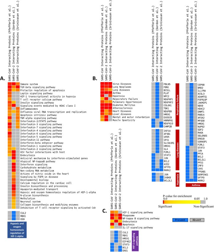
www.nature.com/scientificreports/
Figure 1. Enrichment analysis and comparison between dysregulated genes in SARS-CoV, SARS-CoV-2
(NHBE cells) and SARS-CoV-2 (Lung biopsy) infections using (A) GOBP41 module, (B) Bioplanet p athway24
28 25 108
module, (C) Reactome pathway module, (D) DisGeNet module, (E) HumanCyc module. Selected
significant terms are represented in heatmap. Significance of enrichment in terms of adjusted p-value (< 0.05)
is represented in color-coded P-value scale for all heatmaps. Color towards red indicates higher significance
and color towards yellow indicates less significance, while grey means non-significant. The column headers are
indicating the dysregulation status of the genes used for the enrichment analysis (DE: differentially expressed/
Upregulated/Downregulated), while the row labels are pointing the enriched terms.
Scientific Reports | (2020) 10:19395 | https://doi.org/10.1038/s41598-020-76404-8 3
Vol.:(0123456789)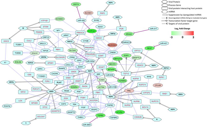
www.nature.com/scientificreports/

                                            Figure 2.  Enrichment analysis and comparison between dysregulated genes and the genes of some selected
                                            processes in SARS-CoV, SARS-CoV-2 (NHBE cells) and SARS-CoV-2 (Lung biopsy) infections using combined
                                            module. Selected significant terms are represented in heatmap in upper panel. Color schemes are similar as
                                            Fig. 1. Lower panel heatmaps presents enriched genes for some selected terms from upper panel enrichment
                                            analysis. For individual processes, blue means presence (significantly differentially expressed gene) while grey
                                            means absence (not significantly differentially expressed genes for this module for this experimental condition).
                                            The column headers are indicating the dysregulation status of the genes used for the enrichment analysis (DE:
                                            differentially expressed, Upregulated and Downregulated), while the row labels are pointing the enriched terms.

          Scientific Reports |   (2020) 10:19395 |              https://doi.org/10.1038/s41598-020-76404-8                                                  4

Vol:.(1234567890)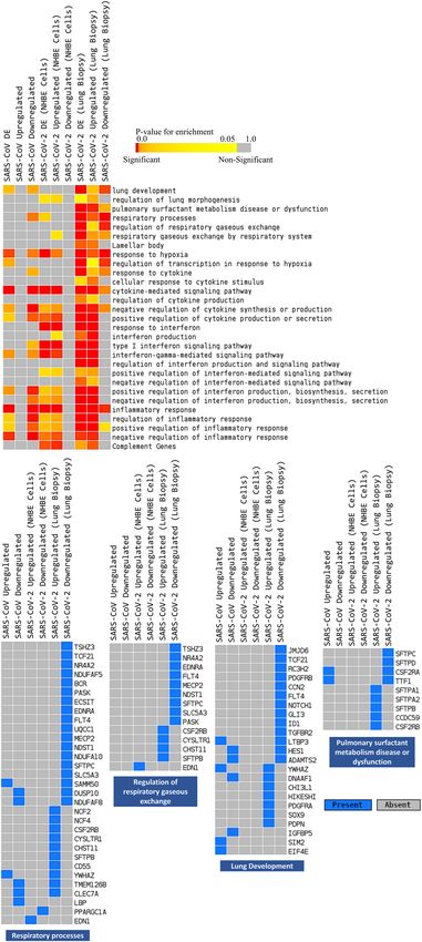
www.nature.com/scientificreports/
Figure 3. Schematic representation of lung surfactant metabolism pathway from Reactome pathway d
atabase28.
Color towards yellow indicates upregulation and while blue indicates downregulation.
for efficient gaseous exchange at the air–liquid interface in the alveoli, and can also modulate functions of lung’s
innate immune cells to eliminate p athogens27. Additionally, they have been shown to impede inflammatory
responses and the clearance of apoptotic cells in lungs following viral infection27. Taking cue from the results
of previous section, it was apparent that surfactant metabolism could be a potential target of viral modulation.
Therefore, we sought to decipher the probable routes of SARS-CoV-2-induced dysregulations of this pathway
and its downstream signaling. To attain this, we mapped the differentially expressed genes into this pathway
using the Reactome pathway b rowser28. Next, we scrutinized the mechanisms of this pathway to elucidate the
probable alterations happening in COVID-19-affected lung.
In normal lung, TTF1-CCDC59 complex can transactivate SFTPB and SFTPC gene expression, which play
an important role by regulating the alveolar surface t ension29. But in COVID-19-affected lung, TTF1 and SFTPC
genes were found to be downregulated, whereas SFTPB was upregulated (Fig. 3). GATA6 transcription factor
promotes the transcription of SFTPA gene30 which is involved in immune and inflammatory responses, and
lowers the surface tension in the a lveoli31. Both GATA6 and SFTPA genes were upregulated in SARS-CoV-
2-infected lung, while the GATA6 antagonist LMCD1 was downregulated (Fig. 3). The CSF2RA-CSF2RB complex
can bind GM-CSF to induce activation of macrophages32 and helps in the degradation of STFPs in the alveo-
lar macrophages33. We found CFSF2RA downregulated and CSF2RB upregulated in the lung of the COVID-
19-affected patient (Fig. 3). Pro-SFTPB and Pro-SFTPC are cleaved by NAPSA, CTSH, and PGA3-5 to produce
active SFTPB and SFTPC34,35. Surprisingly, NAPSA was dysregulated in the COVID-19-affected lung (Fig. 3).
Overall, the transcription of surfactant genes, production of active surfactant proteins, and their turnover may be
dysregulated in the COVID-19 patient’s lung, which could have resulted in the severely lethal disease complica-
tions. As these mechanisms are found to be altered upon SARS-CoV-2 infections, the virus may be positively
facilitating these anomalies.
Host proteins which interact with the virus are involved in different respiratory‑func‑
tion‑related pathways and diseases. In previously reported human coronavirus infections, SARS and
MERS coronaviruses are often found to commandeer host machineries, suppressing host immune responses
and other important biological processes for their continued existence within the infected cells36. We performed
functional enrichment analyses using previously reported SARS-CoV and SARS-CoV-2 host factor proteins37–39
Scientific Reports | (2020) 10:19395 | https://doi.org/10.1038/s41598-020-76404-8 5
Vol.:(0123456789)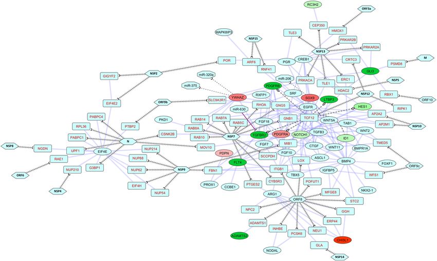
www.nature.com/scientificreports/
to illuminate the pathways which may be targeted by viral proteins, causing the lung injuries in COVID-19
patients.
As anticipated, the enrichment analyses showed association of both SARS-CoV and SARS-CoV-2 infections
with several immune signaling pathways, such as interleukin signaling, interferon signaling, apoptosis, and
inflammasomes (Fig. 4A,C). However, this approach also revealed several vital pathways for respiratory func-
tion such as HIF-1 signaling, and hypoxic and oxygen homeostasis regulation of HIF-1 alpha (Fig. 4A,C). Using
the DisGenNet module25, lung disease, asthma, hyperoxia, respiratory failure, and pulmonary hypertension
were found to be enriched (Fig. 4B). These results enlightened that SARS-CoV-2 is likely utilizing its proteins to
modulate normal lung’s physiological and immune responses, which we further explored by linking these viral-
host protein–protein interactions (PPI) to our previously identified essential lung processes.
SARS‑CoV‑2 proteins and host epigenetic regulators can modulate the functions of lung and
other respiratory processes. Results from the previous sections rationalized that both the dysregulated
genes in COVID-19-affected lung and SARS-CoV-2 interacting proteins are involved in several respiratory func-
tions. Hence, we produced several functional networks of dysregulated genes, viral protein-host protein interac-
tions, and host epigenetic regulators involved in those processes to gain insight on virus-mediated deregulations
and the resulting pathophysiological effects of COVID-19. We have mainly addressed four broad biological
processes that can significantly affect COVID-19 patients: response to hypoxia, lung development, respiratory
processes, and surfactant metabolism.
Numerous genes in hypoxia response and HIF-1 alpha signaling were abruptly dysregulated in the SARS-
CoV-2-infected lung (Supplementary Fig. 5). This included PLAT, a tissue plasminogen activator with pro-
found roles in lung homeostasis, whose aberrant regulation can lead to many lung injuries40. In the PPI map
OBP41 module ‘response to hypoxia’ and SARS-CoV-2 target g enes37,
of differentially-expressed genes of the G
this PLAT protein was found to be directly targeted by viral protein ORF8 (Fig. 5). Moreover, several indirect
responses from the viral protein interactions were also revealed (Fig. 5). SARS-CoV-2 M protein can target STOM
which interacts with SLC2A1. SLC2A1 can also be targeted by host miRNA miR-320a (Fig. 5). SLC2A1 encoding
GLUT1 protein is upregulated by hypoxic responses in alveolar epithelial c ells42.
Viral ORF8 interaction with OS9 can modulate EGLN1 and EGLN2, which might disrupt the functions of the
EGLN-HIF oxygen sensing s ystem43. KCNMA1 was found to interact with host proteins ATP1B1 and PRKACA
that interact with viral M and NSP13 proteins, respectively (Fig. 5). ORF9c may modulate EDNRA indirectly
through F2RL1 (Fig. 5) and can alter the vasoconstrictor effect resulting from E DNRA44. PML functions might
be altered through the viral N and MOV10 proteins’ interaction. Functions of SLCBA1 may be modulated by
NSP13 through PRKACA (Fig. 5). NSP7 can regulate ALDH3A1 which can affect CYP1A1 and NR4A2 (Fig. 5).
NSP5 interacting HDAC2 can curb PML and REST which have hypoxia responsive functions45,46 (Fig. 5). NSP12
can modulate a wide range of hypoxia functions related proteins, namely TGFBR2, MECP2, MTHFR, CBFA2T3,
EGR1, and ANGPTL4 by interacting with the transcription factor TCF12 (Fig. 5). NSP12 through RIPK1 interac-
tions can dampen the apoptosis regulating function of C FLAR47 (Fig. 5). Apart from this, several host miRNAs
could possibly downregulate the expression of some genes, namely- miR-320a, miR-3188, miR-3661, miR-217,
miR-421 and miR-429 (Fig. 5). These viral mediated deviations found in the hypoxia responses might be a deci-
sive factor in the lung injury found in COVID-19 p atients48.
In the lung development network, viral protein NSP13 is found to indirectly target RC3H2, SOX9, GLI3
through CEP350, PRKACA, PRKAR2A proteins (Fig. 6). While GLI3 can also be modulated through several
viral-host interactions, for instance ORF10-RBX1, NSP5-HDAC2, and the M-PSMD8 interactions (Fig. 6). ORF8
can indirectly modulate ADAMTS2, CHI3L1, NOTCH1, TCF12, and FLT4 (Fig. 6). Transcription factor TCF12
is targeted by viral NSP12 protein which can in turn affect transcriptions of PDGFRA, PDGFRB, TGFBR2, ID1,
HES1, and LTBP3 genes (Fig. 6). NSP5 can modulate NOTCH1 through HDAC2 (Fig. 6). Moreover, host miRNAs
such as miR-630 can downregulate TGFBR2; while miR-206, miR-320a and miR-375 are downregulated and so
SOX9, and WYHAZ are overexpressed (Fig. 6). Virus could hamper all these components of different growth
factor signaling pathways which are crucial for various lung injury repair m echanisms49,50.
From the respiratory process network, we can delineate that several transcription factors are dysregulated.
Many of these are associated with ECSIT that can directly interact with viral ORF9c and indirectly by viral ORF8,
and NSP7 (Fig. 7A). Moreover, ECSIT itself is directly targeted by ORF9c (Fig. 7A). NSP12 can modulate SFTPB,
SFTPC, SLC5A3, DUSP10, and SAMM50 by targeting TCF12 (Fig. 7A). Also, in this network we have observed
suppressive actions of miRNAs- miR-206, miR-217, miR-375 on NR4A2 and NDST1; as well as upregulation
of YWHAZ due to the probable downregulation of miR-320a (Fig. 7A). Virus might be dampening the host
immune response in the lung by targeting E CSIT51 and by diminishing, respiratory gaseous exchange by nega-
tively modulating the surfactant proteins52.
Surfactant metabolism is found to be modulated not only by viral proteins, but also through irregular host
responses (Fig. 7B). Viral proteins NSP12 and NSP5 can target transcription factor TCF12 and epigenetic reg-
ulator HDAC2, respectively, which in turn modulate important members of surfactant metabolism process,
namely- TTF1, CCDC59, SFTPB, SFTPC, CSF2RA, CSF2RB, NAPSA, SFTPD, and DMBT1 (Fig. 7B). These
can also be modulated through CKAP4 which is observed to interact with viral M, NSP2, NSP9, E, and ORF8
proteins (Fig. 7B). Furthermore, we have observed that viral M and S proteins can interact with proteins of this
process both directly and indirectly (Supplementary Fig. 6). Host miRNA miR-421 was found to downregulate
LMCD1; while miR-137, miR-375, and miR-429 fail to modulate CCDC59 and GATA6 because of their prob-
able inactivation/suppression by differential expression of the regulating TFs (Fig. 7B). As SFTPD and SFTPC
are downregulated along with several regulatory partners, their primary function of immunomodulation and
Scientific Reports | (2020) 10:19395 | https://doi.org/10.1038/s41598-020-76404-8 6
Vol:.(1234567890)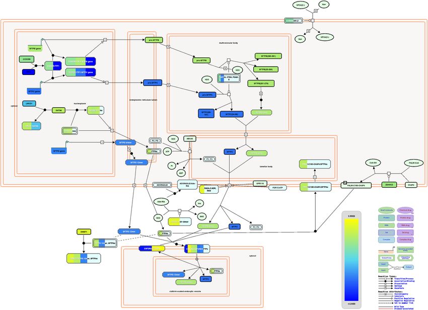
www.nature.com/scientificreports/
Figure 4. Enrichment analysis and comparison between host proteins interacting SARS-CoV proteins (Pfefferle
et al.38), SARS-CoV-2 proteins (Srinivasan et al.39) and SARS-CoV-2 proteins (Gordon et al.37) and the genes of
some selected processes using (A) Bioplanet pathway24 module, (B) DisGeNet25 module, (C) KEGG pathway109
module. Selected significant terms are represented in heatmap. Color schemes are similar as Figs. 1 and 2.
Scientific Reports | (2020) 10:19395 | https://doi.org/10.1038/s41598-020-76404-8 7
Vol.:(0123456789)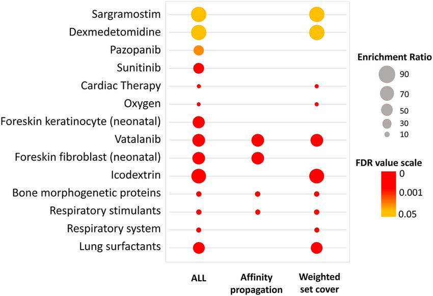
www.nature.com/scientificreports/
Figure 5. Network representing the interactions between proteins in response to hypoxia process (combined
module) along with SARS-CoV-2 proteins (Gordon et al.37), and host miRNAs. Hexagon, ellipse, rounded
rectangle, octagon represents viral proteins, process related genes, proteins that interacts viral proteins and
host miRNAs, respectively. Blunted arrow indicates suppression by miRNAs, dotted arrow pointed with open
half-circle indicates downregulated miRNAs failing to modulate host gene, arrowed line pointed with open half-
circle indicates targets of viral proteins, and arrowed line pointed with open diamond indicates transcription
factors of a gene.
efficient air exchange in lung53,54 might be seriously hindered by the viral proteins; which could further lead to
pathogenic lung injury55.
SARS-CoV-2 proteins can target several epigenetic factors, such as HDAC2, DNMT1, CUL2, MOV10, RBX1
and TLE1, to alter the above-mentioned processes. Epigenetic factors play a key role in balancing normal lung
pathobiology, and anomalies in their regulation can lead to many lung diseases56. For instance, HDAC2 and
DNMT1 have significant roles in chronic obstructive pulmonary disease (COPD) p rogression57,58.
Drug enrichment suggests lung surfactant replacement therapy as a potential treatment
option. Our results pointed towards the probable SARS-CoV-2 directed dysregulation in several important
respiratory processes and surfactant metabolism pathway in COVID-19-affected lung. We then sought to dig
out possible therapies/drugs for the improvement of the lung and respiratory conditions of COVID-19-affected
patients. In this regard, we performed drug enrichment analysis for differentially expressed genes present in the
combined module terms ‘response to hypoxia’, ‘lung development’, ‘respiratory process’ and ‘surfactant metabo-
lism pathway’ using the WebGestalt functional enrichment tool59. We have found significant enrichment scores
for lung surfactants, respiratory stimulants, sargramostim, and oseltamivir (Fig. 8, Supplementary Fig. 7).
Discussion
Though there are a wide variety of symptoms and clinical features seen in COVID-19 patients, almost every
mild-to-critically affected patient showed respiratory and breathing complications, ranging from pneumonia to
acute lung injury60. Severely ill patients are greatly supported by artificial ventilation, as no therapeutic drugs for
mitigating these complications have been discovered. This is due to the lack of understanding on the molecular
aspects of lung-related abnormalities in COVID-19. We have identified dysregulated genes related to respiratory
and lung-related processes upon the SARS-CoV-2-infection. We have prioritized these pathways and searched
for potential therapies/drugs for the treatment which could lessen the resultant effects of gene dysregulation.
Several SARS-CoV-2 entry receptor (ACE2)/entry associated proteins (TMPRSS2, BSG, CTSL, and DPP4)
have been previously d escribed61. All of these viral entry associated factors are readily expressed in the lung,
except ACE2 (Supplementary Fig. 8A). While analyzing the expression profiling in SARS-CoV-2-infected lung,
we discovered a quite unexpected scenario. Astoundingly, in COVID-19-affected lung, ACE2 was upregulated
Scientific Reports | (2020) 10:19395 | https://doi.org/10.1038/s41598-020-76404-8 8
Vol:.(1234567890)www.nature.com/scientificreports/
Figure 6. Network representing the interactions between proteins in lung development process (combined
module) along with SARS-CoV-2 proteins (Gordon et al.37), and host miRNAs. Legends are similar as Fig. 5.
while the genes of the entry associated proteins were downregulated (Supplementary Fig. 8B). More COVID-19
patient data must be analyzed to validate this striking finding.
“Cytokine storm” is a much discussed phenomenon in previously reported pathogenic human coronavirus
infections that can be lethal for the host due to the destruction of its own respiratory systems62. Similar responses
can also occur following SARS-CoV-2 i nfection63. We observed that several inflammatory and antiviral responses
are readily dysregulated in the SARS-CoV-2-infected lung that could lead to the abnormalities in overall respira-
tory functions.
Apart from this, our study identified dysregulation in several lung function related vital processes, namely-
response to hypoxia, lung development, respiratory process, and surfactant metabolism; alterations in these
pathways lead to abnormal lung pathobiology in COVID-19. Networks generated from combining the viral-host
protein interactions suggest that viral proteins might be actively involved in these dysregulations along with
several host factors, which might also be decontrolled due to the viral infections.
Hypoxic conditions are common in respiratory infections due to reduced oxygenation of the blood64. Several
genes involved in hypoxia-induced responses were found to be dysregulated (Fig. 5) in the COVID-19-affected
lung. ALDH3A1 can protect airway epithelial cells from destruction65. Though it is upregulated, its functions
can be impeded through viral proteins (Fig. 5). CFLAR functions in shutting down apoptotic responses by
interacting with RIPK147, but viral interactions with RIPK1 might prevent this (Fig. 5). SARS-CoV-2 can induce
the transcription of ANGPTL4 by utilizing TCF12 transcription factor (Fig. 5) which in turn could cause pul-
monary tissue d amage66. Viral proteins could promote the activity of EGLN1 and EGLN2 (Fig. 5) to suppress
the transcription of HIF-induced genes in hypoxia67; on the other hand the regulation through these proteins
might be hampered due to viral interactions. Moreover, the constant overexpression of HIF-mediated inflamma-
tory genes could also occur, which could lead to inflammation-induced lung damage. Severe hypoxia-induced
responses can occur through the downregulation of MECP268 in SARS-CoV-2 patients (Fig. 5). In hypoxia,
REST is induced and can act as a negative regulator of gene expression to maintain a balance between different
processes45, that were found to be downregulated and can be targeted through miR-421 in COVID-19 (Fig. 5).
GLUT1 (SLC2A1 gene) promotes increased glucose transport into hypoxic cells for its prolonged adaptation
during this condition69, but it was found to be downregulated in lung of COVID-19 patient and this can occur
through miR-320a (Fig. 5). During hypoxia, TGFβ signaling regulates inflammation and vascular r esponses70;
but attenuated TGFβ expression might lead to increased disease severity (Fig. 5). EGR1 transcription factor can
be modulated through viral proteins (Fig. 5) that might prevent the hypoxia induced EGR1 activation of HIF-1
alpha71. This probable route of viral-mediated HIF-1 signaling inhibition could curb the whole hypoxia induced
survival responses. Moreover, defective hypoxia response can occur due to the overactivity of EGR1 that can
result in anomalous t hrombosis72. Also, PLAT is a crucial factor in splitting down c lots73; this function might be
directly targeted by SARS-CoV-2 ORF8 (Fig. 5). Many COVID-19 patients were reported to have pulmonary
embolism and thrombosis74, which could be the effect of altered hypoxic responses.
Scientific Reports | (2020) 10:19395 | https://doi.org/10.1038/s41598-020-76404-8 9
Vol.:(0123456789)www.nature.com/scientificreports/
Figure 7. Network representing the interactions between proteins in (A) respiratory processes (combined
module), (B) surfactant metabolism along with SARS-CoV-2 proteins (Gordon et al.37), and host miRNAs.
Legends are similar as Fig. 5.
Scientific Reports | (2020) 10:19395 | https://doi.org/10.1038/s41598-020-76404-8 10
Vol:.(1234567890)www.nature.com/scientificreports/
Figure 8. Bubble plot of drug enrichment results using WebGestalt t ool59. Color towards red indicates higher
significance and color towards yellow indicates less significance. Bubble size indicates the enrichment ratio.
Similarly, several lung development and respiratory process genes/proteins were found to be dysregulated in
the COVID-19 patient’s lung (Fig. 6, 7). SOX9, an important regulator for the recovery from acute lung injury75,
can be down-modulated by the virus (Fig. 6). Though the host miRNAs cannot target the YWHAZ pro-survival
protein76, they can induce expression surfactant protein A277, which might be indirectly modulated through
viral protein NSP7 (Fig. 6). Likewise, viral protein ORF8 can indirectly modulate CHI3L1 (Fig. 6) that nor-
mally suppresses lung epithelium i njury78. Virus can block TCF12 and inhibit the expression of PDGFRA and
PDGFRB (Fig. 6) which could play a role in lung maturation and injury response79. GLI3, which exerts essential
role in developing the lung and regulating the innate immune c ells80, is downregulated in COVID-19 lungs
(Fig. 6). LTBP3 can promote lung a lveolarization81, but can be modulated by SARS-CoV-2 protein NSP12 (Fig. 6).
Abnormal NOTCH signaling contributes significantly in various lung d iseases82, and in SARS-CoV-2 infec-
tion NOTCH1 and HES1 are found downregulated (Fig. 6). CD55, a member of the complement system which
plays crucial role in host defense in airway epithelium83, can be modulated by viral protein NSP12 (Fig. 7A).
CYSLTR1 is upregulated in SARS-CoV-2 infection (Fig. 7A), which is correlated to COPD84. This protein can
be modulated by viral ORF9c and ORF3a (Fig. 7A). ECSIT was found to directly interact with viral ORF9c
(Fig. 7A) which might stop ECSIT-mediated antiviral innate immune response51. Several mitochondrial genes,
for instance NDUFA10, NDUFAF5, and SAMM50 were downregulated in COVID-19-affected lung (Fig. 7A).
As mitochondria plays important role in cellular respiration and lung diseases85, aberrant expression of these
genes might also lead to lung-related complications. DUSP10 can regulate unusual inflammatory responses
upon viral infections86, but in SARS-CoV-2-infected lung this gene was found downregulated, possibly through
NSP12-TCF12 interactions (Fig. 7A).
Pulmonary surfactant proteins are lipoproteins that mainly function to lower the alveolar surface tension87
and can elicit immune stimulatory roles against some respiratory pathogens88. Among the surfactant proteins,
SP-A and SP-D mainly evoke immune responses, while SP-B and SP-D play roles in maintaining efficient respira-
tory gas e xchange54. Several lung diseases including asthma, acute respiratory distress syndrome (ARDS), COPD
are reportedly associated with aberration in the function of surfactant p roteins55,89. In SARS-CoV-2-affected
lung, production of the surfactant proteins is found to be dysregulated (Fig. 7B). Viral protein NSP5 can recruit
HDAC2 and downregulate the expression of TTF1 which is needed for the expression of SP-B and SP-C (Fig. 7B).
SP-A and SP-D can be targeted indirectly by several viral proteins (Fig. 7B) which might dampen the production
of surfactant proteins in lungs, thus complicating the disease condition. The CSF2RA-CSF2RB complex modu-
late the surfactant recycling that maintains the overall balance of surfactant content33. We found that CSF2RA is
dysregulated in SARS-CoV-2-infected lung, which could lead to abnormal surfactant recycling (Fig. 7B). While
performing enrichment analysis, we witnessed a significant downregulation of several cholesterol biosynthesis
pathways in COVID-19 patient’s lung cells (Fig. 1E) that could lead to low accumulation of phospholipids in
lungs. As phosphatidylcholine (PC) and phosphatidylglycerol (PG) are the principal phospholipids of surfactant
proteins90, disrupted production of lipids might make surfactant proteins non-functional.
Considering all these, lung surfactants might be useful in the treatment of COVID-19 patients, as lung sur-
factant therapies were previously reported to be successful in other respiratory infections and acute lung injury
to reduce the lung damage of the patients87, 91,92. Also, other drugs including respiratory stimulants for COPD93,
sargramostim for treating pulmonary alveolar p roteinosis94, and oseltamivir in curing influenza-related lower
respiratory tract c omplications95 showed potential for improving the lung’s and respiratory system’s overall
condition.
From our results, we can suggest that surfactant protein production along with other respiratory responses in
lung could be dysregulated in COVID-19. However, further experimental proteomic analyses of this dysregula-
tion are required for the functional implications of this study. Along with the antiviral drugs to mitigate the viral
responses, drugs that can improve lung conditions in COVID-19 could also be considered as a treatment option
for the patients. Our generated results can be useful for obtaining greater insight on the probable dampened
Scientific Reports | (2020) 10:19395 | https://doi.org/10.1038/s41598-020-76404-8 11
Vol.:(0123456789)www.nature.com/scientificreports/
surfactant production upon SARS-CoV-2 infection, and the potential implications of surfactant therapy as a
therapeutic agent for COVID-19 treatment.
Methods
Analysis of microarray expression data. Microarray expression data from both SARS-CoV-infected
2B4 cells and uninfected controls (both maintained for 24 h) obtained from Gene Expression Omnibus (GEO)
(https://www.ncbi.nlm.nih.gov/geo)96, accession: GSE17400. Raw Affymatrix CEL files were background cor-
rected, normalized using Bioconductor package “affy v1.28.1” using ’rma’ algorithm. Quality of microarray
experiment (data not shown) was verified by Bioconductor package “arrayQualityMetrics v3.44.0”97. Differen-
tially expressed (DE) between two experimental conditions were called using Bioconductor package Limma98.
Probe annotations were converted to genes using in-house python script basing the Ensembl gene model
(Biomart 99)99. The highest absolute expression value was considered for the probes that were annotated to the
same gene. We have considered the genes to be differentially expressed, which have FDR100 p-value ≤ 0.05 and
Log2 fold change value ≥ 0.25 (Supplementary file 1).
Analysis of RNA‑seq expression data. Illumina sequenced RNA-seq raw FastQ reads were extracted
from GEO database96, accession: GSE147507. This data includes independent biological triplicates of primary
human lung epithelium (NHBE) cell lines which were mock treated or infected with SARS-CoV-2 for 24hrs. This
data also contains two technical replicate of post-mortem lung biopsy sample of a deceased COVID-19 patient,
along with lung biopsy samples of two different healthy persons as control. We have checked the raw sequence
quality using FastQC program (v0.11.9)101, and found that the "Per base sequence quality" and "Per sequence
quality scores" were high over the threshold for all sequences (data not shown). Mapping of reads was done
with TopHat (tophat v2.1.1 with Bowtie v2.4.1)102. Short reads were uniquely aligned allowing at best two mis-
matches to the human reference genome (GRCh38) as downloaded from UCSC database103. Sequences matched
exactly more than once with equal quality were discarded to avoid b ias104. The reads that were not mapped
to the genome were utilized to map against the transcriptome (junctions mapping). Ensembl gene m odel105
(version 99, as extracted from UCSC) was used for this mapping. After mapping, we used SubRead package
featureCount (v2.21)106 to calculate absolute read abundance (read count, rc) for each transcript/gene associated
to the Ensembl genes. For differential expression (DE) analysis we used DESeq2 (v1.26.0) with R (v3.6.2; 2019-
07-05)107 that uses a model based on the negative binomial distribution. To avoid false positives, we considered
only those transcripts where at least 10 reads are annotated in at least one of the samples used in this study and
also applied a minimum Log2 fold change of 0.5 for to be differentially expressed transcripts apart from adjusted
p-value cut-off of ≤ 0.05 by FDR. Raw read counts of this experiment are provided in supplementary file 2. To
assess the fidelity of the RNA-seq data used in this study and normalization method applied here, we checked
the normalized Log2 expression data quality using R/Bioconductor package “arrayQualityMetrics (v3.44.0)”97.
From this analysis, in our data no outlier was detected by “Distance between arrays”, “Boxplots”, and “MA plots”
methods and replicate samples were clustered together (Supplementary file 3).
Retrieval of the host proteins that interact with SARS‑CoV and SARS‑CoV‑2 proteins. We
have obtained the list of human proteins that form high confidence interactions with SARS-CoV and SARS-
CoV-2 proteins from previously conducted studies37–39 and processed their provided protein names into the
associated HGNC official gene symbol.
Functional enrichment analysis. We utilized Gitools (v1.8.4) for enrichment analysis and heatmap
g eneration23. We have utilized the Gene Ontology Biological Processes (GOBP)41, Reactome pathway28, Bio-
planet pathways24, HumanCyc database108, DisGeNet25, KEGG pathway109 modules, and a custom in-house built
combined module (Supplementary file 4) for the overrepresentation analysis. This combined module was gener-
ated from related modules with few genes to a parent term/process which otherwise would have been left out
from analysis due to statistical stringency cutoff (module with at least 10 genes are selected) during enrichment
analysis. Resulting p-values were adjusted for multiple testing using the Benjamin and Hochberg’s method of
False Discovery Rate (FDR)100.
Mapping of the differentially expressed genes from SARS‑CoV‑2‑infected lung onto biological
pathways. We utilized the Reactome pathway browser28 for the mapping of dysregulated genes of SARS-
CoV-2 infection onto different biological pathways. We then focused on the pathways which were found to be
enriched for lung-related functions.
Obtaining the transcription factors which can modulate the differential gene expression. We
obtained the transcription factors (TFs) which bind to a given differentially expressed gene using a custom TFs
module created using ENCODE110, TRRUST111, and C hEA112 databases.
Obtaining human miRNAs target genes. We extracted the experimentally validated target genes of
human miRNAs from miRTarBase database113.
Extraction of transcription factors modulate human miRNA expression. We downloaded exper-
imentally validated TFs which bind to miRNA promoters and modules from TransmiR (v2.0) database which
provides regulatory relations between TFs and miRNAs114. Because miRNAs play roles in transcriptional regu-
Scientific Reports | (2020) 10:19395 | https://doi.org/10.1038/s41598-020-76404-8 12
Vol:.(1234567890)www.nature.com/scientificreports/
lation, we considered TFs that are expressed (upregulated) and can ‘activate’ or ‘regulate’ miRNAs, or in the
absence of TFs (downregulation), those could otherwise ‘suppress’ miRNAs.
Identification of the host epigenetic factors genes. We used the EpiFactors database115 to find
human genes related to epigenetic activity.
Construction of biological networks. Construction, visualization and analysis of biological networks
with differentially expressed genes, their associated transcription factors, associated human miRNAs, and inter-
acting viral proteins were executed in the Cytoscape software (v3.8.0)116. We used the STRING117 database to
extract only the highest confidences (0.9) edges for protein–protein interactions to reduce any false positive
connection.
Drug enrichment analysis. We used the WebGestalt tool59 for predicting potential drugs targeting the
given differentially expressed genes. We selected the drugs based on FDR (BH) ≤ 0.05100, using both D
rugBank118
and GLAD4U119 drug database combined.
Data availability
Publicly available data were utilized. Analyses generated data are deposited as supplementary files.
Received: 9 June 2020; Accepted: 28 October 2020
References
1. Worldometer. 1–22 (2020).
2. Lu, R. et al. Genomic characterisation and epidemiology of 2019 novel coronavirus: implications for virus origins and receptor
binding. Lancet 395, 565–574 (2020).
3. NCBI-Gene. (2020)
4. Jiang, S., Du, L. & Shi, Z. An emerging coronavirus causing pneumonia outbreak in Wuhan, China: calling for developing
therapeutic and prophylactic strategies. Emerg. Microb. Infect. 9, 275–277 (2020).
5. Ren, L.-L. et al. Identification of a novel coronavirus causing severe pneumonia in human: a descriptive study. Chin. Med. J.
133(9), 1015–1024. https://doi.org/10.1097/CM9.0000000000000722 (2020).
6. Lauer, S. A. et al. The incubation period of coronavirus disease 2019 (COVID-19) from publicly reported confirmed cases:
estimation and application. Intern. Med. Ann. https://doi.org/10.7326/m20-0504 (2020)
7. Lan, L. et al. Positive RT-pcr test results in patients recovered from COVID-19. JAMA 323, 1502–1503. https://doi.org/10.1001/
jama.2020.2783 (2020).
8. Huang, C. et al. Clinical features of patients infected with 2019 novel coronavirus in Wuhan China. Lancet 395, 497–506 (2020).
9. Galiatsatos, P. What Coronavirus Does to the Lungs, https://www.hopkinsmedicine.org/health/conditions-and-diseases/coron
avirus/what-coronavirus-does-to-the-lungs (2020).
10. Mao, L. N. et al. Neurologic manifestations of hospitalized patients with coronavirus disease 2019 in Wuhan. JAMA Neurol
China. https://doi.org/10.1001/jamaneurol.2020.1127 (2020).
11. Zheng, Y.-Y., Ma, Y.-T., Zhang, J.-Y. & Xie, X. COVID-19 and the cardiovascular system. Nat. Rev. Cardiol. 17, 259–260. https
://doi.org/10.1038/s41569-020-0360-5 (2020).
12. Rotzinger, D. C., Beigelman-Aubry, C., von Garnier, C. & Qanadli, S. D. Pulmonary embolism in patients with COVID-19: time
to change the paradigm of computed tomography. Thromb. Res. 190, 58–59. https://doi.org/10.1016/j.thromres.2020.04.011
(2020).
13. Yoshikawa, T. et al. Dynamic innate immune responses of human bronchial epithelial cells to severe acute respiratory syndrome-
associated coronavirus infection. PLoS ONE 5, e8729–e8729. https://doi.org/10.1371/journal.pone.0008729 (2010).
14. Ye, Q., Wang, B. & Mao, J. The pathogenesis and treatment of the `Cytokine Storm’ in COVID-19. J. Infect. https://doi.
org/10.1016/j.jinf.2020.03.037 (2020).
15. Mehta, P. et al. COVID-19: consider cytokine storm syndromes and immunosuppression. Lancet 395, 1033–1034 (2020).
16. Gu, J. & Korteweg, C. Pathology and pathogenesis of severe acute respiratory syndrome. Am. J. Pathol. 170, 1136–1147. https
://doi.org/10.2353/ajpath.2007.061088 (2007).
17. Schäfer, A. & Baric, R. S. Epigenetic landscape during coronavirus infection. Pathogens 6, 8. https://doi.org/10.3390/pathogens6
010008 (2017).
18. Fung, S.-Y., Yuen, K.-S., Ye, Z.-W., Chan, C.-P. & Jin, D.-Y. A tug-of-war between severe acute respiratory syndrome coro-
navirus 2 and host antiviral defence: lessons from other pathogenic viruses. Emerg. Microb. Infect. 9, 558–570. https://doi.
org/10.1080/22221751.2020.1736644 (2020).
19. Blanco-Melo, D. et al. Imbalanced host response to SARS-CoV-2 drives development of COVID-19. Cell https: //doi.org/10.1016/j.
cell.2020.04.026 (2020).
20. Butler, D. J. et al. Host, Viral, and Environmental Transcriptome Profiles of the Severe Acute Respiratory Syndrome Coronavirus
2 (SARS-CoV-2). bioRxiv. https://doi.org/10.1101/2020.04.20.048066 (2020).
21. Xiong, Y. et al. Transcriptomic characteristics of bronchoalveolar lavage fluid and peripheral blood mononuclear cells in COVID-
19 patients. Emerg. Microb. Infect. 9, 761–770. https://doi.org/10.1080/22221751.2020.1747363 (2020).
22. Manjarrez-Zavala, M. E., Rosete-Olvera, D. P., Gutiérrez-González, L. H., Ocadiz-Delgado, R. & Cabello-Gutiérrez, C. Patho-
genesis of viral respiratory infection. In Respiratory Disease and Infection - A New Insight (ed Mahboub, B. H.) (IntechOpen,
2013). https://doi.org/10.5772/54287.
23. Perez-Llamas, C. & Lopez-Bigas, N. Gitools: analysis and visualisation of genomic data using interactive heat-maps. PLoS ONE
6, e19541 (2011).
24. Huang, R. et al. The NCATS bioplanet—an integrated platform for exploring the universe of cellular signaling pathways for
toxicology, systems biology, and chemical genomics. Front. Pharmacol. https://doi.org/10.3389/fphar.2019.00445 (2019).
25. Pinero, J. et al. The DisGeNET knowledge platform for disease genomics: 2019 update. Nucleic Acids Res. 48, D845-d855. https
://doi.org/10.1093/nar/gkz1021 (2020).
26. Fessler, M. B. A new frontier in immunometabolism. Cholesterol in lung health and disease. Ann. Am. Thorac. Soc. 14, 399–405.
https://doi.org/10.1513/AnnalsATS.201702-136AW (2017).
27. Glasser, J. R. & Mallampalli, R. K. Surfactant and its role in the pathobiology of pulmonary infection. Microbes Infect. 14, 17–25.
https://doi.org/10.1016/j.micinf.2011.08.019 (2012).
Scientific Reports | (2020) 10:19395 | https://doi.org/10.1038/s41598-020-76404-8 13
Vol.:(0123456789)www.nature.com/scientificreports/
28. Jassal, B. et al. The reactome pathway knowledgebase. Nucleic Acids Res. 48, D498-d503. https://doi.org/10.1093/nar/gkz1031
(2020).
29. Yang, M. C., Guo, Y., Liu, C. C., Weissler, J. C. & Yang, Y. S. The TTF-1/TAP26 complex differentially modulates surfactant
protein-B (SP-B) and -C (SP-C) promoters in lung cells. Biochem. Biophys. Res. Commun. 344, 484–490. https: //doi.org/10.1016/j.
bbrc.2006.03.158 (2006).
30. Bruno, M. D., Korfhagen, T. R., Liu, C., Morrisey, E. E. & Whitsett, J. A. GATA-6 activates transcription of surfactant protein A.
J. Biol. Chem. 275, 1043–1049. https://doi.org/10.1074/jbc.275.2.1043 (2000).
31. Silveyra, P. & Floros, J. Genetic complexity of the human surfactant-associated proteins SP-A1 and SP-A2. Gene 531, 126–132.
https://doi.org/10.1016/j.gene.2012.09.111 (2013).
32. Martinez, F. O. & Gordon, S. The M1 and M2 paradigm of macrophage activation: time for reassessment. F1000Prime Rep. 6,
13–13. https://doi.org/10.12703/P6-13 (2014).
33. Ikegami, M. Surfactant catabolism. Respirology (Carlton, Vic.) 11, 24–27. https://doi.org/10.1111/j.1440-1843.2006.00803.x
(2006).
34. Johansson, J., Jornvall, H. & Curstedt, T. Human surfactant polypeptide SP-B. Disulfide bridges, C-terminal end, and peptide
analysis of the airway form. FEBS Lett. 301, 165–167. https://doi.org/10.1016/0014-5793(92)81239-i (1992).
35. Johansson, J. et al. Hydrophobic 3.7 kDa surfactant polypeptide: structural characterization of the human and bovine forms.
FEBS Lett. 232, 61–64. https://doi.org/10.1016/0014-5793(88)80386-7 (1988).
36. Fung, T. S. & Liu, D. X. Human coronavirus: host-pathogen interaction. Annu. Rev. Microbiol. 73, 529–557. https://doi.
org/10.1146/annurev-micro-020518-115759 (2019).
37. Gordon, D. E. et al. A SARS-CoV-2 protein interaction map reveals targets for drug repurposing. Nature https: //doi.org/10.1038/
s41586-020-2286-9 (2020).
38. Pfefferle, S. et al. The SARS-coronavirus-host interactome: identification of cyclophilins as target for pan-coronavirus inhibitors.
PLoS Pathog. 7, e1002331–e1002331. https://doi.org/10.1371/journal.ppat.1002331 (2011).
39. Srinivasan, S. et al. Structural genomics of SARS-CoV-2 indicates evolutionary conserved functional regions of viral proteins.
Viruses 12, 360 (2020).
40. Sisson, T. H. & Simon, R. H. The plasminogen activation system in lung disease. Curr. Drug Targets 8, 1016–1029. https://doi.
org/10.2174/138945007781662319 (2007).
41. Ashburner, M. et al. Gene ontology: tool for the unification of biology. Nat. Genet. 25, 25–29 (2000).
42. Ouiddir, A., Planès, C., Fernandes, I., VanHesse, A. & Clerici, C. Hypoxia upregulates activity and expression of the glucose
transporter GLUT1 in alveolar epithelial cells. Am. J. Respir. Cell Mol. Biol. 21, 710–718. https: //doi.org/10.1165/ajrcmb
.21.6.3751
(1999).
43. Ivan, M. & Kaelin, W. G. Jr. The EGLN-HIF O(2)-sensing system: multiple inputs and feedbacks. Mol. Cell 66, 772–779. https
://doi.org/10.1016/j.molcel.2017.06.002 (2017).
44. Calabrò, P. et al. Analysis of endothelin-1 and endothelin-1 receptor A gene polymorphisms in patients with pulmonary arterial
hypertension. Intern. Emerg. Med. 7, 425–430. https://doi.org/10.1007/s11739-011-0643-2 (2012).
45. Cavadas, M. A. S. et al. REST is a hypoxia-responsive transcriptional repressor. Sci. Rep. 6, 31355. https://doi.org/10.1038/srep3
1355 (2016).
46. Salsman, J. et al. PML nuclear bodies contribute to the basal expression of the mTOR inhibitor DDIT4. Sci. Rep. 7, 45038. https
://doi.org/10.1038/srep45038 (2017).
47. Faiz, A. et al. Cigarette smoke exposure decreases CFLAR expression in the bronchial epithelium, augmenting susceptibility for
lung epithelial cell death and DAMP release. Sci. Rep. 8, 12426. https://doi.org/10.1038/s41598-018-30602-7 (2018).
48. Shimoda, L. A. & Semenza, G. L. HIF and the lung: role of hypoxia-inducible factors in pulmonary development and disease.
Am. J. Respir. Crit. Care Med. 183, 152–156. https://doi.org/10.1164/rccm.201009-1393PP (2011).
49. Gouveia, L., Betsholtz, C. & Andrae, J. Exploring the effect of PDGF-A deletion in the adult lung: implications in homeostasis and
injury. Retrieved from http://urn.kb.se/resolve?urn=urn:nbn:se:uu:diva-347031 (n.d.).
50. Olajuyin, A. M., Zhang, X. & Ji, H.-L. Alveolar type 2 progenitor cells for lung injury repair. Cell Death Discov. 5, 63. https://doi.
org/10.1038/s41420-019-0147-9 (2019).
51. Lei, C. Q. et al. ECSIT bridges RIG-I-like receptors to VISA in signaling events of innate antiviral responses. J. Innate Immun.
7, 153–164. https://doi.org/10.1159/000365971 (2015).
52. Veldhuizen, E. J. A. & Haagsman, H. P. Role of pulmonary surfactant components in surface film formation and dynamics.
Biochim. Biophys. Acta 1467, 255–270. https://doi.org/10.1016/S0005-2736(00)00256-X (2000).
53. Mulugeta, S. & Beers, M. F. Surfactant protein C: its unique properties and emerging immunomodulatory role in the lung.
Microbes Infect. 8, 2317–2323. https://doi.org/10.1016/j.micinf.2006.04.009 (2006).
54. Nayak, A., Dodagatta-Marri, E., Tsolaki, A. & Kishore, U. An insight into the diverse roles of surfactant proteins, SP-A and SP-D
in innate and adaptive immunity. Front. Immunol. https://doi.org/10.3389/fimmu.2012.00131 (2012).
55. Sorensen, G. L. Surfactant protein D in respiratory and non-respiratory diseases. Front. Med. (Lausanne) 5, 18–18. https://doi.
org/10.3389/fmed.2018.00018(2018).
56. Mortaz, E., Masjedi, M. R., Barnes, P. J. & Adcock, I. M. Epigenetics and chromatin remodeling play a role in lung disease.
Tanaffos 10, 7–16 (2011).
57. Barnes, P. J. Role of HDAC2 in the pathophysiology of COPD. Annu. Rev. Physiol. 71, 451–464. https://doi.org/10.1146/annur
ev.physiol.010908.163257 (2009).
58. He, X., Chen, L., Chen, Y. & Zeng, H. In A28. Advances in Copd and Asthma A1200-A1200.
59. Liao, Y., Wang, J., Jaehnig, E. J., Shi, Z. & Zhang, B. WebGestalt 2019: gene set analysis toolkit with revamped UIs and APIs.
Nucleic Acids Res. 47, W199–W205 (2019).
60. Cascella, M., Rajnik, M., Cuomo, A., Dulebohn, S. C. & Di Napoli, R. In StatPearls [Internet] (StatPearls Publishing, 2020).
61. Sungnak, W. et al. SARS-CoV-2 entry factors are highly expressed in nasal epithelial cells together with innate immune genes.
Nat. Med. https://doi.org/10.1038/s41591-020-0868-6 (2020).
62. Channappanavar, R. & Perlman, S. Pathogenic human coronavirus infections: causes and consequences of cytokine storm and
immunopathology. Semin. Immun. 39, 529–539. https://doi.org/10.1007/s00281-017-0629-x (2017).
63. Pedersen, S. F. & Ho, Y. C. SARS-CoV-2: a storm is raging. J. Clin. Investig. 130, 2202–2205. https://doi.org/10.1172/jci137647
(2020).
64. Schaible, B., Schaffer, K. & Taylor, C. T. Hypoxia, innate immunity and infection in the lung. Respir. Physiol. Neurobiol. 174,
235–243. https://doi.org/10.1016/j.resp.2010.08.006 (2010).
65. Jang, J.-H. et al. Aldehyde dehydrogenase 3A1 protects airway epithelial cells from cigarette smoke-induced DNA damage and
cytotoxicity. Free Radic. Biol. Med. 68, 80–86. https://doi.org/10.1016/j.freeradbiomed.2013.11.028 (2014).
66. Li, L. et al. Angiopoietin-like 4 increases pulmonary tissue leakiness and damage during influenza pneumonia. Cell Rep. 10,
654–663. https://doi.org/10.1016/j.celrep.2015.01.011 (2015).
67. To, K. K. & Huang, L. E. Suppression of hypoxia-inducible factor 1α (HIF-1α) transcriptional activity by the HIF prolyl hydroxy-
lase EGLN1. J. Biol. Chem. 280, 38102–38107 (2005).
68. Kron, M., Zimmermann, J. L., Dutschmann, M., Funke, F. & Muller, M. Altered responses of MeCP2-deficient mouse brain stem
to severe hypoxia. J. Neurophysiol. 105, 3067–3079. https://doi.org/10.1152/jn.00822.2010 (2011).
Scientific Reports | (2020) 10:19395 | https://doi.org/10.1038/s41598-020-76404-8 14
Vol:.(1234567890)You can also read