Ibuprofen alters epoxide hydrolase activity and epoxy oxylipin metabolites associated with different metabolic pathways in murine livers
←
→
Page content transcription
If your browser does not render page correctly, please read the page content below
www.nature.com/scientificreports

                OPEN             Ibuprofen alters epoxide hydrolase
                                 activity and epoxy‑oxylipin
                                 metabolites associated
                                 with different metabolic pathways
                                 in murine livers
                                 Shuchita Tiwari1, Jun Yang2, Christophe Morisseau2, Blythe Durbin‑Johnson3,
                                 Bruce D. Hammock2 & Aldrin V. Gomes 1,4*

                                 Over the last decade oxylipins have become more recognized for their involvement in several
                                 diseases. Non-steroidal anti-inflammatory drugs (NSAIDs) such as ibuprofen are known to inhibit
                                 cyclooxygenase (COX) enzymes, but how NSAIDs affect oxylipins, in addition to COX products,
                                 in animal tissues is not well understood. Oxylipins in livers from male and female mice treated
                                 with 100 mg/kg/day of ibuprofen for 7 days were investigated. The results showed that ibuprofen
                                 treated male livers contained 7 times more altered oxylipins than ibuprofen treated female livers.
                                 In male and female livers some prostaglandins were altered, while diols, hydroxy fatty acids and
                                 epoxides were significantly altered in male livers. Some soluble epoxide hydrolase (sEH) products,
                                 such as 9,10-DiHODE were found to be decreased, while sEH substrates (such as 9(10)-EpODE and
                                 5(6)-EpETrE) were found to be increased in male livers treated with ibuprofen, but not in ibuprofen
                                 treated female livers. The enzymatic activities of sEH and microsomal epoxide hydrolase (mEH) were
                                 elevated by ibuprofen in both males and females. Analyzing the influence of sex on the effect of
                                 ibuprofen on oxylipins and COX products showed that approximately 27% of oxylipins detected were
                                 influenced by sex. The results reveal that ibuprofen disturbs not only the COX pathway, but also the
                                 CYP450 and lipoxygenase pathways in male mice, suggesting that ibuprofen is likely to generate sex
                                 related differences in biologically active oxylipins. Increased sEH activity after ibuprofen treatment
                                 is likely to be one of the mechanisms by which the liver reduces the higher levels of EpODEs and
                                 EpETrEs.

                                 Abbreviations
                                 AA	Arachidonic acid
                                 ALA	α-Linolenic acid
                                 COX	Cyclooxygenase
                                 CYP	Cytochrome P450
                                 DHA	Docosahexaenoic acid
                                 EPA	Eicosapentaenoic acid
                                 CYPe	Cytochrome P450 epoxygenase
                                 CYPh	Cytochrome P450 hydroxylase
                                 DiHDPE	Dihydroxy-docosapentaenoic acid
                                 DiHETrE	Dihydroxy-eicosatrienoic acid
                                 DiHOME	Dihydroxy-octadecenoic acid
                                 EPA	Eicosapentaenoic acid

                                 1
                                  Department of Neurobiology, Physiology, and Behavior, University of California, Davis, CA 95616,
                                 USA. 2Department of Entomology and Nematology, and Comprehensive Cancer Center, University of
                                 California, Davis, CA 95616, USA. 3Department of Public Health Sciences, University of California, Davis, CA,
                                 USA. 4Department of Physiology and Membrane Biology, University of California, Davis, CA, USA. *email:
                                 avgomes@ucdavis.edu

Scientific Reports |   (2021) 11:7042               | https://doi.org/10.1038/s41598-021-86284-1                                            1

                                                                                                                                        Vol.:(0123456789)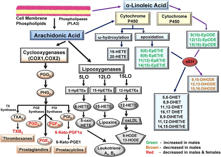
www.nature.com/scientificreports/
EpETrE Epoxy-eicosatrienoic acid
EpOME Epoxy-octadecenoic acid
GLA Gamma-linolenic acid
HDoHE Hydroxy-docosahexaenoic acid
HEPE Hydroxy-eicosapentaenoic acid
HETE Hydroxy-eicosatetraenoic acid
HETrE Hydroxy-eicosatrienoic acid
HHTrE Hydroxy-heptadecatrienoic acid
HODE Hydroxy-octadecadienoic acid
HOTrE Hydroxy-octadecatrienoic acid
LA Linoleic acid
LOX Lipoxygenase
mEH Microsomal epoxide hydrolase
sEH Soluble epoxide hydrolase
Ibuprofen is the most common over-the-counter nonsteroidal anti-inflammatory drug (NSAID) known for its
potent analgesic, anti-inflammatory, and antipyretic e ffects1. The therapeutic actions of ibuprofen are mediated
by its ability to inhibit the synthesis and release of prostaglandins (PGs) E2 (PGE2) and I2 (PGI2) by blocking
cyclooxygenase (COX) enzymes COX-1 and COX-2 which are involved in the generation of pain, fever, and
inflammation2,3. COX-1 and COX-2 are the rate-determining enzymes for the synthesis of prostaglandins and
other prostanoids including thromboxanes and prostacyclins4. These molecules play important roles in the
immune, cardiovascular, gastrointestinal and renal system3. While COX-1 is important for the protection of
gastric mucosa and platelet homeostasis through the formation of protective PGE2 and prostacyclin PGI2,
COX-2 is involved with prostaglandin mediated pain and i nflammation4.
Although NSAIDs are some of the most commonly used medicines worldwide, they are associated with
various adverse side effects which are generally dose dependent5. The use of NSAIDs has been associated with
gastrointestinal injury, hepatotoxicity, renal and cardiovascular disorders and hypertension6,7. The NSAIDs
benoxaprofen and bromfenac were withdrawn from the market due to severe hepatic toxicity8,9. Similarly, higher
incidence of myocardial infarction and stroke led to the withdrawal of rofecoxib and valdecoxib (Bextra)10. In
addition, the NSAIDs nimesulide, diclofenac and sudoxicam were reported to cause acute liver injury11,12. In April
2019, The French National Agency for the Safety of Medicines and Health Products (ANSM) issued a warning
regarding the use of NSAIDs (ibuprofen and ketoprofen) for the treatment of patients with infectious diseases13,14.
Another concern about ibuprofen is its presence in the environment. Data suggests that fish health could be
adversely affected by the levels of ibuprofen found in rivers15,16. Ibuprofen is also found in drinking water and
surface wells. Concerns exist about its possible biological effects when taken regularly in small amounts17,18.
Limited studies have investigated the effect of NSAIDs on oxylipins in the P450 branch of the arachidonic acid
cascade and these studies largely utilized blood samples. In general, oxylipins regulate a variety of physiological
functions by acting as important signaling molecules for specific receptors or by modulating transcription fac-
tors and ion channels in an autocrine manner19. Perturbations in the P450 generated oxylipin concentrations
have been shown to be associated with various pathological conditions including chronic pain20,21, diabetes22,
Alzheimer’s disease23,24, and cardiovascular d iseases25. Oxylipins are bioactive oxidized fatty acid metabolites
synthesized by the oxidation of polyunsaturated fatty acids (PUFAs).
In a placebo-controlled phase III selenium/celecoxib trial to prevent colorectal adenomatous polyps, blood
oxylipins from celecoxib treated participants showed higher levels of individual CYP450 and LOX metabolites
and lower levels of COX-derived metabolites compared to placebo treated participants26. However, since circulat-
ing oxylipin profiles are unlikely to represent tissue profiles, and as we have previously shown that ibuprofen was
associated with proteasome and metabolic dysfunction in l ivers27, the oxylipin profiles of livers from ibuprofen
treated mice were investigated.
We hypothesized that ibuprofen would significantly reduce the prostaglandins and thromboxanes that are
produced by COX1 and COX2 and would have few effects on non-COX related arachidonic acid pathways. Our
data suggest that a moderate dose of ibuprofen treatment for 7 days significantly altered oxylipins related to dif-
ferent metabolic pathways and revealed significant sex specific differences in livers from ibuprofen treated males
and females relative to their vehicle controls.
Materials and methods
Animal studies. C57BL/6 J male and female mice (8-week age) were used for the study. The animal experi-
ment was performed in accordance with the protocols approved by the Institutional Animal Care and Use Com-
mittee (IACUC) of the University of California, Davis. This study is reported in accordance with the ARRIVE
guidelines (https://arriveguidelines.org). Mice were maintained at controlled temperature and humidity and had
free access to food and water. Mice were treated with 100 mg/kg/day of ibuprofen for 7 days in drinking water.
The dose of ibuprofen given to mice would be approximately equivalent to a human taking 486 mg/day as previ-
ously described27. This dose of ibuprofen is significantly lower than the maximum suggested over-the-counter
dose (800–1200 mg/day). The mice were sacrificed, and livers were collected and quickly washed twice in ice-
cold phosphate buffered saline (PBS). The tissues were then pulverized in liquid nitrogen, collected in clean
microcentrifuge tubes, and stored at − 80 °C until needed.
LC–MS/MS‑based lipidomic analysis. Extraction and analyses of the regulatory lipid mediator: The
extraction process is similar to the protocol as described in a previously published paper28. 10 µL of methanol
Scientific Reports | (2021) 11:7042 | https://doi.org/10.1038/s41598-021-86284-1 2
Vol:.(1234567890)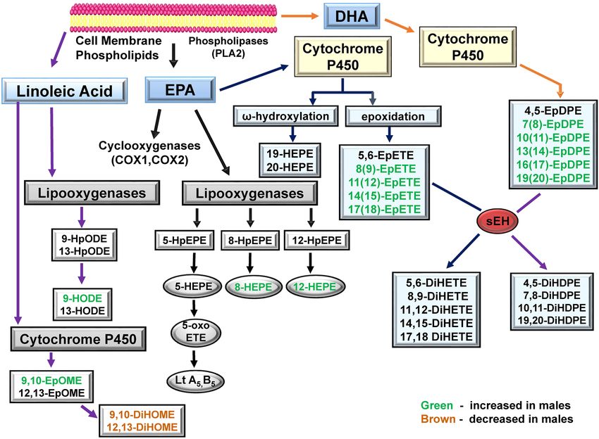
www.nature.com/scientificreports/
containing deuterated internal standard solution, a mixture of d4 PGF1α, d4 TXB2, d4 PGE2, d4 LTB4, d11
14,15 DiHETrE, d6 20 HETE, d4 9 HODE, d8 5 HETE, d11 11,12 EpETrE (Cayman Chemical, Ann Arbor, MI),
was added to approximately 100 mg of liver tissue. 400 µL of cold methanol containing 0.1% acetic acid and 0.1%
butylated hydroxytoluene, BHT (Sigma-Aldrich, St. Louis, MO) solution was added to these tissue samples and
stored at − 80 °C for 30 min. After freezing, samples were homogenized using Retsch MM301 ball mills (Retsch
Gmbh, Germany) at 30 Hz for 10 min and then kept at − 80 °C overnight. The homogenates were centrifuged at
16,000 g for 10 min, the supernatants were collected, and remaining pellets were washed with 100 µL of ice-cold
methanol with 0.1% acetic acid and 0.1% BHT and centrifuged at 16,000 g for 10 min. The supernatants of each
sample were combined and diluted with 2 mL of H2O and loaded onto Waters Oasis HLB 3 cc (Waters, Milford,
MA) solid phase extraction (SPE) cartridges.
The oxylipins were measured on a 1200 SL ultra-high-performance liquid chromatography (UHPLC) (Agilent,
Santa Clara, CA) interfaced with a 4000 QTRAP mass spectrometer (Sciex, Redwood City, CA). The separation
conditions for LC were optimized to separate the critical pairs of oxylipins, which share the same multiple reac-
tion monitor (MRM) transitions. In brief, separation was achieved on an Agilent Eclipse Plus C18 150 × 2.1 mm
1.8 um column with mobile phases of water with 0.1% acetic acid as mobile phase A and acetonitrile/methanol
(84/16) with 0.1% acetic acid as mobile phase B. All the parameters on the mass spectrometer were optimized
with pure standards (purchased from Cayman Chemical, Ann Arbor, MI) under negative m ode28,29. A scheduled
multiple reaction monitoring (MRM) scan mode was employed to increase the sensitivity of the measurement.
The analytical variance and lower limit of quantification for oxylipins are shown in the supplementary data.
LC–MS/MS‑based proteomic analysis. Proteomic analysis was carried out as previously described and
used the same data as published in Shuchita et al. 202027. Scaffold version 4.10 was used to analyze, quantify, and
display the primary amino acid sequence of the sEH and mEH (Proteome Software Inc. Oregon).
sEH and mEH activity assay. Pulverized liver samples were weighed (50 mg) and homogenized in ice
cold 1X PBS buffer containing 0.1% ethylenediaminetetraacetic acid (EDTA), protease inhibitor (PMSF 1 mM)
and 1 mM DTT. The supernatant was separated and quantified by the bicinchoninic acid (BCA) method using
bovine serum albumin (BSA) as standard. The samples were flash frozen and sEH activity was measured as
reported previously using [3H]-t-DPPO as substrate with [S] = 50 μM and incubation for 10 to 30 min at 30 °C30.
Activity measurement. sEH‑H like activity. To measure the residual soluble epoxide hydrolase (sEH-H)
activity, [3H]-trans-diphenyl-propene oxide (t-DPPO) was used as s ubstrate31. Briefly, 1 µL of a 5 mM solution
of t-DPPO in DMSO was added to 100 µL of the tissue extracts diluted in sodium phosphate buffer (100 mM,
pH 7.4) containing 0.1 mg/mL BSA ([S]final = 50 µM). The mixture was incubated at 37 °C for 5–20 min, and the
reaction was quenched by addition of 60 µL of methanol and 200 µL of isooctane, which extracts the remaining
epoxide from the aqueous phase. Extractions of an identical reaction with 1-hexanol were performed in paral-
lel to assess the possible presence of glutathione transferase activity which could also transform the s ubstrate31.
Hexanol extracts epoxides and diols into the hyperphase while glutathione conjugates remain in the hypophase.
The activity was followed by measuring the quantity of radioactive diol formed in the aqueous phase using a
scintillation counter (TriCarb 2810 TR, Perkin Elmer, Shelton, CT). Assays were performed in triplicate.
mEH like activity: The presence of microsomal epoxide hydrolase (mEH) activity was determined using
[3H]-cis-stilbene oxide (c-SO) as s ubstrate32. The tissue extracts were diluted in Tris/HCl buffer (0.1 M, pH 9.0)
containing 0.1 mg/mL BSA. The reaction was started by adding 1 µL of a 5 mM solution of c-SO in ethanol to
100 µL of diluted extracts([S]final = 50 µM). The mixture was incubated at 37 °C for 5–30 min, and then at 30 °C
before substrate introduction. The reaction was then quenched by adding 250 μL of isooctane, which extracts
the remaining epoxide from the aqueous phase. Extractions with 1-hexanol were performed in parallel to assess
the possible presence of glutathione transferase activity which could also transform the substrate REF 1. The
activity was followed by measuring the quantity of radioactive diol formed in the aqueous phase using a scintil-
lation counter. Assays were performed in triplicate.
Western blotting. Sample preparation. Liver tissues were pulverized in liquid nitrogen and 20 mg were
weighed and homogenized in ice cold 1X RIPA buffer (50 mM Tris, 150 mM sodium chloride (NaCl), 1% NP40,
0.5% sodium deoxycholate and 0.1% SD, pH 8) using a glass Dounce homogenizer. The supernatants were sepa-
rated after centrifugation at 12,000 g for 15 min at 4 °C. The quantification of the proteins was done using the
BCA Protein Assay (Bio-Rad, Cat. #500–0119) and the samples were further diluted to equal protein concentra-
tions. Liver samples were then mixed with 4X Laemmli sample buffer (8% SDS, 40% glycerol, 0.4% bromophenol
blue, 240 mM Tris, pH 6.8). The β-mercaptoethanol was added freshly and samples were then boiled for 5 min
at 95 °C.
Electrophoresis and western blotting. 4–20% 18-well TGX precast gels (Cat. # 567–1094, Bio-Rad) were used to
separate 20 µg of protein in each lane. The proteins on the TGX precast gel were then transferred to a nitrocel-
lulose membrane (Trans-Blot Turbo Midi Nitrocellulose, #170–4159, Bio-Rad) using the Bio-Rad Trans-Blot
Turbo Transfer System (Cat. # 170–4155, Bio-Rad). After staining the membranes with Ponceau S they were
imaged and used as a loading control for total protein normalization of Western blots. 3% nonfat dry milk
(NFM) (Cat. # 170–6404, Bio-Rad) in Tris-buffered saline (TBS) (50 mM Tris, 150 mM NaCl, pH 7.5) containing
0.05% (wt/vol) Tween 20 (TTBS) was used to block empty sites on the membranes for 1 h at room temperature.
The membranes were then incubated in 1% TTBS with the primary antibodies diluted to 1:5000 for mouse anti-
sEH33 or 1:2000 for mouse anti-mEH34 and left overnight at 4 °C with gentle shaking. Thereafter, membranes
Scientific Reports | (2021) 11:7042 | https://doi.org/10.1038/s41598-021-86284-1 3
Vol.:(0123456789)www.nature.com/scientificreports/
were washed in TTBS three times for 5 min each, incubated with horseradish peroxidase conjugated rabbit anti-
mouse IgG secondary antibody (Sigma-Aldrich, anti-mouse Cat. # A9044) diluted 1:10,000 in 1% TTBS for 1 h
at room temperature and washed three more times in 1X TTBS for 5 min each with gentle shaking. Blots were
then developed using enhanced commercial chemiluminescent reagent (Clarity, Bio-Rad, 170–5061) and were
imaged in ChemiDoc MP (Bio-Rad). Image Lab 5.0 was used to quantify the blots.
Data analysis. Results in Table 1 are expressed as means ± standard deviations (SD) from at least four inde-
pendent experiments. To determine the differences between control and ibuprofen treated groups and male
versus female differences the raw data were first log2 transformed. The data was then normalized using cyclic
loess normalization35, as implemented in the Bioconductor package l imma36, version 3.44.3. Limma was initially
developed for microarray data, but the fitting routines of this program are not limited to microarrays. Differen-
tial oxylipin abundance analyses were conducted using limma, which fits a linear model to data from each lipid
using empirical Bayes smoothing to improve estimates of standard errors of log fold changes37, and calculates
Benjamini-Hochberg (BH) false-discovery rate p-values for each lipid38. The BH procedure decreases the false
discovery rate and helps avoid false positives. The linear model used in limma for this analysis was a two-factor
ANOVA model including effects for group, sex, and their interaction. Analyses were conducted using R version
4.0.2 (2020-06-22)39. Adjusted p-values of < 0.05 were defined as statistically significant.
Results
The livers from male and female mice treated with ibuprofen or vehicle were analyzed by mass spectrometry.
Oxylipin profiles of ibuprofen treated mice showed that 35 oxylipins in male livers and 5 oxylipins in female livers
were significantly altered compared to livers from vehicle treated male and female mice respectively (Table 1).
Substrates of sEH were increased in livers from ibuprofen treated male mice (Table 1). To determine if the
increased sEH substrate levels may be due to changes in the protein levels of sEH the Scaffold program was
used to extract sEH information from previously analyzed d ata27. In these earlier studies, livers from 5 vehicle
control and 5 ibuprofen treated male mice were used for Tandem Mass Tag (TMT) proteomics. The proteomic
experiments were only carried out on male mice. The mass spectrometry results suggest that both sEH and mEH
were significantly upregulated in livers of male mice treated with ibuprofen (Figs. 1A and 2A). A representative
spectrum of sEH, the intensity of the TMTs for that peptide and the amino acid residues that were detected by
mass spectrometry are shown in supplemental Fig. 1. A similar representative spectrum for mEH, the TMT
intensities for that peptide, and the mEH residues detected by mass spectrometry are shown in supplemental
Fig. 2. As far we know, this is the first report of sEH and mEH being upregulated by ibuprofen treatment.
Validation of the mass spectrometry data using Western blotting showed that sEH was upregulated in livers
from females treated with ibuprofen (Fig. 1B). However, no change in the expression level of sEH was observed
in ibuprofen treated male livers compared to normal control livers (Fig. 1C). The livers used for the Western
blotting were different from the livers used for mass spectrometry and may show intrinsic variability between
mice. Validation of the mass spectrometry data for mEH using Western blotting showed that the livers from
ibuprofen treated female mice had elevated mEH expression, but not statistically significant higher levels when
compared to controls (Fig. 2B). The male mice treated with ibuprofen had higher levels of liver mEH than controls
(Fig. 2C), consistent with the proteomic data.
Effect of ibuprofen on sEH and mEH activities in mice liver. sEH is a promising target for the treat-
ment of hypertension, inflammatory diseases, pain, diabetes, and stroke40–43. Since the proteomic results suggest
that sEH and mEH may not be elevated under some conditions of ibuprofen treatment, we decided to investigate
if ibuprofen altered sEH and mEH activities. Using larger numbers of liver samples, the activities of sEH and
mEH in ibuprofen treated samples were found to be statistically increased in both males and females (Fig. 3).
These results suggest that moderate ibuprofen treatment to mice significantly increased sEH and mEH activities
in both male and female livers relative to their controls.
CYP‑derived oxylipins. Heat maps of the oxylipin changes observed in male and female mice are shown in
Fig. 4A. Partial least squares-discriminant analysis (PLS-DA) score plots of male and female mice liver oxylipin
data suggest that males and females respond very differently to ibuprofen treatment (Fig. 4B). PLS-DA is utilized
to show discrimination between the control and ibuprofen treated samples. Among the three major oxylipin
metabolizing pathways (COX, LOX, and CYP), COX derived components showed the largest decreases in
amounts while the CYP-derived oxylipins showed the most sex specific changes in ibuprofen treated mice livers
relative to their normal controls (Table 1). In total, 69 different oxylipins were detected in quantifiable amounts
in the mouse liver samples.
Effect of ibuprofen on CYP‑derived oxylipins through the AA pathway. The CYP pathway utilizes AA as a substrate
to produce oxylipins by the epoxygenase and ω-hydroxylase pathways. Epoxy fatty acids (EpFA) are produced
in response to vascular endothelial inflammation and are anti-inflammatory and vasodilators in function44,45.
The data suggest that ibuprofen altered CYP-derived EpFA oxylipins, including 5(6)-EpETrE, 8(9)-EpETrE,
11(12)-EpETrE and 14(15)-EpETrE. These oxylipins were significantly elevated in ibuprofen treated male livers
relative to their controls (Table 1. Supplementary Fig. 3). No statistically significant differences in the levels of
CYP-derived oxylipins were observed between the control and ibuprofen treated female groups, suggesting sex
specific differences (Table 1).
Scientific Reports | (2021) 11:7042 | https://doi.org/10.1038/s41598-021-86284-1 4
Vol:.(1234567890)www.nature.com/scientificreports/
Male Female
Oxylipin Control (n = 13) IB (n = 15) Oxylipin Control (n = 12) IB (n = 14)
PG & Others
6-keto-PGF1a 138 ± 89.0 2.60 ± 2.55* 6-keto-PGF1a 184 ± 99.0 6.12 ± 4.53*
TXB2 29.8 ± 13.2 3.87 ± 0.69* TXB2 39.5 ± 26.8 5.82 ± 3.77*
PGF2a 52.1 ± 23.2 1.06 ± 0.43* PGF2a 59.3 ± 45.3 1.84 ± 0.90*
PGE2 5.76 ± 8.81 0.09 ± 0.15* PGE2 6.00 ± 3.43 0.69 ± 0.34*
PGD2 10.9 ± 6.5 0.78 ± 0.31* PGD2 12.1 ± 7.87 1.01 ± 0.41*
5-oxo-ETE 3.95 ± 2.12 4.43 ± 2.11 5-oxo-ETE 73.7 ± 64.2 160 ± 366
12-oxo-ETE 39.7 ± 36.1 50.3 ± 20.0 12-oxo-ETE 395 ± 258 596 ± 1011
15-oxo-ETE 6.30 ± 5.28 6.16 ± 1.96 15-oxo-ETE 30.1 ± 19.9 27.2 ± 12.9
LXA4 1.80 ± 0.70 1.80 ± 0.45 LXA4 3.52 ± 2.40 3.43 ± 2.42
15-deoxy-PGJ2 37.9 ± 12.9 39.2 ± 11.1 15-deoxy-PGJ2 29.6 ± 11.6 30.5 ± 14.2
Diols
9,10-DiHOME 37.8 ± 9.39 21.8 ± 54.9* 9,10-DiHOME 35.6 ± 27.3 27.7 ± 6.53
12,13-DiHOME 171 ± 242 96.6 ± 33.3* 12,13-DiHOME 155 ± 72.4 128 ± 30.8
9,10-DiHODE 3.88 ± 8.64 1.11 ± 0.46* 9,10-DiHODE 1.98 ± 1.10 1.46 ± 0.40
12,13-DiHODE 7.60 ± 20.0 1.63 ± 0.53* 12,13-DiHODE 3.45 ± 2.00 2.45 ± 0.81
15,16-DiHODE 49.6 ± 97.1 18.8 ± 6.56* 15,16-DiHODE 50.4 ± 22.2 44.5 ± 23.2
5,6-DiHETrE 3.02 ± 1.61 3.20 ± 1.23 5,6-DiHETrE 2.85 ± 1.22 2.95 ± 1.09
8,9-DiHETrE 6.59 ± 3.95 6.54 ± 2.13 8,9-DiHETrE 12.3 ± 5.29 14.4 ± 3.86
11,12-DiHETrE 14.3 ± 6.62 14.1 ± 6.38 11,12-DiHETrE 22.8 ± 10.17 27.9 ± 7.93
14,15-DiHETrE 42.2 ± 15.3 43.8 ± 17.2 14,15-DiHETrE 49.9 ± 26.41 55.2 ± 17.6
8,15-DiHETE 8.76 ± 4.37 10.2 ± 3.85 8,15-DiHETE 15.0 ± 10.76 14.4 ± 6.23
11,12-DiHETE 1.47 ± 0.56 1.72 ± 0.62 11,12-DiHETE 1.86 ± 0.67 2.35 ± 1.22
14,15-DiHETE 4.27 ± 1.28 5.20 ± 1.95 14,15-DiHETE 4.87 ± 2.16 5.60 ± 2.09
17,18-DiHETE 10.3 ± 1.95 10.8 ± 3.63 17,18-DiHETE 12.5 ± 4.39 14.5 ± 5.23
4,5-DiHDPE 28.0 ± 24.9 25.5 ± 12.3 4,5-DiHDPE 13.89 ± 13.69 5.28 ± 3.98
7,8-DiHDPE ND ND 7,8-DiHDPE 2.02 ± 0.84 2.23 ± 0.39
10,11-DiHDPE 2.94 ± 1.26 3.15 ± 1.06 10,11-DiHDPE 4.68 ± 2.05 5.41 ± 1.97
13,14-DiHDPE 4.61 ± 1.60 5.17 ± 2.38 13,14-DiHDPE 7.67 ± 3.47 8.30 ± 3.56
16,17-DiHDPE 13.8 ± 4.10 14.8 ± 6.18 16,17-DiHDPE 16.9 ± 8.40 16.7 ± 4.95
19,20-DiHDPE 55.2 ± 11.7 55.6 ± 15.7 19,20-DiHDPE 62.8 ± 23.5 70.8 ± 23.0
Hydroxy Fatty acids
9-HODE 1047 ± 605 623 ± 220* 9-HODE 992 ± 647 907 ± 455
13-HODE 1415 ± 810 1046 ± 385 13-HODE 1743 ± 1139 1445 ± 450
9-HOTrE 20.1 ± 30.3 9.76 ± 2.93* 9-HOTrE 27.2 ± 34.9 23.6 ± 16.7
13-HOTrE 18.7 ± 27.5 9.07 ± 4.24* 13-HOTrE 39.6 ± 43.5 47.2 ± 49.4
15-HETrE 74.3 ± 34.8 113 ± 54.8 15-HETrE 213 ± 181 569 ± 1373
5-HETE 30.2 ± 21.2 30.7 ± 7.8 5-HETE 89.26 ± 74.3 103.1 ± 89.6
8-HETE 28.5 ± 16.1 32.9 ± 11.0 8-HETE 269 ± 389 429 ± 1013
9-HETE 29.7 ± 17.2 34.2 ± 10.9 9-HETE 154 ± 191 131 ± 167
11-HETE 114 ± 43.0 97.1 ± 34.0* 11-HETE 501 ± 424 410 ± 542
12-HETE 283 ± 113 380 ± 195 12-HETE 2014 ± 1197 3319 ± 7329
15-HETE 252 ± 117 291 ± 99.0 15-HETE 1036 ± 811 976 ± 1309
20-HETE 3.07 ± 1.5 3.14 ± 1.7 20-HETE 1.79 ± 0.9 2.02 ± 0.84
5-HEPE 6.21 ± 5.1 5.59 ± 1.9 5-HEPE 18.1 ± 12.8 12.1 ± 5.1
8-HEPE 6.84 ± 5.2 28.5 ± 35.7* 8-HEPE 14.4 ± 17.7 17.1 ± 15.6
12-HEPE 125 ± 113 668 ± 592* 12-HEPE 217 ± 27 221 ± 500
15-HEPE 7.01 ± 3.1 5.95 ± 3.0 15-HEPE 48.7 ± 54.2 126 ± 301
17-HDoHE 56.4 ± 34.8 55.3 ± 25.5 17-HDoHE 289.82 ± 389.9 770 ± 2186
Epoxides
9(10)-EpOME 78.3 ± 79.1 188 ± 164 9(10)-EpOME 88.19 ± 99.0 65.5 ± 38.4
12(13)-EpOME 73.0 ± 68.5 178.3 ± 164* 12(13)-EpOME 67.06 ± 65.7 59.4 ± 40.1
9-HOTrE 20.1 ± 30.3 9.76 ± 2.9* 9-HOTrE 27.18 ± 34.9 23.8 ± 16.7
5(6)-EpETrE 301 ± 249 954 ± 931* 5(6)-EpETrE 305.5 ± 340 225 ± 148
8(9)-EpETrE 14.6 ± 12.5 49.9 ± 47.6* 8(9)-EpETrE 28.2 ± 27.4 25.0 ± 9.0
Continued
Scientific Reports | (2021) 11:7042 | https://doi.org/10.1038/s41598-021-86284-1 5
Vol.:(0123456789)www.nature.com/scientificreports/
Male Female
Oxylipin Control (n = 13) IB (n = 15) Oxylipin Control (n = 12) IB (n = 14)
11(12)-EpETrE 30.0 ± 23.7 112 ± 111* 11(12)-EpETrE 48.7 ± 54.3 42.2 ± 19.6
14(15)-EpETrE 27.0 ± 23.2 103 ± 93.3* 14(15)-EpETrE 41.8 ± 60.4 30.1 ± 16.2
9(10)-EpODE 6.37 ± 6.9 15.4 ± 13.5* 9(10)-EpODE 7.24 ± 8.3 4.59 ± 3.3
12(13)-EpODE 3.79 ± 4.1 8.97 ± 7.6* 12(13)-EpODE 4.48 ± 4.9 3.03 ± 2.0
15(16)-EpODE 18.3 ± 31.1 28.4 ± 23.2* 15(16)-EpODE 23.3 ± 20.6 12.5 ± 8.5
8(9)-EpETE 17.8 ± 22.8 139 ± 201* 8(9)-EpETE 30.7 ± 19.9 23.8 ± 17.0
11(12)-EpETE 1.21 ± 1.1 4.94 ± 4.4* 11(12)-EpETE ND ND
14(15)-EpETE 2.00 ± 1.9 8.95 ± 9.4* 14(15)-EpETE 3.01 ± 3.3 2.09 ± 1.5
17(18)-EpETE 2.52 ± 2.0 10.8 ± 10.3* 17(18)-EpETE 3.61 ± 3.9 2.56 ± 1.9
7(8)-EpDPE 135 ± 94.2 392 ± 306* 7(8)-EpDPE 226 ± 189 185 ± 96.0
10(11)-EpDPE 10.9 ± 9.4 30.1 ± 23.3* 10(11)-EpDPE 14.1 ± 12.7 11.7 ± 5.6
13(14)-EpDPE 7.43 ± 6.4 22.1 ± 16.5* 13(14)-EpDPE 9.00 ± 8.6 7.6 ± 3.6
16(17)-EpDPE 8.35 ± 7.4 23.8 ± 17.9* 16(17)-EpDPE 9.94 ± 10.5 8.00 ± 4.0
19(20)-EpDPE 12.6 ± 9.2 33.7 ± 23.2* 19(20)-EpDPE 19.3 ± 15.0 14.00 ± 8.9
Keto fatty acids
EKODE 11.2 ± 13.4 8.5 ± 2.7 EKODE 37.6 ± 18.4 29.9 ± 25.1
9-oxo-ODE 57.0 ± 51.3 50.7 ± 14.6* 9-oxo-ODE 205 ± 179 139 ± 84.5
13-oxo-ODE ND ND 13-oxo-ODE 6.04 ± 5.7 5.18 ± 3.5
Trihydroxy Fatty acids
9,12,13-TriHOME ND ND 9,12,13-TriHOME 2302 ± 2213 2077 ± 3612
9,10,13-TriHOME ND ND 9,10,13-TriHOME 1231 ± 1128 1139 ± 1880
Table 1. Levels of oxylipins in liver tissue of mice. ND, not detected. Results are in ng/mL, mean ± standard
deviation (mean ± SD). Significance level for the comparison between control and ibuprofen treated groups
using the procedures described in the methods section. The metabolites are arranged in each section by
chain length, then double bond so that LA, ALA, AA, EPA, and DHA metabolites (if present) are separated. *
Adjusted P value < 0.05.
Effect of ibuprofen on CYP‑derived oxylipins through the EPA pathway. Similar to AA, EPA acts as a substrate for
CYP enzymes to produce fatty acid epoxides (EpETEs) which are further metabolized to dihydroxy fatty acids
(DiHETEs) by sEH31. The ω- hydroxylation of EPA yield HEPEs such as 10-HEPE, 19-HEPE and 20-HEPE19,31.
Our analysis found that CYP derived oxylipins from the EPA pathway such as 8-HEPE, 12-HEPE, 8(9)-EpETE,
11(12)-EpETE, 14(15)-EpETE, and 17,18 EpETE were significantly increased in male livers treated with ibu-
profen compared to their control counterparts (Table 1, Fig. 5A). Interestingly, 8(9)-EpETE and 17(18)-EpETE
in ibuprofen treated female livers trended toward a decrease relative to controls (Table 1, Fig. 5B). No differ-
ence was observed in levels of EPA derived oxylipins such as 5-HEPE, 15-HEPE, 8,9-DiHETE, 11,12-DiHETE,
14,15-DiHETE and 17,18-DiHETE in both male and female livers from ibuprofen treated mice relative to con-
trols (Table 1).
Effect of ibuprofen on CYP‑derived oxylipins through DHA pathway. The fatty acid epoxides obtained from DHA
via CYP enzymes generate epoxides such as 4,5-EpDPE, 7,8-EpDPE, and 16,17-EpDPE19,46,47. These epoxides
can be further metabolized by sEH to diols such as 16,17-dihydroxy-docosapentaenoic acid (16,17 DiHDPE)31.
Similarly, ω-hydroxylase activity of CYP produces HDoHE from DHA with hydroxyl groups near the methyl
end of D HA48. Similar to EpETE, CYP-derived EpFA from EPA and DHA have anti-inflammatory, vasodilatory,
and anticancer effects49,50. The levels of 7(8)-EpDPE, 11(12)-EpDPE, 13(14)-EpDPE, 16(17)-EpDPE, and 19(20)-
EpDPE were all increased in ibuprofen treated male livers but not in livers form ibuprofen treated female mice
(all relative to their respective controls) (Fig. 5C and D). No change was observed in the levels of DiHDPEs in
livers from either male or female mice treated with ibuprofen compared to controls (Table 1).
Effect of ibuprofen on CYP‑derived oxylipins through α‑linoleic acid (ALA) pathway. αLA via CYP P450 activity
produces EpFA such as 12,13-EpODE which can be further metabolized to dihydroxy fatty acids 12,13-DiHODE
via sEH activity51. The lipidomic data show that the levels of 9(10)-EpODE, 12(13)-EpODE, and 15(16)-EpODE
were significantly upregulated in ibuprofen treated male livers relative to controls (Table 1, Fig. 6A). However,
a trend towards reduced 9(10)-EpODE, 12(13)-EpODE, and 15(16)-EpODE levels was observed in ibuprofen
treated female livers compared to control groups (Table 1, Fig. 6B).
Effect of ibuprofen on CYP‑derived oxylipins through linoleic acid (LA) pathway. No difference was observed
in the levels of 9,10-DiHODE, 12,13-DiHODE, or 15,16-DiHODE in female livers but they all were reduced
in livers from ibuprofen treated male mice relative to controls (Table 1). LA is also metabolized via CYP to
epoxy-octadecenic acid (EpOME) and is further converted to dihydroxy-octadecenoic acid (DiHOME) by sEH
Scientific Reports | (2021) 11:7042 | https://doi.org/10.1038/s41598-021-86284-1 6
Vol:.(1234567890)www.nature.com/scientificreports/
Figure 1. Characterization of sEH expression in control and ibuprofen treated mouse livers. (A) Abundance of
sEH in ibuprofen treated livers relative to control as determined by TMT mass spectrometry (n = 5), (B) Relative
expression of sEH in livers from female mice as determined by Western blotting (n = 6), (C) Relative expression
of sEH in livers from male mice as determined by Western blotting (n = 6). (B) and (C) are cropped images from
different blots. IB, ibuprofen treated liver samples, (C), control liver samples. Values are mean ± SE; *p < 0.05.
Full length blots are included in the Supplementary Information file.
activity52. In male livers treated with ibuprofen the level of 12(13)-EpOME was significantly greater than con-
trols, while no change was observed in female livers treated with ibuprofen, showing sex-specific differences
(Table 1). 9,10-DiHOME and 12,13-DiHOME diols were decreased in livers from male ibuprofen treated livers.
However, no fatty acid diols were significantly altered in livers from female ibuprofen treated mice.
LOX‑derived oxylipins. The LOX enzymes (5-LOX, 8-LOX, 12-LOX and 15-LOX) catalyze the conver-
sion of LA, AA or EPA to hydroperoxy intermediates such as leukotrienes, lipoxins, hepoxillins and HETEs44,53.
These oxylipins play important functional roles in different cellular processes such as inflammation, cellular
proliferation, and intracellular signaling 53,54. The LA oxylipin, 9-HODE, can be formed non-enzymatically, or
may be formed by the action of LOX or C OX55. Interestingly, the levels of 9-HODE are downregulated in male
livers treated with ibuprofen with no difference in female livers relative to normal controls (Fig. 6C,D). The
metabolism of EPA by 8-LOX yields 8-HpEPE which can then be converted to 8-HEPE. The levels of 8-HEPE
were found to be elevated in male livers treated with ibuprofen, whereas no difference was observed in female
livers treated with ibuprofen compare to control groups (Fig. 6E,F). Similarly, 12-LOX, which plays an important
role in tumor angiogenesis, motility, invasion, and metastasis56, can synthesize 12-HpEPE from EPA metabolism
which can be converted to 12-HEPE. An increase in 12-HEPE levels was observed in ibuprofen-treated male
livers (668.2 ± 591.7) relative to control groups (124.8 ± 113.2) (Table 1). No difference in 12-HEPE was observed
in female livers.
COX‑ derived oxylipins. Prostaglandins derived through the metabolism of AA by enzymatic action
of COX enzymes (COX1 and COX2) are known to play major roles in several biological processes including
inflammation, vascular tone and platelet aggregation57,58. Since ibuprofen is well established to inhibit COX1 and
COX2 it was expected that the levels of prostanoids derived from AA metabolism including PGD2 and PGF2α,
would be significantly reduced in the livers from both male and female mice treated with ibuprofen relative to
their normal controls. (Fig. 7). PGD2, PGE2, PGF2α, 6-keto-PGF1α and TXB2 were all found to be significantly
Scientific Reports | (2021) 11:7042 | https://doi.org/10.1038/s41598-021-86284-1 7
Vol.:(0123456789)www.nature.com/scientificreports/
Figure 2. Characterization of mEH expression in control and ibuprofen treated mouse livers. (A) Abundance
of mEH as determined by TMT mass spectrometry (n = 5), (B) Relative expression of mEH in livers from
female mice as determined by Western blotting (n = 6), (C) Relative expression of mEH in livers from male
mice as determined by Western blotting (n = 6). (B) and (C) are cropped images from different blots. Full length
blots are included in the Supplementary Information file. IB, ibuprofen treated liver samples, (C), control liver
samples. Values are mean ± SE; *p < 0.05.
decreased in livers from ibuprofen treated male and female mice. These results suggest that the moderate con-
centrations of ibuprofen used in the mice inhibited liver COX1 and COX2 as expected.
Other oxylipins altered. A summary of some of the changes in oxylipins observed are shown in Figs. 8
and 9. Figure 8 shows AA and ALA-derived oxylipins that were altered by ibuprofen treatment. Figure 9 shows
LA, EPA and DHA- derived oxylipins which were significantly changed by ibuprofen treatment. These diagrams
indicate ibuprofen induced more changes to oxylipins in male livers compared to female livers.
Effect of ibuprofen on CYP450 enzymes. Using the same mass spectrometry data and Scaffold pro-
gram that was utilized to determine the protein levels of sEH, all data related to CYP enzymes was extracted and
analyzed27. The results obtained suggest that most liver CYP enzymes were not significantly altered by ibuprofen
(Supplemental Fig. 6, Supplemental Table 1). However, CYP4A14, CYP 4A10, CYP 2A5, CYP3A13, CYP2A12,
CYP2B9 were increased while CYP2C70, CYP2C39, CYP1A2 were decreased in ibuprofen treated mice com-
pared to vehicle treated mice. A limitation of these results is that proteomics was only done on male mice.
Sex dependent differences. After a search of the literature to determine the best way to statistically com-
pare sex dependent differences caused by a drug we were surprised that the publications we observed did not
do any specialized statistical tests and just compared the results from male animals with female animals. There-
fore, to test for differential ibuprofen effects between sexes, we used the sex-ibuprofen interaction effect from a
two-factor ANOVA model (see Methods) to test for differential ibuprofen effects between sexes. The script used
for the R program to run the analyses is included in the supplemental file. A total of 18 lipids were found to be
statistically different when the log of the fold change of the ibuprofen treated group (LogFC.IB) versus the log of
the fold change of the control group (logFC.CTRL) for livers from female mice were compared to the LogFC.IB
versus logFC.CTRL for livers from male mice (Table 2).
Discussion
This study analyzed the effects of ibuprofen treatment on oxylipin metabolites. The ibuprofen dose used in this
animal study was similar to concentrations used in previous s tudies2,59,60. The dose of 100 mg/kg/day of ibuprofen
used in this study would correspond to a moderate daily dose of ibuprofen in humans. We profiled more than 69
oxylipins originating from different precursors including epoxides (EpOME, EpETrE, EpDPE), diols (DiHOME,
DiHETE) and hydroxy fatty acids (HETE, HEPE, HODE, DiHODE, HOTrE) that play major regulatory roles
Scientific Reports | (2021) 11:7042 | https://doi.org/10.1038/s41598-021-86284-1 8
Vol:.(1234567890)www.nature.com/scientificreports/
Figure 3. Characterization of sEH and mEH activities in control and ibuprofen treated murine livers. (A) sEH
activity and mEH activity in male mouse liver. (B) sEH activity and mEH activity of female liver lysates. Values
are mean ± SE; n = 12–15 per group. *p < 0.05.
in several biological processes 19,53,61. Moderate treatment with ibuprofen (100 mg/kg for 7 days) significantly
changed oxylipin profiles of 35 oxylipins out of the 69 detected in quantifiable amounts. These oxylipins were
derived from different precursors including AA, EPA, DHA and αLA via the three major enzymatic pathways
(COX, LOX and CYP). In livers for male mice 36 oxylipins were altered while 5 oxylipins were altered in livers
from females. These results suggest that sex specific differences in oxylipin content occurs as a result of ibuprofen
treatment.
High dose and long term NSAID treatments are associated with adverse side effects that include cardiac,
epatotoxicity62,63. Long term administration of aspirin and ibuprofen to rats altered
gastrointestinal, renal and h
liver ultrastructure and increased the metabolic activity of some CYP450 e nzymes64. Previous reports from
our lab suggest that at physiological concentrations NSAIDs such as diclofenac, naproxen and meclofenamate
sodium altered mitochondrial and proteasome function in cardiac tissue65,66. Ibuprofen also alters mitochondrial
and proteasome function in liver tissue27. As expected, ibuprofen (a non-selective COX inhibitor) treatment
Scientific Reports | (2021) 11:7042 | https://doi.org/10.1038/s41598-021-86284-1 9
Vol.:(0123456789)www.nature.com/scientificreports/
Figure 4. Short-term ibuprofen treatment (7 days) altered oxylipin profiles in murine liver. (A) Heatmap
showing the eicosanoid profile shifted by ibuprofen in control and ibuprofen treated groups in female and male
mice liver. The relative intensities of variables in control and ibuprofen treated livers were shown by color bars.
(B) Partial least squares-discriminant analysis (PLS-A) score plots of female and male mice livers (n = 12–15 per
group).
significantly decreases the levels of prostaglandins (PGE2, PGD2, PGF2a, 6-keto-PGF1a) and TXB2 derived from
AA metabolism in both ibuprofen treated male and female mice livers relative to their normal controls, thereby
exerting its anti-inflammatory and anti-analgesic effects. It is well acknowledged that COX-derived prostaglan-
dins are key regulators of inflammation, cellular proliferation and intracellular signaling where PGE2 and TXA2
are pro-inflammatory products of this pathway that activate NF-κB, to promote leukocyte infiltration53,57,67,68.
Thus, pharmacologic inhibition of PGE2 and TXA2 will promote vasodilatory effects. AA acts as a common
substrate for the parallel biosynthesis of eicosanoids derived from COX, LOX and CYP pathways. Epoxy lipids
can also be stored in the cell membrane and released in response to certain stimuli69.
CYPs are a family of enzymes from 18 gene families, some of which encode enzymes related to eicosa-
noid metabolism while others are predominantly involved in the detoxification process due to xenobiotics and
the biosynthesis of other chemical mediators such as steroid hormones, and other products of endogenous
metabolism44,70,71. The EETs, being the AA products of lipid–metabolizing CYP enzymes and other EpFAs, are
reported to play important regulatory roles in several biological functions including vascular tone, mitogenesis,
platelet aggregation, and endothelial cell activation44,72. EETs derived from AA via the CYP pathway were reported
to produce vasodilatory effects through the activation of smooth muscle large-conductance Ca2+-activated
K + channels and attenuated endothelial inflammatory responses by inhibiting NF-kB a ctivation61,73. EET levels
are largely regulated by sEH activity which converts active EETs to physiologically inactive DHETs. The lev-
els of different EETs such as 5(6)-EpETrE, 8(9)-EpETrE 11(12)-EpETrE, 14(15)-EpETrE (derived from AA),
9(10)-EpOME, 12(13)-EpOME, 9(10)-EpODE, 12(13)-EpODE (derived from ALA) 8(9)-EpETE, 11(12)-EpETE,
14(15)-EpETE,17(18)-EpETE (derived from EPA), 7(8)-EpDPE, 13(14)-EpDPE and 16(17)-EpDPE (derived
from DHA) were significantly increased in ibuprofen treated male livers compared to controls. No significant
Scientific Reports | (2021) 11:7042 | https://doi.org/10.1038/s41598-021-86284-1 10
Vol:.(1234567890)www.nature.com/scientificreports/
Figure 5. Schematic diagram showing CYP450 derived oxylipins from EPA and DHA which are altered by
ibuprofen treatment. Boxplots showing the change in the levels of CYP-derived oxylipins from EPA in (A) male
liver and (B) female liver, and CYP-derived oxylipins from DHA in male (C) and female (D) liver. Values are
mean ± SE; n = 12–15 per group. *p < 0.05.
changes in the levels of EETs in ibuprofen treated female livers were observed even though sEH activity was also
enhanced in livers from ibuprofen treated female mice. Interestingly, consistent with our results, in cows exposed
to respiratory syncytial virus, the use of ibuprofen increased lymph node epoxides but decreased plasma d iols74.
Sex differences related to fatty acid metabolism have been previously shown75–77, but these studies are limited
to a few oxylipins or to in vitro or ex vivo studies75–79. These results have sometimes been conflicting. Our current
results strongly suggest that sex related differences with respect to oxylipin changes due to ibuprofen treatment is
substantial (Table 2). From a mechanistic point of view it has been suggested that inhibition of COX1 and COX2
by NSAIDs shifts the availability of free AA towards other pathways including the P450 p athway80,81. Another
possibility is that since ibuprofen is rapidly metabolized in the liver by oxidative metabolism involving multiple
cytochrome P450 (CYPs) enzymes (CYP2C9, CYP2C8)70,71, the metabolism of ibuprofen itself could be affect-
ing AA metabolism by CYP. While this could partly explain why livers from male mice show altered CYP P450
derived oxylipins it does not explain why livers from female mice do not show similar changes. The expression
Scientific Reports | (2021) 11:7042 | https://doi.org/10.1038/s41598-021-86284-1 11
Vol.:(0123456789)www.nature.com/scientificreports/
Figure 6. Effect of ibuprofen treatment on CYTP450 derived oxylipins from ALA, LA, and EPA in liver tissue
of mice. Boxplots showing the change in the levels of CYP-derived oxylipins from ALA in (A) male liver and (B)
female liver, LA (C) male and (D) female liver, and EPA-derived oxylipins in male (E) and female (F) liver. Values
are mean ± SE; n = 12–15 per group. *p < 0.05.
data from mass spectrometry suggest that at least nine CYPs are altered in ibuprofen treated mice compared
to vehicle treated mice. However, the role of these altered CYPs in oxylipin metabolism is not well understood.
The elevated EETs may possibly be due to changes in the activity of CYPs associated with the conversion of dif-
ferent lipids to EETs. One of the CYP enzymes that was upregulated in livers from ibuprofen treated mice was
CYP4A14. CYP4A14 knockout mice are utilized as a model of 20-HETE o verproduction82. It could also be that
ibuprofen is activating a transcription factor which increase expression of CYP enzymes and sEH as Nrf2 has
been shown to significantly alter levels of Cyp2a5 and mEH83.
sEH is a target for the treatment of several diseases including hypertension, diabetes, and s troke40–43. It has
both C-terminal epoxide hydrolase and N-terminal lipid phosphatase activity, and the epoxide hydrolase has a
high affinity for epoxides of fatty acids84. It is suggested that inhibition of the hydrolase activity of sEH enhances
levels of EETs which in turn reduce blood pressure and prevent and resolve inflammatory d iseases85,86. The higher
expression levels and activity of sEH observed in the livers from the ibuprofen treated mice may be important
in the liver reducing the elevated levels of EETs. Several sEH inhibitors have been developed for therapeutic
applications. Some NSAIDs, such as naproxen and indomethacin, have been associated with higher blood pres-
sure in humans 87. In the studies shown in this manuscript, we did not measure the blood pressure of these mice
as studies done on normotensive women showed that a high dose of ibuprofen (2400 mg/day) for up to 7 days
did not have an effect on blood pressure 88. However, inhibition of sEH activity has been shown to reduce blood
pressure in an angiotensin II model of h ypertension89. In general, sEH seems to reduce high blood pressure but
does not change normal blood pressure89. Since we observed that sEH activity was significantly increased in both
males and females (> twofold in females), in future studies we will measure blood pressure in NSAID treated mice
to determine if sEH activity changes are associated with blood pressure changes. If it seems to be associated, we
will test if inhibition of sEH with inhibitors could prevent these changes.
Future studies on NSAIDs and sEH in blood pressure regulation are also important since studies on Swedish
senior primary care patients showed that drug-disease interactions (a drug prescribed for a disease exacerbates
an associated disease) occurred in 10% of patients, with changes in hypertension occurring in some, and the most
common interactions with other drugs being interactions with N SAIDs90. Although some experimental data
Scientific Reports | (2021) 11:7042 | https://doi.org/10.1038/s41598-021-86284-1 12
Vol:.(1234567890)www.nature.com/scientificreports/
Figure 7. Effect of ibuprofen treatment on COX-derived oxylipins in liver tissue of mice. Boxplots showing the
change in the levels of 6-keto-PGF1a, TXB2, PGF2α, PGE2, and PGD2 in (A) male liver and (B) female liver.
Values are mean ± SE; n = 12–15 per group. *p < 0.05.
suggest that oxylipins are involved in blood pressure regulation this aspect of blood pressure modulation is not
well understood. Clinical trials with flaxseed supplementation, which contains high amounts of ALA, suggest that
oxylipins derived from ALA have positive effects on blood p ressure91. Studies on animals and humans have also
implicated higher levels of 18-carbon or 20- and 22-carbon ω-3 fatty acids in the diet with slowing of some age
related changes92. Another aspect of increased sEH activity is the potential for ibuprofen to eventually increase
pain or prolong pain as inhibition of sEH activity has been shown in several reports to reduce pain93. Although
ibuprofen works well at relieving pain, using it long term could possibly result in a person becoming dependent
on pain relievers since ibuprofen may be inducing sEH activity which causes an underlying pain in that person.
Conclusion
We performed lipidomic profiling of oxylipins originating from different precursors including epoxides, diols
and hydroxy fatty acids. Our main finding is that moderate amounts of ibuprofen for seven days significantly
modulated the profiles of lipid mediators in mice liver when compared to control groups. The data suggest that
among the three major fatty acid metabolizing pathways in the arachidonic acid cascade (COX, LOX and CYP),
oxylipins derived from the COX and CYP pathways are the most altered metabolites and showed ibuprofen-
mediated sex-specific differences in male and female mice liver. The data also show that ibuprofen increases sEH
activity in livers of both male and female mice suggesting that altered oxylipins may be another mechanism by
which ibuprofen can cause side effects. Increased sEH activity is associated with inflammation of the kidney and
other organs as well as higher blood pressure and greater pain levels. AA metabolites have been linked to the
pathogenesis of certain types of fibrosis94. Interestingly, dual COX2/sEH inhibitor has been shown to alleviate
experimentally induced pulmonary fibrosis in m ice94. Overall, oxylipins are likely to be an important group of
molecules that can be modified by diet to reduce disease and age-related problems. Understanding differences
between males and females will be essential in optimizing how diet and drugs would influence health and aging.
Limitations of study. Although 69 oxylipins were detected in our study, many more oxylipins exist but
their standards are not easily available or unavailable. It is very likely that many other oxylipins are not yet
discovered. Also, although we are using state of the art lipidomic techniques28,29 all lipidomic techniques utilize
extraction methods that favor some types of oxylipins over o thers95. Hence, it is not possible to conclude if more
oxylipins from CYP pathways are altered than oxylipins from the COX pathways. Since these studies were done
in mice and it has shown that some human and mouse CYPs have similar metabolic p roperties96, it is appealing
to suggest that humans will also show similar differences as observed in mice. It has also been suggested that the
laboratory mouse model is an indispensable model for exploring human CYP-mediated activities96. However, it
has been shown that mice have several more putatively functional CYP genes and pseudogenes than humans97.
Although the mouse results suggest that humans treated with ibuprofen may also show significant differences in
CYPs and oxylipins, it is also likely that there are differences in how CYPs and oxylipins are altered in response
to ibuprofen treatment in mice and humans.
Scientific Reports | (2021) 11:7042 | https://doi.org/10.1038/s41598-021-86284-1 13
Vol.:(0123456789)www.nature.com/scientificreports/
Figure 8. Schematic diagram showing AA and ALA-derived oxylipins that were altered by ibuprofen
treatment. Green indicates that oxylipin is increased in males. Red indicates that oxylipin is decreased
in both males and females. Brown indicates that oxylipin is decreased in males. Abbreviations: COX1
(Cyclooxegenase-1); COX2 (Cyclooxegenase-2); AA (Arachidonic acid); ALA (α-Linoleic Acid); LA (Linoleic
Acid); LO (lipoxygenase); HETE (hydroxyeicosatetraenoic acid); oxo-ETE, oxoeicosatetraenoic acid); HpETE,
(hydroperoxyeicosatetraenoic acid); DiHETrE dihydroxyeicosatrienoic acid; EpETrE (epoxy-eicosatrienoic
acid); DiHODE (dihydroxy-octadecadienoic acid); EpODE (epoxyoctadecadienoic acid); PG (Prostaglandin).
Scientific Reports | (2021) 11:7042 | https://doi.org/10.1038/s41598-021-86284-1 14
Vol:.(1234567890)www.nature.com/scientificreports/
Figure 9. Schematic diagram showing LA, EPA and DHA- derived oxylipins which get altered by ibuprofen
treatment. Green indicates that oxylipin is increased in males. Brown indicates that oxylipin is decreased in
males. Abbreviations: HEPE (Hydroxyeicosapentaenoic acid); HpEPE (Hydroperoxyeicosapentaenoic acid; oxo-
EPE (Oxoeicosapentaenoic acid); DiHETE (Dihydroxyeicosatetraenoic acid); EpETE (Epoxyeicosatetraenoic
acid); LA (linoleic acid); EPA (Eicosapentaenoic acid); DHA (Docosahexaenoic acid); DiHDPE
(Dihydroxydocosapentaenoic acid); EpDPE (Epoxydocosapentaenoic acid).
Scientific Reports | (2021) 11:7042 | https://doi.org/10.1038/s41598-021-86284-1 15
Vol.:(0123456789)www.nature.com/scientificreports/
Lipid logFC.IB_v_CTR_F logFC.IB_v_CTR_M P. Value adj. P. Val
17(18)-EpETE − 0.08929 1.44439 0.00004 0.00288
19(20)-EpDPE − 0.25530 1.13161 0.00012 0.00426
14(15)-EpETE − 0.08011 1.44210 0.00043 0.00884
11(12)-EpETE 0.18567 1.37971 0.00056 0.00884
8(9)-EpETrE 0.10853 1.37898 0.00063 0.00884
10(11)-EpDPE 0.08053 1.15134 0.00171 0.01583
15(16)-EpODE − 0.55614 0.75520 0.00180 0.01583
11(12)-EpETrE − 0.02230 1.46678 0.00181 0.01583
13(14)-EpDPE 0.12494 1.11949 0.00225 0.01748
9(10)-EpODE − 0.21526 0.94107 0.00298 0.02089
16(17)-EpDPE 0.10111 1.07893 0.00345 0.02195
12(13)-EpODE − 0.07269 0.94164 0.00418 0.02303
9-HODE 0.05956 − 0.65337 0.00428 0.02303
9-HOTrE 0.21301 − 0.77013 0.00474 0.02370
8(9)-EpETE − 0.49665 1.92816 0.00703 0.03089
13-HOTrE 0.28959 − 0.84880 0.00750 0.03089
8,9-DiHETrE 0.47225 − 0.26017 0.00845 0.03285
14(15)-EpETrE − 0.09894 1.61670 0.00892 0.03286
Table 2. Male and female liver oxylipins from control and ibuprofen treated mice grouped by sex interaction
that showed statistically significant differences. logFC.IB_v_CTR_ < sex > : log2 fold change for IB/CTR for the
indicated sex (M, male; F, female). P. Value: Raw p-value from the test that the log fold changes differ from each
other. IB, ibuprofen. CTRL, control. Adjusted P values—Benjamini–Hochberg false discovery rate adjusted
p-value.
Received: 15 November 2020; Accepted: 11 March 2021
References
1. Gunaydin, C. & Bilge, S. S. Effects of nonsteroidal anti-inflammatory drugs at the molecular level. Eurasian J. Med. 50, 116–121.
https://doi.org/10.5152/eurasianjmed.2018.0010 (2018).
2. Irvine, J., Afrose, A. & Islam, N. Formulation and delivery strategies of ibuprofen: challenges and opportunities. Drug Dev. Ind.
Pharm. 44, 173–183. https://doi.org/10.1080/03639045.2017.1391838 (2018).
3. Curiel, R. V. & Katz, J. D. Mitigating the cardiovascular and renal effects of NSAIDs. Pain. Med. 14(Suppl 1), S23-28. https://doi.
org/10.1111/pme.12275 (2013).
4. Wongrakpanich, S., Wongrakpanich, A., Melhado, K. & Rangaswami, J. A Comprehensive review of non-steroidal anti-inflam-
matory drug use in the elderly. Aging Dis. 9, 143–150. https://doi.org/10.14336/AD.2017.0306 (2018).
5. Varga, Z., Sabzwari, S. R. A. & Vargova, V. Cardiovascular risk of nonsteroidal anti-inflammatory drugs: an under-recognized
public health issue. Cureus 9, e1144. https://doi.org/10.7759/cureus.1144 (2017).
6. Gao, Y. et al. Proteomic analysis of acetaminophen-induced hepatotoxicity and identification of heme oxygenase 1 as a potential
plasma biomarker of liver injury. Proteomics Clin. Appl. https://doi.org/10.1002/prca.201600123 (2017).
7. Ghosh, R., Alajbegovic, A. & Gomes, A. V. NSAIDs and cardiovascular diseases: role of reactive oxygen species. Oxid. Med. Cell.
Longev. 2015, 536962. https://doi.org/10.1155/2015/536962 (2015).
8. Duthie, A., Nicholls, A., Freeth, M., Moorhead, P. & Triger, D. Fatal cholestatic jaundice in elderly patients taking benoxaprofen.
Br. Med. J. (Clin. Res. Ed.) 285, 62. https://doi.org/10.1136/bmj.285.6334.62(1982) (1982).
9. Hunter, E. B., Johnston, P. E., Tanner, G., Pinson, C. W. & Awad, J. A. Bromfenac (Duract)-associated hepatic failure requiring
liver transplantation. Am. J. Gastroenterol. 94, 2299–2301. https://doi.org/10.1111/j.1572-0241.1999.01321.x (1999).
10. Conaghan, P. G. A turbulent decade for NSAIDs: update on current concepts of classification, epidemiology, comparative efficacy,
and toxicity. Rheumatol. Int. 32, 1491–1502. https://doi.org/10.1007/s00296-011-2263-6 (2012).
11. Tarazi, E. M., Harter, J. G., Zimmerman, H. J., Ishak, K. G. & Eaton, R. A. Sulindac-associated hepatic injury: analysis of 91 cases
reported to the food and drug administration. Gastroenterology 104, 569–574. https://doi.org/10.1016/0016-5085(93)90428-f
(1993).
12. Traversa, G. et al. Cohort study of hepatotoxicity associated with nimesulide and other non-steroidal anti-inflammatory drugs.
BMJ 327, 18–22. https://doi.org/10.1136/bmj.327.7405.18 (2003).
13. Taylor. France’s ANSM warns about NSAIDs following safety review. Regulatory Affairs Professionals. EU Regulatory Roundup.
Web site. 2019.https://www.raps.org/news-and-articles/news-articles/2019/4/eu-regulatory-roundup-frances-ansm-warns-about-
n4/eu-regulatory-roundup-frances-ansm-warns-about-n. (4 Jun 2019.).
14. ANSM. [Anti-inflammatoires non ste´roı¨diens (AINS) et complications infectieuses graves—Point d’Information]. Agence nation-
ale de securite du medicament et des produits de sante. 2019. https://a nsm.s ante.f r/S-i nform er/P
oints-d
-i nform
ation-P
oints-d
-i nfor
mation/Anti-inflammatoires-non-steroidiens-AINS-et-complications-infectieuses-graves-Point-d-Information. (Accessed 4 Jun
2019.).
15. Christensen, A. M., Markussen, B., Baun, A. & Halling-Sorensen, B. Probabilistic environmental risk characterization of phar-
maceuticals in sewage treatment plant discharges. Chemosphere 77, 351–358. https://doi.org/10.1016/j.chemosphere.2009.07.018
(2009).
16. Boxall, A. B. et al. Exploiting monitoring data in environmental exposure modelling and risk assessment of pharmaceuticals.
Environ. Int. 73, 176–185. https://doi.org/10.1016/j.envint.2014.07.018 (2014).
17. Tixier, C., Singer, H. P., Oellers, S. & Muller, S. R. Occurrence and fate of carbamazepine, clofibric acid, diclofenac, ibuprofen,
ketoprofen, and naproxen in surface waters. Environ. Sci. Technol. 37, 1061–1068. https://doi.org/10.1021/es025834r (2003).
Scientific Reports | (2021) 11:7042 | https://doi.org/10.1038/s41598-021-86284-1 16
Vol:.(1234567890)You can also read