Therapeutic Potential of Hydroxysafflor Yellow A on Cardio-Cerebrovascular Diseases - Frontiers
←
→
Page content transcription
If your browser does not render page correctly, please read the page content below
REVIEW
                                                                                                                                     published: 29 September 2020
                                                                                                                                     doi: 10.3389/fphar.2020.01265

                                          Therapeutic Potential of
                                          Hydroxysafflor Yellow A on Cardio-
                                          Cerebrovascular Diseases
                                          Xue Bai , Wen-Xiao Wang , Rui-Jia Fu , Shi-Jun Yue *, Huan Gao , Yan-Yan Chen
                                          and Yu-Ping Tang *
                           Edited by:
          M. Carmen González-Mas,        Key Laboratory of Shaanxi Administration of Traditional Chinese Medicine for TCM Compatibility, and State Key Laboratory
         University of Valencia, Spain    of Research & Development of Characteristic Qin Medicine Resources (Cultivation), and Shaanxi Key Laboratory of Chinese
                     Reviewed by:         Medicine Fundamentals and New Drugs Research, and Shaanxi Collaborative Innovation Center of Chinese Medicinal
                              Li Yu,      Resources Industrialization, Shaanxi University of Chinese Medicine, Xi’an, China
 Zhejiang Chinese Medical University,
                              China
                      Ya-nan Yang,        The incidence rate of cardio-cerebrovascular diseases (CCVDs) is increasing worldwide,
        Chinese Academy of Medical        causing an increasingly serious public health burden. The pursuit of new promising
 Sciences and Peking Union Medical
                     College, China
                                          treatment options is thus becoming a pressing issue. Hydroxysafflor yellow A (HSYA) is
                 *Correspondence:
                                          one of the main active quinochalcone C-glycosides in the florets of Carthamus tinctorius
                         Shi-Jun Yue      L., a medical and edible dual-purpose plant. HSYA has attracted much interest for its
                shijun_yue@163.com        pharmacological actions in treating and/or managing CCVDs, such as myocardial and
                        Yu-Ping Tang
           yupingtang@sntcm.edu.cn        cerebral ischemia, hypertension, atherosclerosis, vascular dementia, and traumatic brain
                                          injury, in massive preclinical studies. In this review, we briefly summarized the mode and
                   Specialty section:     mechanism of action of HSYA on CCVDs based on these preclinical studies. The
         This article was submitted to
          Experimental Pharmacology
                                          therapeutic effects of HSYA against CCVDs were presumed to reside mostly in its
                  and Drug Discovery,     antioxidant, anti-inflammatory, and neuroprotective roles by acting on complex
               a section of the journal
                                          signaling pathways.
           Frontiers in Pharmacology

           Received: 06 June 2020         Keywords: hydroxysafflor yellow A, quinochalcone C-glycoside, cardio-cerebrovascular diseases, Carthami
            Accepted: 30 July 2020        Flos, ischemia
     Published: 29 September 2020

                           Citation:
            Bai X, Wang W-X, Fu R-J,
      Yue S-J, Gao H, Chen Y-Y and
                                          INTRODUCTION
Tang Y-P (2020) Therapeutic Potential
                                          Cardio-cerebrovascular diseases (CCVDs) are characterized by ischemic or hemorrhagic lesions of
        of Hydroxysafflor Yellow A on
   Cardio-Cerebrovascular Diseases.
                                          the heart, brain, and peripheral circulatory tissues (Liu Z. et al., 2018). It is the high incidence,
        Front. Pharmacol. 11:01265.       recurrence, and disability rates of CCVDs that directly aggravate the global burden of public health
      doi: 10.3389/fphar.2020.01265       and hinder socio-economic development (Minno et al., 2019). Although much progress has been

Frontiers in Pharmacology | www.frontiersin.org                                  1                                    September 2020 | Volume 11 | Article 01265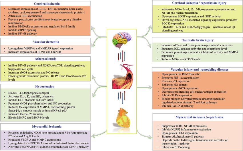
Bai et al. Advances of HSYA Against CCVDs
made in understanding the pathological mechanisms of CCVDs, pharmacological activities in CCVDs that have aroused great
there is still no effective therapy to prevent or stop the epidemic interest worldwide (Zhang et al., 2019), and massive preclinical
trend of CCVDs, resulting in the urgent need to identify studies have aimed to prove the pharmacological effects and
novel therapeutic options (Kazantsev and Outeiro, 2010; dissect the mechanisms of actions of HSYA in treating CCVDs.
Upadhyay, 2014). Safflower yellow injection, a purified yellow pigment extract from
Traditional Chinese medicine (TCM), a cost-effective and Carthami Flos containing no less than 70% of HSYA, is
safe remedy, has been widely used in China and surrounding commercially available for stable exertional angina pectoris of
countries (including Japan and Korea) for the treatment and coronary heart disease with a marked curative effect in Chinese
management of CCVDs with exact and prominent efficacy. clinic (Liu et al., 2014; Xuan et al., 2018). Here, we briefly
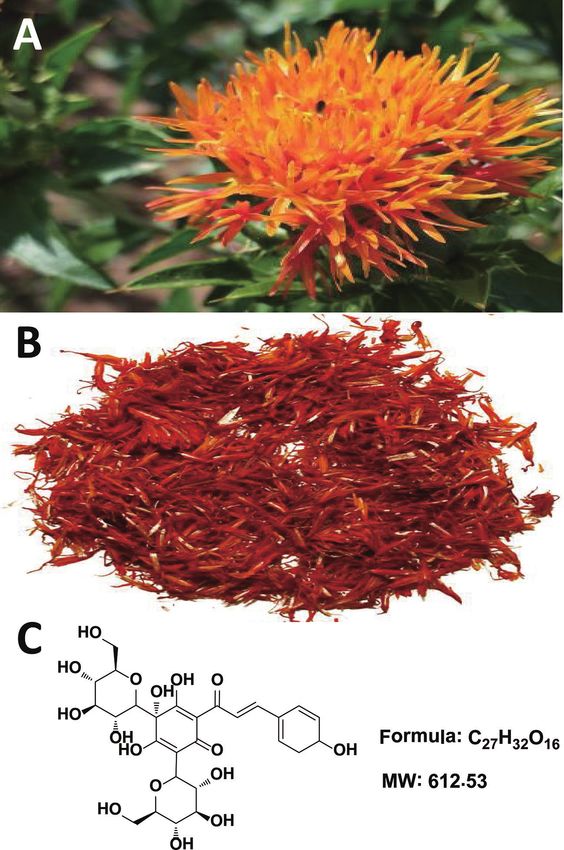
Carthamus tinctorius L. (Compositae) (Figure 1A) seeds are summarize the existing evidence to provide valuable references
known to be rich in a-linoleic acid and have been used since and implications for the clinical uses of HSYA.
ancient times as a source of cooking oil. Meanwhile, its flowers
are widely used for coloring and flavoring foods and
manufacturing dyes (Hu et al., 2018; Guo et al., 2019).
Notably, the medical use of Carthami Flos (Figure 1B, the THERAPEUTIC POTENTIAL OF HSYA
dried florets of C. tinctorius) was first documented in the Golden ON CCVDS
Chamber Synopsis (Han Dynasty, ~2000 years ago) (Ma et al.,
2014), and also described in the Compendium of Materia Effects on Myocardial Ischemia (MI)
Medica (Ming Dynasty, ~500 years ago) as being able to It is acknowledged that MI results from insufficient blood-
“invigorate the blood circulation”, suggestive of its potential oxygen supply (Thiagarajan et al., 2017) and the improved
uses against circulatory system diseases. In modern Chinese vasomotor and circulatory functions exert beneficial effects on
clinic, Honghua injection (made from the water extract of MI (Ribeiro et al., 2019). The vasoconstrictor endothelin and the
Carthami Flos) and Danhong injection (extracted and refined vasodilator nitric oxide (NO) are known as two common
from Salviae miltiorrhizae Radix et Rhizoma and Carthami Flos regulators modulating vasomotor function. HSYA can reverse
herb pair) are widely used for the treatment of coronary heart the circulating levels of both in acute MI animals (e.g., dogs and
disease, angina pectoris, myocardial infarction, ischemic rats), thereby elevating myocardial blood-oxygen supply and
encephalopathy, and cerebral thrombosis (Fan et al., 2019; reducing myocardial injury and apoptosis (Li et al., 2006;
Zhang et al., 2019). Wang et al., 2007). Other vasomotor function-related factors,
The chemical constituents of Carthami Flos are plentiful, and such as 6-keto-prostaglandin F1a, thromboxane B2, and
include flavonoids (e.g. quinochalcone C-glycosides), alkaloids, angiotensin II (Ang II), were also of great importance for
phenolic acids, fatty acids, and more (Yue et al., 2013). Among HSYA (Wang et al., 2007). Angiogenesis participates in the
them, hydroxysafflor yellow A (HSYA, Figure 1C) is both a circulatory function recovery from MI, and HSYA exerts the
representative water-soluble quinochalcone C-glycoside pigment pro-angiogenic effects in two main ways: (1) nucleolin-mediated
and the quality marker of Carthami Flos. It produces remarkable post-transcriptional regulation of vascular endothelial growth
factor-A (VEGF-A) and matrix metalloproteinase (MMP) -9
expressions (Zou et al., 2018); and (2) the up-regulation of heme
oxygenase-1 (HO-1)/VEGF-A/stromal cell-derived factor-1a
Abbreviations: Akt, the protein kinase B; AMPK, 5’-monophosphate -activated cascade (Wei et al., 2017).
protein kinase; Ang II, angiotensin II; AS, atherosclerosis; Bax, Bcl-2 associated X A specialized piece of in vivo research demonstrated that the
protein; BBB, blood-brain barrier; Bcl-2, B-cell lymphoma-2; BDNF, brain- antioxidant effect of HSYA was involved in the prevention of
derived neurotrophic factor; CCVDs, cardio-cerebrovascular diseases; CI,
Ang II-induced myocardial hypertrophy (a compensatory
cerebral ischemia; CI/R, cerebral ischemia/reperfusion; CYP, cytochrome P450;
eNOS, endothelial nitric oxide synthase; GSSG, oxidized glutathione; H/R, response to MI), which may act through the activation of the
hypoxia/reoxygenation; HIF-1, hypoxia inducible factor-1; HO-1, heme nuclear factor erythroid-2-related factor 2 (Nrf2)/NAD(P)H:
oxygenase-1; HSYA, hydroxysafflor yellow A; HUVECs, human umbilical vein quinone oxidoreductase 1/HO-1 signaling pathway (Ni et al.,
endothelial cells; IL, interleukin; JAK2, janus kinase 2/signal transducer; LDL, low- 2018). Nrf-2, as the main regulator of the antioxidant system
density lipoprotein; LPS, lipopolysaccharide; MCAO, middle cerebral artery
present on the cardiovascular system, is becoming a very
occlusion; MDA, malondialdehyde; MI, myocardial ischemia; MI/R, myocardial
ischemia/reperfusion; MMP, matrix metalloproteinase; mTOR, mammalian target promising pharmacological target for cardiovascular diseases
of rapamycin; mtPTP, mitochondrial permeability transition pore; NF-kB, nuclear (McSweeney et al., 2016). Our research group has found that
factor kappa beta; NLRP3, NOD-like receptor 3; NMDARs, NR2B-containing N- HSYA possessed significant antioxidant activity in vitro (Yue
methyl-d-aspartate receptors; NO, nitric oxide; Nrf2, nuclear factor erythroid-2- et al., 2014). Thus, the antioxidant effect of HSYA may be
related factor 2; OGD, oxygen-glucose deprivation; OGD/R, OGD/reoxygenation;
ox-LDL, oxidized low-density lipoprotein; PAF, platelet activating factor; PI3K,
essential to improve the outcomes of cardiovascular diseases.
phosphoinositide 3-kinase; RCT, randomized controlled clinical trial; ROS,
reactive oxygen species; SCFA, short-chain fatty acid; SD, Sprague-Dawley; Effects on Myocardial Ischemia/
SOCS3, suppressor of cytokine signaling protein 3; SOD, superoxide dismutase; Reperfusion (MI/R) Injury
TBI, traumatic brain injury; TCM, traditional Chinese medicine; TLR, toll like
receptor; TNF-a, tumor necrosis factor-a; VaD, vascular dementia; VEGF,
Thrombolytic or percutaneous coronary intervention
vascular endothelial growth factor; VSMCs, vascular smooth muscle cells; WRP, reperfusion for acute myocardial infarction is favorable in most
washed rabbits platelet. cases, but can also cause MI/R injury, resulting in excessive pro-
Frontiers in Pharmacology | www.frontiersin.org 2 September 2020 | Volume 11 | Article 01265Bai et al. Advances of HSYA Against CCVDs
observed effects of HSYA, which suggests that HSYA
suppresses NLRP3 inflammasome activation via the AMPK
pathway (Ye et al., 2020). Together, the TLR4 signaling
pathway and NLRP3 inflammasome impact on the anti-
inflammatory action of HSYA in MI/R.
Apoptosis is initiated shortly after the onset of myocardial
infarction and is enhanced markedly during reperfusion. In H/R-
induced H9c2 cells, the anti-apoptotic effect of HSYA not only
depends on the up-regulation of HO-1 expression through the
phosphoinositide 3-kinase (PI3K)/the protein kinase B (Akt)/
Nrf2 signaling pathway, a compensatory mechanism limiting the
apoptotic events in the presence of aggressive factors (Liu et al.,
2012), but also targets the Akt/hexokinase II pathway to activate
the hexokinase II protein and restore mitochondrial energy to
reduce intracellular reactive oxygen species (ROS) generation
(Min and Wei, 2017). Additionally, Zhou et al. reported that
the anti-apoptotic effect of HSYA might be largely dependent on
the Janus kinase 2/signal transducer (JAK2) and activation of the
transcription 1 pathway (Zhou et al., 2019).
Beside inflammation and apoptosis, MI/R damages
cardiomyocytes in part via the opening of the mitochondrial
permeability transition pore (mtPTP), a non-selective pore that
penetrates the inner and outer mitochondrial membranes
(Bhosale and Duchen, 2019). HSYA has the capability to enter
the cardiomyocytes and then inhibit mtPTP opening to alleviate
H/R-induced myocardial injury through the enhanced
endothelial nitric oxide synthase (eNOS)-produced NO (Liu
et al., 2008; Huber et al., 2018).
Effects on Hypertension
Hypertension is a major global health challenge and an
important risk factor of CCVDs. The blood pressure control
rate of hypertensive patients in developing countries remains at
FIGURE 1 | Carthamus tinctorius L. (A), Carthami Flos (the dried florets of C.
unacceptably low levels (Mills et al., 2016). There is evidence that
tinctorius) (B), and hydroxysafflor yellow A (C). the conspicuous antihypertensive effect of HSYA is attributed to
the inhibition of voltage-gated channels, the renin-angiotensin-
aldosterone system, and the sympathetic nervous system.
inflammatory cytokines in myocardial tissue (Xie et al., 2018). Specifically, HSYA inhibited the endotehlin-independent
HSYA has been reported to possess significant anti- contraction of the thoracic aorta rings of rats through the
inflammatory activity in vitro (Yue et al., 2016). In neonatal rat blockade of inositol 1,4,5-triphosphate receptor in vascular
ventricular myocytes induced by hypoxia/reoxygenation (H/R) smooth muscle cells (VSMCs), leading to the decrease of
and lipopolysaccharide (LPS), HSYA not only inhibited the extracellular Ca2+ influx (Zhang et al., 2011). Beside VSMCs,
excessive secretion of pro-inflammatory cytokines but also endothelial cells participate in vasoconstriction and relaxation.
suppressed the over-expression of toll like receptor (TLR) 4 Yang et al. found that oral HSYA has a concentration-dependent
and nuclear factor kappa beta (NF-kB). Importantly, HSYA was antihypertensive effect. It reversed the constriction of mesenteric
found to alleviate cardiac damage caused by MI/R in normal rats arteries induced by a thromboxane A2 mimetic agent, the
instead of TLR4-knockout mice (Han et al., 2016). It is important potential mechanism of which might be associated with the
to note that TLR4 is the receptor of LPS, the component of the TRPV4 channel-dependent Ca2+ influx, protein kinase A-
outer membrane of Gram-negative bacteria. There is evidence dependent eNOS phosphorylation, and NO production (Yang
that NOD-like receptor 3 (NLRP3) inflammasome activation et al., 2020). A further study disclosed that HSYA could
promotes myocardial injury and apoptosis via inducing the normalize blood pressure and heart rate dose-dependently in
production of pro-inflammatory cytokines. HSYA improved spontaneously hypertensive rats, which might be related to
H/R-induced H9c2 cell viability, maintained mitochondrial activating KATP and BKCa channels, inhibiting L-type Ca
membrane potential, and inhibited NLRP3 inflammasome channels, decreasing Ca2+ influx, and subsequently inhibiting
activation, while the adenosine 5’-monophosphate-activated cardiac contractility (Nie et al., 2012; Wang et al., 2020).
protein kinase (AMPK) inhibitor partially abolished these However, HSYA reduced blood pressure and heart rate in
Frontiers in Pharmacology | www.frontiersin.org 3 September 2020 | Volume 11 | Article 01265Bai et al. Advances of HSYA Against CCVDs
normotensive rats, on which more focus needs to be placed in the interesting to identify the effect of HSYA against ox-LDL formation
future. Moreover, HSYA can increase the reduced diastolic in vivo. In ox-LDL-induced foamy macrophages, HSYA displayed
response of the thoracic aortic to acetylcholine and sodium obvious repairing effects on the de novo fatty acid biosynthesis
nitroprusside, and thus attenuate the vascular contractile effect pathway, among which oleoyl-(acyl-carrier-protein) hydrolase was
of phenylephrine (Jin et al., 2013). HSYA can also inhibit postulated to be a target of HSYA (Wei et al., 2018). HSYA also
proliferative activity and collagen synthesis of Ang II-induced exerted protective effects against ox-LDL-induced VSMCs
adventitial fibroblasts, reduce the expressions of MMP-1, proliferation via increasing mitogen-activated protein kinase
transforming growth factor-b1, a-smooth muscle actin, and phospholipase-1 expression and the proportion of cells in the G0/
NF-kB p65, and thereby decreasing vascular adventitia G1 phase, followed by reducing p-extracellular regulated protein
proliferation and hyperplasia during vascular remodeling kinases activity (Sheng et al., 2012). Moreover, HSYA has been
(Yuan et al., 2014). Obviously, it could be drawn that HSYA shown to significantly improve ox-LDL-induced human endothelial
might be potentially useful as an antihypertensive drug via injury, partially via the anti-apoptotic effect of the mitochondrial
multiple mechanisms mainly involved in both cardiac output membranous voltage-dependent anion-selective channel protein 2
and peripheral resistance. (Ye et al., 2017). Recently, Miao et al. revealed that HSYA could
Hypertension may cause ventricular hypertrophy, which inhibit the high ox-LDL-induced human coronary artery
produces mechanical stimulation to the heart, further causing endothelial cell injury, possibly via increasing eNOS expression
arrhythmia, heart failure, coronary occlusion, and sudden death and NO release, while inhibiting LDL receptor-1 expression and
(Pearson et al., 1991). A study by Wang et al. indicated that oral lactate dehydrogenase release (Miao et al., 2019).
HSYA exhibited anti-apoptotic effects on hypertensive ventricular During the development of AS, platelets can accelerate
hypertrophy in rats by increasing the B-cell lymphoma-2 (Bcl-2)/ activation and release a variety of active substances, such as
Bcl-2 associated X protein (Bax) ratio and blocking serum MMP-2 platelet-activating factor (PAF) and thromboxane B2, conversely
and MMP-9 levels (Wang et al., 2014). Moreover, pulmonary promoting platelet adhesion and aggregation, and even
arterial hypertension is a common combination of congenital damaging vascular endothelial cells (Wang et al., 2008). HSYA
heart disease with systemic-to-pulmonary artery shunt diseases, was able to inhibit PAF-induced platelet aggregation in rabbits
leading to right ventricular heart failure and premature death by blocking PAF-mediated washed rabbit platelets (WRPs) and
(Huang et al., 2018). Voltage-gated K+ channel (KV channel) is an polymorphonuclear leukocyte aggregation (Zang et al., 2002).
important channel for maintaining normal membrane potential Collectively, the above in vitro studies have manifested the
and muscle tension of VSMCs, while HSYA could activate the Kv potential anti-AS effects of HSYA, which should go through
channel of pulmonary artery VSMCs to reduce vascular tension, additional in vivo studies to determine its clinical implications.
suggesting that HSYA may be a potential medication for pulmonary
arterial hypertension (Bai et al., 2012). Effects on Vascular Injury and Remodeling
Diseases
Effects on Atherosclerosis (AS) The vascular endothelium plays an important role in modulating
AS could precipitate the onset of myocardial infarction and numerous aspects of vascular homeostasis (Scarabelli et al., 2002).
inflammation and is being increasingly recognized as the main HSYA was capable of promoting the survival and proliferation of
pathogenic mechanism through the narrowing and blockage of vascular endothelial cells under both normoxic and hypoxic
arteries and the increased risk of blood vessel rupture (Kotla conditions, and its effect was stronger under hypoxia via up-
et al., 2017). HSYA could inhibit ROS-induced inflammation in regulating the Bcl-2/Bax ratio and accumulating hypoxia-
THP-1 macrophages (Jiang et al., 2017), but could also suppress inducible factor-1 (HIF-1) a, which was related to VEGF and its
tumor necrosis factor-a (TNF-a)-induced inflammatory receptor system (Song et al., 2005; Ji et al., 2008). Also, HSYA could
responses through inhibiting the TNF-a receptor type 1- protect human umbilical vein endothelial cells (HUVECs) from
mediated classical NF-kB pathway in arterial endothelial cells hypoxia-induced injury by reducing p53 expression in the cell
(Wang H. F. et al., 2016). Jiang et al. discovered that HSYA- nucleus and up-regulating eNOS expression to produce NO in
mediated sonodynamic therapy induced an autophagic response cell supernatant (Ji et al., 2009).
to inhibit inflammation via the PI3K/Akt/mammalian target of Abnormal proliferation of VSMCs is a crucial cytopathological
rapamycin (mTOR) signaling pathway in THP-1 macrophages basis for the development and progression of vascular remodeling
(Jiang et al., 2017). Nevertheless, it is very interesting to justify diseases (Ivey et al., 2008). HSYA could inhibit platelet-derived
the potential biphasic effect of HSYA under excessive growth factor-BB induced VSMCs proliferation by decreasing
macrophage autophagy, which can drive the instability of proliferating cell nuclear antigen expression and blocking
atherosclerotic plaque (Petrovski et al., 2011). mitogen-activated protein kinase/extracellular regulated protein
In fact, the oxidation of low-density lipoprotein (LDL) and kinases and Akt signaling pathways (Song et al., 2014; Zhao et al.,
oxidized LDL (ox-LDL)-induced vascular damage are key events 2015). In the LPS-induced VSMCs proliferation and migration
in early AS. Although its LDL-lowering effect remains unknown, model, HSYA inhibits the up-regulation of TLR4 expression and the
HSYA was able to reduce the susceptibility of LDL to copper- activation of Ras-related C3 botulinum toxin substrate 1/Akt
induced lipid peroxidation in vitro (Bacchetti et al., 2020). It is pathway (Yang et al., 2015).
Frontiers in Pharmacology | www.frontiersin.org 4 September 2020 | Volume 11 | Article 01265Bai et al. Advances of HSYA Against CCVDs
Effects on Cerebral Ischemia (CI) neurons and resumed eroxisome proliferator-activated receptor g
CI is one of the leading causes of death worldwide, and patients who activity stimulated by either 15-deoxy-delta prostaglandin J2 or
survive CI often experience paralysis, impaired speech, or loss of rosiglitazone (Sun L. et al., 2018).
vision (Moskowitz et al., 2010). HSYA appears to treat focal CI
injury in rats through its anti-coagulation effects on thrombosis Effects on Cerebral Ischemia/Reperfusion
formation and platelet aggregation, as well as beneficial regulation (CI/R) Injury
on prostacyclin/thromboxane and blood rheological changes (Zhu A growing body of research has evidenced that oxidative stress is
et al., 2005). HSYA could also preserve cortex mitochondrial implicated in the pathogenesis of CI/R injury. Wei et al. showed
function of CI rats via scavenging free radicals, reducing lipid that HSYA might oppose CI/R injury of MCAO rats through
peroxides, and antagonizing Ca2+ (Tian et al., 2004). Importantly, attenuating the elevation of malondialdehyde (MDA) level and
HSYA possessed a better effect on cerebrovascular vasodilatation decreasing superoxide dismutase (SOD) activity in the ipsilateral
than on cardiovascular vasodilatation (Sun Y. et al., 2018), but the hemisphere and serum (Wei et al., 2005). HSYA could also
differential molecular mechanism remains to be discovered. reduce CI/R-induced protein oxidation and nitration, attenuate
The blood-brain barrier (BBB) essentially maintains a stable BBB destruction, and importantly inhibit the up-regulation of
cerebral homeostasis, while the destruction or increased 12/15-lipoxygenase, which is implicated in the oxidative stress of
permeability of BBB are common pathological processes during CI/R (Sun et al., 2012). In an in vitro assay, HSYA was shown to
many serious cerebrovascular diseases (Chen Z. X. et al., 2019). A block OGD/reoxygenation (OGD/R)-induced PC12 cells
study by Lv et al. revealed that HSYA significantly attenuated BBB apoptosis through the suppression of intracellular oxidative
dysfunction in anti-inflammatory patterns in ischemia stroke via stress (Fan et al., 2011).
the tight junction pathway, especially the NMMHC IIA, TLR4/ An inflammatory reaction is a recognized player in CI/R
PI3K/Akt/Jun N-terminal kinase 1/2/14-3-3ϵ pathway while damage. Through suppressing TLR4 pathway-mediated
inhibiting the expressions of occludin, claudin-5, and zonula signaling responses, HSYA could up-regulate brain-derived
occludens-1 (Lv and Fu, 2018). Since MMPs are the main neurotrophic factor (BDNF) in MCAO mice at post-ischemia/
endoproteinases involved in BBB destruction (Romanic et al., reperfusion (Lv et al., 2015), but also exert neurotrophic and
1998), the prominent inhibitory effects of HSYA on MMP-2 and anti-inflammatory functions in LPS-activated co-existence
MMP-9 mentioned in cardiovascular diseases may also contribute systems for microglia and neurons (Lv et al., 2016). In the
to BBB improvement. microglia of the ischemic cortex after acute CI/R, HSYA
It is worth mentioning that CI plays a causal role in facilitating exerted anti-inflammatory effects by activating the TLR9
neuronal death (Martin and Wang, 2010). A metabonomic study signaling pathway and suppressing the NF-kB pathway (Gong
revealed that HSYA could attenuate excitatory amino acid-induced et al., 2018). Further studies demonstrated that HSYA
neurotoxicity, at least partially, through inhibiting the NF-kB significantly inhibited NF-kB p65 nuclear translation and p65
pathway in the cerebral tissues of the middle cerebral artery binding activity, both mRNA and protein levels of intercellular
occlusion (MCAO) model rats (Liu et al., 2013). Other protective adhesion molecule 1, and the infiltration of neutrophils (Sun
mechanisms of HSYA against excitotoxic neuronal death include et al., 2010).
the inhibition of NR2B-containing N-methyl-d-aspartate receptors Cognitive impairment is becoming a serious mental deficit that
(NMDARs) expression and the Bcl-2 family regulation in cortical severely affects the life quality of patients following CI/R (Jokinen
cultures, and the inhibition of the N-methyl-d-aspartate-induced et al., 2006). HSYA has the capacity to improve neurological deficit
and NMDARs-mediated intracellular Ca2+ increase in hippocampal scores and increase the surviving hippocampal CA1 pyramidal cells
cultures (Yang et al., 2010; Wang X. T. et al., 2016). In oxygen- in focal CI/R rats (Sun et al., 2010). HSYA injected via the common
glucose deprivation (OGD)-induced BV2 microglia, the carotid artery significantly rescued the neurological and cognitive
neuroprotective action of HSYA involves the decreased functional deficits of MCAO rats against CI/R injury. Meanwhile,
expressions of pro-inflammatory cytokines, including interleukin HSYA could markedly down-regulate JAK2-mediated signaling,
(IL) -1b, TNF-a, inducible nitric oxide synthase, cyclooxygenase-2, while promoting the expression of the suppressor of cytokine
and monocyte chemotactic protein-1, as well as the reserved signaling protein 3 (SOCS3) (Yu et al., 2018; Yu et al., 2020).
phosphorylation of p38 and nuclear translocation of p65 (Li et al., Furthermore, the neuroprotective effect of HSYA against CI/R
2013). Peroxynitrite-mediated protein tyrosine nitration and injury might be conferred through activating the Akt-dependent
nitrosative stress represent the crucial pathogenic mechanisms of autophagy pathway (Qi et al., 2014). In both OGD/R-induced
CI, while the anti-nitrative pathway might contribute to the primary mouse neurons and PC12 cells, HSYA inhibited
neuroprotective efficacy of HSYA. Specifically, Sun et al. phenylalanine biosynthesis to enhance mitochondrial function
discovered that HSYA blocked authentic peroxynitrite-induced and biogenesis for neuroprotection (Chen S. N. et al., 2019).
tyrosine nitration in primary cortical neurons by the reduction of PI3K-mediated signaling pathways are also involved in the
inducible nitric oxide synthase expression and NO content, protective effects of HSYA against apoptosis and autophagy
suggestive of its peroxynitrite scavenging abilities (Sun et al., during CI/R. Chen et al. reported that HSYA critically reduced
2013). They further reported that HSYA prevented peroxisome CI/R-mediated apoptosis through the PI3K/Akt/glycogen
proliferator-activated receptor g nitrative modification in primary synthase kinase 3b signaling pathway (Chen et al., 2013).
Frontiers in Pharmacology | www.frontiersin.org 5 September 2020 | Volume 11 | Article 01265Bai et al. Advances of HSYA Against CCVDs
HSYA protected the cerebral microvascular endothelial cells And there is no doubt that HSYA is a promising lead drug
against OGD/R-induced injury by inhibiting autophagy via the candidate in designing new multi-targeted therapeutic agents
Class I PI3K/Akt/mTOR signaling pathway (Yang et al., 2018). against CCVDs. The other analogues of HSYA, safflor yellow A
Proteomic analysis showed that mTOR, Eftud2, Rab11, Ppp2r5e, (Duan et al., 2013) and safflor yellow B (Wang et al., 2009; Wang
and HIF-1 signaling pathways were the key hub proteins and et al., 2013), showed similar protective effects against CCVDs.
pathways in HSYA against CI/R injury (Xu et al., 2019). Further structural modification of HSYA should be extensively
Therefore, the PI3K/Akt/mTOR signaling pathway needs to be made and coupled with quantitative structure-activity relationship
further studied to clarify the mechanisms of actions of HSYA studies to develop more selective and safe drugs.
against CI/R injury. The oral bioavailability of HSYA is extremely low (~1.2%)
Similar to MI/R, CI/R also results in mtPTP opening. (Ekin, 2005) and oral administration of HSYA accounts for
Mechanically, HSYA could inhibit mtPTP opening by inhibiting about 0.9% of all in vivo experiments from Table 1. However,
Ca2+-induced ROS generation and H2O2-induced swelling of among many administration routes, oral administration is of
mitochondria isolated from rat brains, improving mitochondrial great significance in drug formation because of its convenience
energy metabolism and enhancing ATP levels and the respiratory and safety. Thus, the microemulsion, nanoemulsion, and
control ratio in the ischemia brain (Tian et al., 2008). nanoparticles of HSYA have been developed to overcome the
low oral bioavailability (Li et al., 2010; Qi et al., 2011; Lv et al.,
Effects on Vascular Dementia (VaD) 2012; Shi et al., 2018; Zhao B. X. et al., 2018). Considering the
VaD is characterized by pathological damage and a decline in weak ability of HSYA to penetrate the BBB in physiologic
intelligence resulting from hypoxic-ischemic or hemorrhagic condition (He et al., 2008), Borneolum Syntheticum and Acori
brain injury (Zhao X. X. et al., 2018). As mentioned before, Talarinowii Rhizoma were used to enhance its BBB permeability
HSYA has the capacity to improve cognitive impairment. In VaD (Wu et al., 2011). Nevertheless, more attention should be payed
rats, Zhang et al. revealed that HSYA promoted angiogenesis and to the overall efficacy and safety evaluation of HSYA before and
increased synaptic plasticity via up-regulating the hippocampal after improving oral bioavailability and BBB permeability.
expressions of VEGF-A, NMDAR type-1, BDNF, and GluN2B (a In the past decade, cohesive evidence showed that gut
subunit of NMDAR), thus improving spatial learning and microbiota may serve as a therapeutic target of natural
memory (Zhang et al., 2014; Xing et al., 2016). Although no compounds derived from TCM (Yue et al., 2019a; Yue et al.,
drug is approved, the above findings may shed light on the 2019b). Oral HSYA is mostly retained in the intestinal tract as its
therapeutic potential of HSYA for managing the progress prototype, which inevitably interacts with gut microbiota. Obesity
of VaD. is considered to be one of the most important risk factors of
CCVDs. Our research group reported that HSYA mediated its
Effects on Traumatic Brain Injury (TBI) anti-obesity effects by reversing gut microbiota dysbiosis in obese
TBI refers to the injury of cerebral tissue structure/function caused mice, followed by increasing short-chain fatty acid (SCFA)-
by various kinds of mechanical violence in the outside world (Xiu producing bacteria (Liu J. et al., 2018). For SCFAs, it has a well-
et al., 2018). HSYA has the potential to be utilized as a established role in maintaining host immune function after MI
neuroprotective agent in cases of TBI. Firstly, TBI enables HSYA (Tang et al., 2019). The potential for microbial synthesis of
to distribute in the cerebral tissues of rats (Bie et al., 2010). Then, the SCFAs, including propionate and butyrate, was low in patients
antioxidant effect of HSYA in the brain of the TBI rats could explain with atherosclerotic cardiovascular disease (Jie et al., 2017).
the TBI improvement via increasing SOD, catalase and glutathione Recently, the microbiota-gut-brain axis has shown to influence
levels, while reducing MDA and oxidized glutathione (GSSG) levels BBB permeability and the pathological process of TBI (Braniste
(Bie et al., 2010; Wang Y. et al., 2016). Lastly, HSYA could increase et al., 2014; Ma et al., 2017). Thus, it is feasible to unveil the
mitochondrial ATPase (i.e., Na+, K+-ATPase, Ca2+-ATPase, and underlying mechanisms of oral HSYA on CCVDs from the new
Mg2+-ATPase) and tissue plasminogen activator activities, while perspective of gut microbiota modulation.
decreasing plasma plasminogen activator inhibitor-1 activity and Carthami Flos is a common part of preparations used in TCM
MMP-9 expression in the hippocampus of TBI rats (Wang Y. and other traditional medicinal systems. It is necessary to
et al., 2016). strengthen the compatibility research of HSYA and other
In summary, the above findings buttress the assertion that TCM-derived components. As an example, HSYA and
HSYA exerts cardio-cerebrovascular protective activities through Danshensu synergistically enhanced the antioxidant defense
complex pathways and exhibits a definite curative effect in the system and anti-apoptotic effects on MI/R injury through the
application of CCVDs (Table 1 and Figure 2). Akt/Nrf2/HO-1 signaling pathway (Hu et al., 2016). Their
combination further achieved enhanced neuroprotective effects
on CI/R injury by alleviating pro-inflammatory and oxidative
CONCLUSIONS AND PROSPECTS stress reactions via the TLR4/NF-kB and Nrf2/HO-1 pathways
(Xu et al., 2017). On the other hand, combinations with existing
It is becoming clear that the important mechanisms by which western medications may also provide new therapy options for
HSYA exerts extensive biological activities in CCVDs are through CCVDs patients. For example, HSYA as an add-on therapy to
its antioxidant, anti-inflammatory, and neuroprotective effects. acetylglutamine could synergistically modulate the neuronal
Frontiers in Pharmacology | www.frontiersin.org 6 September 2020 | Volume 11 | Article 01265Bai et al. Advances of HSYA Against CCVDs
TABLE 1 | Summary of pharmacological effects and mechanisms of HSYA on cardio-cerebrovascular diseases.
Disease Species/Strains Effective dose/ Route Mechanism of action Reference
concentration
Myocardial Acute MI model in mongrel 14, 28 mg·kg-1 i.v. Inhibits endothelin release, increases myocardial blood flow, (Li et al., 2006)
ischemia (MI) dogs and improves the cardiac oxygen metabolism
Acute MI model in male Wistar 5, 10, 20 mg·kg-1 i.v. Increases the activity of serum NO synthase, the content of (Wang et al., 2007)
rats NO and 6-keto-prostaglandin F1a, and decreases the levels
of creatine kinase-MB, lactate dehydrogenase, thromboxane
B2 and Ang II
Acute MI model in male C57 25 mg·kg-1 i.p. Promotes the migration and tube formation of HUVECs, (Zou et al., 2018)
mice enhances the expressions of nucleolin, VEGF-A and MMP-9
MI model in male C57BL/6 15, 30, 60 mg·kg-1 i.v. Promotes endothelial progenitor cells function through the (Wei et al., 2017)
mice HO-1/VEGF-A/stromal cell-derived factor-1a signaling
cascade
MI model in male SD rats 2, 5 mg·kg-1 i.p. Activates the Nrf2/NAD(P)H:quinone oxidoreductase 1/HO-1 (Ni et al., 2018)
signaling pathway
Ang II-induced H9c2 cells 80 mmol·L-1 / Increases the cell viability, however, reduces protein synthesis (Ni et al., 2018)
rate, mitigates cell surface area and decreases the expression
of brain natriuretic factor and b-myosin heavy chain
Myocardial MI/R model in male SD rats 5 mg·kg-1 i.p. Decreases JAK2/signal transducer and activator of (Zhou et al., 2019)
ischemia/ transcription 1 activity, enhances antioxidant capacity and
reperfusion (MI/ decreases apoptosis
R) injury Hyperlipidemia combined with 8, 16, 32 mg·kg-1 i.p. Suppresses the over-expression of TLR4 (Han et al., 2016)
MI/R model in male Wistar rats
H/R and LPS-induced neonatal 1, 3, 10 mmol·L-1 / Decreases excessive secretion of inflammatory cytokines, (Han et al., 2016)
rat ventricular myocytes down-regulates over-expression of TLR4 and NF-kB
H/R-induced H9c2 cells 6.25, 12.5, 25 / Improves cardiomyocyte viability, maintains mitochondrial (Ye et al., 2020)
mmol·L-1 membrane potential, reduces apoptotic cardiomyocytes,
decreases Caspase-3 activity, and inhibits NLRP3
inflammasome activation
H/R-induced H9c2 cells 1.25, 5, 20 mmol·L-1 / Activates the hexokinase II proteins, restores mitochondrial (Min and Wei,
energy, reduces ROS generation 2017)
H/R-induced H9c2 cells 20 mmol·L-1 / Inactivates the JAK2/signal transducer and activator of the (Zhou et al., 2019)
transcription 1 pathway
H/R-induced H9c2 cells 5, 20, 80 mmol·L-1 / Up-regulates HO-1 expression through the PI3K/Akt/Nrf2 (Liu et al., 2012)
signaling pathway
MI/R model in hearts isolated 50, 100, 200 / Enhances NO production by eNOS activation (Liu et al., 2008)
from male SD rats mmol·L-1
H/R-induced cardiomyocytes 100, 200 mmol·L-1 / Modulates the reduction of viability and the loss of rod- (Huber et al., 2018)
isolated from SD rat hearts shaped cells, and interacts with the mtPTP
Hypertension Male spontaneous 0.1-3 mg·kg-1 i.v. Reduces blood pressure and heart rate, activates BKCa and (Nie et al., 2012)
hypertension rat and KATP channels
normotensive Wistar-Kyoto rats
Male spontaneous 0.6-2.4 mg·kg-1 i.v. Activates BKCa channels, inhibits Ca-L channels, and reduces (Wang et al., 2020)
hypertension rat and intracellular free Ca2+ level
normotensive Wistar-Kyoto rats
Ang II-induced vascular 0.5, 1 ml/kg i.p. Reduces matrix metallopeptidase-1, transforming growth (Yuan et al., 2014)
adventitial fibroblasts in male factor-b1, a-smooth muscle actin and NF-kB p65 expression
SD rats
AT1 receptor-induced 10 mg·kg-1 i.g. Reverses the vascular structure and function and improves (Jin et al., 2013)
hypertension in male Wistar plasma biochemical parameters
rats
Pressure overload-induced 20, 40 mg·kg-1 i.g. Increases the Bcl-2/Bax ratio, blocks the levels of MMP-2 (Wang et al., 2014)
cardiac hypertrophy in male and MMP-9 in serum
Wistar rats
PE-induced pulmonary artery 0.01-10 mmol·L-1 / Activates the KV channel in placental VSMCs and relaxes rat (Bai et al., 2012)
rings of Wistar rats pulmonary artery
KCl-precontracted thoracic EC50 = 18.2 mmol·L- / Induces relaxation in endothelium-intact/endothelium- (Zhang et al., 2011)
aorta rings of male Wistar rats 1
/17.4 mmol·L-1 precontracted aortas precontracted by KCl
PE-precontracted thoracic EC50 = 18.7 mmol·L- / Induces relaxation in endothelium-intact/endothelium- (Zhang et al., 2011)
aorta rings of male Wistar rats 1
/17.9 mmol·L-1 precontracted aortas precontracted by phenylephrine
CaCl2-precontracted thoracic 20 mmol·L-1 / Reduces Ca2+ influx and inhibits inositol 1,4,5-triphosphate (Zhang et al., 2011)
aorta rings of male Wistar rats receptor
U46619-induced mesenteric 1-100 mmol·L-1 / Reverses the constriction, promotes Ca2+ influx, eNOS (Yang et al., 2020)
arteries of male Wistar rats phosphorylation and NO production
(Continued)
Frontiers in Pharmacology | www.frontiersin.org 7 September 2020 | Volume 11 | Article 01265Bai et al. Advances of HSYA Against CCVDs
TABLE 1 | Continued
Disease Species/Strains Effective dose/ Route Mechanism of action Reference
concentration
Atherosclerosis Human THP-1 monocytes 400-800 mmol·L-1 / Induces an autophagic response via the PI3K/Akt/mTOR (Jiang et al., 2017)
(AS) signaling pathway and inhibits inflammation by ROS
Human coronary artery 200-1600 mmol·L-1 / Up-regulates the eNOS gene and protein expression, (Miao et al., 2019)
endothelial cells injury model increases NO release, inhibits lactate dehydrogenase release
and down-regulates LDL receptor 1 expression
TNF-a-stimulated primary 120 mmol·L-1 / Inhibits the TNF-a receptor type 1-mediated classical NF-kB (Wang H. F. et al.,
mouse kidney arterial pathway 2016)
endothelial cells and
RAW264.7 macrophage cells
Ox-LDL-induced foamy 65.30 mmol·L-1 / Up-regulates the abnormal metabolism of C12:0, C14:0, (Wei et al., 2018)
macrophages C18:1
Ox-LDL-induced human 1, 5, 25 mmol·L-1 / Inhibits cell apoptosis by voltage-dependent anion-selective (Ye et al., 2017)
endothelial cells channel protein 2
Ox-LDL-induced VSMCs 10 mmol·L-1 / Increases mitogen-activated protein kinase phospholipase-1 (Sheng et al., 2012)
expression and the proportion of cells in G0/G1 phase,
reduces p-extracellular signal-regulated protein kinase 1/2
activity, and suppresses cell cycle
PAF-induced WRP suspension 250-1470 mmol·L-1 / Inhibits PAF binding to WRP receptors (Zang et al., 2002)
of male New Zealand white
rabbits
PAF-induced WRP suspension IC50 = 990 mmol·L-1 / Reduces PAF-mediated WRP aggregation (Zang et al., 2002)
of male New Zealand white
rabbits
PAF-induced IC50 = 700 mmol·L-1 / Reduces PAF-mediated polymorphonuclear leukocytes (Zang et al., 2002)
polymorphonuclear leukocytes aggregation
suspension of male New
Zealand white rabbits
Vascular Injury Hypoxia-induced canine aortic 10, 100, 1000 / Promotes vein endothelial cells proliferation via promoting (Song et al., 2005)
Diseases endothelial cell mmol·L-1 vascular endothelial growth factor and its receptor secretion
Hypoxia-induced HUVECs 1, 10, 100 mmol·L-1 / Up-regulates the Bcl-2/Bax ratio and promotes HIF-1a (Ji et al., 2008)
protein accumulation
Hypoxia-induced HUVECs 1, 10, 100 mmol·L-1 / Inhibits cell apoptosis and cell cycle G1 arrest induced by (Ji et al., 2009)
hypoxia
Vascular Platelet-derived growth factor 1-60 mmol·L-1 / Reduces the expression of proliferating cell nuclear antigen, (Zhao et al., 2015)
Remodeling -induced VSMCs blocks signal transduction of mitogen-activated protein
Diseases kinase/extracellular regulated protein kinases
Platelet-derived growth factor 20 mmol·L-1 / Suppresses Akt signaling activation (Song et al., 2014)
-induced VSMCs
LPS-induced VSMCs 0.1-100 mmol·L-1 / Inhibits TLR4/Ras-related C3 botulinum toxin substrate 1/Akt (Yang et al., 2015)
pathway
Cerebral MCAO model in male SD rats 10, 50 mg·kg-1 i.v. Corrects the impaired metabolic pathways, suppresses pro- (Liu et al., 2013)
ischemia (CI) inflammatory cytokine expression and p65 translocation and
binding activity
MCAO model in male SD rats 10, 20 mg·kg-1 i.v. Reduces lipid peroxides, inhibits Ca2+ overload, scavenges (Tian et al., 2004)
free radicals
MCAO model in male Wistar- 3, 6 mg·kg-1 i.v. Inhibits thrombosis formation and platelet aggregation, (Zhu et al., 2005)
Kyoto rats regulates prostacyclin/thromboxane and blood rheological
changes
MCAO model in C57BL/6J 1, 2, 4 mg·kg-1 i.p. Attenuates BBB dysfunction via the tight junction pathway, (Lv and Fu, 2018)
mice and attenuates the expression of occludin, claudin-5, and
zonula occludens-1
Nitrified bovine serum albumin 10, 100, 1000 / Blocks authentic peroxynitrite-induced tyrosine nitration (Sun et al., 2013)
and primary cultured male SD mmol·L-1
rats cortical neurons exposed
to peroxynitrite
Peroxynitrite donor SIN-1- 10, 100, 1000 / Blocks peroxisome proliferator-activated receptor g nitration (Sun et al., 2018)
induced primary cultured male mmol·L-1
SD rats cortical neurons
Primary cultured SD rats 10 mmol·L-1 / Inhibits the expression NR2B-containing NMDA receptors (Yang et al., 2010)
cortical neurons exposed to and regulates Bcl-2 family
NMDA
10-80 mmol·L-1 / (Tian et al., 2008)
(Continued)
Frontiers in Pharmacology | www.frontiersin.org 8 September 2020 | Volume 11 | Article 01265Bai et al. Advances of HSYA Against CCVDs
TABLE 1 | Continued
Disease Species/Strains Effective dose/ Route Mechanism of action Reference
concentration
Ca2+ and H2O2-induced brain Inhibits Ca2+-induced generation of ROS, and mtPTP opening
mitochondrial suspension of by a free radical scavenging action
male SD rats
NMDA-induced and NMDA IC50 = 17.60 / Protects hippocampal neurons from excitotoxic damage (Wang X. T. et al.,
receptors-mediated C57BL/6 mmol·L-1 through the inhibition of NMDA receptors 2016)
mice hippocampal neurons
OGD-induced BV2 microglia 40-1280 mmol·L-1 / Suppresses inflammatory responses by inhibiting the NF-kB (Li et al., 2013)
signaling pathway and phosphorylation of p38
Phenylephrine-precontracted 5 mg into the 20 ml / Attenuates the contractile responsiveness (Sun et al., 2018)
Coronary artery and basilar Krebs’-Henseleit
artery of beagle dogs buffer every 5 min
for 4–5 times
Cerebral MCAO/R model in male SD 1, 5, 10 mg·kg-1 i.v. Reduces protein oxidation and nitration, inhibits the up- (Sun et al., 2012)
ischemia/ rats regulation of 12/15-lipoxygenase, and attenuates BBB
reperfusion (CI/ breakdown
R) injury MCAO/R model in male SD 8, 16 mg·kg-1 CCAI Protects cognitive function and synaptic plasticity (Yu et al., 2018)
rats
MCAO/R model in male SD 8, 16 mg·kg-1 CCAI Downregulates the expression of JAK2-mediated signaling, (Yu et al., 2020)
rats while promotes the expression of SOCS3
MCAO/R model in male SD 6 mg·kg-1 i.p. Regulates Eftud2, Rab11, Ppp2r5e, and HIF-1 signaling (Xu et al., 2019)
rats pathway
Acute MCAO/R model in male 6 mg·kg-1 i.p. Activates TLR9 in the microglia of ischemic cortex and (Gong et al., 2018)
SD rats suppresses the NF-kB pathway
Acute I/R stroke model in male 2 mg·kg-1 i.v. Activates the Akt autophagy pathway in penumbra tissue (Qi et al., 2014)
SD rats
MCAO/R model in male Wistar 2, 4, 8 mg·kg-1 i.v. Attenuates the elevation of MDA content, the decrease in (Wei et al., 2005)
rats SOD activity, and the total antioxidative capability
MCAO/R model in male Wistar 2, 4, 8 mg·kg-1 i.v. Suppresses thrombin generation and thrombin-induced (Sun et al., 2010)
rats inflammatory responses by reducing Ang II content
MCAO/R model in male Wistar 4, 8 mg·kg-1 i.v. Reduces apoptosis via PI3K/Akt/glycogen synthase kinase 3b (Chen et al., 2013)
rats signaling pathway
MCAO/R model in C57BL/6J 2 mg·kg-1 i.v. Inhibits TLR4 pathway-mediated signaling responses (Lv et al., 2015)
mice
OGD/R induced human brain 10-80 mmol·L-1 / Inhibits autophagy via the Class I PI3K/Akt/mTOR signaling (Yang et al., 2018)
microvascular endothelial cells pathway
LPS-stimulated non-contact 50, 100 mmol·L-1 / Exerts neurotrophic and anti-inflammatory functions in (Lv et al., 2016)
transwell co-culture system response to LPS stimulation by inhibiting TLR4 pathway-
comprised microglia and mediated signaling
neurons
OGD-induced PC12 cells 10, 100 mmol·L-1 / Suppresses the intracellular oxidative stress and (Fan et al., 2011)
mitochondria-dependent caspase cascade
OGD/Reduced primary 1, 10 mmol·L-1 / Reduces phenylalanine level, promotes mitochondrial function (Chen S. N. et al.,
neurons and PC12 cells and biogenesis for neuroprotection 2019)
Vascular VaD model in male SD rats 6 mg·kg-1 i.v. Promotes angiogenesis and increases synaptic plasticity (Zhang et al., 2014)
dementia (VaD) VaD model in male SD rats 6 mg·kg-1 i.v. Increases in the expression levels of BDNF and GluN2B (Xing et al., 2016)
Traumatic brain TBI model in male SD rats 2, 4 mg·kg-1 i.v. Increases mitochondrial ATPase and tissue plasminogen (Bie et al., 2010)
injury (TBI) activator activities, decreases plasma plasminogen activator
inhibitor-1 activity and MMP-9 expression
TBI model in male SD rats 10, 30 mg·kg-1 i.g. Enhances SOD and catalase activities, glutathione level and (Wang Y. et al.,
the glutathione/GSSG ratio while reduces MDA and GSSG 2016)
levels
/, no information in the original paper; i.v., intravenous injection; i.g., intragastrical administration; i.p., intraperitoneal injection; CCAI, common carotid artery injection.
apoptosis and inflammation process during CI/R (Deng et al., The randomized controlled clinical trial (RCT) is an essential
2018). However, it is important to note that HSYA is able to step in confirming the efficacy and safety of drugs. In contrast with
inhibit cytochrome P450 (CYP) enzymes’ (i.e. CYP1A2 and the large numbers of preclinical experiments, only a few completed
CYP2C11) activities but induces CYP3A1 activity (Xu et al., RCTs of HSYA were reported, which were mainly reflected in
2014). Hence, more detailed and advanced research should be evaluating the efficacy and safety of HSYA injection in the treatment
done in the future to develop new compound formulas with of acute ischemic stroke with blood stasis syndrome (Qin et al.,
HSYA, which may bring about important benefits for CCVDs 2016; Hu et al., 2020), followed by a currently ongoing phase III
patients and TCM modernization. RCT (No. CTR20150839, http://www.chinadrugtrials.org.cn/).
Frontiers in Pharmacology | www.frontiersin.org 9 September 2020 | Volume 11 | Article 01265Bai et al. Advances of HSYA Against CCVDs
FIGURE 2 | HSYA acts on the functional targets and signaling pathways of cardio-cerebrovascular diseases.
However, none of the existing RCTs were of high methodological review. All authors contributed to the article and approved the
quality, and the conclusions need to be further verified by large submitted version.
sample, multicenter, and double-blind RCTs (as compared to
traditional treatment regimens). In addition, clinical evidence
supporting the application of HSYA for the management of
CCVDs other than acute ischemic stroke with blood stasis
FUNDING
syndrome is still lacking. This work was supported by the National Natural Science
Foundation of China (81903786, 81773882), the Natural Science
Foundation of Shaanxi Province (2019JQ-054), the Young Talent
AUTHOR CONTRIBUTIONS Support Program from the Association for Science and Technology
of Colleges in Shaanxi Province (20190306), grants from the Key
S-JY and Y-PT conceived and designed the review. XB searched Research and Development Program of Shaanxi (2019ZDLSF04-
the literature and drafted the manuscript. W-XW and R-JF 05), Shaanxi Administration of Traditional Chinese Medicine
examined the literature and made the figures. HG edited the (2019-ZZ-JC018), and Subject Innovation Team of Shaanxi
manuscript. S-JY, Y-YC, and Y-PT made a critical revision of the University of Chinese Medicine (2019-YL10).
REFERENCES Bie, X. D., Han, J., and Dai, H. B. (2010). Effects of hydroxysafflor yellow A on the
experimental traumatic brain injury in rats. J. Asian Nat. Prod. Res. 12, 239–
Bacchetti, T., Morresi, C., Bellachioma, L., and Ferretti, G. (2020). Antioxidant and 247. doi: 10.1080/10286020903510636
pro-oxidant properties of Carthamus tinctorius, hydroxy safflor yellow A, and Braniste, V., AlAsmakh, M., Kowal, C., Anuar, F., Abbaspour, A., Toth, M., et al.
safflor yellow A. Antioxidants 9, 119. doi: 10.3390/antiox9020119 (2014). The gut microbiota influences blood-brain barrier permeability in
Bai, Y. H., Lu, P., Han, C. H., Yu, C. Y., Chen, M. G., He, F., et al. (2012). mice. Sci. Transl. Med. 6, 263ra158. doi: 10.1126/scitranslmed.3009759
Hydroxysafflor yellow A (HSYA) from flowers of Carthamus tinctorius L. and Chen, L., Xiang, Y. X., Kong, L. J., Zhang, X. M., Sun, B. Z., and Wei, X. B. (2013).
its vasodilatation effects on pulmonary artery. Molecules 17, 14918–14927. Hydroxysafflor yellow A protects against cerebral ischemia-reperfusion injury
doi: 10.3390/molecules171214918 by antiapoptotic effect through PI3K/Akt/GSK3b pathway in rat. Neurochem.
Bhosale, G., and Duchen, M. R. (2019). Investigating the mitochondrial Res. 38, 2268–2275. doi: 10.1007/s11064-013-1135-8
permeability transition pore in disease phenotypes and drug screening. Curr. Chen, S. N., Sun, M., Zhao, X. H., Yang, Z. F., Liu, W. X., Cao, J. Y., et al. (2019).
Protoc. Pharmacol. 85, e59. doi: 10.1002/cpph.59 Neuroprotection of hydroxysafflor yellow A in experimental cerebral ischemia/
Frontiers in Pharmacology | www.frontiersin.org 10 September 2020 | Volume 11 | Article 01265Bai et al. Advances of HSYA Against CCVDs
reperfusion injury via metabolic inhibition of phenylalanine and mitochondrial Ji, D. B., Zhang, L. Y., Li, C. L., Ye, J., and Zhu, H. B. (2009). Effect of
biogenesis. Mol. Med. Rep. 19, 3009–3020. doi: 10.3892/mmr.2019.9959 hydroxysafflor yellow A on human umbilical vein endothelial cells under
Chen, Z. X., Xu, Q. Q., Shan, C. S., Shi, Y. H., Wang, Y., Chang, R. C. C., et al. hypoxia. Vasc. Pharmacol. 50, 137–145. doi: 10.1016/j.vph.2008.11.009
(2019). Borneol for regulating the permeability of the blood-brain barrier in Jiang, Y. Q., Kou, J. Y., Han, X. B., Li, X. S., Zhong, Z. Y., Liu, Z. N., et al. (2017).
experimental ischemic stroke: preclinical evidence and possible mechanism. ROS-dependent activation of autophagy through the PI3K/Akt/mTOR
Oxid. Med. Cell Longev. 2019:2936737. doi: 10.1155/2019/2936737 pathway is induced by hydroxysafflor yellow A-sonodynamic therapy in
Deng, L., Wan, H. T., Zhou, H. F., Yu, L., and He, Y. (2018). Protective effect of THP-1 macrophages. Oxid. Med. Cell. Longev. 2017, 8519169. doi: 10.1155/
hydroxysafflor yellow A alone or in combination with acetylglutamine on 2017/8519169
cerebral ischemia reperfusion injury in rat: A PET study using 18F- Jie, Z. Y., Xia, H. H., Zhong, S. L., Feng, Q., Li, S. H., Liang, S. S., et al. (2017). The
fuorodeoxyglucose. Eur. J. Pharmacol. 825, 119–132. doi: 10.1016/ gut microbiome in atherosclerotic cardiovascular disease. Nat. Commun. 8,
j.ejphar.2018.02.011 845. doi: 10.1038/s41467-017-00900-1
Duan, J. L., Wang, J. W., Guan, Y., Yin, Y., Wei, G., Cui, J., et al. (2013). Safflor Jin, Z., Zhang, W. H., Chai, W. R., Zheng, Y. Q., and Zhi, J. M. (2013). Antibodies
yellow A protects neonatal rat cardiomyocytes against anoxia/reoxygenation against AT1 receptors are associated with vascular endothelial and smooth
injury in vitro. Acta Pharmacol. Sin. 34, 487–495. doi: 10.1038/aps.2012.185 muscle function impairment: Protective effects of hydroxysafflor yellow A. PloS
Ekin, Z. (2005). Resurgence of safflower (Carthamus tinctorius L.) utilization: a One 8, e67020. doi: 10.1371/journal.pone.0067020
global view. J. Agron. 4, 83–87. doi: 10.3923/ja.2005.83.87 Jokinen, H., Kalska, H., Mäntylä, R., Pohjasvaara, T., Ylikoski, R., Hietanen, M.,
Fan, L. H., Dang, X. Q., Shi, Z. B., Zhang, C., and Wang, K. Z. (2011). et al. (2006). Cognitive profile of subcortical ischaemic vascular disease.
Hydroxysafflor yellow A protects PC12 cells against the apoptosis induced J. Neurol. Neurosurg. Psychiatry 77, 28–33. doi: 10.1136/jnnp.2005.069120
by oxygen and glucose deprivation. Cell. Mol. Neurobiol. 31, 1187–1194. Kazantsev, A. G., and Outeiro, T. F. (2010). Drug discovery for CNS disorders:
doi: 10.1007/s10571-011-9720-3 from bench to bedside. CNS Neurol. Disord-DR. 9, 668. doi: 10.2174/
Fan, J. X., Qin, X. M., and Li, Z. Y. (2019). Molecular docking and multivariate 187152710793237395
analysis studies of active compounds in the safflower injection. J. Liq. Kotla, S., Singh, N. K., and Rao, G. N. (2017). ROS via BTK-p300-STAT1-PPARg
Chromatogr. R. T. 42, 673–680. doi: 10.1080/10826076.2019.1665540 signaling activation mediates cholesterol crystals-induced CD36 expression and
Gong, Z., Pan, J. R., Li, X. P., Wang, H. X., He, L., and Peng, Y. (2018). foam cell formation. Redox Biol. 11, 350–364. doi: 10.1016/j.redox.2016.12.005
Hydroxysafflor yellow A reprograms TLR9 signalling pathway in ischaemic Li, X. Z., Liu, J. X., Shang, X. H., and Fu, J. H. (2006). Protective effects of
cortex after cerebral ischaemia and reperfusion. CNS Neurol. Disord-DR. 17, hydroxysafflor yellow A on acute myocardial ischemia in dogs. Chin. Pharm.
370–382. doi: 10.2174/1871527317666180502110205 Bull. 22, 533–537.
Guo, X. J., Zheng, M., Pan, R. Y., Zang, B. X., Gao, J. W., Ma, H. Y., et al. (2019). Li, J. R., Sun, M. J., Ping, Q. N., Chen, X. J., Qi, J. P., and Han, D. E. (2010).
Hydroxysafflor yellow A (HSYA) targets the platelet-activating factor (PAF) Metabolism, excretion and bioavailability of hydroxysafflor yellow A after oral
receptor and inhibits human bronchial smooth muscle activation induced by administration of its lipid-based formulation and aqueous solution in rats.
PAF. Food Funct. 10, 4661–4673. doi: 10.1039/c9fo00896a Chin. J. Nat. Med. 8, 233–240. doi: 10.3724/SP.J.1009.2010.00223
Han, D., Wei, J., Zhang, R., Ma, W., Shen, C., Feng, Y. D., et al. (2016). Li, J., Zhang, S. Y., Lu, M. R., Chen, Z. B., Chen, C., Han, L. J., et al. (2013).
Hydroxysafflor yellow A alleviates myocardial ischemia/reperfusion in Hydroxysafflor yellow A suppresses inflammatory responses of BV2 microglia
hyperlipidemic animals through the suppression of TLR4 signaling. Sci. Rep. after oxygen-glucose deprivation. Neurosci. Lett. 535, 51–56. doi: 10.1016/
6:35319. doi: 10.1038/srep35319 j.neulet.2012.12.056
He, P. P., Fu, F. H., Wang, T., Li, C. K., Xin, W. Y., and Zhang, X. M. (2008). Effect of Liu, Y. N., Zhou, Z. M., and Chen, P. (2008). Evidence that hydroxysafflor yellow A
cerebral ischemia/reperfusion injury on hydroxysafflor yellow A penetrating across protects the heart against ischaemia-reperfusion injury by inhibiting
the blood-brain barrier. Sci. Pharm. 76, 713–724. doi: 10.3797/scipharm.0811-01 mitochondrial permeability transition pore opening. Clin. Exp. Pharmacol. P.
Hu, T. X., Wei, G., Xi, M. M., Yan, J. J., Wu, X. X., Wang, Y. H., et al. (2016). 35, 211–216. doi: 10.1111/j.1440-1681.2007.04814.x
Synergistic cardioprotective effects of Danshensu and hydroxysafflor yellow A Liu, S. X., Zhang, Y., Wang, Y. F., Li, X. C., Xiang, M. X., Bian, C., et al. (2012).
against myocardial ischemia-reperfusion injury are mediated through the Akt/ Upregulation of heme oxygenase-1 expression by hydroxysafflor yellow A
Nrf2/HO-1 pathway. Int. J. Mol. Med. 38, 83–94. doi: 10.3892/ijmm.2016.2584 conferring protection from anoxia/reoxygenation-induced apoptosis in H9c2
Hu, Z. C., Xie, Z. J., Tang, Q., Li, X. B., Fu, X., Feng, Z. H., et al. (2018). cardiomyocytes. Int. J. Cardiol. 160, 95–101. doi: 10.1016/j.ijcard.2011.03.033
Hydroxysafflor yellow A (HSYA) targets the NF-kB and MAPK pathways and Liu, Y. Y., Lian, Z. Q., Zhu, H. B., Wang, Y. H., Yu, S. S., Chen, T. T., et al. (2013). A
ameliorates the development of osteoarthritis. Food Funct. 9, 4443–4456. systematic, integrated study on the neuroprotective effects of hydroxysafflor
doi: 10.1039/C8FO00732B yellow A revealed by 1H NMR-based metabonomics and the NF-kB pathway.
Hu, M. Z., Zhou, Z. Y., Zhou, Z. Y., Lu, H., Gao, M., Liu, L. M., et al. (2020). Effect Evid. Based. Compl. Alt. 2013, 147362. doi: 10.1155/2013/147362
and safety of hydroxysafflor yellow A for injection in patients with acute Liu, Y. Q., Tian, X. F., Cui, M. Z., and Zhao, S. Z. (2014). Safflower yellow inhibits
ischemic stroke of blood stasis syndrome: A phase II, multicenter, randomized, angiotensin II-induced adventitial fibroblast proliferation and migration.
double-blind, multiple-dose, active-controlled clinical trial. Chin. J. Integr. J. Pharmacol. Sci. 126, 107–114. doi: 10.1254/jphs.14055FP
Med. 26, 420–427. doi: 10.1007/s11655-020-3094-7 Liu, J., Yue, S. J., Yang, Z. R., Feng, W. W., Meng, X. T., Wang, A. T., et al. (2018). Oral
Huang, L., Li, L., Hu, E. C., Chen, G., Meng, X. M., Xiong, C. M., et al. (2018). hydroxysafflor yellow A reduces obesity in mice by modulating the gut microbiota
Potential biomarkers and targets in reversibility of pulmonary arterial and serum metabolism. Pharmacol. Res. 134, 40–50. doi: 10.1016/j.phrs.2018.05.012
hypertension secondary to congenital heart disease: an explorative study. Liu, Z., Xu, Y. Q., and Ji, X. M. (2018). Progress in clinical diagnosis and treatment
Pulm. Circ. 8:204589321875598. doi: 10.1177/2045893218755987 of cardio-cerebrovascular diseases. Chin. J. Geriatr. Heart Brain Vessel Dis. 20,
Huber, G. A., Priest, S. M., and Geisbuhler, T. P. (2018). Cardioprotective effect of 1219–1220. doi: 10.3969/j.issn.1009-0126.2018.11.025
hydroxysafflor yellow A via the cardiac permeability transition pore. Planta Lv, Y. N., and Fu, L. S. (2018). The potential mechanism for hydroxysafflor yellow
Med. 84, 507–518. doi: 10.1055/s-0043-122501 A attenuating blood-brain barrier dysfunction via tight junction signaling
Ivey, M. E., Osman, N., and Little, P. J. (2008). Endothelin-1 signalling in vascular pathways excavated by an integrated serial affinity chromatography and
smooth muscle: Pathways controlling cellular functions associated with shotgun proteomics analysis approach. Neurochem. Int. 112, 38–48.
atherosclerosis. Atherosclerosis 199, 237–247. doi: 10.1016/j.atherosclerosis. doi: 10.1016/j.neuint.2017.10.012
2008.03.006 Lv, L. Z., Tong, C. Q., Lv, Q., Tao, X. J., Li, L. M., Fang, Q. X., et al. (2012).
Ji, D. B., Zhu, M. C., Zhu, B., Zhu, Y. Z., Li, C. L., Ye, J., et al. (2008). Enhanced absorption of hydroxysafflor yellow A using a self-double-
Hydroxysafflor yellow A enhances survival of vascular endothelial cells emulsifying drug delivery system: In vitro and in vivo studies. Int.
under hypoxia via upregulation of the HIF-1a-VEGF pathway and J. Nanomed. 7, 4099–4107. doi: 10.2147/IJN.S33398
regulation of Bcl-2/Bax. J. Cardiovasc. Pharm. 52, 191–202. doi: 10.1097/ Lv, Y. N., Qian, Y. S., Fu, L. S., Chen, X. Y., Zhong, H. L., and Wei, X. H. (2015).
FJC.0b013e318181fb02 Hydroxysafflor yellow A exerts neuroprotective effects in cerebral ischemia
Frontiers in Pharmacology | www.frontiersin.org 11 September 2020 | Volume 11 | Article 01265You can also read