A Ratchet Mechanism of Transcription Elongation and Its Control
←
→
Page content transcription
If your browser does not render page correctly, please read the page content below
Cell, Vol. 120, 183–193, January 28, 2005, Copyright ©2005 by Elsevier Inc.   DOI 10.1016/j.cell.2004.11.045

A Ratchet Mechanism
of Transcription Elongation
and Its Control
Gil Bar-Nahum,1,4 Vitaly Epshtein,1,4                                    these signals within RNAP that controls the basic move-
Andrei E. Ruckenstein,2 Ruslan Rafikov,1                                 ment of the enzyme has been determined.
Arkady Mustaev,3 and Evgeny Nudler 1,*                                      Recent biochemical and structural analyses have gen-
1
 Department of Biochemistry                                              erated a comprehensive model of the elongation com-
New York University Medical Center                                       plex (EC) with a high-resolution map of the catalytic
New York, New York 10016                                                 center and nucleic acid binding sites (Korzheva et al.,
2
  BioMaPS Institute for Quantitative Biology and                         2000; Gnatt et al., 2001). In this model, the two largest
Department of Physics                                                    subunits of RNAP hold the EC together by clamping the
Rutgers University                                                       8 bp RNA:DNA hybrid and downstream DNA duplex
Piscataway, New Jersey 08854                                             (Nudler, 1999). These interactions allow the EC to slide
3
  Public Health Research Institute                                       along DNA and RNA during elongation and reverse
Newark, New Jersey 07103                                                 translocation (backtracking) (Komissarova and Kashlev,
                                                                         1997; Nudler et al., 1997). Based on comparative analy-
                                                                         sis of bacterial and yeast RNAP structures, Kornberg
Summary                                                                  and coworkers proposed an elementary transcription
                                                                         cycle in which bending of the F bridge helix (F bridge)
RNA chain elongation is a highly processive and accu-                    at the 3⬘ face of the RNA:DNA hybrid induces transloca-
rate process that is finely regulated by numerous in-                    tion of the nucleic acid by one nucleotide within the
trinsic and extrinsic signals. Here we describe a gen-                   RNAP mainframe, while a subsequent relaxation of the
eral mechanism that governs RNA polymerase (RNAP)                        F bridge opens the substrate binding site for the next
movement and response to regulatory inputs such as                       complementary NTP (Gnatt et al., 2001). A bent F bridge
pauses, terminators, and elongation factors. We show                     helix has only been observed in bacterial RNAP struc-
that E.coli RNAP moves by a complex Brownian ratchet                     tures (Zhang et al., 1999; Vassylyev et al., 2002) and a
mechanism, which acts prior to phosphodiester bond                       straight F bridge only in the yeast RNAP II structure
formation. The incoming substrate and the flexible F                     (Cramer et al., 2001). However, protein-RNA crosslinking
bridge domain of the catalytic center serve as two                       data obtained using the E. coli EC strongly suggest the
separate ratchet devices that function in concert to                     ability of the F bridge to change its conformation within
drive forward translocation. The adjacent G loop do-                     the same enzyme (Epshtein et al., 2002).
main controls F bridge motion, thus keeping the proper                      We used a genetic screen to isolate dominant lethal
balance between productive and inactive states of the                    mutants in E. coli RNAP and selected two point muta-
elongation complex. This balance is critical for cell                    tions (G1136S and I1134V) in the G helix-loop-helix do-
viability since it determines the rate, processivity, and                main (G loop) of the RNAP largest subunit. These two
fidelity of transcription.                                               mutants potentiate and restrict F bridge motion, respec-
                                                                         tively. G1136S renders RNAP fast, inaccurate, and poorly
Introduction                                                             responsive to pauses and terminators. Conversely,
                                                                         I1134V renders the enzyme slow and overreactive at
                                                                         regulatory signals. Both mutations impair the respon-
The control of transcription elongation occurs at differ-
                                                                         siveness of the EC to general elongation factors. Based
ent levels in all organisms (Uptain et al., 1997; Reines
                                                                         on comparative biochemical, protein chemical, and ki-
et al., 1999). In E. coli, increasing or decreasing the
                                                                         netic analysis of wild-type (wt) and mutant RNAPs, we
overall rate of elongation plays an important role in cell
                                                                         propose a model that explains all the basic elongation
adaptation to nutrient conditions (Vogel and Jensen,
                                                                         properties of the enzyme. The model incorporates the
1994; Condon et al., 1995). Nus factors along with cis-
                                                                         concept of a ratchet and pawl, a classical engineering
acting RNA signals participate in this type of regulation.
                                                                         device in which a cogged wheel is restricted by a wedge
In vitro, NusG and NusA accelerate and decelerate elon-
                                                                         known as a pawl such that its movement is unidirec-
gation, respectively, by altering pausing at various sites
                                                                         tional. Depending on its design, a pawl may be station-
(Schmidt and Chamberlin, 1984; Burova et al., 1995).                     ary, restricting reverse movement by insertion of its
The eukaryotic homolog of NusG, DSIF, has also been                      wedge between the cogged teeth. Alternatively, the pawl
shown to increase the rate of RNAP II elongation (Wada                   may reciprocate in such a way as to generate unidirec-
et al., 1998). Another level of regulation uses specific                 tional movement of the wheel. In our model of the EC,
pauses to control transcription termination and antiter-                 both types of pawl are envisaged: the F bridge acts as
mination (Landick et al., 1996; McDowell et al., 1994;                   a reciprocating pawl, pushing RNAP forward in relation
Roberts et al., 1998; Nudler and Gottesman, 2002). There                 to the nucleic acid scaffold, while the incoming substrate
has been much recent progress in understanding the                       acts as a second, stationary pawl, preventing RNAP
structure/functional organization of RNAP. However,                      from slipping backward. We present evidence that these
neither the process by which these regulatory signals                    two pawls compete for the same substrate binding site.
are transduced to RNAP nor the ultimate acceptor of                      Thus, RNAP acts as a unique molecular motor, combin-
                                                                         ing two types of ratchet device within the same catalytic
*Correspondence: evgeny.nudler@med.nyu.edu                               center. Such a superposition of two Brownian ratchets,
4
  These authors contributed equally to this work.                        while limiting the overall elongation rate, allows the F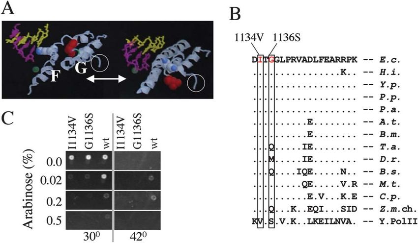
Cell
184
Figure 1. Dominant Lethal Mutations in the RNAP ⬘ Subunit G Loop Domain
(A) Dynamic structure of the F bridge/G loop domain. The ribbon model shows two alternating states of the F bridge (F): bent (left) as in the
T. aquaticus structure (Zhang et al., 1999) and straight (right) as in the S. cereviciae structure (Gnatt et al., 2001). In the diagram, Lys789 (shown
in blue) in the F bridge clashes with the 3⬘ face of the RNA:DNA hybrid in the bent configuration. Note that the hybrid is present only in the
yeast structure. The G helix-loop-helix domain (G) changes its conformation as represented by rotation of Met932 (Leu1081 in the yeast structure)
(shown in red). White circles highlight the location of I1134V and G1136S mutations in the G loop. Location of the catalytic Mg2⫹ ion is shown
in green.
(B) Evolutionary conservation of mutated residues. I1134 and G1136 are shown in red. The sequence alignment spans part of the G loop of
E. coli (E.c.), H. influenzae (H.i.), Y. pestis (Y.p.), P. putida (P.p), P. aeruginosa (P.a.), A. tumefaciens (A.t.), B. melitensis (B.m.), T. aquaticus
(T.a.), D. radiodurans (D.r.), B. subtilis (B.s.), M. tuberculosis (M.t.), C. pneumoniae (C.p.), Z. mays chroloplast (Z.m.ch), and S. cerevisiae
RNAP II (Y. Pol II). Dots denote amino acids that are identical between species.
(C) Dominant-negative phenotype of I1134V and G1136S. Both mutations were selected from an error-prone PCR-generated library generated
from a cloned sequence encoding rpoC under the control of the pBAD promoter. A screen for dominant-negative phenotypes was conducted
in host cells carrying a temperature-sensitive chromosomal copy of rpoC. LB agar plates display wt, I1134V, and G1136S clones grown at
permissive (30⬚C) and nonpermissive (42⬚C) temperatures under various pBAD-inducing conditions (shown as arabinose %). Note that wt, but
not I1134V or G1136S, complement rpoCts at 0.02%–0.2% arabinose and that wt rpoC expression becomes toxic at 0.5% arabinose.
bridge/G loop unit to mediate exceptional control and fi- The ⴕ I1134V and G1136S Mutations Have
delity. Opposite Effects on Elongation and Termination
The dominant phenotype of I1134V and G1136S and the
conservation of wt residues (Figure 1) argue for their
Results critical role in RNAP. To determine which step in the
transcription cycle was affected by these mutations, a
Dominant-Negative Mutations in the G Loop series of tests was performed with purified enzymes.
Domain of the ⴕ Subunit We observed little, if any, effect on promoter binding
Structural analysis of the catalytic site of cellular RNAPs and open complex formation (data not shown). However,
suggests that the G loop influences the conformation both mutants displayed drastic changes at the elonga-
of the F bridge (Korzheva et al., 2000; Epshtein et al., tion and termination stages. The overall elongation rate
2002; Zhang et al., 1999; Cramer et al., 2001; Figure 1A). of G1136S was about two times greater than that of wt
To test this hypothesis and to address its physiological (Figure 2A). Conversely, I1134V was more than three
significance, we developed a genetic screen for domi- times slower than wt (Figure 2A). Furthermore, NusG,
nant-negative mutations in the G region of the ⬘ subunit. which normally accelerates the elongation rate, was un-
The rpoC gene was placed under the control of the able to do so in the case of the fast G1136S mutant.
finely regulated pBAD promoter, and a library of point Moreover, both NusG and NusA (which normally slows
substitutions in the ⬘ segment comprising the entire G the elongation rate) failed to show any significant effect
region was generated by error-prone PCR (see Experi- on the slow, I1134V enzyme. These data suggest that
mental Procedures). Cells harboring the chromosomal the two mutations in the G loop of ⬘ affect the function
copy of ⬘ that is inactive at 42⬚C were transformed with of the RNAP enzyme in the same manner as NusG and
wt and mutant pBAD⬘ plasmids. A parallel screen of NusA. G1136S and I1134V also responded differently to
2380 colonies at permissive and nonpermissive temper- specific pause signals, including the T stretch of the
atures yielded 15 dominant-negative isolates, two of tR2 terminator (Gusarov and Nudler, 1999), ops (Artsi-
which, I1134V and G1136S (Figures 1B and 1C), were movitch and Landick, 2000), and his and trp (Landick et
selected for further analysis. al., 1996) (Figure 2B). G1136S almost completely failed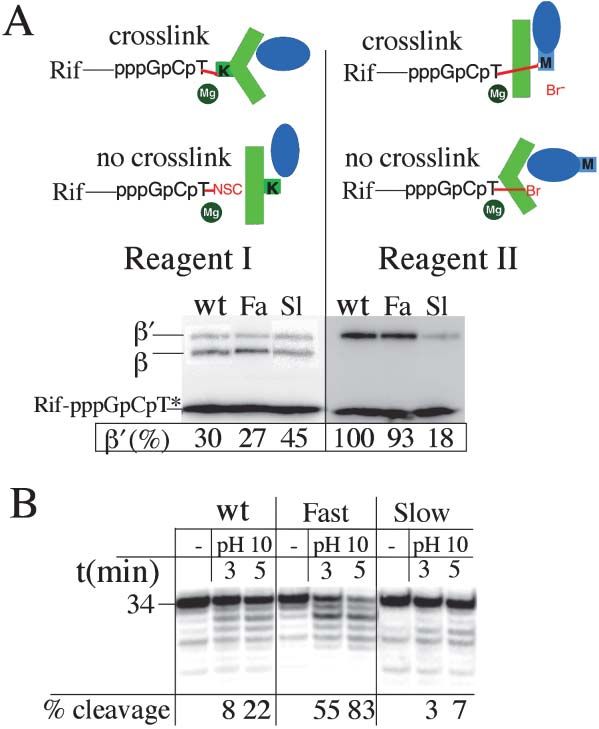
RNA Polymerase as a Ratchet Machine
185
Figure 2. Effect of G Loop Mutations on
Transcription Elongation and Its Regulation
(A) Effect of I1134V and G1136S mutations
on the overall elongation rate and respon-
siveness to negative (NusA) and positive
(NusG) general elongation factors. Elongation
rates (nucleotides/second) were measured
on a linear T7A1 promoter template under
conditions where only a single round of tran-
scription can occur. The initial elongation
complex ([␣-32P] CMP-labeled EC32) was
formed and immobilized on Co2⫹ beads as
described in the Experimental Procedures.
After washing the beads, the complete set of
NTPs was added to 10 M, with or without
NusA (300 nM) or NusG (250 nM). Rates were
calculated by measuring the time-dependent
extension of the transcript in each reaction
to the run-off position (EC254).
(B) Effect of I1134V or G1136S on specific
regulatory pauses. Natural sequences of hair-
pin-dependent his and trp leader pauses and
hairpin-independent ops and T stretch (Ts)
pauses were fused to the initial transcribed
sequence of the T7A1 promoter. Preformed
[␣-32P] CMP EC32 was immobilized as in (A), washed with standard transcription buffer (TB100), and chased to the pause signals by addition
of 150 M NTPs (in the case of ops and T stretch) or 150 M CTP, ATP, UTP, and 10 M GTP (in the case of trp and his) for the indicated
time periods prior to quenching and resolution of the reaction products on a 12% polyacrylamide gel containing 8 M urea. Pause half-lives
were calculated as fractions of [32P] elongated transcripts interrupted at the pause positions (marked P in each panel).
(C) Effect of I1134V or G1136S on intrinsic termination. Experiments were performed on a linear DNA template carrying the T7A1 promoter
fused to the tR2 terminator. Preformed [␣-32P] CMP EC32 was generated as described in (A) and chased to the termination site with 150
M NTPs for 20 min at 25⬚C. The efficiency of termination (%T) was calculated by dividing the amount of radioactivity in a terminated band
by the total radioactivity present in the sum of the terminated and readthrough bands. Transcription termination, i.e., RNA dissociation from
the EC and hence from the beads, was confirmed in each case by the loss of radioactive RNA corresponding to the termination band after
washing the beads with TB100. A representative example is shown for the I1134V mutant in the lane marked W. In this experiment, the runoff
EC serves as an internal control, which remained unchanged after washing.
to recognize these regulatory sequences under normal III (ExoIII) DNA footprinting. Comparison of the front-
chase conditions. In contrast, I1134V stalled for much edge position of wt EC33 (wtEC33) relative to G1136SEC33
longer than wt at each of these pause sites (Figure 2B). and I1134VEC33 (Figure 3A, lanes 1–3) revealed a major
Since pausing at the T stretch is crucial for termination difference between these three complexes. wtEC33 was
(Gusarov and Nudler, 1999), we also compared the abil- distributed quite evenly over a three nucleotide span
ity of both mutants to terminate at the tR2 terminator. (namely ⫹1, ⫺1, ⫺2). G1136SEC33 was mostly forward
As expected, G1136S was almost completely resistant translocated, i.e., it shifted downstream toward the ⫹1
to termination at physiological NTP concentrations, boundary, while I1134VEC33 was primarily backtracked,
while I1134V terminated much more efficiently than wt locating around the ⫺2 position. Since in each case
(Figure 2C). Taken together, these results establish the EC33 remained fully active during the experiment (Figure
G loop as an essential modulator of the basic elongation 3A, lower panel), the complex must oscillate between
properties of RNAP and its responsiveness to regula- the three adjacent positions, with the most downstream
tory factors. position (⫹1) corresponding to the active (forward-
translocated) state. In the case of EC34 (lanes 4–6), the
The G Loop Controls Lateral Movement of the EC amplitude of oscillation was larger (five nucleotides), yet
In general, increased or decreased pausing can account the relative distribution of wt and mutant ECs was similar
for the altered ability of mutant RNAP to acquire an to that of EC33. G1136SEC34 spent most of its time in
active (nonbacktracked) state, defined as the state in the forward-translocated (active) state (lane 5), while
I1134V
which the i⫹1 site of the catalytic center is capable of EC34 remained mainly in the backtracked (inactive)
accepting the next-required NTP. To test this hypothe- state (lane 6). These results explain the fast and slow
sis, we analyzed the dynamic positioning of wt and mu- phenotype of the mutants and establish the G loop as
tant ECs stalled at two consecutive positions (⫹33 a modulator of the lateral movement of RNAP.
and ⫹34) of the T7A1 promoter DNA template. These
two positions do not arrest or terminate transcription Factor-Mediated EC Translocation
and thus reflect the “ordinary” behavior of the EC. A and the Role of the G Loop
His6 tag at the C terminus of recombinant wt and mutant A correlation between the elongation rate and the ability
⬘subunit was used to obtain homogeneous EC33 and of EC to adopt the forward-translocated (active) state
EC34 by step-wise transcription (“walking”) in a solid suggests that positive (NusG) and negative (NusA) elon-
support (see Experimental Procedures; Nudler et al., gation factors facilitate forward and backward translo-
2003). To probe EC boundaries, we utilized exonuclease cation, respectively. Indeed, NusG has a dramatic effect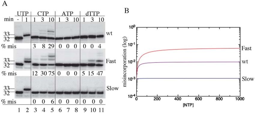
Cell
186
Figure 3. Dynamic Positioning of wt and G Loop Mutant ECs on Template DNA
(A) Effect of fast (G1136S, denoted Fa) and slow (I1134V, denoted Sl) mutations on translocation of EC33 and EC34. Top panel shows protection
of the 32P-end-labeled nontemplate DNA strand from ExoIII digestion (see Experimental Procedures), limited by occlusion at the front edge
of the EC. Arrow and accompanying schematic at the left shows the direction of EC forward translocation. The histogram displays the relative
radioactive content of each band within a given lane. “% (top)” refers to the relative radioactive content of the upper band in each lane. The
lower panel shows a control experiment done to demonstrate that in each case, the ECs remained active after ExoIII digestion. Paired lanes
of radiolabeled RNA from EC33 and EC34 were analyzed after treatment with ExoIII either before (left-hand lane) or after (right-hand lane) the
chase with 1 mM NTPs. The shift of the signals in the right-hand lanes to a position corresponding to a much larger species (not shown)
implies extension of each transcript.
(B) Effect of elongation factors NusA (A, 300 nM) and NusG (G, 250 nM) on translocation of EC34 formed with wt, fast, or slow RNAP. Reaction
conditions were as described in (A).
(C) Effect of the next complementary NTP on translocation 3⬘ terminated EC formed with wt, fast, or slow RNAP. Upper panel shows the
protection of the 32P-end-labeled template DNA strand from ExoIII digestion, limited by occlusion at the rear edge of the EC. Arrow and
accompanying schematic at the left shows the direction of forward translocation. EC33 and EC34 were each generated carrying terminating
3⬘-deoxy-UMP (33dU) and 3⬘-deoxy-CMP (34dC) incorporated at the 3⬘ terminus. Where indicated, the next-required CTP (C) or GTP (G) were
added to 500 M prior to ExoIII treatment. “% bottom” refers to the relative radioactive content of the bottom band in each lane. The lower
panel shows radiolabeled RNA after treatment with ExoIII. This serves as a control that demonstrates a lack of incorporation of correct NTP
into the transcripts.
(D) Effect of nonhydrolyzable complementary NTP on translocation of active EC34 formed with wt, fast, or slow RNAP. Reaction conditions
were as described in (A). Where indicated, GMPcPP was added to 250 M prior to ExoIII treatment. Arrow and accompanying schematic at
the left shows the direction of forward translocation. The lower panel shows radiolabeled RNA from each reaction analyzed after treatment
with ExoIII.
(E) Effect of complementary and noncomplementary substrates on translocation of wt EC34. Reaction conditions were as described in (A).
Where indicated, NMP and/or NTP and/or GMPcPP were each added to 500 M prior to ExoIII treatment. NusG (lane 10) was added to 250
nM. The lower panel shows radiolabeled RNA from each reaction analyzed after treatment with ExoIII. This served to confirm that the RNA
was not extended and that it remained intact.
on the lateral mobility of wtEC33 by shifting all its bound- data shown in Figure 2, where NusG accelerated wt
aries forward to the “⫹1” position (Figure 3B, lane 3). RNAP but had little effect on the fast and slow mutants.
The effect of NusG on G1136SEC34 positioning was less The effect of NusA on the lateral mobility of the EC
pronounced (Figure 3B, lane 6), apparently because further corroborates the kinetic results. NusA strongly
G1136S
EC34 was already forward translocated. NusG had and moderately promoted backtracking of G1136SEC34
no visible effect on the translocation of I1134VEC34 (Figure and wtEC34, respectively (Figure 3B, lanes 2 and 5) but
3B, lane 9). These results correlate well with the kinetic had no effect on I1134VEC34 (Figure 3B, lane 8). TakenRNA Polymerase as a Ratchet Machine
187
together, these results argue that NusA and NusG con- bending, two F bridge parameters, the bent/straight ra-
trol the rate of elongation by modulating the lateral mo- tio and the transition rate between the two conforma-
tion of EC via the G loop. tions, were compared between wt and G loop mutants.
To measure the bent/straight ratio, the catalytic site was
Substrate-Mediated EC Translocation probed with two crosslinkable UTP derivatives (which
Recent kinetic studies suggest that, during elongation, we designate rTTP*, where the asterisk denotes the
binding of the next complimentary NTP facilitates the crosslinkable group) carrying either a lysine-specific
process, apparently by stabilizing the EC in an active isothiocyano group (reagent I) or a nucleophile-specific
state (Foster et al., 2001; Sousa, 2001; Nedialkov et al., bromoacetate group (reagent II) (Figure 4A). These re-
2003; Holmes and Erie, 2003). To test this hypothesis agents discriminate between bent and straight F bridge
directly and to establish the role of the G loop/F bridge conformations because it has been shown that reagent
in this process, we compared the ability of the next I forms an adduct with Lys789 in the ⬘ subunit only if the
obligate NTP substrate to shift wtEC, G1136SEC, and I1134VEC F bridge is bent, whereas reagent II reacts with ⬘ Met932
forward without being incorporated into RNA. Two ap- only if the F bridge is straight. Met932 is the major nucleo-
proaches, each complementing one another, were used. philic target for the latter reagent; in the absence of
In the first approach, the next-required NTP was added Met932, this crosslinker reacts less discriminately, tar-
to the EC carrying a terminating 3⬘-deoxy analog geting various nearby residues in both  and ⬘ (Epshtein
(ECd⫹NTP) (Guajardo and Sousa, 1997). Figure 3C dem- et al., 2002). To constrain each reagent to reaction at
onstrates the effect of CTP and GTP on the rear-edge the active site and prevent ambiguous crosslinks from
positioning of EC33dU and EC34dC, respectively. backtracked EC, we employed the chimerical primer Rif-
G1136S
EC33dU and G1136SEC34dC shifted forward almost com- GTP, in which rifampicin is covalently attached to GTP
pletely upon addition of the corresponding substrates (Mustaev et al., 2003). Following its incorporation, Rif-
(Figure 3C, lanes 6 and 8). wtEC33dU⫹CTP and GTP was extended to Rif-GCT by reaction with [␣-32P]
wt
EC34dC⫹GTP shifted by ⵑ35% and 15%, respectively CTP and rTTP*. This places the crosslinker in the cata-
(Figure 3C, lanes 2 and 4), while I1134VEC333⬘d⫹CTP and lytic site, while Rif occupies its nearby pocket and stabi-
I1134V
EC343⬘d⫹GTP barely shifted at all (Figure 3C, lanes lizes the complex (Mustaev et al., 2003). Mapping the
10 and 12). In a second approach, a complementary crosslinking sites (done as described by Epshtein et al.
nonhydrolyzable NTP or NMP was added to normal EC [2002]) in each case confirmed their identity, i.e., their
(EC⫹NMPcPP/NMP). Similar results to those described reaction with Lys789 and Met932 in the case of reagents I
above using 3⬘-deoxy analogs were obtained in these and II, respectively (data not shown). In the case of
experiments (Figure 3D). The majority of wtEC34 and G1136S, the relative efficiency of the ⬘ subunit cross-
G1136S
EC34 shifted forward upon addition of GMPcPP linking was similar to that of wt (Figure 4A, lanes 1 and
(Figure 3C, lanes 2 and 4). In contrast, I1134VEC34 did not 2 and lanes 4 and 5), suggesting that the fast mutation
respond to the analog (Figure 3C, lane 6). We conclude did not alter the preferred conformation of the F bridge.
In contrast, the I1134V mutation changed the ratio of
that the G loop is directly implicated in modulating the
Lys789/Met943 crosslinking significantly. The relative
responsiveness of EC to NTP-induced translocation.
crosslinking to ⬘ (Lys789) was increased in comparison
To examine the specificity of NTP-induced transloca-
to wt (Figure 4A, lane 3), whereas crosslinking to ⬘
tion, we compared the effect of complementary and
(Met932) was strongly reduced (Figure 4A, lane 6). This
noncomplementary substrates on wtEC34 (Figure 3E).
result suggested that in the I1134V enzyme, the equilib-
The complementary monophosphate nucleotide, GMP,
rium between the two conformations of the F bridge is
induced forward translocation to a similar extent as
shifted toward the bent state. Note that the Rif linker
GMPcPP, implying that phosphates do not play a major
allows crosslinks to occur only from the i⫹1 site and
role in inducing translocation. The noncomplementary
not from backtracking positions, thus directly reflecting
substrates AMP and CMP failed to induce any forward conformational changes in the catalytic center.
translocation. Remarkably, however, these noncomple- To estimate the effect of G loop mutations on the
mentary nucleotides stimulated backtracking (Figure kinetics of F bridge fluctuations between bent and
3E, lanes 3 and 4). Equimolar pairs of correct and incor- straight conformations, we utilized the intrinsic property
rect substrates had a reduced effect on forward translo- of RNAP to perform transcript cleavage. Current models
cation, regardless of whether the incorrect substrates of the mechanism of the transcription cleavage reaction
were complementary to the i⫹2 position (AMP or ATP). suggest that the bent F bridge compromises this reac-
This result implies that in each case there is direct com- tion (Opalka et al., 2003; Kettenberger et al., 2003; Lap-
petition for the i⫹1 site, but not for other (i⫹n) sites. tenko et al., 2003). Normally, the cleavage reaction is
These experiments demonstrate that correct base pair- stimulated by cleavage factors such as GreB; however,
ing at the i⫹1 site alone is sufficient to drive forward these factors may actively interfere with F bridge bend-
translocation by stabilizing the EC in the active state. ing (Laptenko et al., 2003; Sosunova et al., 2003; Opalka
et al., 2003). We therefore elected to use high pH condi-
The G Loop Sets the Parameters of the F Bridge tions as an alternative means to induce intrinsic tran-
It has been proposed that the F bridge helix can adopt script cleavage (Orlova et al., 1995; Figure 4B). At least
two distinct conformations, bent and straight, and that two events must occur for the cleavage reaction to take
the transition between the two drives forward transloca- place: backtracking and straightening of the F bridge.
tion (Gnatt et al., 2001). To determine whether the G In the event of backtracking, if the F bridge were in its
loop affects the lateral motion of the EC via F bridge bent conformation, it would melt the RNA/DNA hybridCell
188
connection between the conformational state of the F
bridge and the cleavage reaction, we found that
G1136S
EC34 is more sensitive to pH cleavage than wtEC34
(Figure 4A, lanes 1–6), even though it is forward translo-
cated in relation to wtEC34, as detected by ExoIII (Figure
3). Our crosslinking data (Figure 4A) showed that the
bent/straight ratio of the F bridge in G1136S was the
same as that in wt, implying the same equilibrium kinet-
ics between bent and straight conformations in the two
ECs. We therefore propose that the energy barrier be-
tween the two F bridge conformations is lower (and that
the F bridge oscillates faster) in G1136SEC34 than in wtEC34.
This explains the faster rate of translocation of the for-
mer (see below and Supplemental Data at http://www.
cell.com/cgi/content/full/120/2/183/DC1/) and also im-
plies that G1136SEC34 assumes the cleavage-competent
state more frequently than wtEC34.
Taken together, these data suggest that the G loop
affects the elongation properties of RNAP by modulating
two F bridge parameters: the equilibrium between bent
and straight conformations and the energy barrier (and
thus the rate of oscillation) between the two conforma-
tions.
Discussion
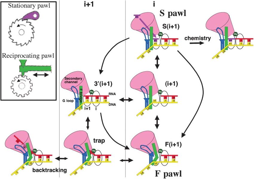
Figure 4. Effect of G Loop Mutations on F Bridge Conformation. A Two Pawl Ratchet Mechanism
(A) F bridge conformational probing by protein-RNA crosslinking. of Transcription Elongation
Two types of crosslinking derivatives of TTP (T*) were allowed to Recent structural and kinetic studies of transcription
react with ternary complexes carrying rifampicin (Rif) covalently have offered various models of elongation by multisub-
linked to the trinucleotide G[32P]CT*. The reactive isothiocyano
unit RNAPs ranging from a powerstroke mechanism,
group (NSC) of reagent I crosslinks specifically to two Lys residues
in the vicinity of the catalytic site: Lys789 (K) in the F bridge within (i.e., forward translocation that is linked to substrate
the ⬘subunit (shown in green) and  Lys1065. The alkylating group hydrolysis), to different Brownian ratchet type mecha-
(Br) of reagent II crosslinks specifically to ⬘ Met932 (M) in the G loop nisms (i.e., translocation occurs before phosphodiester
(Epshtein et al., 2002; shown in blue). Lower panels show analysis bond formation) (Foster et al., 2001; Nedialkov et al.,
by SDS-PAGE of crosslinking reaction products containing  and 2003; Holmes and Erie, 2003; Temiakov et al., 2004; Yin
⬘ subunits carrying [32P]-labeled RNA adducts. Note that the lower
and Steitz, 2004). To define the actual mechanism, we
part of each gel was intentionally underexposed so as to be able
to confirm that each reaction contained comparable quantities of combined genetic and biochemical tools to understand
radiolabeled crosslinking probe (Rif-pppGpCpT*). “⬘ (%)” refers to all the basic elongation functions of RNAP in terms of
the relative amount of ⬘ crosslinking calculated from densitometric the dynamic structure of its catalytic center. Our data
quantitation. “Fa” and “Sl” denote the fast (G1136S) and slow support a model in which no energy other than that
(I1134V) mutants, respectively. The identity of the crosslinked sites provided by thermal fluctuations is needed for RNAP
was confirmed by limited chemical degradation at Met and Cys
translocation. The model features a two pawl ratchet
residues as described in the Experimental Procedures.
(B) Effect of G loop mutations on the transcript cleavage reaction. mechanism (Figure 5). The two pawls differ in an impor-
[␣-32P] CMP-labeled EC32 was formed with wt, fast, or slow RNAP, tant respect. In one case (stationary), the pawl functions
immobilized on Co2⫹ beads, walked to position ⫹34, and washed by preventing reverse movement of the ratchet wheel.
with 40 mM CAPS at pH 10 to promote cleavage (see Experimental In the second, (reciprocating), the pawl itself generates
Procedures). The time-dependent spontaneous cleavage reaction unidirectional movement of the wheel by lateral oscilla-
was assayed at the intervals shown in the figure. Reaction mixtures
tion (Figure 5, inset). In mechanical devices, the two
were resolved on 12% polyacrylamide gels containing 8 M urea so
as to display the 5⬘ [32P]-labeled products of pH-mediated transcript kinds of pawl would normally exist as separate entities.
cleavage in wt and mutant EC34. The percent efficiency of cleavage As applied to RNAP, however, the two kinds of pawl
was calculated as the ratio of all cleavage product to the intact 34- coexist within the same catalytic site, thus providing a
mer transcript. unique opportunity for both regulation and fidelity. The
coupled pawls limit the stochastic lateral motion of the
nucleic acid scaffold in RNAP by stabilizing the enzyme
base pair in the i⫹1 site, thus rendering this RNA site in the forward translocated active (“i”) state. The first
refractory to cleavage. Indeed, in spite of the preferred (stationary) pawl (S pawl) represents the incoming com-
existence of I1134VEC34 in the backtracked state (Figure plementary substrate. Loading of substrate into the i⫹1
3), we found that this mutant was considerably less site prevents the RNA 3⬘ terminus from occupying this
sensitive to high pH-induced cleavage than wtEC (Figure site, thus suppressing the first step of backtracking. A
4B, lanes 4–9). This is consistent with the notion that similar ratchet mechanism was originally proposed for
the bent conformation of the F bridge that predominates T7 RNAP (Guajardo and Sousa, 1997; von Hippel, 1998).
in I1134V inhibits cleavage (Figure 4A). Reinforcing this In the model proposed here, however, the S pawl isRNA Polymerase as a Ratchet Machine 189 Figure 5. A Two Pawl Ratchet Model of Transcription Elongation The model invokes two common types of mechanical ratchet, one driven by a stationary pawl and a second driven by a reciprocating pawl (inset). In the stationary-type ratchet, the pawl (shown in pink) serves to prevent reverse motion of the wheel, which could otherwise oscillate back and forth randomly. The geometry and positioning of the stationary pawl determine the unidirectional movement of the wheel. In the reciprocating-type ratchet, the pawl (shown in green) oscillates back and forth, pushing the wheel in a single direction. The transcription EC combines both types of pawl in the same catalytic center of RNAP. The incoming complementary substrate (S pawl, pink) mimics a stationary pawl by preventing reverse sliding of RNAP relative to the nucleic acid scaffold. The F bridge (F pawl, green) mimics a reciprocating pawl by oscillating between bent and straight conformations, thus pushing RNAP forward. In the EC, RNA (red) base paired with the DNA template strand (yellow) oscillates in the active center (indicated by the Mg2⫹ ion flanked by the i and i⫹1 sites). Two coupled pawls (S and F) drive forward translocation by limiting the stochastic movement of the nucleic acid scaffold. The S pawl acts when the complementary substrate (pink) enters the i⫹1 site, stabilizing the EC in the S(i⫹1) state. From this state, the EC executes the chemical reaction (i.e., phosphodiester bond formation), extending its transcript by 1 nucleotide. Fluctuations of the F bridge (green) between bent and straight configurations define the F pawl action. Bending of the F bridge pushes the EC into the F(i⫹1) state, from which it proceeds to S(i⫹1). Although the F pawl drives the EC in the forward translocated state by restricting access of the RNA 3⬘ end to the i⫹1 site, it also limits the entry of the incoming substrate to the i⫹1 site, thus restricting the rate of elongation under conditions of saturating NTPs. Under limiting NTPs, i.e., when the function of the S pawl is compromised, the F bridge has a greater chance of shortening the hybrid by breaking and/or preventing 3⬘ base pairing in the i⫹1 site (trap). This would promote backtracking (i.e., reverse sliding of the RNA through the NTP delivery [secondary] channel). The trap intermediate is resistant to transcript cleavage and represents the initial step leading to pausing and termination. This model explains the biphasic elongation rate curves observed in transient state kinetic studies with E. coli and human RNAPs (Foster et al., 2001; Nedialkov et al., 2003; Holmes and Erie, 2003) without invoking any additional hypothetical substrate binding sites (see Supplemental Data on the Cell website). The model implies that principal regulation occurs through the G loop (shown in blue) that supports the F bridge. The G loop controls the F pawl in response to external signals and determines the rate, processivity, and fidelity of transcription. coupled to a second, reciprocating pawl (F pawl) repre- faster the F bridge oscillates, the greater the rate of senting the F bridge domain; this oscillates between elongation, as illustrated by the fast mutant enzyme. bent and straight configurations. The F pawl shifts the With a certain probability, however, clashing between EC forward upon bending via thermally assisted ejection the F bridge and the hybrid breaks the 3⬘ terminal hybrid of the RNA 3⬘ terminus from the i⫹1 site. Upon returning base pair (Figure 5, “trap state”), thus facilitating back- to its straight conformation, the F pawl allows the next- tracking instead of forward translocation, since the for- required substrate (S pawl) to enter the empty i⫹1 site. mer depends on the stability of the hybrid (Nudler et al., The RNA 3⬘ terminus slips back to the i⫹1 site if the 1997). It follows that the longer the F bridge spends substrate is not readily available, or while the F bridge in its bent conformation, the higher the probability of remains straight. In summary, the S pawl (the incoming backtracking. This explains the backtracking-prone substrate) acts to prevent backward slippage of RNAP phenotype of the slow mutant (Figure 3). It also explains in relation to the nucleic acid scaffold, while the F pawl why backtracking is particularly sensitive to the stability (F bridge) acts to push RNAP one nucleotide forward to of the 3⬘ base pair of the hybrid (Nudler et al., 1997). initiate the next cycle of substrate addition. Kinetic simulations of the two pawl ratchet model de- The model described above serves to explain (inter scribed here predict that the elongation rate would de- alia) how RNAP maintains its rate of elongation. The crease sharply if the NTP concentration falls below a
Cell
190
Figure 6. Effect of G Loop Mutations on Transcription Fidelity
(A) Time course of misincorporation by slow (I1134V) and fast (G1136S) RNAP. Immobilized EC32 washed free of NTPs and carrying a 32P-
labeled transcript was incubated with 300 M complementary UTP, noncomplementary CTP or ATP, or 2⬘-deoxy-TTP for the times shown in
the figure. Washed, intact EC32 is shown in the first (left) lane as a marker. Misincorporation (% mis) was calculated as the ratio of all extended
products to 32 nucleotide RNA.
(B) Theoretically predicted rate of misincorporation based on a kinetic analysis of the two pawl model. The measure of misincorporation is
taken as the ratio of the steady-state incorporation rate for incorrect NTP to the steady-state incorporation rate for correct NTP. This ratio
was computed as a function of NTP concentration (assuming the same NTP concentration of correct and incorrect nucleotides) for slow, wt,
and fast mutants. The analysis was done using steady-state solutions of a full kinetic scheme of the two pawl model (see Supplemental Data
on the Cell website). Parameters were chosen to reproduce the experimental findings shown in (A).
certain threshold level (see Supplemental Data on the phenotype. In contrast, the slow mutant showed much
Cell website). This is consistent with data from recent greater fidelity than wt. These results provide indepen-
transient state kinetic studies (Foster et al., 2001; Nedial- dent support for our two pawl ratchet mechanism and
kov et al., 2003; Holmes and Erie, 2003). However, our establish the G loop/F bridge as a key determinant of
model does not invoke any hypothetical allosteric and/ transcriptional fidelity.
or template-specific NTP binding sites other than i⫹1 The two pawl ratchet model also reveals an intrinsic
to explain the biphasic rate curves. Simply, under sub- mechanism for proofreading. Our kinetic description of
strate-limiting conditions, the F bridge has a higher this model predicts that the presence of an incorrect
probability to melt the 3⬘ end of the hybrid, thus facilitat- substrate in the i⫹1 site would facilitate backtracking
ing backtracking (Figure 5, trap state). On the other hand, (see Supplemental Figure S2 on the Cell website). This
at saturating NTP concentration, the action of the F pawl situation can be readily understood in mechanical terms:
becomes the rate-limiting step. With a certain probabil- the incorrect substrate represents a stationary pawl with
ity, the F bridge blocks the binding of NTP at the i⫹1 a geometry that favors movement of the ratchet in the
site, thus serving as a throttle that limits the S pawl. reverse direction. Consistent with this notion, a noncom-
Irregularities between F bridge transitions are likely to plementary substrate (e.g., GMP or AMP) promoted
represent force-independent pauses detected at the backtracking of wtEC33 (Figure 3E, lanes 3 and 4). Fur-
single-molecule level (Neuman et al., 2003). thermore, addition of complementary GMPcPP together
with saturating amounts of incorrect CTP or ATP (i⫹2
Mechanism of Transcription Fidelity substrate) suppressed GMPcPP-mediated forward trans-
Our kinetic representation of the two pawl model (see location (Figure 3E, lanes 8 and 9). We conclude that
Supplemental Data on the Cell website) argues that the these nucleotides all compete for the same i⫹1 site.
faster the F bridge oscillates between bent and straight During backtracking, the 3⬘ terminus of RNA would eject
conformations, the greater the probability of misincor- the wrong substrate from the i⫹1 site through the sec-
poration. Indeed, the probability of a correct choice of ondary channel before phosphodiester bond formation
NTP in the i⫹1 site is diminished by the rapidly oscillating had occurred, thus diminishing the probability of an er-
F bridge: this limits the time needed for discrimination ror. Remarkably, this mechanism of proofreading does
between correct and incorrect substrates, which de- not require any energy input or any special site for se-
pends on base-pairing energies and structural criteria. lecting a substrate other than i⫹1.
To test these considerations and our model, we com-
pared wtEC, G1136SEC, and I1134VEC for their ability to incor- A Unified Model for Elongation Control
porate incorrect substrates. Misincorporation was mon- According to our two pawl ratchet model, the rate of
itored by incubating wt and mutant EC32 with CTP, ATP, elongation could be modulated at any given position by
or 2⬘-deoxy-TTP instead of the correct substrate, i.e., changing the parameters of the reciprocating F pawl,
complementary UTP (Figure 6A). As predicted by the i.e., the kinetics of F bridge fluctuation between bent
kinetic model (Figure 6B and Supplemental Data on the and straight conformations. The same point mutation in
Cell website), the fast mutant exhibited an error-prone the G loop that modulates F bridge conformation (FigureRNA Polymerase as a Ratchet Machine
191
4) alters the responsiveness of RNAP to various pauses translocation, suggesting that NusA was unable to sup-
(Figure 2B) and elongation factors (Figure 2A). Thus, all press F bridge oscillation in this mutant any further.
major types of regulatory signals seem to converge on Although one can argue that G loop mutations might
the same ultimate target—the F bridge. Indeed, the fast alter the affinity of Nus factors for RNAP, we find this a
(G1136S) mutation stabilizes the EC in the forward trans- highly unlikely possibility. Neither mutation is exposed;
located (active) state (Figure 3A), presumably by accel- each is buried deep within the enzyme. They are located
erating F bridge oscillation (Figure 4 and Supplemental essentially adjacent to each other, and yet have opposite
Figure S2 on the Cell website). This mutation severely effects on NusA. Moreover, NusA could still stimulate
compromises the response to both hairpin-dependent intrinsic termination of the slow mutant (Figure 2C), un-
(such as those present in the his and trp synthetic genes) derscoring its independent function in modulating RNA
and hairpin-independent (such as those present in intrin- folding during termination and antitermination (Gusarov
sic terminators) pauses (Figure 2B), suggesting that all and Nudler, 2001). Finally, we were unable to detect any
pauses have a common intermediate that is controlled differences in NusA or NusG binding to wt and G loop
by the F bridge. Furthermore, the backtracking-prone mutant enzymes in chromatographic assays (data not
(I1134V) mutant is characterized by a predominantly shown).
bent F bridge (Figure 4A). This slow mutant is extremely It remains to be determined exactly how extrinsic and
sensitive to both hairpin-dependent and hairpin-inde- intrinsic signals are transmitted through RNAP to the F
pendent pauses and intrinsic termination (Figures 2B bridge. With the exception of transcript cleavage factors
and 2C). Taken together, these results suggest that the that reach the catalytic site directly through the second-
basic intermediate for all major types of pauses and ary channel (Laptenko et al., 2003; Sosunova et al., 2003;
termination is as shown in Figure 5 as a trap intermedi- Opalka et al., 2003; Kettenberger et al., 2003), other
ate. In this case, F bridge bending does not induce elongation factors and RNA signals are likely to employ
forward translocation, but melts the 3⬘ base pair in the an allosteric mode of action. NusA and the hairpin-
i⫹1 site, directing the RNA 3⬘ terminus to the secondary dependent pause signals have been proposed to act
channel. In the case of hairpin-dependent pauses, this via the  subunit flap domain (Toulokhonov et al., 2001),
intermediate is stabilized by the hairpin, which traps the while NusG may directly or indirectly utilize the G loop
F bridge in its bent conformation. The pause hairpin also for this purpose. Indeed, the whole G helix-loop-helix
blocks further backtracking. A similar trapped intermedi- domain may be a universal adaptor for various factors,
ate can be detected during intrinsic termination just since it is large and unstructured in most RNAPs. Parts
prior to EC dissociation (Gusarov and Nudler, 1999). This of the G loop are exposed and can be targeted by anti-
model of hairpin-dependent pausing is consistent with bodies that inhibit catalytic activity of the EC (Zakharova
recent crosslinking and Fe2⫹ cleavage data demonstra- et al., 1998). Even seemingly mild perturbations in the
ting the presence of a 3⬘ terminus in the i⫹1 site and G loop, as exemplified here by two adjacent point muta-
resistance of the paused complexes to transcript cleav- tions, could change parameters of F bridge oscillation,
age and pyrophosphorolysis (Toulokhonov and Landick, thereby dramatically affecting basic elongation proper-
2003). However, our model does not involve any unusual ties with lethal consequences for the cell. Curiously,
conformational changes in the catalytic center leading wt RNAP II from S. cerevisiae carries both I1134V and
to various hypothetical “frayed” states (Artsimovitch and G1136S “substitutions” in its G loop domain (Figure 1B).
Landick, 2000; Toulokhonov et al., 2001). In every re- We speculate that the simultaneous presence of these
spect, the trapped intermediate is simply a pretranslo- “substitutions” in the yeast enzyme is possible because
cated state, which is resistant to cleavage and pyro-
they neutralized each other’s detrimental effect. Since
phosphorolysis due to the bent F bridge. In the case of
the key elements of the catalytic center, including the F
hairpin-independent pauses, e.g., a T stretch-, F bridge-
bridge and G loop, are similar in bacterial and eukaryotic
mediated hybrid melting is facilitated because of the
RNAPs (Gnatt et al., 2001), there is little doubt that the
intrinsically weak hybrid. In contrast to a hairpin-depen-
same structural principles of elongational control and
dent pause, hairpin-independent pauses do not freeze
proofreading operate in higher organisms.
the F bridge in its bent conformation, thereby allowing
the cleavage reaction to occur. Experimental Procedures
Our data suggest that elongation factors also act via
the F pawl. Specifically, we propose that NusG and NusA Isolation of Dominant-Negative G Loop Mutants
accelerate and decelerate elongation by accelerating The wt rpoC was amplified by PCR with a high-fidelity Pwo enzyme
and decelerating F bridge oscillation, respectively. Thus, (Roche) using ⬘L 5⬘-gggattaaccatggctatgaaagatttattaaagtt-3⬘ and
⬘R 5⬘-ttcgggcccaagcttctcgttatcagaaccgcc-3⬘ primers and cloned
NusG modifies RNAP the same way as the “fast” G1136S
into the Nco I and Hind III sites of the pBAD-A plasmid (Invitrogen).
mutation does, i.e., by stabilizing the EC in the forward- The 960–1408 aa fragment of rpoC was amplified by PCR using
translocated (active) state (Figure 3B). Significantly, the primers: 5⬘-cccggcaggaagtactgcg-3⬘ and ⬘R. The resulting frag-
fast mutant almost completely lost its responsiveness ment was gel purified and used as a template in error-prone PCR
to NusG during elongation or translocation (Figures 2A (Fromant et al., 1999) with Taq polymerase (Eppendorf) and 0.51
and 3B), suggesting that NusG was unable to accelerate mM dATP, 0.2 mM dCTP, 1.15 mM dGTP, 3.76 mM dTTP, 1 mM
F bridge oscillation any further in this mutant enzyme. Mn2⫹, 10 mM Mg2⫹. The products of three separate reactions were
gel purified and subcloned into the BlpI and Hind III sites. The
NusA works in an opposite manner. It decreases the
products of the ligation reaction were electroporated into TOP10
elongation rate of the fast mutant (Figure 2A) and stimu- competent cells (Invitrogen), and plated on LB agar supplemented
lates its backward translocation to an even greater ex- with 2% glucose and 40 g/ml ampicillin. Transformants were
tent than wt (Figure 3B). In contrast, the slow (I1134V) picked with a cell scraper and the plasmids were isolated with a
mutant was resistant to NusA-mediated pausing and miniprep kit (Qiagen). The plasmid library was then used to transformCell
192
strain RL602 (leu [am], trp [am], lacZ2110 [am], galK [am], galE, rpsL, band by the total radioactivity present in it and all read-through
tsx, supD43,47 [Ts], sueB, sueC, rpoC325 [am], del [recA-srl] 306 bands (Nudler and Gusarov, 2003).
srl-301::Tn10-84 [rpoCTS])—provided by Robert Landick (University
of Wisconsin, Madison). RL602 transformants were grown on LB DNA Footprinting
agar supplemented with 2% glucose (to ensure suppression of the The T732del3tR2 template for ExoIII footprinting (front edge) was
arabinose promoter) and 40 g/ml of ampicillin. Colonies were inoc- obtained by PCR using nonphosphorylated (left) and 5⬘-phosphory-
ulated into 1 ml LB supplemented with 2% glucose and 40 g/ml lated (right) primers to produce a 5⬘-OH group in the nontemplate
ampicillin and grown overnight at 30⬚C. Aliquots of individual cul- strand for subsequent enzymatic phosphorylation. For rear edge
tures were replica plated in duplicate onto LB agar containing 2% footprinting, the opposite set of 5⬘ terminally modified oligos was
glucose, 0.002%, 0.02%, 0.2%, or 0.5% arabinose, and 40 g/ml used for PCR. DNA was labeled with T4 polynucleotide kinase (25
ampicillin. The remainder of the overnight culture was suspended units) and [␥-32P] ATP (ICN) for 10 min at room temperature in TB100.
in 40% glycerol, flash frozen, and stored at ⫺70⬚C. In each case, EC33 and EC34 were obtained by walking as described above.
RL602 transformed with a wt rpoC was also plated. Plates were Treatment with ExoIII was performed for 5 min using 15 units of
grown overnight at 30⬚C and 42⬚C. Colonies that failed to grow under ExoIII (New England Biolabs) at room temperature followed by
expression conditions (⫹arabinose) when the wt was viable were quenching with SS. DNA products were separated by 8% sequenc-
marked as dominant negative. As a positive control for the domi- ing PAGE.
nant-negative phenotype, the previously characterized dominant-
negative rpoC mutant (DFDGD→AFAGA) (Zaychikov et al., 1996) Crosslinking and Mapping of Crosslinked Sites
was used under the same conditions. Colonies that failed to grow at Crosslinking reagents I and II were synthesized as described pre-
42⬚C were marked recessive. Plasmids from colonies that appeared viously (Epshtein et al., 2002). Crosslinks were performed as follows:
dominant in two separate experiments were isolated, transformed 5 pmol RNAP, 8 pmol T7A2 template, and 10 M Rif-(CH2)5-GTP
into TOP10, and stored at ⫺70⬚C. were mixed with 20 l preequilibrated Talon beads and 20 l of
TB-H (20 mM HEPES-HCl [pH 7.9], 50 mM NaCl, 10 mM MgCl2).
DNA Templates, Proteins, and Nucleotides After 5 min incubation at 37⬚C, the beads were washed three times
All templates were generated by PCR using Deep Vent DNA poly- with 1 ml TB-H. [32P] CTP (3000 Ci/mmol) was added and the mixture
merase (New England Biolabs) and synthetic DNA oligos (IDT). The incubated for 10 min at 37⬚C followed by repetitive washes with
template T732del3tR2 used for footprinting experiments and termi- TB-H containing 2.5 mM MnCl2 in place of MgCl2. The crosslinkable
nation assays had the following initial transcribed sequence: (⫹1) 3⬘-dTTP analog was added to each mixture to a final concentration
ATCGAGAGGG CCACGGCGAA CAGCCAACCC AATCGAACAG of 200 M for reagent I or 50 M for reagent II, and incubation
(⫹40) (Bar-Nahum and Nudler, 2001). All other templates had the continued for 30 min at 37⬚C. The reaction was quenched with an
same T7A1 promoter and initial transcribed sequence up to position equal volume of stop buffer (100 mM Tris-HCl [pH 6.8], 200 mM
⫹10 (counting from the ⫹1 start of the transcription). They contain DTT, 20% glycerol, 4% SDS, 0.2% bromphenol blue). Mapping of
E.coli sequences of trp, his, ops, and tR2 T stretch pause sites. All crosslinking sites was performed by limited chemical degradation.
templates were purified from low-melting agarose gels and diluted in Briefly, the ⬘ subunit carrying radioactive adduct was excised from
TE buffer to a concentration ⵑ1 pmol/l. His6-tagged mutant and the gel and eluted with 3 vol of 0.2% SDS at 37⬚C for 1h. The eluate
wt RNAPs were purified as described (Nudler et al., 2003), except was freeze-dried with a SpeedVac and redissolved in water to a
that TOP10 cells carrying pBADrpoC were inoculated into LB sup- final concentration of 1%–2% SDS. Protein degradation reactions
plemented with 0.2% glucose and 40 g/ml ampicillin and grown with CNBr (at Met residues) and NTCBA (at Cys residues) were
overnight at 30⬚C. 100 l of the overnight culture were then used performed according to Mustaev et al. (2003). Gels containing radio-
to inoculate 500 ml LB and grown to OD600 0.5, at which point rpoC active materials were scanned on a PhosphoImager.
was induced with 0.4% arabinose for 1.5 hr. NusA and NusG were
from Asis Das (University of Connecticut Health Center). ExoIII and Kinetic Modeling
T4 polynucleotide kinase were from New England Biolabs. rNTPs The kinetic equations presented in the Supplemental Data on the
were from Pharmacia, 3⬘-dNTPs were from Trilink Biotech, GMPcPP Cell website were solved numerically and the solutions checked for
was from Jena Biosciense, and NMPs were from Sigma. All sub- consistency against analytical calculations in a number of tractable
strates were further purified by ion exchange chromatography limits. The global fits to the biochemical data of Holmes and Erie
(Nudler et al., 2003). (2003) were obtained by using the data analysis software GraphPad
(www.graphpad.com). In all cases, the global fit based on the two
pawl mechanism was preferred over a single exponential form for
Solid Phase Transcription Reactions and Walking
which global fits never converged.
His6-RNAP (ⵑ2 pmol) was mixed with a 2-fold molar excess of DNA
in 20 l of TB50 (10 mM MgCl2, 40 mM Tris-HCl [pH 7.9], 50 mM
Acknowledgments
KCl) for 5 min at 37⬚C followed by addition of ApUpC (10 M, Oligos
Etc.), GTP, and ATP (25 M) for 7 min. Next, 5 l TB100-equilibrated
We thank S. Borukhov, A. Das, R. Landick, and R. Mooney for
Talon Co2⫹ affinity bead suspension (Clontech) equilibrated in tran-
materials. We thank N.J. Cowan, W. Jelinek, and J. Borowiec for
scription buffer (TB) was added for 5 min at room temperature fol-
invaluable help in editing the manuscript and comments. This work
lowed by washing with 2 ⫻ 1.5 ml of TB1000 (1 M KCl) and 2 ⫻ 1.5
was supported by NIH grants R01 GM58750 (to E.N.) and P20
ml of TB200 (200 mM KCl). To produce EC32, ATP, GTP (5 M), and
GM64375 (to A.E.R.).
1 l of [␣-P32] CTP (3000 Ci/mmol; NEN Life Science Products) were
added for 5 min at room temperature followed by CTP (5 M) for a
Received: July 26, 2004
further 2 min. Beads were washed five times with TB100. The walk-
Revised: September 14, 2004
ing reactions done to obtain EC33 and EC34 were performed using
Accepted: November 23, 2004
limited NTP sets (Nudler et al., 2003). Washed EC32 was incubated
Published: January 27, 2005
with 5 M UTP for 5 min at room temperature to obtain EC33, or
5 M UTP⫹CTP to obtain EC34. The beads were washed four times
References
with TB100, divided into equal aliquots, and processed as described
in the figure legends. The reactions were quenched by addition of
Artsimovitch, I., and Landick, R. (2000). Pausing by bacterial RNA
an equal volume of stop solution (SS): 8 M urea, 20 mM EDTA, 1 ⫻
polymerase is mediated by mechanistically distinct classes of sig-
TBE, 0.25% bromphenol blue, 0.25% xylene cyanol. The products
nals. Proc. Natl. Acad. Sci. USA 97, 7090–7095.
were separated by 12% sequencing PAGE containing 8 M urea.
Relative amounts of [32P] RNA and DNA species were determined Bar-Nahum, G., and Nudler, E. (2001). Isolation and characterization
using a PhosphorImager and software from Molecular Dynamics. of sigma(70)-retaining transcription elongation complexes from
The efficiency of termination (%T) or pausing (%P) was calculated Escherichia coli. Cell 106, 443–451.
by dividing the amount of radioactivity in a terminated or paused Burova, E., Hung, S.C., Sagitov, V., Stitt, B.L., and Gottesman, M.E.RNA Polymerase as a Ratchet Machine
193
(1995). Escherichia coli NusG protein stimulates transcription elon- RNA-DNA hybrid maintains the register of transcription by pre-
gation rates in vivo and in vitro. J. Bacteriol. 177, 1388–1392. venting backtracking of RNA polymerase. Cell 89, 33–41.
Condon, C., Squires, C., and Squires, C.L. (1995). Control of rRNA Nudler, E., Gusarov, I., and Bar-Nahum, G. (2003). Methods of walk-
transcription in Escherichia coli. Microbiol. Rev. 59, 623–645. ing with the RNA polymerase. Methods Enzymol. 371, 160–169.
Cramer, P., Bushnell, D.A., and Kornberg, R.D. (2001). Structural Opalka, N., Chlenov, M., Chacon, P., Rice, W.J., Wriggers, W., and
basis of transcription: RNA polymerase II at 2.8 angstrom resolution. Darst, S.A. (2003). Structure and function of the transcription elonga-
Science 292, 1863–1876. tion factor GreB bound to bacterial RNA polymerase. Cell 114,
335–345.
Epshtein, V., Mustaev, A., Markovtsov, V., Bereshchenko, O., Niki-
forov, V., and Goldfarb, A. (2002). Swing-gate model of nucleotide Orlova, M., Newlands, J., Das, A., Goldfarb, A., and Borukhov, S.
entry into the RNA polymerase active center. Mol. Cell 10, 623–634. (1995). Intrinsic transcript cleavage activity of RNA polymerase.
Proc. Natl. Acad. Sci. USA 92, 4596–4600.
Foster, J.E., Holmes, S.F., and Erie, D.A. (2001). Allosteric binding
of nucleoside triphosphates to RNA polymerase regulates transcrip- Reines, D., Conaway, J.W., and Conaway, R.C. (1999). Mechanism
tion elongation. Cell 106, 243–252. and regulation of transcriptional elongation by RNA polymerase II.
Curr. Opin. Cell Biol. 11, 342–346.
Fromant, M., Blanquet, S., and Plateau, P. (1999). Direct random
mutagenesis of gene-sized DNA fragments using polymerase chain Roberts, J.W., Yarnell, W., Bartlett, E., Guo, J., Marr, M., Ko, D.C.,
reaction. Anal. Biochem. 224, 347–353. Sun, H., and Roberts, C.W. (1998). Antitermination by bacteriophage
lambda Q protein. Cold Spring Harb. Symp. Quant. Biol. 63, 319–325.
Gnatt, A.L., Cramer, P., Fu, J., Bushnell, D.A., and Kornberg, R.D.
Schmidt, M.C., and Chamberlin, M.J. (1984). Amplification and isola-
(2001). Structural basis of transcription: an RNA polymerase II elon-
tion of Escherichia coli nusA protein and studies of its effects on
gation complex at 3.3 Å resolution. Science 292, 1876–1882.
in vitro RNA chain elongation. Biochemistry 23, 197–203.
Guajardo, R., and Sousa, R. (1997). A model for the mechanism of
Sosunova, E., Sosunov, V., Kozlov, M., Nikiforov, V., Goldfarb, A.,
polymerase translocation. J. Mol. Biol. 265, 8–19.
and Mustaev, A. (2003). Donation of catalytic residues to RNA poly-
Gusarov, I., and Nudler, E. (1999). The mechanism of intrinsic tran- merase active center by transcription factor Gre. Proc. Natl. Acad.
scription termination. Mol. Cell 3, 495–504. Sci. USA 100, 15469–15474.
Gusarov, I., and Nudler, E. (2001). Control of intrinsic transcription Sousa, R. (2001). A new level of regulation in transcription elonga-
termination by N and NusA: The basic mechanisms. Cell 107, tion? Trends Biochem. Sci. 26, 695–697.
437–449.
Temiakov, D., Patlan, V., Anikin, M., McAllister, W.T., Yokoyama, S.,
Holmes, S.F., and Erie, D.A. (2003). Downstream DNA sequence and Vassylyev, D.G. (2004). Structural basis for substrate selection
effects on transcription elongation. Allosteric binding of nucleoside by t7 RNA polymerase. Cell 116, 381–391.
triphosphates facilitates translocation via a ratchet motion. J. Biol. Toulokhonov, I., and Landick, R. (2003). The flap domain is required
Chem. 278, 35597–35608. for pause RNA hairpin inhibition of catalysis by RNA polymerase
Kettenberger, H., Armache, K.J., and Cramer, P. (2003). Architecture and can modulate intrinsic termination. Mol. Cell 12, 1125–1136.
of the RNA polymerase II-TFIIS complex and implications for mRNA Toulokhonov, I., Artsimovitch, I., and Landick, R. (2001). Allosteric
cleavage. Cell 114, 347–357. control of RNA polymerase by a site that contacts nascent RNA
Komissarova, N., and Kashlev, M. (1997). RNA polymerase switches hairpins. Science 292, 730–733.
between inactivated and activated states by translocating back and Uptain, S.M., Kane, C.M., and Chamberlin, M.J. (1997). Basic mecha-
forth along the DNA and the RNA. J. Biol. Chem. 272, 15329–15338. nisms of transcript elongation and its regulation. Annu. Rev. Bio-
Korzheva, N., Mustaev, A., Kozlov, M., Malhotra, A., Nikiforov, V., chem. 66, 117–172.
Goldfarb, A., and Darst, S.A. (2000). A structural model of transcrip- Vassylyev, D.G., Sekine, S., Laptenko, O., Lee, J., Vassylyeva, M.N.,
tion elongation. Science 289, 619–625. Borukhov, S., and Yokoyama, S. (2002). Crystal structure of a bacte-
Landick, R., Turnbough, J.R., and Yanofsky, C. (1996). Transcription rial RNA polymerase holoenzyme at 2.6 Å resolution. Nature 417,
attenuation. In Escherichia coli and Salmonella typhimurium, F.C. 712–719.
Neidhardt, ed. (Washington: American Society for Microbiology), Vogel, U., and Jensen, K.F. (1994). Effects of ppGpp on rate of
pp. 1263–1286. transcription elongation in isoleucine-starved Escherichia coli. J.
Laptenko, O., Lee, J., Lomakin, I., and Borukhov, S. (2003). Tran- Biol. Chem. 269, 16236–16241.
script cleavage factors GreA and GreB act as transient catalytic von Hippel, P.H. (1998). An integrated model of the transcription
components of RNA polymerase. EMBO J. 22, 6322–6334. complex in elongation, termination, and editing. Science 281,
McDowell, J.C., Roberts, J.W., Jin, D.J., and Gross, C. (1994). Deter- 660–665.
mination of intrinsic transcription termination efficiency by RNA Wada, T., Takagi, T., Yamaguchi, Y., Ferdous, A., Imai, T., Hirose,
polymerase elongation rate. Science 266, 822–825. S., Sugimoto, S., Yano, K., Hartzog, G.A., Winston, F., et al. (1998).
Mustaev, A. Zaychikov, E., Grachev, M., Kozlov, M., Severinov, K., DSIF, a novel transcription elongation factor that regulates RNA
Epshtein, V., Korzheva, N., Bereshchenko, O., Markovtsov, V., Lukh- polymerase II processivity, is composed of human Spt4 and Spt5
tanov, E., et al. (2003). Strategies and methods of cross-linking of homologs. Genes Dev. 12, 343–356.
RNA polymerase active center. Methods Enzymol. 371, 191–206. Yin, Y.W., and Steitz, T.A. (2004). The structural mechanism of trans-
location and helicase activity in t7 RNA polymerase. Cell 116,
Nedialkov, Y.A., Gong, X.Q., Hovde, S.L., Yamaguchi, Y., Handa, H.,
393–404.
Geiger, J.H., Yan, H., and Burton, Z.F. (2003). NTP-driven transloca-
tion by human RNA polymerase II. J. Biol. Chem. 278, 18303–18312. Zakharova, N., Bass, I., Arsenieva, E., Nikiforov, V., and Severinov,
K. (1998). Mutations in and monoclonal antibody binding to evolu-
Neuman, K.C., Abbondanzieri, E.A., Landick, R., Gelles, J., and
tionary hypervariable region of Escherichia coli RNA polymerase
Block, S.M. (2003). Ubiquitous transcriptional pausing is indepen-
beta⬘ subunit inhibit transcript cleavage and transcript elongation.
dent of RNA polymerase backtracking. Cell 115, 437–447.
J. Biol. Chem. 273, 24912–24920.
Nudler, E. (1999). Transcription elongation: structural basis and
Zaychikov, E., Martin, E., Denissova, L., Kozlov, M., Markovtsov, V.,
mechanisms. J. Mol. Biol. 288, 1–12.
Kashlev, M., Heumann, H., Nikiforov, V., Goldfarb, A., and Mustaev,
Nudler, E., and Gottesman, M.E. (2002). Transcription termination A. (1996). Mapping of catalytic residues in the RNA polymerase
and anti-termination in E. coli. Genes Cells 7, 755–768. active center. Science 273, 107–109.
Nudler, E., and Gusarov, I. (2003). Analysis of the intrinsic transcrip- Zhang, G., Campbell, E.A., Minakhin, L., Richter, C., Severinov, K.,
tion termination mechanism and its control. Methods Enzymol. and Darst, S.A. (1999). Crystal structure of Thermus aquaticus core
371, 369–382. RNA polymerase at 3.3 Å resolution. Cell 98, 811–824.
Nudler, E., Mustaev, A., Lukhtanov, E., and Goldfarb, A. (1997). TheYou can also read