Start Codon Recognition in Eukaryotic and Archaeal Translation Initiation: A Common Structural Core - MDPI
←
→
Page content transcription
If your browser does not render page correctly, please read the page content below
International Journal of
           Molecular Sciences

Review
Start Codon Recognition in Eukaryotic and Archaeal
Translation Initiation: A Common Structural Core
Emmanuelle Schmitt * , Pierre-Damien Coureux, Auriane Monestier, Etienne Dubiez                    and
Yves Mechulam *
 Laboratoire de Biochimie, Ecole polytechnique, CNRS, Université Paris-Saclay, 91128 Palaiseau CEDEX, France;
 pierre-damien.coureux@polytechnique.edu (P.-D.C.); auriane.monestier@inra.fr (A.M.);
 Etienne.dubiez@ed.ac.uk (E.D.)
 * Correspondence: emmanuelle.schmitt@polytechnique.edu (E.S.); yves.mechulam@polytechnique.edu (Y.M.);
   Tel.: +33-1-69-33-4885 (E.S. & Y.M.)

 Received: 5 December 2018; Accepted: 13 February 2019; Published: 21 February 2019

 Abstract: Understanding molecular mechanisms of ribosomal translation sheds light on the
 emergence and evolution of protein synthesis in the three domains of life. Universally, ribosomal
 translation is described in three steps: initiation, elongation and termination. During initiation,
 a macromolecular complex assembled around the small ribosomal subunit selects the start codon on
 the mRNA and defines the open reading frame. In this review, we focus on the comparison of start
 codon selection mechanisms in eukaryotes and archaea. Eukaryotic translation initiation is a very
 complicated process, involving many initiation factors. The most widespread mechanism for the
 discovery of the start codon is the scanning of the mRNA by a pre-initiation complex until the first
 AUG codon in a correct context is found. In archaea, long-range scanning does not occur because of
 the presence of Shine-Dalgarno (SD) sequences or of short 50 untranslated regions. However, archaeal
 and eukaryotic translation initiations have three initiation factors in common: e/aIF1, e/aIF1A and
 e/aIF2 are directly involved in the selection of the start codon. Therefore, the idea that these archaeal
 and eukaryotic factors fulfill similar functions within a common structural ribosomal core complex
 has emerged. A divergence between eukaryotic and archaeal factors allowed for the adaptation to
 the long-range scanning process versus the SD mediated prepositioning of the ribosome.

 Keywords: archaea; eukaryotes; ribosome; translation initiation; evolution

1. Introduction
      In the 1980s, pioneering studies demonstrated that sequence analysis and molecular structures
revealed evolutionary relationships between organisms. From this period, the sequences of 16S/18S
rRNA genes, present in all organisms and slowly evolving, have been used as phylogenetic markers [1].
These works led to the definition of a new taxon called a “domain” and to the division of the tree
of life into three monophyletic domains, bacteria, archaea and eukarya, each comprising several
kingdoms [2,3]. The root of this tree separates bacteria from the two other domains, archaea and
eukarya, that appear as distant relatives. Indeed, most informational genes in archaea, in particular
those related to ribosomal translation (but also those involved in DNA replication and transcription),
are closer to their eukaryotic counterparts than to their bacterial ones [4–6]). During the same
period, another theory has emerged, first from electron microscopy studies of ribosomes from eocytes
(nowadays called crenarchaeotes) and also from the use of alternative strategies for phylogenetic
reconstructions [7,8]. This led to a two-domain tree, called the eocyte tree, with eukaryotes originating
from within an archaeal phylum instead of being a sister lineage, in such a way that archaea would
be paraphyletic [9,10]. The use of new statistical methods for studying phylogeny [11–13] and recent

Int. J. Mol. Sci. 2019, 20, 939; doi:10.3390/ijms20040939                         www.mdpi.com/journal/ijms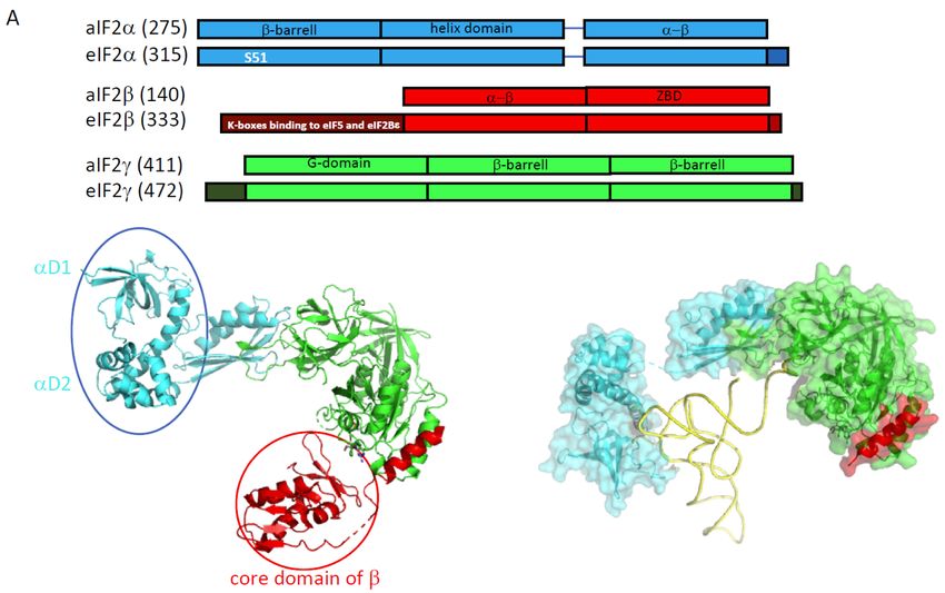
Int. J. Mol. Sci. 2019, 20, 939                                                                     2 of 21

developments of metagenomics or of single-cell genomics [14,15] further reinforce this theory [16–19].
This model is still a matter of debate and other trees of life are defended (see for instance [14,16–21]).
In particular, the monophyly of archaea is privileged in a two-domain tree updated from [3] where
archaea and eukaryotes are members of a clade named “Arkarya” [20]. In an alternative two-domain
model, archaea and bacteria are sister clades, sharing a common “akaryote” ancestor to the exclusion
of eukaryotes, as previously proposed [22–24]. This latter model emerged from reconstructions based
on the genome content of coding sequences for protein domains [25,26]. Overall, depending on
the model used for the tree of life, reductive evolution processes may have played a role [23,26,27].
As pointed out in [20], it is important to focus on biological plausibility and on comparative molecular
biology in order to help resolve phylogenetic impasses. For instance, the sisterhood of eukaryotes and
archaea appears supported by the proximity of the archaeal and eukaryotic ribosomes and further
confirmed by structural studies showing that archaeal ribosomal proteins are either universal or shared
with eukaryotes [28]. These new developments strengthen the importance of studying molecular
mechanisms of fundamental processes in archaeal organisms, such as ribosomal translation, not only
because they are interesting as such but also because their studies can help to better understand
phylogenetic links between archaea and eukaryotes. Finally, because archaeal functional systems are
usually simpler than their counterparts in eukaryotes, biochemical and structural studies in archaea
can also provide valuable models of eukaryotic processes [29–34].
      This review focuses on molecular mechanisms of protein synthesis initiation. Universally, this step
allows for an accurate selection of the start codon on mRNA and consequently the definition of the
open reading frame. In eukaryotes, this process is very complicated with many initiation factors
involved, and it is therefore the target of many regulations (see for instance [35,36]). The mechanism
involves a pre-initiation complex (PIC) made up of the small ribosomal subunit bound to the ternary
complex eIF2-GTP-Met-tRNAi Met (TC), the two small initiation factors eIF1 and eIF1A, as well as two
proteins that have a more regulatory function in the process. These are eIF5, the guanine activating
protein (GAP) of eIF2 and the multimeric factor eIF3. In the presence of factors belonging to the
eIF4 family, the pre-initiation ribosomal complex (PIC) is recruited near the 50 -capped end and
scans the mRNA until an AUG codon (frequently the first) in a correct context (Kozak motif) is
found. The AUG recognition stops the scanning, provokes a factor release and the assembly of an
elongation-proficient 80S complex through large subunit joining, with the help of eIF5B and eIF1A
(Figure 1). Beside this canonical mechanism, a number of alternative initiation routes have been
described (Table 1). This includes non-canonical cap-dependent translation initiations as exemplified
in [37], entry of the ribosome mediated by specific mRNA structures such as IRES (internal ribosome
entry site) [38], cap-independent translation enhancers [39] or eIF4F-resistant translation initiation of
N6-methyladenosine-containing mRNA under stress conditions [40]. In addition, mRNAs lacking 50
untranslated regions (leaderless mRNAs) are translated by various non-canonical mechanisms [41].
      In archaea, there is no long-range scanning because mRNAs have Shine-Dalgarno sequences
or very short 50 UTR (Table 1). However, genomic analyses have shown that three initiation factors
homologous to their eukaryotic counterparts, aIF1, aIF1A and aIF2, are found ([5,6,42], Figure 1 and
Table 1). Thus, even if obvious differences between eukaryotes and archaea exist, in particular for the
recruitment of the PIC, the start codon selection is achieved within a common structural core made up
of the small ribosomal subunit, the mRNA, the methionine initiator tRNA (Met-tRNAi Met ) and the
three initiation factors e/aIF1, e/aIF1A and e/aIF2 (Figure 2).
      This review gathers data from the structural and functional studies of the three eukaryotic and
archaeal initiation factors, e/aIF1, e/aIF1A and e/aIF2. The scanning mechanism in eukaryotes
and the SD-dependent mechanism in archaea are mainly considered (Figure 1). The similarities and
divergences of the initiation factors in both domains are highlighted. Although eukaryotic factors
have been studied for a long time, the studies of their archaeal representatives had been sparser.
Still, they have brought valuable information on understanding eukaryotic translation initiation
mechanisms. Moreover, recent structural studies allowed a description of molecular complexes during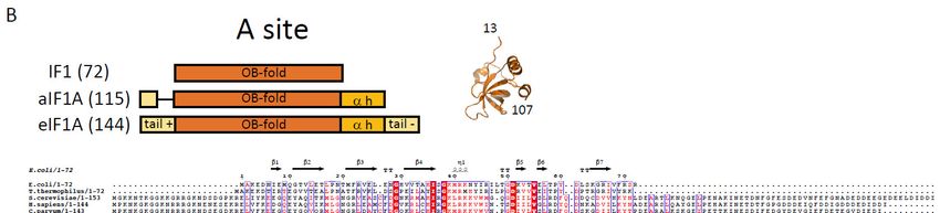
Int. J. Mol. Sci. 2019, 20, 939 3 of 21
Int. J. Mol. Sci. 2019, 20, x FOR PEER REVIEW 3 of 20
the eukaryotic and archaeal translation initiation. At a time when the idea that archaea can be the
ancestors of eukaryotes
the ancestors is reinforced,
of eukaryotes these data
is reinforced, raise
these datanew elements
raise to discuss
new elements the molecular
to discuss evolution
the molecular
of evolution
the translation
of theinitiation
translationprocesses.
initiation processes.
Figure
Figure Schematic
1. 1. Schematicviews viewsofofthe
thetranslation
translation initiation stepsin
initiation steps ineukaryotes
eukaryotesandandininarchaea.
archaea.TheThe figure
figure
illustrates
illustratesthethemain
mainsteps
stepsininthe
theformation
formation ofof the
the pre-initiation complex(PIC)
pre‐initiation complex (PIC)inineukaryotes
eukaryotes (left)
(left) andand
in in
archaea
archaea (right).
(right).TheThetranslation
translationcompetent
competent IC IC is
is formed after the
formed after the release
releaseofofe/aIF1A
e/aIF1Aandand e/aIF5B.
e/aIF5B.
TheThe complex
complex formedbybyeIF4E
formed eIF4E++eIF4G
eIF4G++ eIF4A
eIF4A is known
known as as eIF4F.
eIF4F. Note
Notethat
thatthe
thee/aIF2
e/aIF2heterotrimer
heterotrimer
is representedwith
is represented witha athree-color
three‐colorcode
code (α
(α subunit
subunit in blue,
blue, ββsubunit
subunitininred,
red,γ γsubunit
subunitin in
green)
green)forfor
consistency
consistency withthe
with theother
otherfigures,
figures,highlighting
highlighting the
the role
role ofofeach
eachsubunit
subunitininthe
thetranslation
translationinitiation.
initiation.
eIF3,
eIF3, composed
composed ofof6 6(yeast)
(yeast)toto13
13(mammals)
(mammals) subunits is is represented
representedasasaasingle
singleyellow
yellowoval
ovalto to
simplify
simplify
thethe figure.
figure.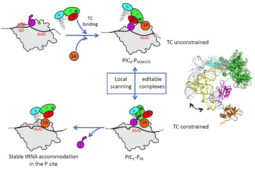
Int. J. Mol. Sci. 2019, 20, 939 4 of 21
Int. J. Mol. Sci. 2019, 20, x FOR PEER REVIEW 4 of 20
Figure 2. Definition
Definition of a common structural
structural core for the start codon selection in eukaryotes and in
archaea. The figure highlights the common elements used in eukaryotes and archaea for the key steps
of the start codon selection. Left, eukaryotic PIC; middle, archaeal PIC; and right, common core.
core.
Table
Table 1.1. The main features of the translation initiation in the three domains of life. The main main mRNA,
mRNA,
tRNAfeatures,
initiator tRNA features,andandcorrespondence
correspondencebetweenbetween the
the initiation
initiation factors
factors in the
in the three
three domains
domains of
of life
life are shown. *: IF3 is a two-domain protein. The correspondence between IF3
are shown. *: IF3 is a two‐domain protein. The correspondence between IF3 and e/aIF1 is based on a and e/aIF1 is based
on a structural
structural and and functional
functional resemblance
resemblance of of
thethe
IF3IF3C‐terminal
C-terminaldomain
domain with
with e/aIF1.
e/aIF1. Despite
Despite this
this
resemblance, the topologies of the two α–β folds are different. This suggests a convergent evolution
resemblance, the topologies of the two α–β folds are different. This suggests a convergent evolution
rather than §
rather than the
the occurrence
occurrence of of aa common
common ancestor.
ancestor. §:: ItIt should
should bebe underlined
underlined that,
that, because
because ofof its
its
homology with the bacterial IF2, aIF5B was misleadingly called aIF2 in some early publications. ##: The
homology with the bacterial IF2, aIF5B was misleadingly called aIF2 in some early publications. : The
catalytic γ
catalytic and εε subunits
γ and subunits ofof eIF2B
eIF2B areare missing
missing in
in archaea.
archaea. The
The function
function of
of the
the eIF2B
eIF2B α,β,δ homologues
α,β,δ homologues
in archaea is not clear [43,44]. ! : aIF4A present in some archaea.
in archaea is not clear [43,44]. : aIF4A present in some archaea.
!
Eukaryotes
Eukaryotes Archaea
Archaea Bacteria
Bacteria
mRNA
mRNA features
features
Canonical capped
Canonical capped dependent,
dependent,Kozak
Kozak motif
motif
Non-canonical
Non‐canonical cappeddependent
capped dependent
SD-dependent
SD‐dependent SD-dependent
SD‐dependent
Non-canonical
Non‐canonical cappedindependent
capped independent
Leaderless Leaderless
IRES-mediated Leaderless Leaderless
IRES‐mediated
Leaderless
Leaderless
main initiator tRNA features
main initiator tRNA features
methionine methionine formyl-methionine
methionine methionine formyl‐methionine
A1 -U72 A1 -U72 mispaired 1-72 bases
G29 -C41A,G1‐U-C72 , G -C
30 40 31 39 G -C
29 41 , G A
-C
30 40
1‐U , 72
G -C
31 39 G mispaired 1‐72 bases
29 41 , G30 -C40 , G31 -C39
-C
G29‐C41, G30‐C40, G31‐C39 G29‐C41, G30‐C40, G31‐C39 G29‐C41, G30‐C40, G31‐C39
Translation initiation factors
Translation initiation factors
eIF2 (α,β,γ) aIF2 (α,β,γ) -
eIF2 (α,β,γ)
eIF1 aIF2
aIF1(α,β,γ) ~IF3 * ‐
eIF1
eIF1A aIF1AaIF1 IF1~IF3 *
eIF5B §
eIF1A aIF1A
aIF5B § IF2 IF1
eIF5BeIF5§ -
aIF5B § - IF2
eIF2B (α,β,γ,δ,ε) aIF2B (α,β,δ) # -
eIF5 ‐ ‐
eIF3 (6 to 13 subunits) - -
eIF2B (α,β,γ,δ,ε)
eIF4F (4A, 4G, 4E) aIF2B
aIF4A (α,β,δ)
! #
- ‐
eIF3 (6 to 13 subunits)
eIF4B - ‐ - ‐
eIF4F (4A, 4G, 4E) aIF4A ! ‐
eIF4B
2. Features of Eukaryotic and Archaeal Translation Initiation Factors ‐ ‐
2. Features
2.1. e/aIF2 of Eukaryotic and Archaeal Translation Initiation Factors
In both domains of life, the Met-initiator tRNA is carried to the small ribosomal subunit by the
2.1. e/aIF2
heterotrimeric (α, β and γ) GTP-binding factor e/aIF2 [45]. The factor is specific to the initiator tRNA,
In both
strongly domainsthe
recognizes ofesterified
life, the Met‐initiator tRNA is
methionine group andcarried to an
prefers theAsmall ribosomal subunit by the
1 -U72 base pair, as found in all
heterotrimeric (α, β and γ) GTP‐binding factor e/aIF2 [45]. The factor is specific
eukaryotic or archaeal initiator tRNAs [46–51]. Early on, the crucial role of eIF2 to the initiator
in the start tRNA,
codon
strongly
selection recognizes
was broughtthetoesterified
light frommethionine group and
the identification prefers
of yeast an Aable
mutants 1‐U72 to
base pair,translation
initiate as found inonalla
eukaryotic
non-AUG codonor archaeal initiator
[52–57]. tRNAs
Further [46–51].
studies showed Early
thaton, the crucial
correct pairingrole of eIF2the
between in start
the start
codoncodon
and
selection was brought to light from the identification of yeast mutants able to initiate translation on
a non‐AUG codon [52–57]. Further studies showed that correct pairing between the start codon and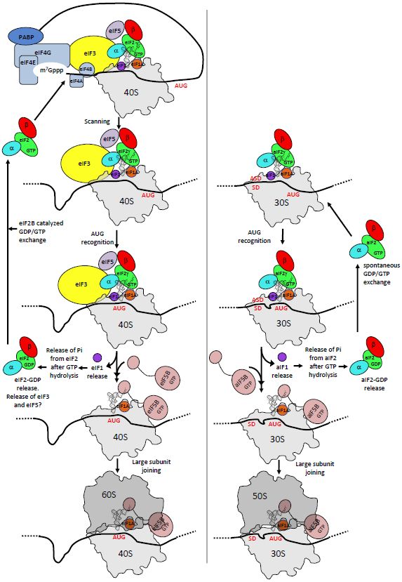
Int. J. Mol. Sci. 2019, 20, x FOR PEER REVIEW                                                     5 of 20
Int. J. Mol. Sci. 2019, 20, 939                                                                    5 of 21
the Met‐tRNAiMet anticodon induced the release from the ribosome of e/aIF2 in a GDP bound state.
Therefore, the selection of the start codon is achieved through the control of the nucleotide state of
the Met-tRNA    Met anticodon induced the release from the ribosome of e/aIF2 in a GDP bound state.
e/aIF2  [58]. i
Therefore, the selection of the start codon is achieved through the control of the nucleotide state of
e/aIF2  [58].
2.1.1. e/aIF2‐tRNA  Complex

2.1.1.At the beginning
       e/aIF2-tRNA       of eIF2 studies, sequence alignments had suggested that the γ subunit shared
                     Complex
homologies with the elongation factor EF1A, responsible for the handling of the aminoacylated
      At the beginning of eIF2 studies, sequence alignments had suggested that the γ subunit shared
elongator tRNA during the translation elongation [59,60]. Structural studies of the eukaryotic
homologies with the elongation factor EF1A, responsible for the handling of the aminoacylated
versions of the heterotrimer or of the isolated eIF2γ subunit had been hampered because of
elongator tRNA during the translation elongation [59,60]. Structural studies of the eukaryotic versions
difficulties in purification. However, structural studies were successfully performed with archaeal
of the heterotrimer or of the isolated eIF2γ subunit had been hampered because of difficulties in
versions of the protein. Numerous crystal structures of isolated subunits and finally of the full
purification. However, structural studies were successfully performed with archaeal versions of
heterotrimeric protein were obtained [45,48,61–66]. Consistent with sequence alignment predictions,
the protein. Numerous crystal structures of isolated     subunits and finally of the full heterotrimeric
aIF2γ is a three‐domain protein binding GTP‐Mg2+ in its domain I, similar to EF1A. Conformations
protein were obtained [45,48,61–66]. Consistent with sequence alignment predictions, aIF2γ is a
of two switch regions (switch 1 and 2) and of the GKT loop control the nucleotidic state of the factor
three-domain protein binding GTP-Mg2+ in its domain I, similar to EF1A. Conformations of two
[67]. aIF2α is made up of three domains (Figure 3, [68]). Its C‐terminal domain is bound to the domain
switch regions (switch 1 and 2) and of the GKT loop control the nucleotidic state of the factor [67].
II of aIF2γ. aIF2β has a long N‐terminal helix linked to a structural module containing an α–β domain
aIF2α is made up of three domains (Figure 3, [68]). Its C-terminal domain is bound to the domain II
and a zinc binding domain. The N‐terminal helix alone ensures the anchoring of the β subunit to the
of aIF2γ. aIF2β has a long N-terminal helix linked to a structural module containing an α–β domain
nucleotidic domain of γ (Figure 3). In the heterotrimer, the peripheral α and β subunits do not
and a zinc binding domain. The N-terminal helix alone ensures the anchoring of the β subunit to the
interact. Notably, comparison of all available structures showed that within the trimer, there is a
nucleotidic domain of γ (Figure 3). In the heterotrimer, the peripheral α and β subunits do not interact.
“rigid unit” (containing γ, the C‐terminal domain of α and the N terminal helix of β) surrounded by
Notably, comparison of all available structures showed that within the trimer, there is a “rigid unit”
two mobile wings: domains 1 and 2 of α, and the core domain of β [63,64,66]. In 2012, a 5 Å crystal
(containing γ, the C-terminal domain of α and the N terminal helix of β) surrounded by two mobile
structure of the TC (aIF2:GDPNP:Met‐tRNA) was solved [69]. This structure showed that the initiator
wings: domains 1 and 2 of α, and the core domain of β [63,64,66]. In 2012, a 5 Å crystal structure of
tRNA was essentially bound to aIF2 via the domain 3 of α and the domains I and II of γ, while the
the TC (aIF2:GDPNP:Met-tRNA) was solved [69]. This structure showed that the initiator tRNA was
aIF2β subunit did not strongly contribute to the tRNA binding. Unexpectedly, the overall tRNA
essentially bound to aIF2 via the domain 3 of α and the domains I and II of γ, while the aIF2β subunit
binding mode to aIF2γ was totally different from that observed for the binding of an elongator tRNA
did not strongly contribute to the tRNA binding. Unexpectedly, the overall tRNA binding mode to
to EF1A [69,70]. The initiator tRNA binding mode by aIF2 observed in the crystal structure was
aIF2γ was totally different from that observed for the binding of an elongator tRNA to EF1A [69,70].
further supported by SAXS experiments and by a Kd value determination using aIF2 variants
The initiator tRNA binding mode by aIF2 observed in the crystal structure was further supported by
[48,49,69,71].
SAXS experiments and by a Kd value determination using aIF2 variants [48,49,69,71].

                                                Figure 3. Cont.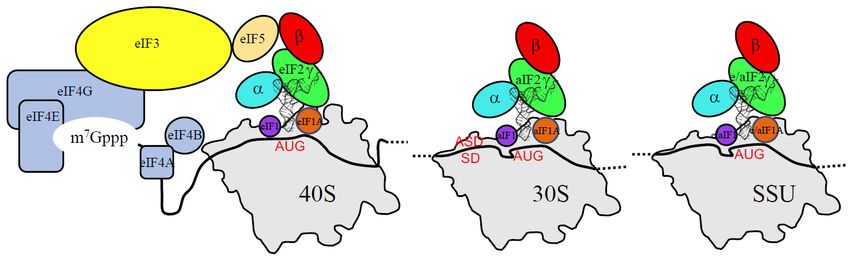
Int. J. Mol. Sci. 2019, 20, 939 6 of 21
Int. J. Mol. Sci. 2019, 20, x FOR PEER REVIEW 6 of 20
Figure 3. The structure of the initiation factors belonging to the core complex. (A) The structural
Figure 3. The structure of the initiation factors belonging to the core complex. (A) The structural
organization of the three subunits of e/aIF2 is shown. Overall, aIF2α, β and γ from P. abyssi have
organization of the three subunits of e/aIF2 is shown. Overall, aIF2α, β and γ from P. abyssi have about
about 28%, 40%, and 48% sequence identity with the eukaryotic eIF2α, β and γ subunits, respectively.
28%, 40%, and 48% sequence identity with the eukaryotic eIF2α, β and γ subunits, respectively. The
The eukaryotic specificities of each factor are schematized by dark boxes or indicated within the block
eukaryotic specificities of each factor are schematized by dark boxes or indicated within the block
with white letters. The eukaryotic eIF2α have an acidic C-terminal extension. The eukaryotic eIF2β
with white letters. The eukaryotic eIF2α have an acidic C‐terminal extension. The eukaryotic eIF2β
have a supplementary N-terminal domain necessary for the binding to eIF5 and eIF2Bε. The eukaryotic
have a supplementary N‐terminal domain necessary for the binding to eIF5 and eIF2Bε. The
eIF2γ have N- and C-terminal extensions compared to the archaeal versions. Note that some eukaryotic
eukaryotic
and/or archaeal eIF2γspecificities
have N‐ and areC‐terminal
also found at extensions compared
some positions in thetosequences
the archaeal(see versions. Note that
text and [66–68] for
some eukaryotic and/or archaeal specificities are also found at some positions
sequence alignments). The structure of the full heterotrimeric aIF2 is shown in a cartoon representation in the sequences (see
text
in itsand [66–68]form
unbound for sequence
(left, PDB alignments).
ID 2AHO and The structure
2QMU) and of the
boundfull heterotrimeric
to Met-tRNAi Met aIF2(right,
is shownPDBinID a
cartoon representation in its unbound form (left, PDB ID 2AHO and
3V11). The α subunit is colored in cyan, the β subunit is colored in red and the γ subunit is colored 2QMU) and bound to Met‐
tRNA iMet (right, PDB ID 3V11). The α subunit is colored in cyan, the β subunit is colored in red and
in green. The two mobile wings of aIF2, the αD12 domains and the core domain of β, are circled. (B)
The structures isofcolored
the γ subunit e/aIF1Ainand green.
of itsThe two mobile
bacterial wings IF1.
homologue of aIF2,
The the αD12 of
structure domains
aIF1A from and the core
P. abyssi
domain of β, are circled. (B) The structures of e/aIF1A and of its bacterial
(PDB ID 4MNO) is drawn in orange cartoons. Overall, eIF1A from P. abyssi has about 40% identity with homologue IF1. The structure
of
theaIF1A from P.
eukaryotic abyssiThe
eIF1A. (PDB basicIDand
4MNO) acidicis eukaryotic
drawn in orange extensionscartoons. Overall,
necessary eIF1A fromscanning
for long-range P. abyssi
has about with
are shown 40% light
identityyellowwith the eukaryotic
boxes. The archaeal eIF1A.
version The of basic and contains
the factor acidic eukaryotic
a helix domain extensions
at the
necessary
C-terminalfor long‐range
extremity but scanning
no acidic are shown with
extension. light ayellow
However, boxes. The archaeal
short N-terminal version
tail is present of the
in aIF1A.
factor
The contains a helix
organization of thedomain
bacterial at IF1
the protein
C‐terminal extremity
is also shown.but As no acidic extension.
described in the text,However,
because of a short
their
N‐terminal tail is present in aIF1A. The organization of the bacterial
structural resemblance, the IF1 and e/aIF1A proteins are considered as universal initiation factors. IF1 protein is also shown. As
described
The secondaryin the text, because
structures are from of their
E. colistructural
IF1 (PDB code resemblance,
1AH9) and thefrom
IF1 and e/aIF1A
P. abyssi aIF1Aproteins
(PDB code are
considered
4MNO). (C)asStructureuniversalofinitiation
e/aIF1. The factors. The secondary
structure of aIF1 from structures are from E. jannaschii
Methanocaldococcus coli IF1 (PDB (PDB codeID
1AH9)
4MO0) and from P.
is drawn in abyssi
magenta aIF1A (PDB code
cartoons, and 4MNO).
that of eIF1 (C) from
Structure of e/aIF1.
S. cerevisiae TheID
(PDB structure
2OGH) of is aIF1 from
in purple.
Methanocaldococcus
Overall, eIF1 from P.jannaschiiabyssi has(PDB aboutID30%4MO0)
sequence is drawn in magenta
identity with the cartoons,
eukaryotic and that
eIF1. of L1
The eIF1 from
loop, S.
also
cerevisiae (PDBloop,
called a basic ID 2OGH)
is located is in purple. Overall,
front of the anticodoneIF1 from
loop ofP. the
abyssi
tRNAhasin about
the P30% sequence
IN states identity
of eukaryotes
with the eukaryotic
and archaea. The loop eIF1.isThe L1 loop,
mobile alsoof
outside called a basic loop,
the ribosome is located
(Figure 4). Thein front
L2 loopof the anticodon
is only present loopin
of the tRNA[72].
eukaryotes in the IN states the
It Pcontacts of eukaryotes
tRNA D-loop andin archaea.
the PINThe stateloop is mobile
[73]. In all theoutside
panels, of the
the ribosome
numbers
(Figure
of residues4). The L2 loopinisparentheses
indicated only presentrefer in eukaryotes
to P. abyssi [72]. It contacts
proteins thearchaea,
for the tRNA D‐loop human the PIN state
in proteins for
[73]. In all the
eukaryotes, andpanels,
E. colithefor numbers
IF1. of residues indicated in parentheses refer to P. abyssi proteins for
the archaea, human proteins for eukaryotes, and E. coli for IF1.
At first glance, the use of similar tRNA binding modes by eukaryotic and archaeal e/aIF2 was not
obvious. Indeed,
At first early
glance, thebiochemical studies
use of similar tRNA using yeast modes
binding eIF2 hadbyshown that the
eukaryotic subunit contributed
andα archaeal e/aIF2 was
onlyobvious.
not slightly to tRNA early
Indeed, binding affinity [74].
biochemical Interestingly,
studies eukaryotic
using yeast eIF2 had α subunits
shown thatspecifically harbor
the α subunit
an acidic C-terminal
contributed extension
only slightly (Figurebinding
to tRNA 3). The removal
affinity of thisInterestingly,
[74]. extension waseukaryotic
sufficient toα reveal the
subunits
positive effect
specifically of the an
harbor α subunit on the tRNAextension
acidic C‐terminal binding affinity
(Figurein3).
eukaryotic complexes
The removal [75].
of this This argued
extension was
in favor oftothe
sufficient samethe
reveal tRNA binding
positive mode
effect in both
of the domains
α subunit of life.
on the tRNAIt has been proposed
binding affinity inthat during
eukaryotic
the translation
complexes [75].initiation
This argued in eukaryotes, the same
in favor of the negative
tRNA effect of themode
binding acidicintail
bothmight be relieved
domains upon
of life. It has
the interaction
been proposed of theduring
that TC withthethe ribosome.
translation Finally, in
initiation aneukaryotes,
adjustment the of the influence
negative of of
effect each
theperipheral
acidic tail
might be relieved upon the interaction of the TC with the ribosome. Finally, an adjustment of the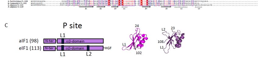
Int. J. Mol. Sci. 2019, 20, 939                                                                        7 of 21

subunit on the tRNA binding affinity was observed depending on the e/aIF2 species studied [76].
This suggested a possible remodeling of the position of the mobile wings of e/aIF2 in the TC during
the translation initiation, as discussed below. It can also be noted that another crystal structure of the
TC was determined but with a very different conformation [77]. However, no confirmation of the
biological significance of this conformation has been obtained yet.

2.1.2. Nucleotide Cycle on e/aIF2
      Other eukaryotic specificities such as added domains or extensions are deduced from sequence
alignments (Figure 3). The most striking ones are those related to the nucleotide cycle on eIF2.
In eukaryotes, two proteins assist eIF2: a guanine activating protein (GAP), eIF5 [58], and a guanine
exchange factor (GEF), eIF2B [78,79]. eIF5 is monomeric and eIF2B is a heterodecameric complex (2 sets
of α, β, γ, δ and ε subunits) with γ and ε having a catalytic function [80,81]. In the β subunit of eIF2,
a eukaryote-specific N-terminal domain containing lysine-rich boxes was shown to be responsible for
the binding of eIF5 and of the catalytic subunit of eIF2B (eIF2Bε) [82,83]. Moreover, the translation
initiation is regulated via the phosphorylation of a conserved serine residue (S51 in yeast) on eIF2α
(see, for instance, [84,85]). This phosphorylation leads to the formation of a highly stable complex
between eIF2B and phosphorylated eIF2. Because cellular concentration of eIF2B is low compared
to that of eIF2, this inhibits the exchange of GDP for GTP in eIF2 thereby preventing formation
of TC and down-regulating the global translation level [86,87]. Interestingly, although the eIF2α
phosphorylation decreases the level of translation of many genes, other genes that possess a short
upstream open reading frame (50 uORFs) [88,89] or are IRES-dependent [90–92] are up-regulated.
When the availability of TC is low, the stabilization of the ribosomal initiation complex at the start
codon of the short uORFs is disfavored and the ribosome scanning is continued up to the ORF
corresponding to the targeted protein (leaky scanning). In yeast and mammals, GCN4 and ATF4 are
two examples of activated transcription factors acting as “master regulators” in response to amino
acid starvation [93–95]. The whole regulation process triggered by the phosphorylation of eIF2α
is termed the integrated stress response (ISR) [84,85]. Many studies have shown that translational
regulations involve alternative routes for translation initiation using a particular set of initiation factors
that can substitute for eIF2, such as MCT-1/DENR [96], eIF2D [97], eIF2A [98]. In addition, under some
stress conditions such as hypoxia [99], eIF5B was proposed to participate in the recruitment of the
initiator tRNA, in addition to its role in subunit joining [90,91,100]. This is reminiscent of the bacterial
homologue of e/aIF5B, IF2, which in bacteria is involved in both initiator tRNA recruitment and
subunit joining [101].
      In archaea, no homologues of eIF5 and of the two catalytic subunits of eIF2B (γ and ε) are found.
Therefore, the nucleotide cycle is thought to be non-assisted by a GAP and a GEF. Consistent with the
absence of GEF, aIF2 from Sulfolobus solfataricus (Ss-aIF2) was found to have similar affinities for GDP
and GTP [67,71]. Moreover, high-resolution crystal structures of Ss-aIF2γ in the presence of GTP-Mg2+
and molecular dynamics simulations suggested that in archaea a second magnesium ion could play a
similar role as the one played in trans by the catalytic arginine residue of the GAP in eukaryotes [67].
In favor of this hypothesis, it is notable that the second magnesium ion is bound by two residues
(in Ss-aIF2γ; D19 from the GKT loop and G44 from switch1) strictly conserved in all archaeal aIF2γ
sequences whereas they are systematically replaced by A and N in eukaryotes [67].
      Finally, in archaea, the equivalent of S51 is not strictly conserved in aIF2α. Phosphorylation at this
position was reported with aIF2α from Pyrococcus horikoshii [102]. However no further confirmation of a
possible role of this phosphorylation in aIF2 regulation was obtained, rather suggesting that translation
is not regulated via aIF2α phosphorylation in archaea [103,104]. By analogy to the eukaryotic case
where eIF5B can substitute for eIF2 (see above), it cannot be excluded that aIF2-independent processes
in which aIF5B ensures tRNA recruitment as well as subunit joining, may also occur in specific
conditions or for specific mRNAs.
Int. J. Mol. Sci. 2019, 20, 939 8 of 21
2.1.3. Regulation of eIF2 Assembly by Cdc123
In archaea, the assembly of the three subunits in the heterotrimeric form of the factor does
not require any assistance. Accordingly, the three subunits can be purified independently and the
heterotrimer assembled in vitro [48,49,71]. In S. cerevisiae, however, the essential eukaryote-specific
Cdc123 protein was shown to be necessary to the formation of the eIF2 heterotrimeric complex, as it
promotes the eIF2αγ assembly step [105–107]. Interestingly, the activity of Cdc123 requires a short
eukaryote-specific C-terminal extension in eIF2γ ([106] and Figure 3). To date, Cdc123 orthologues
appear widely distributed across all major lineages of the eukaryotic tree. Therefore, the phyletic
pattern, together with its essentiality in yeast, indicates that Cdc123 might be a general regulator of
translation initiation in eukaryotes [108].
2.2. e/aIF1A
The role of eIF1A (formerly called eIF4C) in the translation initiation was first evidenced in
ribosomal complex preparations [109,110]. Eukaryotic sequences of eIF1A representatives showed a
high-level of conservation and a “dipole nature” of the protein harboring a basic N-terminal extremity
and an acidic C-terminal one [111,112]. The structure of human eIF1A was determined by NMR [113].
The protein contains an OB-fold with two C-terminal α-helices packed onto it. The N and C-terminal
extensions are disordered. Archaeal aIF1A shares about 40% of sequence identity with its eukaryotic
homologue. The 3D structures of e/aIF1A proteins are superimposable with very good rms values.
For example, the structures of aIF1 from P. abyssi (PDB ID 4MNO) and eIF1A from Cryptosporidium
parvum (PDB ID 2OQK) have an rms value of 0.64 Å for 72 compared C-alpha atoms. Notably,
whereas the core of the protein is well conserved, the archaeal version of the factor does not possess
an acidic C-terminal extension and has a shortened N-terminal basic tail (Figure 3). Hydroxyl radical
probing, and then X-ray and cryo-EM structures, showed that eIF1A and aIF1A occupy similar
positions on the small ribosomal subunit, in front of the A site, thereby precluding any elongator tRNA
binding [114–118].
2.3. e/aIF1
Like eIF2, the role of eIF1 in the start codon selection was first defined genetically [119]. Since then,
a huge amount of data has proven the crucial role of this factor in the dynamics and the accuracy
of the eukaryotic translation initiation process (reviewed in [35]). e/aIF1 is a small protein of about
100 residues in archaea and 110 residues in eukaryotes (Figure 3). The 3D structure of human eIF1
was first determined using NMR [120]. The factor is made up of an α–β domain (29–113) and of an
N-terminal unstructured domain (1–28). The archaeal proteins are highly similar to the eukaryotic
proteins ([72,121] and Figure 3). The presence of a zinc knuckle in the N-domain is suggested for some
archaeal representatives [72], but no 3D structure of this domain has been determined yet. Directed
hydroxyl radical probing, and then crystallographic and cryo-EM structures, have shown the position
of the factor on the small ribosomal subunit, in front of the P site on h44 [115,117,118,122,123].
3. The Three Initiation Factors in the PIC
3.1. The Scanning Model in Eukaryotes
The two small initiation factors eIF1 and eIF1A promote the formation of an open 48S complex
competent for scanning [124,125]. eIF1A acts synergistically with eIF1 to bind the 40S subunit [126],
and both factors facilitate the recruitment of the TC [127,128]. The acidic C-terminal tail of eIF1A
contains two motifs, dubbed scanning enhancers (SE1 and SE2 ), important for mRNA scanning [129].
In contrast, the basic N-terminal extension of eIF1A has an opposite effect, with two regions (the
scanning inhibitors SI1 and SI2) that disfavor the scanning-competent conformation of the ribosome.
Together, these two regions of eIF1A favor the accurate accommodation of the initiator tRNA in
the P site when an AUG codon is found [130–132]. Important advances on the mechanism of startInt. J. Mol. Sci. 2019, 20, 939 9 of 21
codon selection have been obtained from kinetic studies using partially reconstituted yeast translation
initiation systems [133]. Notably, the data have been obtained in the absence of eIF3, in a minimal
eukaryotic initiation system close to the archaeal one. These studies showed that rather than via GTP
hydrolysis by eIF2, the release of inorganic phosphate (Pi) from eIF2 controls the recognition of the
start codon. This has led to a new description of the dynamics of the system. AUG recognition stops
the scanning of the PIC, triggers the eIF1 departure, provokes the Pi release from eIF2, and then the
further departure of eIF2-GDP. Consistent with this model, the full accommodation of the initiator
tRNA in the P site upon the start codon recognition would cause a clash with eIF1 and therefore trigger
its
Int.release,
J. Mol. Sci.as suggested
2019, 20, x FORby cryo-EM
PEER REVIEWstudies [117,118,123]. 10 of 20
Figure
Figure 4. 4. AA comparison
comparison of of archaeal
archaeal and and eukaryotic
eukaryotic initiation
initiation complexes.
complexes. The figure shows shows partial
partial
views (P site region) of eukaryotic and archaeal initiation complexes. (A)
views (P site region) of eukaryotic and archaeal initiation complexes. (A) The eukaryotic 48S‐openThe eukaryotic 48S-open POUT
complex.
POUT complex.(B) The (B)eukaryotic 48S-closed
The eukaryotic PIN complex.
48S‐closed (A) and
PIN complex. (A)(B) are(B)
and from
are[134]
from(PDB[134]ID(PDB3JAQ,
ID 3JAP).
3JAQ,
(C) The(C)
3JAP). archaeal PIC0-PREMOTE
The archaeal conformation.
PIC0‐PREMOTE (D) The
conformation. (D)archaeal PIC1-PPIC1‐P
The archaeal IN conformation. The color
IN conformation. The
code
colorfor
codetheforinitiation factorsfactors
the initiation is the same
is theas in Figure
same 3. The small
as in Figure 3. Theribosomal helix h44
small ribosomal is inh44
helix dark purple
is in dark
and
purpletheand
mRNA is in dark
the mRNA blue.
is in dark The eIF3The
blue. subunits in viewsin(A)
eIF3 subunits and(A)
views (B) and
are in(B)black.
are inThe twoThe
black. mobile
two
wings of e/aIF2, as defined in the legend of Figure 3, are encircled. Archaeal
mobile wings of e/aIF2, as defined in the legend of Figure 3, are encircled. Archaeal aIF1 and aIF1A aIF1 and aIF1A have
positions similarsimilar
have positions to thoseto of theirofeukaryotic
those orthologues
their eukaryotic eIF1 andeIF1
orthologues eIF1A.
andIneIF1A.
eukaryotes, the initiator
In eukaryotes, the
tRNA
initiatoris bound
tRNA istobound the γ subunit
to the γof eIF2 and
subunit to the
of eIF2 domain
and to the3domain
of eIF2α, 3 ofas eIF2α,
observed in the archaeal
as observed in the
TC [69]. However,
archaeal eIF2αD12eIF2αD12
TC [69]. However, has moved and
has is found
moved andinisthe E site,
found while
in the the while
E site, core domain
the coreofdomain
β is closeof
to the tRNA. No interaction is observed between the γ subunit and h44.
β is close to the tRNA. No interaction is observed between the γ subunit and h44. In archaea, the In archaea, the structure of
the TC bound
structure of the toTCthebound
ribosome corresponds
to the ribosome to that observed
corresponds outside
to that the ribosome
observed outside forthethe IC0-PREMOTE
ribosome for the
complex.
IC0‐PREMOTE The γ subunit
complex. Thecontacts
γ subunit h44contacts
and aIF1. h44Inand
theaIF1.
IC1-PIn conformation,
INthe the structure
IC1‐PIN conformation, theof the TC
structure
is
of constrained, but the energetic
the TC is constrained, cost would
but the energetic costbewould
compensated by the codon:anticodon
be compensated by the codon:anticodonbase pairing.
base
The movement of the two mobile wings of aIF2 may help the start
pairing. The movement of the two mobile wings of aIF2 may help the start codon selection. codon selection.
Recent ofcryo-EM
3.2. Control structures
AUG Selection of in
by aIF2 partial
Archaeayeast PIC (py-48S; 40S:mRNA:TC:eIF1:eIF1A:eIF5 and
eIF3) further illustrate molecular events during the transition between the open scanning-competent
The full Pyrococcus
conformation of the 48Sabyssi translation
complex and a pre‐initiation complexstalled
closed conformation (30S:mRNA:TC:GDPNP:aIF1:aIF1A)
on the start codon ([116,134],
was reconstituted and studied by cryo‐EM. The PIC contains
Figure 4). In the open scanning-competent state of the PIC, the mRNA the 30S,is an mRNA
loosely withinaits
bound strong SD
channel
sequence,
and the TCtRNA
the initiator madeiswith GDPNP,
not fully and the
engaged two
in the P small initiation
site. The factors aIF1 and
POUT conformation aIF1A
of the tRNA [115]. Two
appears
conformations of the full PIC were observed (Figure 4). The two small initiation
to be stabilized around the anticodon loop by interactions with the core domain of eIF2β, and with factors aIF1 and
aIF1A are bound at positions similar to their eukaryotic homologues. In
eIF1 and eIF1A. Because the complex is scanning the mRNA, GTP hydrolysis on eIF2, activatedthe major conformation,
dubbed
by eIF5, PIC0‐P REMOTE
can occur, , the
but theanticodon
Pi remainsstem‐loop
held ontoofeIF2.
the tRNA is out of
Base pairing in the
the PP site
site (Figure
induces4). The TC
a rotation
structure is similar to that of the free TC, showing that the ribosome does not constrain the TC. In
contrast to the eukaryotic PIC, the γ subunit is tightly bound to the 30S. The contacts involve the
aIF2γ domain III (aIF2γ‐DIII) and a long L2 loop of its domain II ([115] and Figure 4). Moreover,
aIF2γ‐DIII is bound to h44 and interacts with aIF1. In particular, the N‐terminal domain of aIF1
contacts aIF2γ at the level of the two switch regions which control the nucleotide binding. ThisInt. J. Mol. Sci. 2019, 20, 939 10 of 21
of the head of the small ribosomal subunit towards the body, thereby closing the so-called latch
region. This movement contributes to further stabilizing the mRNA in its channel, as well as the
tRNA. In addition, the basic N-terminal tail of eIF1A favors the codon:anticodon interaction. Finally,
the adjustment of the tRNA in the P site interferes with the eIF1 binding site at the level of its basic
loop, in front of the anticodon, and at the level of loop 2, near the D-stem loop region of the tRNA.
These close contacts disfavor the binding of eIF1 and trigger both its departure and the following
events [73,135].
In the cryo-EM structures of eukaryotic PIC [116,134,136,137], the TC was modeled from the X-ray
structure of its archaeal representative [69]. The rigid unit of eIF2 (γ:αD3:β:anchoring-helix) is bound
to the acceptor helix of the tRNA as in the crystallographic structure of the archaeal TC, although the
two mobile wings (the αD12 domain and the core domain of β) are repositioned. No contact exists
between the eIF2 rigid unit, containing the GTP binding site, and the ribosome. However, in the
PIN closed state [116], eIF2αD1 is bound in the E site and interacts with the mRNA nucleotides -2
to -3, as previously proposed from hydroxyl radical probing experiments [138]. These interactions,
which help stabilize the mRNA in its channel, are absent in the POUT conformation where mRNA is
not visible in the E site (Figure 4). Moreover, the core module of β is seen in contact with the anticodon
stem-loop of the POUT tRNAi , whereas it would rather interact with the upper part of the tRNA in the
PIN complex. Finally, the eukaryote-specific lysine-rich N-terminal domain of eIF2β is visible in neither
of the two structures. Accordingly, the position of eIF5 could only be tentatively assigned. Altogether,
these observations reinforce the idea that the versatility of the two mobile wings of e/aIF2 plays an
important role in the dynamics of the translation initiation process. Intriguingly, no contact between
eIF1 and eIF2 is seen in the available structures, and therefore the structural basis showing how the
eIF1 departure triggers the release from eIF2 is still missing. The possibility that the “hub” protein
eIF3 relays information from eIF1 to eIF2 is not excluded. Indeed, this factor participates in all of the
initiation steps [139–141], and a close proximity between eIF3b, h44 and eIF2γ has been proposed from
the cryo-EM structures ([134,142], Figure 4). Finally, recent work also suggested that the dynamics of
the eIF5 interaction network also plays an important role in the release of eIF2-GDP [143].
3.2. Control of AUG Selection by aIF2 in Archaea
The full Pyrococcus abyssi translation pre-initiation complex (30S:mRNA:TC:GDPNP:aIF1:aIF1A)
was reconstituted and studied by cryo-EM. The PIC contains the 30S, an mRNA with a strong SD
sequence, the TC made with GDPNP, and the two small initiation factors aIF1 and aIF1A [115].
Two conformations of the full PIC were observed (Figure 4). The two small initiation factors aIF1
and aIF1A are bound at positions similar to their eukaryotic homologues. In the major conformation,
dubbed PIC0-PREMOTE , the anticodon stem-loop of the tRNA is out of the P site (Figure 4). The TC
structure is similar to that of the free TC, showing that the ribosome does not constrain the TC.
In contrast to the eukaryotic PIC, the γ subunit is tightly bound to the 30S. The contacts involve the
aIF2γ domain III (aIF2γ-DIII) and a long L2 loop of its domain II ([115] and Figure 4). Moreover,
aIF2γ-DIII is bound to h44 and interacts with aIF1. In particular, the N-terminal domain of aIF1 contacts
aIF2γ at the level of the two switch regions which control the nucleotide binding. This network of
interactions was never observed before in eukaryotes or in archaea. Still, the contacts between
eIF2γ-DIII and h44 had been suggested in the eukaryotic PIC using directed hydroxyl radical
probing [144]. In the second conformation, called PIC1-PIN , the anticodon stem-loop of the initiator
tRNA is bound within the P site, while the position of aIF2γ on h44 has not changed. Therefore,
the structure of the TC is constrained but it is thought that the codon-anticodon pairing in the P site
compensates for the structural constraint. Altogether, the data led to a novel view of the role of aIF2 in
the start codon selection. The resulting model shows that during the archaeal translation initiation,
the tRNA bound to aIF2 oscillates between the two positions observed in PIC0-PREMOTE and PIC1-PIN
(Figure 5). These two positions are in equilibrium and the transition from one position to the other,
modeled by superimposing PIC0 to PIC1, reflects the dynamics of the PIC during the testing for thepairing (Figure 5 and [115]). When a proper start codon is found, the base pairing with the initiator
tRNA would stabilize the PIC1‐PIN conformation by compensating for the structural constraint on the
TC. This would ensure a longer stay of the initiator tRNA in the P site and thereby trigger the aIF1
departure because of steric hindrance. The aIF1 release will also relieve contacts between aIF1 and
the switch regions of aIF2γ. Therefore, it is tempting to imagine that the aIF1 departure causes the
Int. J. Mol. Sci. 2019, 20, 939 11 of 21
release of the Pi group from aIF2 and renders the process irreversible, as observed in eukaryotes [133].
The role of aIF1‐induced dynamics of the PIC in the start codon selection was recently supported by
toeprinting
presence of aexperiments
start codon in[72]. the PThe
site.oscillation
The TC complex between actsthe
as atwo conformations
spring, is favored
pulling the initiator tRNAby outthe
presence
of the P site of asaIF1
longwhich
as its prevents
position isa notfullstabilized
tRNA accommodation
by a codon-anticodon throughpairing
a competition
(Figure 5foranda[115]).
same
binding
When site (Figure
a proper start5). In theisabsence
codon found,of theaIF1,
basethepairing
PIC becomes
with the more stable,tRNA
initiator as observed
wouldby a restricted
stabilize the
toeprinting
PIC1-P IN signal
conformation [72], and
by the tRNA
compensating is fully
for accommodated
the structural within
constraint theon P site,
the as
TC. observed
This would in a cryo‐
ensure
aEM structure
longer stay ofof the
a PIC devoidtRNA
initiator of aIF1in (to
thebe P published).
site and thereby Importantly,
trigger thein this
aIF1structure
departure thebecause
anticodon of
stem‐loop
steric of the initiator
hindrance. The aIF1tRNA releaseis will
stabilized by interactions
also relieve involvingaIF1
contacts between the and
C‐terminal tail of
the switch three
regions
universal
of aIF2γ. ribosomal
Therefore, proteins, illustrating
it is tempting a key role
to imagine that of
thetheaIF1
ribosome itselfcauses
departure in the start codon selection.
the release of the Pi
groupFinally,
from aIF2it should be underlined
and renders the processthat the two mobile
irreversible, as wings
observed of aIF2 also move [133].
in eukaryotes duringThe therole
searchof
for a start codon
aIF1-induced in the Pofsite
dynamics the(Figure
PIC in 4).theTheir movements
start codon maywas
selection contribute
recentlytosupported
the start codon selection
by toeprinting
process. Notably,
experiments the αD12
[72]. The domain
oscillation is never
between theobserved in the E site,isinfavored
two conformations contrast bytothe
thepresence
eukaryotic case
of aIF1
which prevents a full tRNA accommodation through a competition for a same binding site (Figurethe
(Figure 4). Because in eukaryotes, the interaction involving αD12 and the mRNA plays a role in 5).
selection
In the absenceof the of start
aIF1, codon
the PICcontext
becomes [138],
moreitstable,
is possible that the
as observed bySD:antiSD
a restrictedinteraction
toeprintingrenders the
signal [72],
interaction
and the tRNA of αD12
is fullyinaccommodated
the E site unnecessary
within the in this archaeal
P site, case. Itinisa also
as observed interesting
cryo-EM to note
structure that
of a PIC
aIF1A possesses
devoid of aIF1 (toan beN‐tail (FigureImportantly,
published). 3). Although in shorter than that
this structure the of its eukaryotic
anticodon orthologue,
stem-loop the tail
of the initiator
might isalso
tRNA stabilize
stabilized bythe codon‐anticodon
interactions involving interaction in the tail
the C-terminal P site. However,
of three suchribosomal
universal an interaction has
proteins,
not yet been observed in the archaeal cryo‐EM structures,
illustrating a key role of the ribosome itself in the start codon selection. and the question remains open.
Figure 5. Local scanning for the start codon selection in archaea. The figure schematizes the different
Figure 5. Local scanning for the start codon selection in archaea. The figure schematizes the different
steps of the start codon selection for SD containing archaeal mRNAs. Note that aIF1 facilitates the
steps of the start codon selection for SD containing archaeal mRNAs. Note that aIF1 facilitates the
mRNA binding [72,145]. On the right of the figure, the positions of the TC in PIC0–PREMOTE and
mRNA binding [72,145]. On the right of the figure, the positions of the TC in PIC0–PREMOTE and PIC1–
PIC1–PIN are compared after superimposition of the 30S bodies. The color code is the same as that
PIN are compared after superimposition of the 30S bodies. The color code is the same as that in Figures
in Figures 3 and 4, with light colors for PIC0–PREMOTE and dark colors for PIC1–PIN . For clarity,
3 and 4, with light colors for PIC0–PREMOTE and dark colors for PIC1–PIN. For clarity, the mobile wings
the mobile wings of aIF2 are omitted. The view shows that aIF2γ does not significantly move in the
of aIF2 are omitted. The view shows that aIF2γ does not significantly move in the two conformations,
two conformations, leading to a structural constraint in PIC1-PIN .
leading to a structural constraint in PIC1‐PIN.
Finally, it should be underlined that the two mobile wings of aIF2 also move during the search
for a start codon in the P site (Figure 4). Their movements may contribute to the start codon selection
process. Notably, the αD12 domain is never observed in the E site, in contrast to the eukaryotic case
(Figure 4). Because in eukaryotes, the interaction involving αD12 and the mRNA plays a role in the
selection of the start codon context [138], it is possible that the SD:antiSD interaction renders the
interaction of αD12 in the E site unnecessary in this archaeal case. It is also interesting to note that
aIF1A possesses an N-tail (Figure 3). Although shorter than that of its eukaryotic orthologue, the tail
might also stabilize the codon-anticodon interaction in the P site. However, such an interaction has not
yet been observed in the archaeal cryo-EM structures, and the question remains open.Int. J. Mol. Sci. 2019, 20, 939 12 of 21
4. Evolution of Translation Initiation Mechanisms and Concluding Remarks
The recent cryo-EM studies of archaeal and eukaryotic translation initiation complexes have shed
light on their structural features in the two domains of life. Within a similar minimal translation
initiation complex, the core domains of the three initiation factors 1, 1A and 2 fulfill similar functions.
The archaeal system can be viewed as the simplest translation initiation process that involves the tRNA
binding protein aIF2. The equilibrium between the PIC0-PREMOTE and PIC1-PIN conformations reflects
a local scanning of the PIC to search for a start codon in the P site. aIF2 would play an active role in
this process by pulling the tRNA out of the P site if no start codon was present. Such a mechanism
can therefore appear as an ancestor of long-range scanning, as observed in eukaryotes. Strikingly,
the emergence of the long-range scanning process along evolution is accompanied by the appearance
of sequence specificities in the initiation factors, such as the well-characterized N and C extensions
of eIF1A [131,132] or the L2 loop of eIF1 ([73], Figure 3). In e/aIF2, it appears that the two mobile
wings have evolved differently in the two systems. Therefore, the position of eIF2αD12 in the E site
seems to be related to the recognition of specific mRNA elements during scanning. Also, the role of
the eukaryote-specific acidic C-terminal tail of eIF2α in the PIC remains to be determined. Starting
from the minimal core complex, eukaryotes have also evolved towards more sophisticated processes
that allow the emergence of additional regulation mechanisms. This is illustrated by the regulation
of GTP hydrolysis by eIF5 and of GDP/GTP exchange on eIF2 by the GEF eIF2B. The available data
are not sufficient to establish whether the active role of eIF2 in pulling the tRNA out of the P site also
operates in eukaryotes. If this turned out to be the case, it would be another indication in favor of the
emergence of eukaryotes from within an archaeal phylum.
Finally, the late step of translation initiation occurring after the e/aIF2-GDP departure involves
the two e/aIF1A and e/aIF5B factors for a final check of the presence of an initiator tRNA and large
subunit joining ([146–148], Figure 1). It is striking that two closely related factors, IF1 (homologous
to e/aIF1A) and IF2 (homologous to e/aIF5B) also operate for large ribosomal subunit joining in
bacteria [101,149,150]. This joining step therefore has a universal character (Table 1). Moreover, several
studies have shown that in some cases eukaryotic translation initiation uses eIF5B instead of eIF2
to ensure the initiator tRNA recruitment [90,91,99,100]. This further argues in favor of an ancestral
translation initiation mechanism involving e/aIF5B/IF2, with e/aIF1A/IF1 having relics in all domains
of life and a possible representative of that being used in the LUCA. Evolution might then have selected
the formylation of the initiator tRNA in bacteria to favor specificity, whereas this improvement would
have been gained in eukaryotes and archaea thanks to the emergence of e/aIF2.
The evolution of translation initiation mechanisms is also related to the evolution of the 50
untranslated regions of mRNA. For instance, mRNAs having very short or no 50 UTRs, called leaderless
mRNAs, are present in all domains of life [151,152]. Such leaderless mRNAs are abundant in some
archaea such as S. solfataricus [153–156] and Haloferax volcanii [157], though less abundant in others
such as P. abyssi [158] and Aeropyrum pernix [153]. The translation initiation mechanism for the archaeal
leaderless mRNA remains a matter of debate. In S. solfataricus, the formation of a stable complex
between a leaderless mRNA and the 30S subunit was shown to depend on the presence of the initiator
tRNA [104,159]. However, the requirement for initiation factors and even a possible start with 70S
ribosomes remain to be studied in detail. The translation initiation of leaderless mRNAs is more
documented in bacteria [151,160,161]. Although a favorable action of IF2 and an unfavorable one of
IF3 were observed, both pathways involving either 30S or 70S IC are plausible, and further studies are
required to determine which of the two routes is prevalent in vivo.
Finally, diversity in possible translation initiation pathways is also observed for leaderless mRNA
translation in eukaryotes [41]. This raises the intriguing question of whether these leaderless mRNAs,
possibly translated from an initiation mechanism involving the assembled 70S/80S ribosome, are relics
of what happened in LUCA, or whether, on the contrary, they represent an evolved form of translation
initiation allowing a diversification of the mechanisms for regulation. The same question is raised by the
increasing number of alternative translation initiation routes that are being identified [37,41,96,162,163].You can also read