Ribosome Biogenesis in Archaea
←
→
Page content transcription
If your browser does not render page correctly, please read the page content below
REVIEW
                                                                                                                                               published: 22 July 2021
                                                                                                                                      doi: 10.3389/fmicb.2021.686977

                                             Ribosome Biogenesis in Archaea
                                             Paola Londei 1* and Sébastien Ferreira-Cerca 2*
                                             1
                                               Department of Molecular Medicine, University of Rome Sapienza, Rome, Italy, 2 Biochemistry III – Regensburg Center
                                             for Biochemistry, Institute for Biochemistry, Genetics and Microbiology, University of Regensburg, Regensburg, Germany

                                             Making ribosomes is a major cellular process essential for the maintenance of functional
                                             ribosome homeostasis and to ensure appropriate gene expression. Strikingly, although
                                             ribosomes are universally conserved ribonucleoprotein complexes decoding the genetic
                                             information contained in messenger RNAs into proteins, their biogenesis shows an
                                             intriguing degree of variability across the tree of life. In this review, we summarize
                                             our knowledge on the least understood ribosome biogenesis pathway: the archaeal
                                             one. Furthermore, we highlight some evolutionary conserved and divergent molecular
                             Edited by:      features of making ribosomes across the tree of life.
                        Eveline Peeters,
      Vrije University Brussel, Belgium      Keywords: archaea, ribosome, ribosome biogenesis, ribosomal RNA, ribosomal proteins, RNA modifications

                        Reviewed by:
                             Ute Kothe,
     University of Lethbridge, Canada       RIBOSOMAL SUBUNIT COMPOSITION: ARCHAEAL
                     Fabian Blombach,       SPECIFICITY AND COMMON FEATURES
            University College London,
                       United Kingdom       The ribosome is a universally conserved ribonucleoprotein (RNP) complex required for the
                        Henrik Nielsen,
                                            synthesis of polypeptides from the intermediate molecule carrying the genetic information, the
  University of Copenhagen, Denmark
                                            messenger RNA (Melnikov et al., 2012; Bowman et al., 2020). The birth of a ribosome itself is a
                  *Correspondence:          highly energy-consuming and complicated orchestrated molecular dance that culminates in the
             Sébastien Ferreira-Cerca
                                            formation of translation-competent mature ribosomal subunits (Nomura, 1999; Warner, 1999).
        sebastien.ferreira-cerca@ur.de
     orcid.org/0000-0002-0522-843X
                                            The mature ribosome is composed of two ribosomal subunits, the small and large ribosomal
                          Paola Londei      subunits (hereafter SSU and LSU, respectively). These ribosomal subunits can be further divided
             paola.londei@uniroma1.it       into two main classes of structural components, the ribosomal RNA (rRNA) and the ribosomal
                                            proteins (r-protein). Despite its universality, the sequence and composition of the ribosomal
                   Specialty section:       subunits’ structural components diverge across and within the different domains of life (Melnikov
         This article was submitted to      et al., 2012; Ban et al., 2014; Bowman et al., 2020). Notably, the sequence variabilities seen among
                   Biology of Archaea,      the universally conserved ribosome structural components were recognized and harnessed at the
               a section of the journal
                                            end of the 1970s by the pioneering studies of Carl Woese and his collaborators and are still the
             Frontiers in Microbiology
                                            cornerstone of modern molecular phylogenetic analysis and microbial taxonomy (Fox et al., 1977;
           Received: 28 March 2021
                                            Woese and Fox, 1977; Woese et al., 1990; Albers et al., 2013; Bahram et al., 2019).
            Accepted: 14 May 2021
                                                Similar to their bacterial counterparts, archaeal ribosomes are composed of three types of
            Published: 22 July 2021
                                            rRNAs: the SSU 16S rRNA and the LSU 23S and 5S rRNAs, which interact with 60–70 r-proteins,
                             Citation:
                                            establishing an intricate macromolecular network (Melnikov et al., 2012; Ban et al., 2014; Bowman
       Londei P and Ferreira-Cerca S
         (2021) Ribosome Biogenesis
                                            et al., 2020; Figure 1).
                           in Archaea.          Up to now and due to the size and sequence similarities among organisms lacking a cell
         Front. Microbiol. 12:686977.       nucleus, the archaeal rRNA molecules have been largely seen as being of a prokaryotic nature
    doi: 10.3389/fmicb.2021.686977          (Figure 1). Particularly and in contrast to canonical prokaryotic rRNAs, most eukaryotic rRNAs

Frontiers in Microbiology | www.frontiersin.org                                    1                                             July 2021 | Volume 12 | Article 686977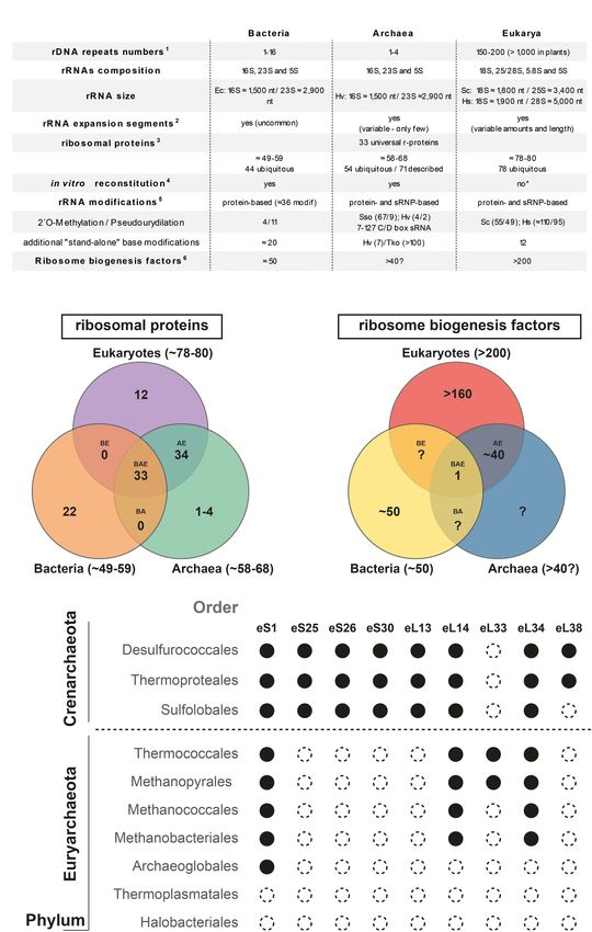
Londei and Ferreira-Cerca                                                                                                                    Making Ribosomes in Archaea

  FIGURE 1 | Ribosome and ribosome biogenesis key features overview across the tree of life. (A) Summary of ribosome and ribosome biogenesis key features.
  Modified from Ferreira-Cerca (2017) according to 1 (Hadjiolov, 1985; Warner, 1999; Klappenbach et al., 2001; Stoddard et al., 2015); 2 (Gerbi, 1986, 1996; Armache
  et al., 2013; Parker et al., 2015; Petrov et al., 2015); 3 (Lecompte et al., 2002; Nakao et al., 2004; Yutin et al., 2012); 4 (Londei et al., 1986; Sanchez et al., 1990,
  1996; Nierhaus, 1991; Mangiarotti and Chiaberge, 1997; Culver, 2003; Nierhaus and Lafontaine, 2004); 5 (Lafontaine and Tollervey, 1998; Grosjean et al., 2008;
  Dennis et al., 2015; Sharma and Lafontaine, 2015; Krogh et al., 2016; Sloan et al., 2016; Taoka et al., 2018; Coureux et al., 2020; Grünberger et al., 2020;
  Sas-Chen et al., 2020); and 6 (Hage and Tollervey, 2004; Thomson et al., 2013; Woolford and Baserga, 2013; Ebersberger et al., 2014; Grosjean et al., 2014; Henras
  et al., 2015). The detailed list of putative eukaryotic ribosome biogenesis factors conserved in archaea is depicted in Ebersberger et al. (2014). Abbreviations used:
  Sso, Saccharolobus solfataricus; Hv, Haloferax volcanii; Tko, Thermococcus kodakarensis; Hs, Homo sapiens; Sc, Saccharomyces cerevisiae. (B,C) Summary of
  shared ribosomal proteins (B) and ribosome biogenesis factors (C) across the three domains of life. Numbers of r-proteins and putative ribosome biogenesis factors
  sequence homologs shared between bacteria, archaea, and eukarya (BAE); bacteria, archaea (BA), archaea and eukarya (AE), bacteria and eukarya (BE), or unique
  to bacteria (B), or archaea (A), or eukarya (E), are indicated [based on (Lecompte et al., 2002; Hage and Tollervey, 2004; Nakao et al., 2004; Márquez et al., 2011;
  Yutin et al., 2012; Thomson et al., 2013; Woolford and Baserga, 2013; Ban et al., 2014; Ebersberger et al., 2014; Grosjean et al., 2014; Henras et al., 2015;
  Coureux et al., 2020; Nürenberg-Goloub et al., 2020)]. (D) Exemplary gene distribution of selected archaeal ribosomal proteins shared between archaea and
  eukaryotes across two major archaeal Phyla. Black circle denotes the presence and open circle denotes the absence of sequence homolog for the indicated
  ribosomal protein of the small (S) or large (L) ribosomal subunits, respectively. Adapted from Lecompte et al. (2002); Yutin et al. (2012) using the nomenclature
  proposed in Ban et al. (2014).

Frontiers in Microbiology | www.frontiersin.org                                      2                                               July 2021 | Volume 12 | Article 686977
Londei and Ferreira-Cerca Making Ribosomes in Archaea
are characterized by the presence of so-called expansion segments adaptation of ribosome biogenesis and function (Ferreira-Cerca
(ES), which are additional RNA elements of various sizes et al., 2007; Melnikov et al., 2018; Dao Duc et al., 2019).
incorporated into the universal prokaryotic rRNA core (Gerbi, However, the functional contributions of the additional archaeal-
1996; Bowman et al., 2020; Figure 1). These ES increase the specific r-protein features for ribosome assembly and function
size and complexity of the respective rRNAs; however, recent remain to be explored.
analyses have provided evidence for the presence of such ES Another particularity of the r-protein composition of some
in both bacteria and archaea (Armache et al., 2013; Penev archaeal ribosomal subunits is the presence of intra- and inter-
et al., 2020; Tirumalai et al., 2020; Stepanov and Fox, 2021). subunit promiscuous r-proteins, which leads to an increase of the
Although most of these sequence additions are limited in size respective r-protein stoichiometry and to the presence of shared
and number (Armache et al., 2013; Penev et al., 2020; Tirumalai structural components of both the SSU and LSU (Armache et al.,
et al., 2020; Stepanov and Fox, 2021), larger ES, similar in size 2013). This peculiarity is in stark contrast to what is typically
to those commonly observed in eukaryotes, have been recently observed in the bacterial and eukaryotic systems, in which
described in the Asgard archaeal phylum (Penev et al., 2020), r-proteins are thought to be exclusive structural components of
which is proposed to be the cradle of the eukaryotic lineage one or the other ribosomal subunit present in one copy per
(Spang et al., 2015; Zaremba-Niedzwiedzka et al., 2017; Liu Y. ribosomal subunit, with the exception of the LSU stalk r-proteins
et al., 2021). However, a common evolutionary relationship— (Armache et al., 2013). The functional implications of these
based on sequence and/or structure homology—of the larger molecular peculiarities remain to be analyzed.
archaeal and eukaryotic ES could not be established (Penev In conclusion, the core structural components of the archaeal
et al., 2020). Recently, a role of some of these ES in ribosomal ribosomal subunits are of prokaryotic origin, to which archaeal-
biogenesis and/or function has been established in eukaryotes specific and shared archaeal-eukaryotic features have been
(Ramesh and Woolford, 2016; Fujii et al., 2018; Díaz-López added. Together, the structural and functional constraints and/or
et al., 2019; Shankar et al., 2020). Accordingly, determining both advantages of these structural and compositional idiosyncrasies
the respective function(s) and evolutionary origin(s) of these for ribosome biogenesis and function remain to be explored.
additional rRNA segments in archaea is of general interest for the
field and will be crucial to distinguish between the archaeal origin
of eukaryotic features from the independent but convergent rRNA ORGANIZATION, SYNTHESIS, AND
evolution trajectories of structural elements present in both PROCESSING IN ARCHAEA
archaea and eukaryotes.
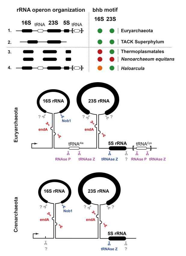
The archaeal ribosomal proteins can be divided into three The organization of the rRNA genes and the maturation of the
different groups: (1) the universally conserved r-proteins that transcripts thereof to yield mature rRNA molecules is the most
form, with the rRNAs, the universal ribosomal core (Melnikov widely studied and best understood aspect of ribosome biogenesis
et al., 2012), (2) the r-proteins exclusively shared between in archaea (Yip et al., 2013; Ferreira-Cerca, 2017; Clouet-d’Orval
archaea and eukaryotes, and (3) the archaeal-specific r-proteins et al., 2018). Because a large literature, including a number of
(Lecompte et al., 2002; Márquez et al., 2011; Yutin et al., 2012; Ban excellent reviews, exist on this topic, here only the features most
et al., 2014; Coureux et al., 2020; Nürenberg-Goloub et al., 2020; relevant from an evolutionary point of view are described.
Figure 1). The absence of exclusively shared r-proteins between As described, archaeal ribosomes are composed of one
bacteria and archaea remains an intriguing observation. 30S and one 50S ribosomal subunit, the former containing a
Among the 70 different r-proteins described in archaea, only 16S rRNA and the latter 23S and 5S rRNAs. The genomic
54 are known to be ubiquitous across archaea; among them, organization of the rRNA genes, however, presents marked
33 are universally conserved (Lecompte et al., 2002; Yutin differences in the different archaeal groups. Most euryarchaeota
et al., 2012; Ban et al., 2014; Figure 1). The composition have a typically bacterial operon organization with the 16S-
variability of the r-protein complement also correlates with a 23S-5S rRNA genes linked in this order, separated by spacer
general decrease in complexity of the r-proteins composition sequences, and transcribed all together. In most cases the spacer
at the domain scale (Lecompte et al., 2002; Yutin et al., 2012; separating the 16S and the 23S rRNA genes contains an Ala-tRNA
Figure 1). In other words, the r-protein counterpart of the gene; some euryarchaea also have a second tRNA gene, Cys-
last archaeal common ancestor was likely more complex than tRNA, in the 30 ETS downstream of the 5S rRNA gene (Figure 2).
that of most of its descendent lineages (Lecompte et al., 2002; By contrast, in the crenarchaeota and probably in most members
Yutin et al., 2012). The functional consequences and additional of the TACK superphylum, the 5S rRNA genes are physically
adaptations underlying such r-protein reductive evolution for separated from the other two larger rRNAs and transcribed
archaeal ribosome biogenesis and function is currently unknown. independently (Figure 2). There are also a few special situations,
Furthermore, recent studies also indicate the presence of such as that of the euryarchaeon Themoplasma acidophilum,
archaeal-specific ribosomal proteins (Márquez et al., 2011; where the three 16S, 23S, and 5S rRNA genes are unlinked
Coureux et al., 2020; Nürenberg-Goloub et al., 2020), suggesting and separately transcribed (Yip et al., 2013; Brewer et al., 2020;
that the discovery of new additional archaeal-specific r-proteins Figure 2).
is still incomplete. Last, organism-specific insertion, extension, The primary rRNA transcripts are maturated following
deletion, or sequence variations within conserved r-proteins are pathways that follow neither the bacterial nor the eukaryal
not unusual, and may play an important role for the cellular paradigm, albeit having features reminiscent of both.
Frontiers in Microbiology | www.frontiersin.org 3 July 2021 | Volume 12 | Article 686977Londei and Ferreira-Cerca Making Ribosomes in Archaea FIGURE 2 | rDNA gene organization and processing of pre-rRNA in archaea. (A) Ribosomal DNA gene organization and rRNA BHB motif conservation across archaea. A selected survey of archaeal rRNA operon organizations suggests two predominant classes of linked rRNA organization found in representative organisms of the Euryarchaeota and TACK Superphylum (Thaumarchaeota–Aigarchaeota–Crenarcheota–Korarchaeota) and one minor class of unlinked organization (e.g., Thermoplasmata class/Nanoarchaeum equitans). 16S and 23S rRNAs processing stem secondary structures were predicted using the ViennaRNA Web servers. Presence of predicted BHB is indicated in black. Presence of heterogeneous rRNA operons with heterogeneous presence of BHB motif within the processing stem is depicted in orange (Haloarcula genus). Absence of predictable BHB motifs is depicted by a red circle (e.g., Thermoplasmata class/Nanoarchaeum equitans). Modified from Jüttner et al. (2020) under CC-BY License. (B) Schematic representation of exemplary rRNA processing sites and the known respective ribonuclease activities required for the maturation or the pre-rRNA are indicated. Unknown activities are indicated in gray, putative activities in lilac, activities base on in vitro analysis in blue, and activities based on in vivo analysis in red. Upper panel represents common organization found in Euryarchaeota and lower panel in Crenarchaeota. Modified from Ferreira-Cerca (2017); Jüttner et al. (2020). As in bacteria, the sequences flanking the rRNA genes Bulge-helix-Bulge (BHB) motifs that are recognized and cleaved have extended complementarity and pair, forming double-helical by the archaeal-specific endA splicing endonuclease (Tang et al., stems that are the target of certain endonucleases starting rRNA 2002; Ferreira-Cerca, 2017; Clouet-d’Orval et al., 2018; Qi maturation. However, although, in bacteria, these stems are et al., 2020; Schwarz et al., 2020; Figure 2). Consequently, cleaved by RNAse III, in most archaea, they typically contain the pre-16S and pre-23S rRNAs are ligated and first released Frontiers in Microbiology | www.frontiersin.org 4 July 2021 | Volume 12 | Article 686977
Londei and Ferreira-Cerca                                                                                            Making Ribosomes in Archaea

in a circular pre-rRNA form, which is subsequently opened                  Omer et al., 2003; Yip et al., 2013). Notably, these RNP complexes
and matured by other enzymes that have not yet been                        are ubiquitous in both archaea and eukaryotes but are absent
characterized (Tang et al., 2002; Ferreira-Cerca, 2017; Clouet-            from bacteria and are responsible for the two major types of
d’Orval et al., 2018; Jüttner et al., 2020; Qi et al., 2020;               rRNA modifications, i.e., 20 O-methylation of the ribose moiety
Schwarz et al., 2020). For a comprehensive review of the rRNA              by the C/D box sRNPs and isomerization of the uridine base
maturating/modifying enzymes, see Clouet-d’Orval et al. (2018)             into pseudouridine by the H/ACA box sRNPs (Lafontaine
and Ferreira-Cerca (2017).                                                 and Tollervey, 1998; Omer et al., 2003; Yip et al., 2013).
    In certain members of the crenarchaeota, the processing of             Moreover, in eukaryotes, few snoRNPs do not have any known
16S rRNA has features that present some homology with the                  rRNA modification function but are instead required for pre-
eukaryotic process; specifically, there are endonucleases that             rRNA processing (Lafontaine and Tollervey, 1998; Sharma and
introduce 1-2 cuts within the 50 ETS (Durovic and Dennis, 1994;            Lafontaine, 2015; Sloan et al., 2016). Among these, the snoRNA
Figure 2). The most distal of these processing sites, termed               U3 is required for early processing steps of the SSU and to
site 0, lies some 70 nucleotides ahead of the 16S mature 50                avoid premature folding of the SSU central pseudoknot structure
end, is probably conserved in most crenarchaeota, and has                  (Baßler and Hurt, 2019; Klinge and Woolford, 2019). In archaea,
similarity to the processing site termed A0 in eukaryotes. Site            U3 and snoRNPs facilitating rRNA processing and folding
A0 is generally present in eukaryotic pre-rRNAs and is one of              independently of rRNA modification activity are not known.
the earliest processing sites starting its maturation (Mullineux           More details about these two classes of RNPs and their rRNA
and Lafontaine, 2012). In archaea, endonucleolytic cleavage at             modifications in archaea can be found in the two accompanying
site 0 is independent of the formation of the processing stems             reviews in this special issue by Randau and collaborators (C/D
containing the BHB motifs. Instead, its recognition is guided              box sRNPs; Breuer et al., 2021) and Kothe and collaborators
by a specific sequence containing a conserved CUU motif also               (H/ACA box sRNPs; Czekay and Kothe, 2021).
found in the eukaryotic counterpart. This CUU motif is shown                   In addition to the two main types of rRNA modifications
to be essential for cleavage in S. solfataricus (Ciammaruconi              mentioned, additional base modifications are also found.
and Londei, 2001). Notably, in the eukarya, cleavage at site A0            Commonly, base methylations (m1, m3, m5, m6A, . . .) and
requires a RNP particle containing the small nucleolar RNA                 also acetylation or larger types of modifications (e.g., acp3)
U3, but in the archaea this does not seem to be the case. The              are decorating the rRNAs (Piekna-Przybylska et al., 2008;
archaeal endonuclease cutting at site 0 has not yet been identified;       Boccaletto et al., 2018). Generally, most of these modifications
interestingly, it seems to be closely associated with the 60 kDa           cluster within the ribosomal subunit functional centers (A-,
chaperonin, at least in S. solfataricus (Ruggero et al., 1998).            P-, E-sites, and subunit bridges) and are believed to stabilize
    Although homologs of eukaryotic small nucleolar RNAs do                and/or support the activity of the translation machinery (Piekna-
not seem to be involved in rRNA processing in archaea, they do             Przybylska et al., 2008; Sharma and Lafontaine, 2015; Sloan
participate massively in another prominent feature of archaeal             et al., 2016). Interestingly, the position and/or chemical nature
rRNA maturation, that is, guiding chemical modifications of                of these modifications is apparently flexible across the tree of
specific nucleotides, which is described in the next paragraph.            life, suggesting that the functional contribution of the respective
                                                                           rRNA modification(s) in their respective structural environments
                                                                           prevails over their exact chemical nature and/or relative position
RIBOSOMAL RNA MODIFICATIONS                                                (Piekna-Przybylska et al., 2008; Sharma and Lafontaine, 2015;
                                                                           Sloan et al., 2016; Ferreira-Cerca, 2017).
RNA modifications were discovered in the early 1950s, and since                The total amounts and types of rRNA modifications strongly
then, more than 100 different types of chemical modifications              vary across archaea. For instance, halophilic archaea possess a
have been described (Littlefield and Dunn, 1958; Boccaletto                lower total amount of rRNA modifications (e.g., H. volcanii
et al., 2018). These modifications are expanding the chemical and          ∼10 known modifications; Grosjean et al., 2008). For example,
structural properties of the classical RNA alphabet (Li and Mason,         the archaeal homologs of the eukaryotic methyltransferase Nep1
2014; Kadumuri and Janga, 2018).                                           are not found in the phylogenetically related Methanogen class
    Ribosomal RNA modifications are found in all rRNAs studied             II and Haloarchaea (see also Figure 3). This decrease in the
thus far (Piekna-Przybylska et al., 2008; Boccaletto et al., 2018);        number of RNA modifications also correlates with a generally
however, their diversity (respective chemical nature, number,              reduced amount of r-proteins and ribosome biogenesis factors
and position) can be diverging across archaea (Grosjean et al.,            in these organisms (Lecompte et al., 2002; Yutin et al., 2012;
2008; Dennis et al., 2015; Boccaletto et al., 2018; Coureux et al.,        Ebersberger et al., 2014; see above and below). In contrast,
2020; Sas-Chen et al., 2020). rRNA modifications can be grouped            the total amount of rRNA modifications in thermophiles and
into two main types: (1) base and (2) ribose modifications.                hyperthermophiles is particularly increased (Dennis et al., 2015).
Furthermore, the machineries involved in the rRNA modification             For example, representative organisms of the Thermococcales
process can be also subdivided into two major groups: (1)                  order, which can grow at remarkably high temperatures
stand-alone enzymes, which are found across all domains of                 (near the boiling point of water), contain a large amount
life, and (2) RNA-guided modifications, which utilize RNP                  of base acetylations, presumably introduced by the archaeal
complexes to guide and modify the target rRNA in an RNA                    homolog of the eukaryotic RNA cytidine acetyltransferase
sequence-dependent manner (Lafontaine and Tollervey, 1998;                 Kre33/Nat10 (Sleiman and Dragon, 2019; Coureux et al., 2020;

Frontiers in Microbiology | www.frontiersin.org                        5                                      July 2021 | Volume 12 | Article 686977
Londei and Ferreira-Cerca Making Ribosomes in Archaea
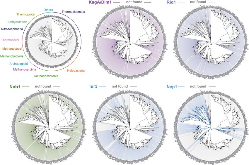
FIGURE 3 | Exemplary conservation of selected putative ribosome biogenesis factors involved in small ribosomal subunit biogenesis in archaea. Phylogenetic
conservation profile of the indicated known or putative small ribosomal subunit ribosome biogenesis factors across 1,500 archaeal genomes were generated using
AnnoTree (http://annotree.uwaterloo.ca; Mendler et al., 2019). Archaeal classes are annotated in a phylogenetic tree (upper left) as provided by AnnoTree. Absence
of sequence homolog in a define organism is indicated by a gray line, whereas its presence is indicated by a colored line. Note the absence of significant homology
for Nep1 (e.g., Thermoplasmata, Halobacteria, and more) or Tsr3 (e.g., Thermococcales) in a large group of organisms, in contrast to the more widespread
distribution of KsgA/Dim1, Rio1, and Nob1 archaeal homologs.
Grünberger et al., 2020; Sas-Chen et al., 2020). Moreover, and Overall, these observations suggest that the relative amount
in contrast to the clustered distribution of rRNA modifications of rRNA modifications and their diversity may reflect organism-
normally observed, these acetylations are scattered throughout specific adaptation to their respective environmental conditions
the rRNA sequences (Coureux et al., 2020; Grünberger et al., and/or organism-specific evolutionary trajectories (Dennis et al.,
2020; Sas-Chen et al., 2020). Furthermore, the total amount 2015; Seistrup et al., 2017; Sas-Chen et al., 2020; Knüppel
of these acetylations seems to vary according to the growth et al., 2021). The functional significance of the variability in
temperature (Sas-Chen et al., 2020). rRNA modifications, of the presence of different modification
Remarkably, among all the known stand-alone enzymes, the machineries on the ribosome biogenesis pathway in archaea,
SSU dimethyltransferase KsgA/Dim1 carrying the dimethylation and how these machineries have contributed to (re-)shape the
of two universally conserved adenosines at the 30 end of the SSU ribosome assembly pathway remains to be determined.
rRNA is the only almost universally conserved factor involved
in ribosome biogenesis (Lafontaine et al., 1994; Connolly et al.,
2008; Seistrup et al., 2017; Knüppel et al., 2021). Despite ASSEMBLY OF ARCHAEAL RIBOSOMES:
its widespread distribution, several functional aspects of the IN VITRO STUDIES
KsgA/Dim1 biology, such as assembly/release mechanisms and
the modification process itself (e.g., completion) strikingly The capability of bacterial ribosomes to assemble spontaneously
diverge between different organisms and across the different in vitro from the separate RNA and protein components was
domain of life (Van Buul et al., 1984; Formenoy et al., 1994; first demonstrated in the late 60’s by the Nomura laboratory
Lafontaine et al., 1994; Connolly et al., 2008; Zorbas et al., 2015; with Escherichia coli 30S subunit (Traub and Nomura, 1968) and
Ghalei et al., 2017; Seistrup et al., 2017; Knüppel et al., 2021; later by the Nierhaus laboratory with the 50S subunit from the
Liu K. et al., 2021). same organism (Nierhaus and Dohme, 1974). Ribosomes from
Frontiers in Microbiology | www.frontiersin.org 6 July 2021 | Volume 12 | Article 686977Londei and Ferreira-Cerca Making Ribosomes in Archaea
other bacterial species were also successfully reconstituted in vitro That archaeal ribosomes were also capable of spontaneous
(Green and Noller, 1999; Agalarov et al., 2016). self-assembly in vitro was demonstrated some years later with
These experiments are important in showing that, even in the particles of two different extremophilic archaea: the 50S
a huge macromolecular complex such as the ribosome, the subunits of Saccharolobus (formerly Sulfolobus) solfataricus
components contain in themselves all the necessary information (Londei et al., 1986), an extreme thermophile, and both 30S and
to interact in an orderly way so as to form a functional particle. 50S subunits of Haloferax mediterranei, a halophilic organism
Even more importantly, they highlight a definite assembly (Sanchez et al., 1990, 1996). The challenge here was not only to
hierarchy, in which a subset of ribosomal proteins starts the obtain spontaneous reassembly of the ribosomal particles from a
ribosome biogenesis process by binding directly to specific sites different domain of life, but also to explore how living in extreme
on the rRNA. These “early assembly” proteins, together with environments affected ribosome biogenesis.
the rRNA, create a “core particle” that has to undergo certain The thermophilic archaeon S. solfataricus is a particularly
conformational changes before binding the missing proteins and interesting case because it thrives optimally at a temperature
being converted into the final functional particle. of 80–85◦ C and because it is known to have more protein-
Experiments of in vitro assembly with purified components, rich ribosomes than its bacterial counterparts (Schmid and
could define an “assembly map,” i.e., the stepwise binding of Böck, 1982; Londei et al., 1983). S. solfataricus 50S subunits
ribosomal proteins to the rRNAs leading to the formation could be functionally reassembled from the separate RNA and
of intermediate particles that are finally converted into a protein components only at high temperatures (80◦ C) and using
complete functional ribosomal subunit (Roth and Nierhaus, high polyamine (thermine) concentrations. Interestingly, the
1980). However, the necessary experimental conditions (e.g., best conditions for Sulfolobus 50S subunits in vitro assembly
time, temperature, and ionic strength, etc.) to enable these entailed a two-step procedure such as for the case of the
in vitro reconstitution experiments are commonly incompatible corresponding E. coli particles. As in E. coli, the first step is
with the physiological conditions of the respective organisms, performed at a relatively low temperature (60◦ C) and yields
thereby suggesting the presence of facilitating molecular complete but functionally inactive particles. Activation is only
mechanisms in vivo. achieved upon incubation at temperatures close to the one
Among these mechanisms, the “assembly gradient” originally optimal for Sulfolobus growth (85◦ C), suggesting the requirement
proposed by Knud Nierhaus suggests that cotranscriptional and for a temperature-driven conformational change. The presence
directional assembly of r-proteins (50 to 30 direction), facilitate of a high concentration of the polyamine thermine, which
the initial steps of ribosomal assembly in vivo (Nierhaus, 1991). is physiologically present in S. solfataricus, is most probably
Similar principles of ribosomal assembly seem to apply in some required to stabilize and promote the RNA/protein interactions
eukaryotes [see, e.g., Cheng et al. (2017); de la Cruz et al. (2015); (Londei et al., 1986).
Ferreira-Cerca et al. (2007), but see also Cheng et al. (2019) Notably, however, it was never possible to achieve in vitro
and references therein] and may, therefore, likely operate in the reconstitution of functional S. solfataricus 30S subunits despite
archaeal context. For example, our recent work suggests a 50 the lower complexity of these particles with respect to the
to 30 coordination of the initial pre-rRNA maturation steps in 50S ones. More precisely, in vitro assembly of 30S particles
H. volcanii (Jüttner et al., 2020). Moreover, recent studies in containing the 16S rRNA and the whole complement of 30S
Sulfolobales suggest local clustering of the rRNA and r-protein ribosomal proteins was easily obtained, but they were not
operon genes, which may potentially have implication for early active in translation (Londei, unpublished). The reason for this
steps of ribosome assembly in some archaea (Takemata and unexpected result is unclear. It may be due to the substantially
Bell, 2021). However, the conservation of the topology and higher protein content of S. solfataricus 30S subunits with respect
organization of the ribosome synthesis machinery remains to be to bacterial particles (28 r-proteins vs 20–21), and/or to the
explored (Cockram et al., 2021; Sobolev et al., 2021). requirement for some additional assembly-promoting factor (see
Furthermore, additional ribosome biogenesis factors below). If so, biogenesis of S. solfataricus 30S subunits may
facilitating or speeding up ribosome assembly were also present interesting homologies with the eukaryotic process that
identified later (Bunner et al., 2010; Nikolay et al., 2018; see would be worth exploring in better detail.
below). Even if the pathways for in vitro ribosome assembly are As to halophilic ribosomes, Haloferax mediterranei 30S and
likely to be at least in part different from those adopted in vivo, 50S subunits could be reassembled successfully only at very high
the results from in vitro studies reveal that ribosome biogenesis concentrations of salt, close to the physiological concentration
is a highly coordinated process that requires a number of specific within the cell. Two types of monovalent cations were the
sequential steps to be completed successfully. most effective in promoting reconstitution, K+ and NH4 + .
In vitro reconstitution experiments were also employed to Unlike what happens for both E. coli and S. solfataricus, H.
explore the degree of conservation of ribosomal components mediterranei ribosomes could be reconstituted using a single-step
among different bacterial species. It was demonstrated that incubation at 42◦ C., i.e., within the optimal temperature range
hybrid, active ribosomes could be successfully reconstituted for physiological growth of this organism. The procedure was
from proteins and rRNA from different sources, thus further similar for 30S and 50S subunits except that reconstitution of 30S
highlighting the high degree of functional and structural subunits had a higher tolerance to ionic strength than that of 50S
conservation of bacterial ribosomes (Higo et al., 1973; Vogel et al., subunits and was independent of the Mg2+ concentration present
1984). in the assay (Sanchez et al., 1990, 1996).
Frontiers in Microbiology | www.frontiersin.org 7 July 2021 | Volume 12 | Article 686977Londei and Ferreira-Cerca Making Ribosomes in Archaea
One important outcome of the in vitro reconstitution in vitro reconstitution of functional eukaryotic ribosomes
experiments with archaeal ribosomes was the possibility of from the separated components was largely unsuccessful. The
studying the assembly pathways and to identify the assembly- one study claiming success in this task was performed with
initiating r-proteins. Indeed, using purified rRNA and r-proteins the ribosomes of Dictyostelium discoideum (Mangiarotti and
from S. solfataricus large ribosomal subunits, it was shown that Chiaberge, 1997). Interestingly, in vitro assembly of functional
the initial RNA–protein interactions leading to the formation D. discoideum ribosomes could not be achieved using 18S
of a definite but still incomplete assembly intermediate did and 28S rRNA species from mature cytoplasmic ribosomes
not require high temperatures, but took place optimally at but required still immature rRNA extracted from nuclear
about 20◦ C (Altamura et al., 1991). High temperatures, plus ribosomes. Furthermore, a small RNA species—presumably
the missing proteins, were instead mandatory to convert the nucleolar—is apparently required for successful reconstitution.
low-temperature assembly intermediate into active complete Although this study was never replicated, it agrees with the
subunits. The assembly intermediate contains 16 of the 34 total fact that ribosome assembly is inherently more complex in
50S subunit r-proteins; among these, the actual primary RNA- eukaryotes, developing along a pathway that makes use of many
binding proteins were identified by experiments of rRNA binding additional extra-ribosomal nuclear/nucleolar factors. Also, the
to membrane-immobilized S. solfataricus large subunit proteins. similarity in operon organization and in processing pathways
These turned out to be 8–9 r-proteins, well in accordance with of archaeal and bacterial rRNAs with respect to the eukaryotic
the number of primary RNA-binding proteins in bacterial 50S ones speaks in favor of a greater evolutionary conservation
ribosomes. It is probable that some, or even all, of these proteins between the two prokaryotic domains. The presence of a
belong to the universally conserved set of r-proteins, but because single cellular compartment in which everything happens, from
their identity was not assessed in the study in question, this transcription of rRNAs, to maturation of rRNA transcripts,
cannot be stated with certainty. In any event, that the r-proteins to ribosome assembly and activation, must have dictated
present in the low-temperature-assembly intermediate are the the need for a simpler and more streamlined process of
innermost in the body of the 50S subunit was also confirmed by ribosome biogenesis than it is the case for eukaryotes. However,
preparing ribosomal “cores,” i.e., stripping the outer r-proteins more work is required to assess these points, especially
with high concentrations of LiCl, a salt known to disrupt weak in vivo experiments, which, at present, are almost completely
RNA/protein interactions (Altamura et al., 1991). lacking in archaea.
Finally, the availability of methods for in vitro reconstitution
of archaeal ribosomes allows exploring the degree of evolutionary
conservation of the assembly pathways and of rRNA/r-protein RIBOSOME BIOGENESIS FACTORS:
interactions. In one study, it was found that incubation of ARCHAEAL SPECIFICITY AND SHARED
S. solfataricus LSU proteins with the 23S rRNAs from a distantly FEATURES
related archaeon (H. mediterranei) or from E. coli led to the
formation of a definite and compact 40S particle, containing most Ribosome biogenesis also requires the participation of additional
of the proteins previously identified as early assembly proteins ribosome biogenesis factors, also known as assembly factors
in S. solfataricus, including all of the primary RNA-binding ones or trans-acting factors. These factors have been analyzed in
(Altamura et al., 1991). These results suggest that the basic great detail in bacteria and eukaryotes. Generally, these factors
architecture of the ribosome and the primary rRNA/r-protein transiently interact with the nascent ribosomal subunits and
interactions are conserved to a large extent in the two prokaryotic are believed to facilitate the ribosome biogenesis process.
domains of life. Among these factors, a significant fraction homes various
Other data in agreement with this surmise is the complete enzymatic activity, mostly NTPase activity (ATPase, GTPase,
functional exchangeability of 5S rRNA between S. solfataricus and and RNA helicases. . .), which may contribute to promote
E. coli LSUs (Teixidò et al., 1989). energy-dependent steps of the ribosomal subunit biogenesis
In contrast, incubation of the S. solfataricus whole process. Interestingly, whereas GTP-dependent processes are
complement of 50S ribosomal proteins with LSU rRNAs from predominant in bacteria, ATP-dependent processes are strikingly
yeast produced no particle, but only an heterogeneous array of more frequent in Eukaryotes (Shajani et al., 2011; Thomson
RNP complexes, further indicating that both ribosome structure et al., 2013; Davis and Williamson, 2017; Baßler and Hurt,
and assembly pathways have undergone a marked divergence 2019; Klinge and Woolford, 2019). Paradoxically, and despite
from the prokaryotic model in the course of eukaryotic evolution the universal conservation of the ribosomal subunits, most of
(Altamura et al., 1991). the ribosome biogenesis factors are (1) not conserved across
In summary, probably the most important lesson to be evolution, and (2) their numbers are dramatically increasing in
learned from the in vitro assembly experiment is that strong eukaryotes (Hage and Tollervey, 2004; Ebersberger et al., 2014;
similarities exist in the basic architecture and assembly pathways Ferreira-Cerca, 2017; Figures 1, 3). This observation suggests
of archaeal and bacterial ribosomes in spite of the presence of that the ribosome biogenesis pathway has been reengineered
unique features in both and of certain “eukaryotic” features in multiple times during evolution and may reflect early adaptation
archaea, especially as regards rRNA structure and maturation. to molecular constraints present within the respective cellular
The greater complexity of ribosome assembly in eukaryotes lineage ancestors. Still, there are remarkable similarities and/or
is best documented by the fact that, despite many efforts, analogies between the different ribosome biogenesis pathways
Frontiers in Microbiology | www.frontiersin.org 8 July 2021 | Volume 12 | Article 686977Londei and Ferreira-Cerca Making Ribosomes in Archaea
that may exist and are worth being highlighted. First, the homologs of Rio1, Fap7, Dim1, Pno1, or Nob1, which form
presence of ribosome biogenesis factor sequence homologs an important functional network involved in the late steps of
between archaea and eukaryotes suggests a common origin of the eukaryotic SSU maturation, remains to be explored. Gaining
archaeal–eukaryotic ribosome biogenesis pathway (Ebersberger information on these points will surely offer important insights
et al., 2014). Intriguingly, these sequence homologs are known on the molecular evolution and adaptation of the ribosome
to predominantly act during the latest steps of eukaryotic biogenesis pathway.
SSU and LSU maturation. Second, the presence of structural Last, the endonuclease endA known to be involved in
and/or functional mimicry conserved across the tree of life the maturation of intron-containing tRNAs (Clouet-d’Orval
suggests that, despite an apparent sequence/structure divergence et al., 2018) has been recently implicated in rRNA processing,
between most ribosome biogenesis factors, some steps have thereby indicating a functional coordination of tRNA and rRNA
similar molecular constraints across the tree of life that maturation in archaea (Qi et al., 2020; Schwarz et al., 2020;
are overcome by functionally equivalent molecular inventions Figure 2).
[discussed in Ferreira-Cerca (2017); Jüttner et al. (2020)]. This
seems to be particularly true in the context of the late steps of
the small ribosomal subunit biogenesis (Ferreira-Cerca, 2017; PERSPECTIVES AND OUTLOOK
Knüppel et al., 2018). Notably, despite the absence of apparent
sequence and structural conservation between bacterial and Among the numerous challenges and outstanding questions
eukaryotic ribosome biogenesis factors, those, for example, ahead, the comprehensive identification and functional
involved in the late steps of SSU maturation remarkably characterization of factors implicated in archaeal ribosome
cluster within an analogous structural region on the nascent biogenesis are a key step to further understanding the common
SSU, i.e., regions that form the future functional centers. This and specific features of archaeal ribosome biogenesis. In addition,
suggests that binding of these ribosome biogenesis factors may recent improvement of cryo-electron microscopy analysis has
ensure functional testing and avoid premature release of the been instrumental to better characterize bacterial and eukaryotic
nascent ribosomal subunits into the translational pool (Strunk ribosome biogenesis pathways (Davis and Williamson, 2017;
et al., 2011, 2012; Ferreira-Cerca, 2017; Ghalei et al., 2017; Baßler and Hurt, 2019; Klinge and Woolford, 2019). A similar
Parker et al., 2019). revolution is still to come in the archaeal ribosome biogenesis
Furthermore, the ribosome biogenesis factors sequence field and will be important to decipher functional and structural
homologs are not evenly distributed across all archaeal genomes, analogies conserved across the tree of life and further improve
but follow the reductive evolution trend observed for the our view on the evolutionary history of the ribosome biogenesis
r-proteins, thereby suggesting a simplification of the ribosome pathway and how molecular and environmental constraints may
biogenesis pathway in these organisms, e.g., euryarchaeota have (re-)shaped the ribosome biogenesis molecular dance.
or nanoarchaeota, whereas ribosome synthesis in the TACK Furthermore, and as discussed, the ribosome biogenesis
superphylum may generally be more complex due to the sequence homologs and r-proteins are not ubiquitously
presence of additional r- proteins or ribosome biogenesis factors distributed across archaea. Therefore, it is of interest to define the
(Lecompte et al., 2002; Yutin et al., 2012; Ebersberger et al., 2014; extent of archaeal ribosome biogenesis diversity and functional
Zaremba-Niedzwiedzka et al., 2017; Figures 1, 3). However, in adaptation (Seistrup et al., 2017; Birkedal et al., 2020; Sas-
organisms showing an apparent reduced ribosome biogenesis Chen et al., 2020; Knüppel et al., 2021). Additionally, future
complexity, the addition or molecular exchange by unknown metagenomics analyses will certainly increase the numbers of
archaeal specific r-proteins and/or ribosome biogenesis factors newly identified archaea. Accordingly, learning from archaeal
cannot be fully excluded. biodiversity, changes and adaptation of the ribosome biogenesis
So far, the functional analysis of archaeal ribosome biogenesis pathway are expected to be discovered; however, the formal
factors is rather limited, and only a few have been established analysis of this biodiversity is only possible with the advance
in vivo. Most of these characterized factors are sequence of culturomics (Bilen et al., 2018; Lewis et al., 2021) and
homologs of genuine eukaryotic ribosome biogenesis factors the fast implementation of genetic manipulation in multiple
[see Ebersberger et al. (2014) for a complete list of candidates]. archaeal organisms.
Among them, the dimethyltransferase KsgA/Dim1 [see above
and Grünberger et al. (2020); Knüppel et al. (2021)], or the Rio
ATPase/Kinase family members are implicated in the late steps
AUTHOR CONTRIBUTIONS
of SSU maturation, where they probably play a role similar to PL and SF-C wrote the manuscript. Both authors contributed to
their eukaryotic counterparts (Knüppel et al., 2018). Similarly, the article and approved the submitted version.
the endonuclease Nob1 is implicated in the maturation of the
16S rRNA 30 end in vitro (Veith et al., 2012; Qi et al., 2020;
Figure 2). Collectively, these analyses suggest that the late steps FUNDING
of archaeal SSU biogenesis is a simplified version of the late steps
of eukaryotes SSU maturation (Ferreira-Cerca, 2017; Knüppel Work in the SF-C laboratory is supported by intramural
et al., 2018). However, the degree of functional conservation and funding from the department of Biochemistry III “House
interactions of ribosome biogenesis factors such as the archaeal of the Ribosome,” by the German Research Foundation
Frontiers in Microbiology | www.frontiersin.org 9 July 2021 | Volume 12 | Article 686977Londei and Ferreira-Cerca Making Ribosomes in Archaea
(DFG): individual research grant (FE1622/2-1), and collaborative ACKNOWLEDGMENTS
research center SFB/CRC 960 (SFB960-AP1 and SFB960-
B13) “RNP biogenesis: assembly of ribosomes and non- The authors would like to apologize to our colleagues whose
ribosomal RNPs and control of their function.” Open access work could not be highlighted and/or discussed in this review.
publishing of this work was partly supported by the German SF-C like to acknowledge Michael Jüttner and Robert Knüppel
Research Foundation (DFG) within the funding program Open (University of Regensburg) for fruitful and inspiring discussions
Access Publishing. on archaeal ribosome biogenesis evolution.
REFERENCES Clouet-d’Orval, B., Batista, M., Bouvier, M., Quentin, Y., Fichant, G., Marchfelder,
A., et al. (2018). Insights into RNA-processing pathways and associated RNA-
Agalarov, S., Yusupov, M., and Yusupova, G. (2016). “Reconstitution of degrading enzymes in Archaea. FEMS Microbiol. Rev. 42, 579–613. doi: 10.
Functionally Active Thermus thermophilus 30S Ribosomal Subunit from 1093/femsre/fuy016
Ribosomal 16S RNA and Ribosomal Proteins,” in Nucleic Acid Crystallography: Cockram, C., Thierry, A., Gorlas, A., Lestini, R., and Koszul, R. (2021).
Methods and Protocols, ed. E. Ennifar (New York, NY: Springer New York), Euryarchaeal genomes are folded into SMC-dependent loops and domains, but
303–314. doi: 10.1007/978-1-4939-2763-0_19 lack transcription-mediated compartmentalization. Mol. Cell 81, 459–472.e10.
Albers, S.-V., Forterre, P., Prangishvili, D., and Schleper, C. (2013). The legacy of Connolly, K., Rife, J. P., and Culver, G. (2008). Mechanistic insight into the
Carl Woese and Wolfram Zillig: from phylogeny to landmark discoveries. Nat. ribosome biogenesis functions of the ancient protein KsgA. Mol. Microbiol. 70,
Rev. Micro. 11, 713–719. doi: 10.1038/nrmicro3124 1062–1075. doi: 10.1111/j.1365-2958.2008.06485.x
Altamura, S., Caprini, E., Sanchez, M., and Londei, P. (1991). Early assembly Coureux, P.-D., Lazennec-Schurdevin, C., Bourcier, S., Mechulam, Y., and Schmitt,
proteins of the large ribosomal subunit of the thermophilic archaebacterium E. (2020). Cryo-EM study of an archaeal 30S initiation complex gives insights
Sulfolobus. Identification and binding to heterologous rRNA species. J. Biol. into evolution of translation initiation. Commun. Biol. 3:58.
Chem. 266, 6195–6200. doi: 10.1016/s0021-9258(18)38103-1 Culver, G. M. (2003). Assembly of the 30S ribosomal subunit. Biopolymers 68,
Armache, J.-P., Anger, A. M., Márquez, V., Franckenberg, S., Fröhlich, T., Villa, 234–249.
E., et al. (2013). Promiscuous behaviour of archaeal ribosomal proteins: Czekay, D. P., and Kothe, U. (2021). H/ACA Small Ribonucleoproteins:
implications for eukaryotic ribosome evolution. Nucleic Acids Res. 41, 1284– structural and Functional Comparison Between Archaea and Eukaryotes. Front.
1293. doi: 10.1093/nar/gks1259 Microbiol. 12:654370. doi: 10.3389/fmicb.2021.654370
Bahram, M., Anslan, S., Hildebrand, F., Bork, P., and Tedersoo, L. (2019). Newly Dao Duc, K., Batra, S. S., Bhattacharya, N., Cate, J. H. D., and Song, Y. S. (2019).
designed 16S rRNA metabarcoding primers amplify diverse and novel archaeal Differences in the path to exit the ribosome across the three domains of life.
taxa from the environment. Environ. Microbiol. Rep. 11, 487–494. doi: 10.1111/ Nucleic Acids Res. 47, 4198–4210. doi: 10.1093/nar/gkz106
1758-2229.12684 Davis, J. H., and Williamson, J. R. (2017). Structure and dynamics of bacterial
Ban, N., Beckmann, R., Cate, J. H. D., Dinman, J. D., Dragon, F., Ellis, S. R., et al. ribosome biogenesis. Philos. Trans. R. Soc. Lond. B Biol. Sci. 372:20160181.
(2014). A new system for naming ribosomal proteins. Curr. Opin. Struct. Biol. doi: 10.1098/rstb.2016.0181
24, 165–169. de la Cruz, J., Karbstein, K., and Woolford, J. L. (2015). Functions of Ribosomal
Baßler, J., and Hurt, E. (2019). Eukaryotic Ribosome Assembly. Annu. Rev. Proteins in Assembly of Eukaryotic Ribosomes In Vivo. Annu. Rev. Biochem.
Biochem. 88, 281–306. doi: 10.1146/annurev-biochem-013118-110817 84, 93–129. doi: 10.1146/annurev-biochem-060614-033917
Bilen, M., Dufour, J.-C., Lagier, J.-C., Cadoret, F., Daoud, Z., Dubourg, G., et al. Dennis, P. P., Tripp, V., Lui, L., Lowe, T., and Randau, L. (2015). C/D box sRNA-
(2018). The contribution of culturomics to the repertoire of isolated human guided 20 -O-methylation patterns of archaeal rRNA molecules. BMC Genomics
bacterial and archaeal species. Microbiome 6:94. 16:632. doi: 10.1186/s12864-015-1839-z
Birkedal, U., Beckert, B., Wilson, D. N., and Nielsen, H. (2020). The 23S Ribosomal Díaz-López, I., Toribio, R., Berlanga, J. J., and Ventoso, I. (2019). An mRNA-
RNA From Pyrococcus furiosus Is Circularly Permuted. Front. Microbiol. binding channel in the ES6S region of the translation 48S-PIC promotes RNA
11:582022. doi: 10.3389/fmicb.2020.582022 unwinding and scanning. Elife 8:e48246.
Boccaletto, P., Machnicka, M. A., Purta, E., Piatkowski,
˛ P., Bagiński, B., Wirecki, Durovic, P., and Dennis, P. P. (1994). Separate pathways for excision and
T. K., et al. (2018). MODOMICS: a database of RNA modification pathways. processing of 16S and 23S rRNA from the primary rRNA operon transcript
2017 update. Nucleic Acids Res. 46, D303–D307. from the hyperthermophilic archaebacterium Sulfolobus acldocaldarius:
Bowman, J. C., Petrov, A. S., Frenkel-Pinter, M., Penev, P. I., and Williams, similarities to eukaryotic rRNA processing. Mol. Microbiol. 13, 229–242. doi:
L. D. (2020). Root of the Tree: the Significance, Evolution, and Origins of the 10.1111/j.1365-2958.1994.tb00418.x
Ribosome. Chem. Rev. 120, 4848–4878. doi: 10.1021/acs.chemrev.9b00742 Ebersberger, I., Simm, S., Leisegang, M. S., Schmitzberger, P., Mirus, O., von
Breuer, R., Gomes-Filho, J.-V., and Randau, L. (2021). Conservation of Archaeal Haeseler, A., et al. (2014). The evolution of the ribosome biogenesis pathway
C/D Box sRNA-Guided RNA Modifications. Front. Microbiol. 12:654029. doi: from a yeast perspective. Nucleic Acids Res. 42, 1509–1523. doi: 10.1093/nar/
10.3389/fmicb.2021.654029 gkt1137
Brewer, T. E., Albertsen, M., Edwards, A., Kirkegaard, R. H., Rocha, E. P. C., and Ferreira-Cerca, S. (2017). “Life and Death of Ribosomes in Archaea,” in RNA
Fierer, N. (2020). Unlinked rRNA genes are widespread among bacteria and Metabolism and Gene Expression in Archaea, ed. B. Clouet-d’Orval (Cham:
archaea. ISME J. 14, 597–608. doi: 10.1038/s41396-019-0552-3 Springer International Publishing), 129–158. doi: 10.1007/978-3-319-65
Bunner, A. E., Nord, S., Wikström, P. M., and Williamson, J. R. (2010). The Effect of 795-0_6
Ribosome Assembly Cofactors on In Vitro 30S Subunit Reconstitution. J. Mol. Ferreira-Cerca, S., Pöll, G., Kühn, H., Neueder, A., Jakob, S., Tschochner, H., et al.
Biol. 398, 1–7. doi: 10.1016/j.jmb.2010.02.036 (2007). Analysis of the In Vivo Assembly Pathway of Eukaryotic 40S Ribosomal
Cheng, J., Baßler, J., Fischer, P., Lau, B., Kellner, N., Kunze, R., et al. (2019). Proteins. Mol. Cell 28, 446–457. doi: 10.1016/j.molcel.2007.09.029
Thermophile 90S Pre-ribosome Structures Reveal the Reverse Order of Formenoy, L. J., Cunningham, P. R., Nurse, K., Pleij, C. W. A., and Ofengand,
Co-transcriptional 18S rRNA Subdomain Integration. Mol Cell 75, 1256– J. (1994). Methylation of the conserved A1518-A1519 in Escherichia coli 16S
1269.e7. ribosomal RNA by the ksgA methyltransferase is influenced by methylations
Cheng, J., Kellner, N., Berninghausen, O., Hurt, E., and Beckmann, R. (2017). 3.2- around the similarly conserved U1512-G1523 base pair in the 30 terminal
Å-resolution structure of the 90S preribosome before A1 pre-rRNA cleavage. hairpin. Biochimie 76, 1123–1128. doi: 10.1016/0300-9084(94)90040-x
Nat. Struct. Mol. Biol. 24, 954–964. doi: 10.1038/nsmb.3476 Fox, G. E., Magrum, L. J., Balch, W. E., Wolfe, R. S., and Woese, C. R.
Ciammaruconi, A., and Londei, P. (2001). In Vitro Processing of the 16S rRNA (1977). Classification of methanogenic bacteria by 16S ribosomal RNA
of the Thermophilic Archaeon Sulfolobus solfataricus. J. Bacteriol. 183, 3866– characterization. Proc. Natl. Acad. Sci. U. S. A. 74, 4537–4541. doi: 10.1073/
3874. doi: 10.1128/jb.183.13.3866-3874.2001 pnas.74.10.4537
Frontiers in Microbiology | www.frontiersin.org 10 July 2021 | Volume 12 | Article 686977Londei and Ferreira-Cerca Making Ribosomes in Archaea
Fujii, K., Susanto, T. T., Saurabh, S., and Barna, M. (2018). Decoding the Function Lecompte, O., Ripp, R., Thierry, J., Moras, D., and Poch, O. (2002). Comparative
of Expansion Segments in Ribosomes. Mol. Cell 72, 1013–1020.e6. analysis of ribosomal proteins in complete genomes: an example of reductive
Gerbi, S. (1996). “Expansion segments: regions of variable size that interrupt evolution at the domain scale. Nucleic Acids Res. 30, 5382–5390. doi: 10.1093/
the universal core secondary structure of ribosomal RNA,” in Ribosomal RNA nar/gkf693
Structure, Evolution, Processing, and Function in Protein Biosynthesis, eds R. A. Lewis, W. H., Tahon, G., Geesink, P., Sousa, D. Z., and Ettema, T. J. G. (2021).
Zimmermann and A. E. Dahlberg (Boca Raton, FL: Telford-CRC Press), 71–87. Innovations to culturing the uncultured microbial majority. Nat. Rev. Microbiol.
Gerbi, S. A. (1986). The evolution of eukaryotic ribosomal DNA. Biosystems 19, 19, 225–240. doi: 10.1038/s41579-020-00458-8
247–258. doi: 10.1016/0303-2647(86)90001-8 Li, S., and Mason, C. E. (2014). The Pivotal Regulatory Landscape of RNA
Ghalei, H., Trepreau, J., Collins, J. C., Bhaskaran, H., Strunk, B. S., and Karbstein, Modifications. Annu. Rev. Genom. Hum. Genet. 15, 127–150. doi: 10.1146/
K. (2017). The ATPase Fap7 Tests the Ability to Carry Out Translocation-like annurev-genom-090413-025405
Conformational Changes and Releases Dim1 during 40S Ribosome Maturation. Littlefield, J. W., and Dunn, D. B. (1958). Natural Occurrence of Thymine and
Mol. Cell 67, 990–1000.e3. Three Methylated Adenine Bases in Several Ribonucleic Acids. Nature 181,
Green, R., and Noller, H. F. (1999). Reconstitution of Functional 50S Ribosomes 254–255. doi: 10.1038/181254a0
from in Vitro Transcripts of Bacillus stearothermophilus 23S rRNA. Liu, K., Santos, D. A., Hussmann, J. A., Wang, Y., Sutter, B. M., Weissman, J. S.,
Biochemistry 38, 1772–1779. doi: 10.1021/bi982246a et al. (2021). Regulation of translation by methylation multiplicity of 18S rRNA.
Grosjean, H., Breton, M., Sirand-Pugnet, P., Tardy, F., Thiaucourt, F., Citti, C., Cell Rep. 34:108825. doi: 10.1016/j.celrep.2021.108825
et al. (2014). Predicting the Minimal Translation Apparatus: lessons from the Liu, Y., Makarova, K. S., Huang, W.-C., Wolf, Y. I., Nikolskaya, A. N., Zhang, X.,
Reductive Evolution of Mollicutes. PLoS Genet. 10:e1004363. doi: 10.1371/ et al. (2021). Expanded diversity of Asgard archaea and their relationships with
journal.pgen.1004363 eukaryotes. Nature 593, 553–557. doi: 10.1038/s41586-021-03494-3
Grosjean, H., Gaspin, C., Marck, C., Decatur, W. A., and de Crécy-Lagard, V. Londei, P., Teichner, A., Cammarano, P., De Rosa, M., and Gambacorta, A. (1983).
(2008). RNomics and Modomics in the halophilic archaea Haloferax volcanii: Particle weights and protein composition of the ribosomal subunits of the
identification of RNA modification genes. BMC Genomics 9:470. doi: 10.1186/ extremely thermoacidophilic archaebacterium Caldariella acidophila. Biochem.
1471-2164-9-470 J. 209, 461–470. doi: 10.1042/bj2090461
Grünberger, F., Knüppel, R., Jüttner, M., Fenk, M., Borst, A., Reichelt, R., Londei, P., Teixidò, J., Acca, M., Cammarano, P., and Amils, R. (1986). Total
et al. (2020). Exploring prokaryotic transcription, operon structures, rRNA reconstitution of active large ribosomal subunits of the thermoacidophilic
maturation and modifications using Nanopore-based native RNA sequencing. archaebacterium Sulfolobus solfataricus. Nucleic Acids Res. 14, 2269–2285.
BioRxiv [Preprint]. doi: 10.1101/2019.12.18.880849 Mangiarotti, G., and Chiaberge, S. (1997). Reconstitution of Functional Eukaryotic
Hadjiolov, A. A. (1985). The Nucleolus and Ribosome Biogenesis. Vienna: Springer- Ribosomes fromDictyostelium discoideum Ribosomal Proteins and RNA.
Verlag Wien. J. Biol. Chem. 272, 19682–19687. doi: 10.1074/jbc.272.32.19682
Hage, A. E., and Tollervey, D. (2004). A Surfeit of Factors: why is Ribosome Márquez, V., Fröhlich, T., Armache, J.-P., Sohmen, D., Dönhöfer, A., Mikolajka,
Assembly So Much More Complicated in Eukaryotes than Bacteria? RNA Biol. A., et al. (2011). Proteomic Characterization of Archaeal Ribosomes Reveals
1, 9–14. doi: 10.4161/rna.1.1.932 the Presence of Novel Archaeal-Specific Ribosomal Proteins. J. Mol. Biol. 405,
Henras, A. K., Plisson-Chastang, C., O’Donohue, M.-F., Chakraborty, A., and 1215–1232. doi: 10.1016/j.jmb.2010.11.055
Gleizes, P.-E. (2015). An overview of pre-ribosomal RNA processing in Melnikov, S., Ben-Shem, A., Garreau de Loubresse, N., Jenner, L., Yusupova,
eukaryotes. Wiley Interdiscip. Rev. RNA 6, 225–242. doi: 10.1002/wrna.1269 G., and Yusupov, M. (2012). One core, two shells: bacterial and eukaryotic
Higo, K., Held, W., Kahan, L., and Nomura, M. (1973). Functional ribosomes. Nat. Struct. Mol. Biol. 19, 560–567. doi: 10.1038/nsmb.2313
correspondence between 30S ribosomal proteins of Escherichia coli and Melnikov, S., Manakongtreecheep, K., and Söll, D. (2018). Revising the Structural
Bacillus stearothermophilus. Proc. Natl. Acad. Sci. U. S. A. 70, 944–948. Diversity of Ribosomal Proteins Across the Three Domains of Life. Mol. Biol.
doi: 10.1073/pnas.70.3.944 Evol. 35, 1588–1598. doi: 10.1093/molbev/msy021
Jüttner, M., Weiß, M., Ostheimer, N., Reglin, C., Kern, M., Knüppel, R., et al. Mendler, K., Chen, H., Parks, D. H., Lobb, B., Hug, L. A., and Doxey, A. C. (2019).
(2020). A versatile cis-acting element reporter system to study the function, AnnoTree: visualization and exploration of a functionally annotated microbial
maturation and stability of ribosomal RNA mutants in archaea. Nucleic Acids tree of life. Nucleic Acids Res. 47, 4442–4448. doi: 10.1093/nar/gkz246
Res. 48, 2073–2090. doi: 10.1093/nar/gkz1156 Mullineux, S.-T., and Lafontaine, D. L. J. (2012). Mapping the cleavage sites on
Kadumuri, R. V., and Janga, S. C. (2018). Epitranscriptomic Code and Its mammalian pre-rRNAs: where do we stand? Biochimie 94, 1521–1532. doi:
Alterations in Human Disease. Trends Mol. Med. 24, 886–903. doi: 10.1016/ 10.1016/j.biochi.2012.02.001
j.molmed.2018.07.010 Nakao, A., Yoshihama, M., and Kenmochi, N. (2004). RPG: the Ribosomal Protein
Klappenbach, J. A., Saxman, P. R., Cole, J. R., and Schmidt, T. M. (2001). rrndb: Gene database. Nucleic Acids Res. 32, D168–D170.
the Ribosomal RNA Operon Copy Number Database. Nucleic Acids Res. 29, Nierhaus, K. H. (1991). The assembly of prokaryotic ribosomes. Biochimie 73,
181–184. doi: 10.1093/nar/29.1.181 739–755. doi: 10.1016/0300-9084(91)90054-5
Klinge, S., and Woolford, J. L. (2019). Ribosome assembly coming into focus. Nat. Nierhaus, K. H., and Dohme, F. (1974). Total reconstitution of functionally active
Rev. Mol. Cell Biol. 20, 116–131. doi: 10.1038/s41580-018-0078-y 50S ribosomal subunits from Escherichia coli. Proc. Natl. Acad. Sci. U. S. A. 71,
Knüppel, R., Christensen, R. H., Gray, F. C., Esser, D., Strauß, D., Medenbach, J., 4713–4717. doi: 10.1073/pnas.71.12.4713
et al. (2018). Insights into the evolutionary conserved regulation of Rio ATPase Nierhaus, K. H., and Lafontaine, D. L. (2004). “Ribosome Assembly,” in Protein
activity. Nucleic Acids Res. 46, 1441–1456. doi: 10.1093/nar/gkx1236 Synthesis and Ribosome Structure, esd K. H. Nierhaus and D. N. Wilson (KGaA:
Knüppel, R., Trahan, C., Kern, M., Wagner, A., Grünberger, F., Hausner, W., et al. Wiley-VCH Verlag GmbH & Co), 85–143. doi: 10.1002/3527603433.ch3
(2021). Insights into synthesis and function of KsgA/Dim1-dependent rRNA Nikolay, R., Hilal, T., Qin, B., Mielke, T., Bürger, J., Loerke, J., et al. (2018).
modifications in archaea. Nucleic Acids Res. 49, 1662–1687. doi: 10.1093/nar/ Structural Visualization of the Formation and Activation of the 50S Ribosomal
gkaa1268 Subunit during In Vitro Reconstitution. Mol. Cell 70, 881–893.e3.
Krogh, N., Jansson, M. D., Häfner, S. J., Tehler, D., Birkedal, U., Christensen- Nomura, M. (1999). Regulation of Ribosome Biosynthesis in Escherichia coli and
Dalsgaard, M., et al. (2016). Profiling of 20 -O-Me in human rRNA reveals a Saccharomyces cerevisiae: diversity and Common Principles. J. Bacteriol. 181,
subset of fractionally modified positions and provides evidence for ribosome 6857–6864. doi: 10.1128/jb.181.22.6857-6864.1999
heterogeneity. Nucleic Acids Res. 44, 7884–7895. doi: 10.1093/nar/gkw482 Nürenberg-Goloub, E., Kratzat, H., Heinemann, H., Heuer, A., Kötter, P.,
Lafontaine, D., Delcour, J., Glasser, A.-L., Desgres, J., and Vandenhaute, J. (1994). Berninghausen, O., et al. (2020). Molecular analysis of the ribosome recycling
The DIM1 Gene Responsible for the Conserved m62Am62A Dimethylation factor ABCE1 bound to the 30S post-splitting complex. EMBO J. 39:e103788.
in the 30 -Terminal Loop of 18 S rRNA is Essential in Yeast. J. Mol. Biol. 241, Omer, A. D., Ziesche, S., Decatur, W. A., Fournier, M. J., and Dennis, P. P. (2003).
492–497. doi: 10.1006/jmbi.1994.1525 RNA-modifying machines in archaea. Mol. Microbiol. 48, 617–629. doi: 10.
Lafontaine, D. L. J., and Tollervey, D. (1998). Birth of the snoRNPs: the evolution 1046/j.1365-2958.2003.03483.x
of the modification-guide snoRNAs. Trends Biochem. Sci. 23, 383–388. doi: Parker, M. D., Collins, J. C., Korona, B., Ghalei, H., and Karbstein, K. (2019).
10.1016/s0968-0004(98)01260-2 A kinase-dependent checkpoint prevents escape of immature ribosomes into
Frontiers in Microbiology | www.frontiersin.org 11 July 2021 | Volume 12 | Article 686977You can also read