Homeostasis of Second Messenger Cyclic-di-AMP Is Critical for Cyanobacterial Fitness and Acclimation to Abiotic Stress - Semantic Scholar
←
→
Page content transcription
If your browser does not render page correctly, please read the page content below
ORIGINAL RESEARCH
                                                                                                                                               published: 29 May 2018
                                                                                                                                       doi: 10.3389/fmicb.2018.01121

                                             Homeostasis of Second Messenger
                                             Cyclic-di-AMP Is Critical for
                                             Cyanobacterial Fitness and
                                             Acclimation to Abiotic Stress
                                             Marco Agostoni 1,2† , Alshaé R. Logan-Jackson 2,3 , Emily R. Heinz 2 , Geoffrey B. Severin 4 ,
                                             Eric L. Bruger 3† , Christopher M. Waters 1,3 and Beronda L. Montgomery 1,2,3,4*
                                             1
                                              Cell and Molecular Biology Graduate Program, Michigan State University, East Lansing, MI, United States, 2 Department of
                             Edited by:      Energy Plant Research Laboratory, Michigan State University, East Lansing, MI, United States, 3 Department of Microbiology
                    Conor P. O’Byrne,        and Molecular Genetics, Michigan State University, East Lansing, MI, United States, 4 Department of Biochemistry and
 National University of Ireland Galway,      Molecular Biology, Michigan State University, East Lansing, MI, United States
                                 Ireland
                         Reviewed by:        Second messengers are intracellular molecules regulated by external stimuli known as
                           Iris Maldener,
       Universität Tübingen, Germany
                                             first messengers that are used for rapid organismal responses to dynamic environmental
                  Juan Carlos Alonso,        changes. Cyclic di-AMP (c-di-AMP) is a relatively newly discovered second messenger
    Centro Nacional de Biotecnología
                                             implicated in cell wall homeostasis in many pathogenic bacteria. C-di-AMP is
                           (CNB), Spain
                        Ronan Sulpice,       synthesized from ATP by diadenylyl cyclases (DAC) and degraded by specific c-di-AMP
 National University of Ireland Galway,      phosphodiesterases (PDE). C-di-AMP DACs and PDEs are present in all sequenced
                                  Ireland
                                             cyanobacteria, suggesting roles for c-di-AMP in the physiology and/or development of
                  *Correspondence:
              Beronda L. Montgomery
                                             these organisms. Despite conservation of these genes across numerous cyanobacteria,
                 montg133@msu.edu            the functional roles of c-di-AMP in cyanobacteria have not been well-investigated. In
                   † Present  address:       a unique feature of cyanobacteria, phylogenetic analysis indicated that the broadly
                      Marco Agostoni,
                                             conserved DAC, related to CdaA/DacA, is always co-associated in an operon with
   California Institute for Quantitative
 Biosciences, University of California,      genes critical for controlling cell wall synthesis. To investigate phenotypes regulated
Berkeley, Berkeley, CA, United States        by c-di-AMP in cyanobacteria, we overexpressed native DAC (sll0505) and c-di-AMP
                         Eric L. Bruger,
  Department of Biological Sciences,
                                             PDE (slr0104) genes in the cyanobacterium Synechocystis sp. PCC 6803 (hereafter
    University of Idaho, Moscow, ID,         Synechocystis) to increase and decrease intracellular c-di-AMP levels, respectively.
                          United States
                                             DAC- and PDE-overexpression strains, showed abnormal aggregation phenotypes,
                   Specialty section:
                                             suggesting functional roles for regulating c-di-AMP homeostasis in vivo. As c-di-AMP
         This article was submitted to       may be implicated in osmotic responses in cyanobacteria, we tested whether sorbitol
Microbial Physiology and Metabolism,
                                             and NaCl stresses impacted expression of sll0505 and slr0104 or intracellular c-di-AMP
               a section of the journal
             Frontiers in Microbiology       levels in Synechocystis. Additionally, to determine the range of cyanobacteria in which
          Received: 22 January 2018          c-di-AMP may function, we assessed c-di-AMP levels in two unicellular cyanobacteria,
            Accepted: 11 May 2018            i.e., Synechocystis and Synechococcus elongatus PCC 7942, and two filamentous
            Published: 29 May 2018
                                             cyanobacteria, i.e., Fremyella diplosiphon and Anabaena sp. PCC 7120. C-di-AMP
                            Citation:
    Agostoni M, Logan-Jackson AR,
                                             levels responded differently to abiotic stress signals in distinct cyanobacteria strains,
   Heinz ER, Severin GB, Bruger EL,          whereas salt stress uniformly impacted another second messenger cyclic di-GMP in
   Waters CM and Montgomery BL
                                             cyanobacteria. Together, these results suggest regulation of c-di-AMP homeostasis in
      (2018) Homeostasis of Second
  Messenger Cyclic-di-AMP Is Critical        cyanobacteria and implicate a role for the second messenger in maintaining cellular
           for Cyanobacterial Fitness        fitness in response to abiotic stress.
    and Acclimation to Abiotic Stress.
             Front. Microbiol. 9:1121.       Keywords: abiotic stresses, c-di-AMP, c-di-GMP, cyanobacteria, ionic stress, osmotic stress, salt stress, second
     doi: 10.3389/fmicb.2018.01121           messengers

Frontiers in Microbiology | www.frontiersin.org                                    1                                                May 2018 | Volume 9 | Article 1121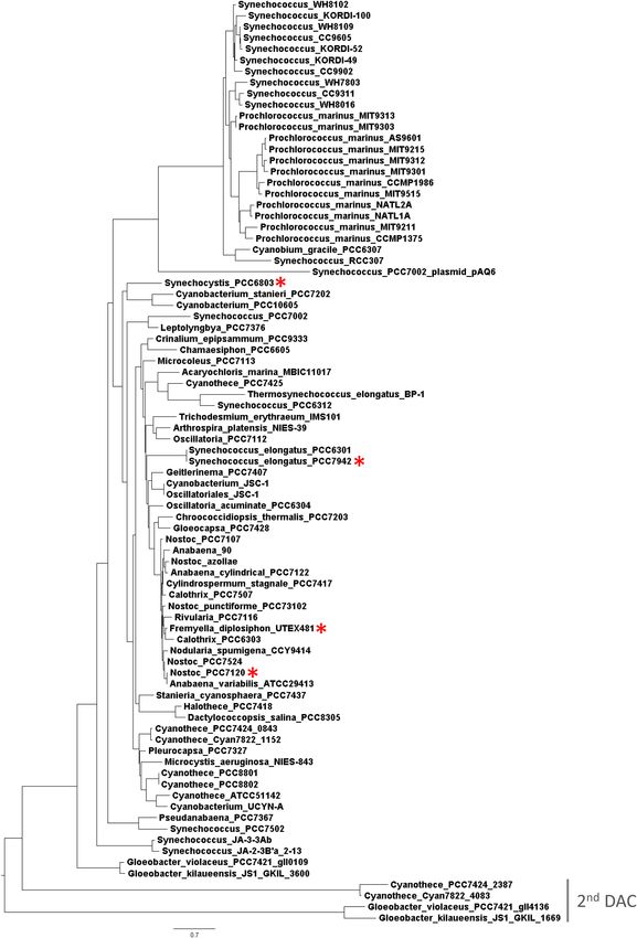
Agostoni et al.                                                                                  Second Messengers in Cyanobacterial Abiotic Stress

INTRODUCTION                                                                 Whereas numerous roles for c-di-AMP have been
                                                                          documented in Gram positive bacteria, limited insights
Cyanobacteria comprise a group of highly diverse, oxygenic                into the roles of this molecule in other bacteria have
photosynthetic bacteria that respond to a range of abiotic                been reported. Notably, cyanobacteria have recently been
and biotic signals in their environment, from light that                  reported to contain c-di-AMP synthesis genes (Agostoni
has direct impacts on photosynthesis and productivity to                  and Montgomery, 2014) and c-di-AMP accumulation has
osmotic and saline stresses. These organisms are highly                   recently been reported in Synechococcus elongatus sp. PCC
abundant in many ecosystems (Garcia-Pichel et al., 2003)                  7942 (Rubin et al., 2018). All sequenced cyanobacteria possess
and as carbon, and sometimes nitrogen fixers, contribute                  at least one DAC, with some exceptions of strains that carry
significantly to global carbon and nitrogen cycles. Second                two DACs (Agostoni and Montgomery, 2014). Unlike those
messengers are critical intracellular molecules that are regulated        DAC proteins reported in many bacteria which include a DAC
in response to specific external stimuli known as first messengers.       domain with fusions to other regulatory domains, the DACs
Control of second messenger homeostasis is used frequently                of cyanobacteria generally contain only the cyclase enzymatic
to initiate physiological changes that occur in microorganisms            domain. Additionally, two specific c-di-AMP PDEs have
as a part of environmental acclimation. A range of second                 been discovered in bacteria: one containing a DHH-DHHA1
messengers have been identified that play key roles in                    domain (Romling, 2008; Corrigan and Grundling, 2013), a
regulating environmentally-controlled physiological responses             class which is likely not present in cyanobacteria; the other
in cyanobacteria (Agostoni and Montgomery, 2014). Among                   with a domain architecture similar to the 7TM_7TMR_HD
second messengers, cyanobacteria commonly rely on cyclic                  protein family (Huynh et al., 2015). The 7TM_7TMR_HD
nucleotide signaling molecules such as cyclic AMP (i.e., cAMP)            is more common than DHH-DHHA1 domain-containing
(Ohmori et al., 1988, 2001, 2002; Katayama and Ohmori, 1997;              PDEs in bacteria and it is also present in cyanobacterial
Terauchi and Ohmori, 1999, 2004; Ohmori and Okamoto, 2004;                genomes (Huynh et al., 2015; Huynh and Woodward,
Okamoto et al., 2004; Imashimizu et al., 2005) and cyclic                 2016).
GMP (i.e., cGMP) (Ochoa De Alda et al., 2000; Cadoret et al.,                Recently, an assessment of regulons of riboswitches involved
2005). However, dicyclic nucleotides such as cyclic dimeric GMP           in binding the second messenger c-di-AMP suggested a function
(hereafter, cyclic di-GMP or c-di-GMP) have only recently been            of c-di-AMP in regulating the synthesis of osmoprotectants
reported in these organisms. In cyanobacteria, c-di-GMP has               under abiotic stress in cyanobacteria (Nelson et al., 2013).
roles in acclimation to light, phototaxis, and cellular aggregation       The addition of organic solutes, which are not permeable to
(Savakis et al., 2012; Agostoni et al., 2013, 2016; Enomoto et al.,       the bacterial cell, to cellular growth medium induces osmotic
2014, 2015; Angerer et al., 2017).                                        stress. Although salt stress is often referred to as osmotic stress
    The second messenger cyclic dimeric AMP (hereafter, cyclic            (Hagemann, 2011), salt stress specifically results in reduced
di-AMP or c-di-AMP) is a relatively newly discovered cyclic               water availability due to dissolved ions that concomitantly
dinucleotide (Romling, 2008; Witte et al., 2008; Fu et al.,               induce osmotic stress (Pade and Hagemann, 2015). Thus,
2016; Jenal et al., 2017; Krasteva and Sondermann, 2017).                 salt stress includes both ionic stress and secondary osmotic
Cyclic di-AMP is synthesized by diadenylyl cyclase (DAC;                  stress. The primary signals induced in response to salt and
PF02457) from two molecules of ATP and degraded by specific               osmotic stress in cyanobacteria are still being elucidated (Pade
phosphodiesterase (PDE) enzymes into pApA (Corrigan and                   and Hagemann, 2015). However, a role for two component
Grundling, 2013). Cyclic di-AMP and its functional roles in vivo          histidine kinase Hik33 in responses to osmotic and salt stress
have been studied primarily in Gram-positive bacteria. In Gram-           in cyanobacteria has been noted (Mikami et al., 2002; Marin
positive species, the regulation of c-di-AMP homeostasis has been         et al., 2003; Paithoonrangsarid et al., 2004; Shoumskaya et al.,
associated with a range of responses. Cyclic di-AMP levels impact         2005). The primary abiotic stress can also induce secondary
growth (Witte et al., 2013; Rismondo et al., 2016; Whiteley et al.,       signals, including second messengers. In cyanobacteria, the
2017), sporulation (Oppenheimer-Shaanan et al., 2011; Mehne               second messenger Ca2+ is involved in organismal responses
et al., 2014; Zheng et al., 2015; Raguse et al., 2017), virulence         to environmental osmotic and salt changes (Torrecilla et al.,
(Bai et al., 2013; Cho and Kang, 2013; Du et al., 2014; Yang              2001). The recognition that potential c-di-AMP binding
et al., 2014; Dey et al., 2015), biofilm formation related to host–       riboswitches may be involved in osmoprotectant production
microbe interactions (Townsley et al., 2018), and DNA repair              in response to osmotic stress in cyanobacteria suggests a
damage responses or the coordination of DNA damage response               potential role for c-di-AMP in cellular responses to abiotic
and stress homeostasis (Bejerano-Sagie et al., 2006; Witte et al.,        stress.
2008; Gándara and Alonso, 2015; Gándara et al., 2017; Raguse                 In this study, we investigated the activity of DAC and
et al., 2017), among other phenotypes in a range of Gram-positive         PDE enzymes in the moderately halotolerant freshwater
strains. A role for c-di-AMP in growth and virulence has also             unicellular cyanobacterium Synechocystis, the potential
been observed for the Gram-negative Borrelia burgdorferi and              for abiotic stresses to alter intracellular c-di-AMP levels,
Chlamydia trachomatis (Barker et al., 2013; Ye et al., 2014).             and the impact of altering c-di-AMP homeostasis on the
Additionally, c-di-AMP has been implicated in cellular responses          physiology and survival of this organism. Furthermore,
to abiotic stresses in multiple Gram-positive bacteria (Dengler           we assessed the impacts of osmotic and salt stresses on
et al., 2013; Bowman et al., 2016; Zhu et al., 2016).                     modulating c-di-AMP homeostasis in several additional

Frontiers in Microbiology | www.frontiersin.org                       2                                          May 2018 | Volume 9 | Article 1121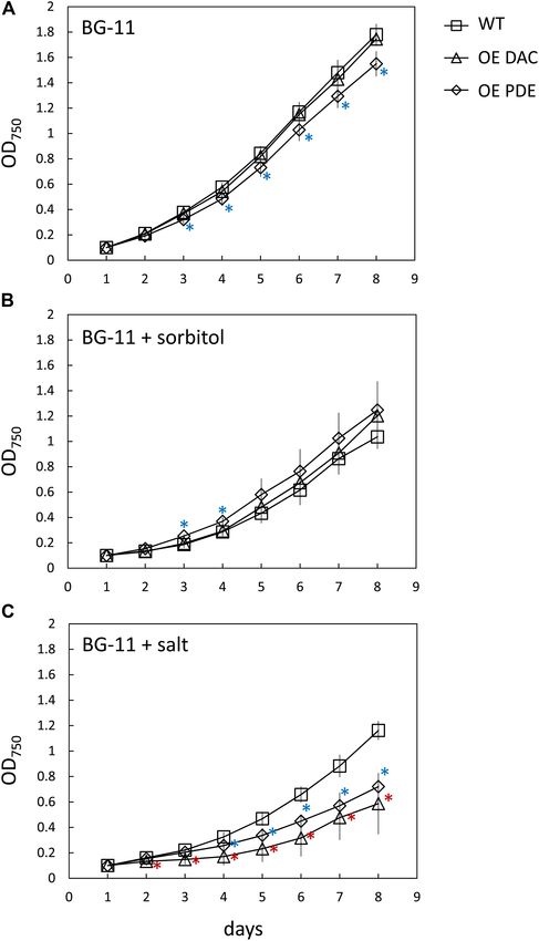
Agostoni et al.                                                                                   Second Messengers in Cyanobacterial Abiotic Stress

cyanobacteria, including another freshwater unicellular strain            0.6. Aliquots of 10 µL of the dilutions up to 1:10,000 were plated
Synechococcus elongatus PCC 7942 (hereafter Synechococcus)                and the plates incubated under 15 µmol m−2 s−1 of WL.
for which c-di-AMP has been recently implicated in
nighttime survival (Rubin et al., 2018), and two filamentous              Abiotic Stresses and c-di-AMP/c-di-GMP
freshwater strains Fremyella diplosiphon [also known as                   Quantification
Tolypothrix sp. PCC 7601 (Yerrapragada et al., 2015)], and                Cells were grown to an optical density at 750 nm (OD750 ) of
Anabaena sp. PCC 7120 (hereafter Anabaena, also known                     1 and transferred to new 250 ml flasks with BG-11/HEPES
as Nostoc sp. PCC 7120) to more broadly understand the                    medium containing 0.2 M sorbitol for the osmotic stress, or
modulation of c-di-AMP homeostasis across a range of                      0.2 M NaCl for the ionic stress, except for halophile Synechocystis
cyanobacteria.                                                            for which the NaCl concentration was 0.6 M. As a control,
                                                                          BG-11/HEPES medium without sorbitol or NaCl added was
                                                                          utilized. Cells were maintained under osmotic or salt stress for
MATERIALS AND METHODS                                                     24 h. After 24 h, c-di-AMP and c-di-GMP were quantified as
                                                                          described (Massie et al., 2012; Agostoni et al., 2013; Barker
Plasmid Construction in Synechocystis                                     et al., 2013). In brief, c-di-AMP and c-di-GMP were quantified
Intracellular levels of c-di-AMP in Synechocystis were targeted           by UPLC-MS/MS. Prior to analysis, an aliquot of each sample
for increase by overexpressing the native DAC protein Sll0505             was dried under vacuum to remove extraction buffer and the
or reduction by overexpressing the native PDE protein Slr0104.            pellet was resuspended in an equal volume of water. A 10-
The open reading frame of native genes encoding the DAC                   µl volume of the resuspended sample was analyzed together
and PDE enzymes were constitutively overexpressed under                   with an eight-point standard curve of purified c-di-AMP or
the control of the apcE (slr0335) promoter using the self-                c-di-GMP (Biolog). C-di-AMP and c-di-GMP concentrations
replicating plasmid pRL1342 (Wolk et al., 2007; GenBank:                  determined for samples were normalized to total soluble protein
AF403427.1). Promoters were added to the DAC- or PDE-                     content from an equal volume of cells from which second
encoding genes by overlap PCR using primers indicated in                  messengers were extracted as previously described (Agostoni
Table 1. The genes were amplified from genomic DNA with                   et al., 2013; Zhu et al., 2016). Growth over time of WT, OE
PrimeSTAR Max DNA polymerase (Clontech Laboratory, Inc.)                  DAC and OE PDE Synechocystis stains in the presence of sorbitol
using primers that encoded XhoI and BamHI restriction sites               (0.5 M) or NaCl (0.6 M) was measured using OD750 as described
(Table 1). The promoter-gene fusion product and pRL1342                   above.
were restricted with XhoI and BamHI and the cleaved products
ligated using DNA Ligation Kit, Mighty Mix (Takara). After                Quantitative Reverse Transcriptase PCR
transformation of the ligation mix into E. coli DH5α competent            (qRT-PCR) and RT-PCR in Synechocystis
cells (Life Technologies, Inc.), transformants were selected on           For RNA extraction, Synechocystis cells from a 10 ml aliquot
LB agar containing chloramphenicol at 50 µg mL−1 (w/v). The               of culture were collected 24 h after the osmotic stress or after
DNA sequences of isolated plasmids were confirmed by Sanger               subculturing. RNA was isolated using Trizol reagent essentially
sequencing. The plasmid was then inserted into Synechocystis              as described (Seib and Kehoe, 2002; Singh and Montgomery,
by triparental mating as previously described (Agostoni et al.,           2013a). Total RNA extracted was treated with a TURBO DNA-
2016).                                                                    free kit (Ambion, Austin, TX, United States). cDNA synthesis
                                                                          was performed as described (Pattanaik and Montgomery, 2010)
Culture Conditions                                                        with 0.5 µg of total RNA using the Reverse Transcription System
Axenic cultures of Synechocystis, F. diplosiphon, Synechococcus,          (Promega Corporation, Madison, WI, United States). Control
and Anabaena were grown at 28◦ C in BG-11 (Allen, 1968)                   reactions were conducted in which no reverse transcriptase (No
containing 20 mM HEPES at pH 8.0 (hereafter BG-11/HEPES)                  RT) was added to the reaction mixtures. Gene rnpB (RNase P
with the indicated antibiotic when needed. F. diplosiphon strain          subunit B), the expression of which is not altered by nutrient
SF33, a shortened-filament mutant strain that displays wild-              or salt stress (Kloft et al., 2005; Zhang et al., 2007; Wang et al.,
type (WT) pigmentation (Cobley et al., 1993), was used as                 2014), was used as an internal control. RT cycling parameters
the WT F. diplosiphon strain. Cultures (25 ml) in 250 ml                  were denaturing at 95◦ C for 20 s, 40 cycles of denaturation at
glass flasks were adapted to fluorescent white light (WL;                 95◦ C for 3 s and annealing/extension at 60◦ C for 30 s, followed
Philips F32T8/TL741/ALTO) at 15 µmol m−2 s−1 with shaking                 by melt-curve analysis starting at 60◦ C and ending at 95◦ C for
at 175 rpm for at least a week. Growth rate of the WT,                    15 min. Table 1 shows primers used for qRT-PCR and RT-
overexpression (OE) DAC, and OE PDE strains was estimated by              PCR.
optical density at 750 nm (OD750 ) every day for strains grown
under WL (Philips F32T8/TL741/ALTO) at 35 µmol m−2 s−1                    Genome Comparisons
with shaking at 175 rpm. A secondary analysis of growth was               Phylogenetic analyses of multiple conserved DAC domain
conducted on BG-11 plates containing 1% (w/v) agar. A 1:10                sequences (PF02457) from 83 finished cyanobacterial genomes
dilution series of cells growing homogenously in liquid culture           present in the IMG database were performed using SeaView4
was plated for each strain with the initial cultures at an OD750 of       software (Galtier et al., 1996). Multiple alignments of amino

Frontiers in Microbiology | www.frontiersin.org                       3                                           May 2018 | Volume 9 | Article 1121
Agostoni et al. Second Messengers in Cyanobacterial Abiotic Stress
TABLE 1 | Primers used in this study.
Forward primer (50 -30 )a Reverse primer (50 -30 ) Purpose
OEsll0505_npapcE CGCGCTCGAGTTAAAACTGCATTATCAG CTGTCAATGGCGACTCCCCGATTGAGGAAA DAC cloning
OEsll0505 TTTCCTCAATCGGGGAGTCGCCATTGACAG CGCGGATCCTCATTTTTTGTCGTT DAC cloning
OEslr0104_npapcE CGCGCTCGAGTTAAAACTGCATTATCAG GGCAAAAATTGCTTTCATTGGATTTCATTATCTCCC PDE cloning
OEslr0104 GGGAGATAATGAAATCCAATGAAAGCAATTTTTGCC CTCGGATCCCTAAAATCTGGTGGTG PDE cloning
sll0504 ACCGGATGAACGACGAAATTA TAGACAATCCTGGCGCAATAG RT-PCR/qRT-PCR
sll0505 GGAGTCGCCATTGACAGTAA TCCTCGGAAACGACAATACAA RT-PCR/qRT-PCR
sll0506 CCGGATTTGGACCAGCA TCCTTTAATTCCCGCCGTAG RT-PCR/qRT-PCR
slr0104 CGCCCAACTCAAACAAGAAAG GTTGCTGCTCCAGGGTAAA RT-PCR/qRT-PCR
rnpB GTGAGGACAGTGCCACAGAA GGCAGGAAAAAGACCAACCT qRT-PCR
a Bold text indicates sequence of restriction site.
acid sequences were generated using MUSCLE (Edgar, 2004). multimerization or in regulating enzymatic activity (Corrigan
Phylogenetic trees were inferred using maximum likelihood- and Grundling, 2013; Commichau et al., 2015). The genus
based method 100 bootstraps, and the Jones-Taylor-Thornton Gloeobacter, which represents primordial cyanobacteria (Turner
model (Jones et al., 1992). The likelihood log was −23144.7. et al., 1999), possesses both DACs suggesting that c-di-AMP
signaling was present early during the evolution of this phylum.
Cell Lysis Assay Phylogenetic analysis based on amino acid sequences of DACs
The cell lysis assay was conducted by pelleting 7 mL of cells in cyanobacteria (Figure 1) indicated that DACs have been
that had been grown in BG-11/HEPES medium under WL at vertically transferred as the typology of the tree is similar to
15 µmol m−2 s−1 as described above and diluted to an OD750 one generated based on phylogenetic diversity of cyanobacterial
of 0.4 and incubated for an additional 24 h. Pelleted cells were genomes (Shih et al., 2013). For cyanobacterial species with two
then resuspended in 4 mL of CelLyticTM B (Millipore Sigma, St. DACs, one of the two copies is extremely divergent from that of
Louis, MO, United States) containing 10 µg/mL (w/v) lysozyme all the other DACs found in species with just one copy (Figure 1,
and shaken on a vortexer for 30 min. After this period, samples indicated as “2nd DAC”). The diverged copy of DAC is related
were centrifuged at 10,000 × g at room temperature for 5 min. to other unknown or hypothetical DAC enzyme-encoding genes
The level of cellular lysis was estimated based on measuring that contain a DisA-N domain, which may indicate a novel class
absorbance at 660 nm of the chlorophyll released into the of DAC enzymes.
supernatant (Mehta et al., 2015). Chlorophyll absorbance values Cyanobacteria exhibit a conserved operon structure for the
were standardized relative to total soluble protein content of cells. DAC gene related to cdaA/dacA that is found broadly across
distinct strains. The Diaminopimelate Decarboxylase (DAPDC
Statistical Analysis or lysA) and Undecaprenyl Pyrophosphate Synthase (UPS or
Experiments were conducted with at least three independent uppS) genes are always downstream and upstream, respectively,
biological replicates. Statistical significance was determined via from the DAC gene which is conserved across cyanobacteria
Student’s t-test or via one way analysis of variance (ANOVA) with (Figure 2A). It appears that the grouping of these three
Fisher post hoc test using OpenStat statistical software (version genes in an operon is a unique feature of cyanobacteria, as
10.01.08; W. G. Miller http://www.Statprograms4U.com). this arrangement is absent in non-cyanobacterial species. In
Statistical analyses were performed utilizing 95% confidence species with two DACs, only one DAC is found in this gene
intervals (p < 0.05). arrangement (data not shown). DAPDC catalyzes the last step
in the biosynthesis of the amino acid lysine, i.e., conversion
of the peptidoglycan precursor DAP to lysine (Bukhari and
RESULTS Taylor, 1971a,b). UPS is a prenyltransferase that catalyzes the
production of undecaprenyl pyrophosphate, which is critical
Bioinformatic and Evolutionary Analyses as a lipid carrier for peptidoglycan synthesis (Apfel et al.,
of c-di-AMP Synthesis Genes in 1999). Thus, both neighboring genes appear to be implicated
Cyanobacteria in peptidoglycan synthesis. In Synechocystis, there is a 42 bp
Nearly all sequenced cyanobacteria assessed contain one copy intergenic region between the DAPDC gene (sll0504) and DAC
of the c-di-AMP synthesis gene DAC, with the exception of gene (sll0505), whereas the DAC overlaps with the UPS gene
Cyanothece sp. PCC 7424, Cyanothece sp. PCC 7822, Gloeobacter (sll0506). We decided to verify whether the three genes were co-
kilaueensis JS1, and Gloeobacter violaceus PCC 7421, which transcribed. RT-PCR analyses of amplicons of portions of the
each carry two copies (Agostoni and Montgomery, 2014). mRNA indicated that the three genes can be transcribed together
Notably, DACs from cyanobacteria lack additional sensor in a single operon in Synechocystis (Figure 2B, lane 6), suggesting
domains (Agostoni and Montgomery, 2014), in contrast to that they may be involved in similar or related functions in the
DACs from other bacteria that contain domains involved in organism.
Frontiers in Microbiology | www.frontiersin.org 4 May 2018 | Volume 9 | Article 1121Agostoni et al. Second Messengers in Cyanobacterial Abiotic Stress FIGURE 1 | Phylogenetic analysis based on multiple putative functionally conserved DAC sequences. For the species with two DACs (see gray line for the second rare DAC), the gene identification number was added. Red asterisks indicate four species investigated in this study. Frontiers in Microbiology | www.frontiersin.org 5 May 2018 | Volume 9 | Article 1121
Agostoni et al. Second Messengers in Cyanobacterial Abiotic Stress
feedback mechanisms on DAC activity in some cases (Savage
et al., 2015). The levels of c-di-AMP in the PDE overexpression
Synechocystis strain were on average half as much as the WT
strain, although this level was not significantly different. To
assess whether the overexpression of the DAC and c-di-AMP-
specific PDE were specific to affecting c-di-AMP levels, we
assessed levels of another second messenger c-di-GMP. We
observed no difference in c-di-GMP levels between WT, DAC-
overexpression strain, and c-di-AMP PDE overexpression strain
(data not shown).
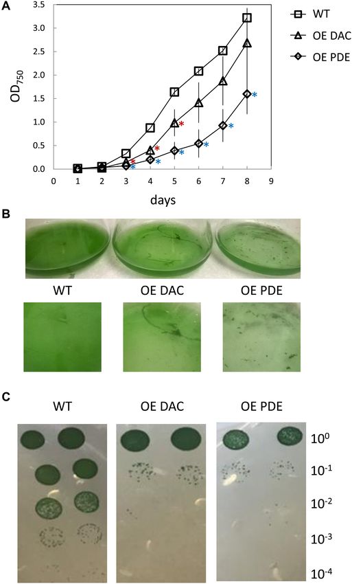
Previously, it has been observed that c-di-AMP homeostasis is
fundamental for optimal growth, with either lower than normal
levels or overaccumulation relative to WT resulting in defects in
growth (Corrigan et al., 2013; Mehne et al., 2013; Ye et al., 2014;
Gundlach et al., 2015; Rubin et al., 2018). Our overexpressing
strains both exhibited a lag in growth compared to WT in
BG-11/HEPES medium (Figure 5A). Additionally, WT grew
FIGURE 2 | Genomic context and RT-PCR analyses of conserved homogenously in the medium, whereas DAC and PDE strains
DAC-containing operon in Synechocystis. (A) Genomic context of conserved formed distinct aggregates in the late part of the growth curve
DAC (sll0505) in Synechocystis. Lines (1–6) below genomic region indicate
analysis (Figure 5B). This aggregation may impact analysis of
PCR products amplified from cDNA in panel B. RT-PCR of DAPDC (sll0504),
DAC (sll0505), and UPS (sll0506) in Synechocystis. (B) RT-PCR analyses of growth by measuring culture optical density. Thus, we also
transcripts produced from the DAC genomic region in Synechocystis: Lanes 1 assessed growth using dilution-based colony growth assays on
and 7, DAPDC; Lanes 2 and 8, DAC; Lanes 3 and 9, UPS; Lanes 4 and 10, agar plates. In this assay, we similarly observed that the OE DAC
DAPDC and DAC; Lanes 5 and 11, DAC and UPS; Lanes 6 and 12, DAPDC, and OE PDE strains exhibited impaired growth relative to WT
DAC, and UPS. Lanes 1–6, RT-PCR reactions; Lanes 7–12, no RT negative
control reactions. M, molecular marker. RNA was isolated from cells grown at
(Figure 5C).
35 µmol m−2 s−1 white light. Given the association of the c-di-AMP synthesis gene
with other genes associated with peptidoglycan synthesis or
modification and the cellular aggregation phenotypes, c-di-
AMP accumulation in cells may impact cell wall synthesis
Overexpression of Native DAC-Encoding or modification. We, thus, tested for alterations in cell wall
sll0505 and PDE-Encoding slr0104 Genes properties for the OE strains relative to WT by conducting
in Synechocystis lysozyme sensitivity assays. Both the OE DAC strain and OE PDE
In Synechocystis there is only one DAC (PF02457, sll0505), which strain had increased sensitivity to lysozyme treatment relative
is related to cdaA/dacA, and one c-di-AMP PDE (PF07698, to WT, as measured by chlorophyll release into the supernatant
slr0104, belonging to the 7TM_7TMR_HD family), which is (Figure 6).
related to pgpH. In many bacteria c-di-AMP is essential for
survival (Commichau et al., 2015), and in accordance with
this observation, we were not able to produce a mutant
Osmotic and Salt Stresses Impact
completely lacking sll0505 in Synechocystis (data not shown). c-di-AMP Levels in Multiple
We, thus, decided to overexpress the c-di-AMP DAC and PDE Cyanobacteria
native enzymes in Synechocystis to investigate their activity Based on the observation that c-di-AMP-binding riboswitches
in vivo. Quantitative RT-PCR showed increased accumulation have a putative role in osmoprotectant synthesis and transport
of the mRNA for DAC and PDE genes in the DAC and PDE in cyanobacteria (Nelson et al., 2013), c-di-AMP could be
overexpression strains, respectively (Figure 3). The DAC strain critical for osmotic or salt stress responses in these organisms.
exhibited an ∼40-fold increase in the level of DAC mRNA Osmoprotectants have recognized roles in cellular responses
accumulation compared to WT (Figure 3A), whereas the PDE to both osmotic and salt stresses (Bougouffa et al., 2014).
strain had a more than 200-fold increase in PDE mRNA levels Thus, we quantified changes in DAC and PDE mRNA
compared to WT (Figure 3B). levels in WT under osmotic and salt stresses, the latter of
Quantification of c-di-AMP levels in the DAC and PDE which induces both osmotic and ionic stress. After 24 h
overexpression strains was conducted to assess whether these two of osmotic or salt stress, levels of DAC and PDE mRNA
native enzymes could modulate intracellular levels of c-di-AMP decreased compared to non-stress, control conditions although
(Figure 4). The levels of c-di-AMP in the DAC overexpression differences were not statistically significant (Figure 3). The
strain were significantly altered, i.e., 1.7-fold higher (p < 0.05), DAC mRNA levels decreased to 0.3- and 0.7-fold relative to
compared to the WT strain. Whereas this is lower than might WT under sorbitol and salt stress, respectively (Figure 3A).
be anticipated based on gene expression data, some bacterial Similarly, the PDE mRNA levels decreased to 0.5- and 0.7-
strains which overaccumulate DAC mRNA show no upregulation fold relative to WT under sorbitol and salt stress, respectively
of intracellular c-di-AMP levels suggesting possible negative (Figure 3B).
Frontiers in Microbiology | www.frontiersin.org 6 May 2018 | Volume 9 | Article 1121Agostoni et al. Second Messengers in Cyanobacterial Abiotic Stress
FIGURE 3 | Quantitative reverse transcriptase PCR (qRT-PCR) analysis of the expression of DAC and PDE genes in Synechocystis. (A) DAC and (B) PDE genes
were analyzed for strains grown under 35 µmol m−2 s−1 white light. Wild-type (WT), overexpression of DAC (sll0505) in WT (OE DAC), overexpression of PDE
(slr0104) in WT (OE PDE), WT under sorbitol stress (sorbitol, 0.2 M sorbitol), WT under salt stress (salt, 0.6 M NaCl). The transcript level of the rnpB gene was used
as an internal control for each sample. Bars represent averages (± standard deviations). Bars marked with different letters are significantly different (p < 0.05).
Numbers below graphs represent fold difference relative to WT grown under non-stress conditions.
was no significant difference in intracellular levels of c-di-
AMP in another unicellular strain Synechococcus in response to
osmotic stress (Figure 7A). In the filamentous cyanobacterium
F. diplosiphon, c-di-AMP levels were much higher than for the
other cyanobacterial strains and increased ∼2-fold under osmotic
stress, whereas sorbitol treatment did not result in an increase
in c-di-AMP under our conditions in Anabaena (Figure 7A).
Changes in the intracellular concentration of c-di-AMP occur
during osmotic stress in Synechocystis and F. diplosiphon. We
also investigated whether c-di-AMP levels varied in these four
cyanobacterial species under salt stress (Figure 7A). Under
salt stress, c-di-AMP levels were not impacted in unicellular
strains, whereas levels were lower in F. diplosiphon and higher
in Anabaena in the presence of salt.
In cyanobacteria, biofilm formation is a protective mechanism
against salt stress (Jittawuttipoka et al., 2013). We previously
FIGURE 4 | Cyclic di-AMP levels in Synechocystis in replete BG-11/HEPES
demonstrated that induction of biofilm formation is under
medium under white light. WT, wild-type; OE DAC, strain overexpressing the the control of c-di-GMP in Synechocystis (Agostoni et al.,
native DAC (sll0505); OE PDE, strain overexpressing the native PDE (slr0104). 2016). To investigate whether c-di-GMP is elevated under
Bars represent averages (± standard deviations). Bars marked with different salt stress conditions that are associated with cyanobacterial
letters are significantly different (p < 0.05). Numbers below graphs represent
biofilm formation, we quantified c-di-GMP levels in response
fold difference relative to WT.
to treatment with salt. Indeed, c-di-GMP levels increased in all
four species after 24 h of salt stress (Figure 7B). In contrast
to stress caused by salt, the levels of c-di-GMP did not vary in
To determine whether these stresses influence intracellular Synechocystis, Synechococcus, and Anabaena under sorbitol stress
c-di-AMP levels and whether stress-induced changes occur in (Figure 7B). However, c-di-GMP levels significantly increased
cyanobacteria beyond Synechocystis, we exposed Synechocystis under sorbitol stress in F. diplosiphon (Figure 7B). Taken
and three additional species of cyanobacteria to sorbitol or salt together, these analyses suggest that c-di-AMP responds to
stress for 24 h. Two were unicellular cyanobacteria, moderately osmotic and salt stress, although generally an increase in c-di-
halotolerant Synechocystis (Reed et al., 1985) and salt-sensitive AMP levels is associated with sorbitol-induced osmotic stress (if
Synechococcus (Kaku et al., 2000), and two were filamentous there is a response). By contrast, levels of c-di-GMP primarily
cyanobacteria, highly salt-sensitive Anabaena (Rai and Tiwari, respond to ionic rather than osmotic stress across cyanobacterial
2001) and salt-sensitive F. diplosiphon (Singh and Montgomery, species, with an increase in c-di-GMP levels observed in all tested
2013b). C-di-AMP levels were threefold higher under sorbitol- strains in response to salt stress. The distinct responses of these
induced osmotic stress in Synechocystis. By comparison, there cyanobacterial strains to salt and osmotic stresses may reflect
Frontiers in Microbiology | www.frontiersin.org 7 May 2018 | Volume 9 | Article 1121Agostoni et al. Second Messengers in Cyanobacterial Abiotic Stress
FIGURE 6 | Comparison of lysozyme-dependent lysis of Synechocystis
wild-type (WT), strain overexpressing diadenylyl cyclase (OE DAC), and strain
overexpressing c-di-AMP phosphodiesterase (OE PDE). All strains were
grown in BG-11/HEPES under 15 µmol m−2 s−1 white light. Cells were
treated with 10 µg mL−1 lysozyme for 30 min. Cellular lysis was measured
based on chlorophyll released from cells at measured by absorbance at
660 nm in the supernatant after centrifugation. Bars represent averages
(± standard deviations). Bars marked with different letters are significantly
different (p < 0.05).
as high light and salt on cyanobacterial growth (Lu and Zhang,
2000). We first noted that, although not directly comparable
due to changing multiple factors, growth under lower light (i.e.,
∼15 µmol m−2 s−1 ) resulted in a significant impairment only
of the OE PDE strain compared to WT compared to growth
under higher white light (i.e., 35 µmol m−2 s−1 ). Additionally,
OE DAC and OE PDE cells exposed to low light lacked a major
lag in growth relative to WT that was observed at higher white
light levels (compare Figure 5A and Figure 8A). Osmotic stress
conditions had minor effects, with only the OE PDE strain having
FIGURE 5 | Growth curve of Synechocystis wild-type (WT), strain
statistically significant, transiently improved growth relative to
overexpressing diadenylyl cyclase (OE DAC), and strain overexpressing
c-di-AMP phosphodiesterase (OE PDE). (A) All strains were grown in WT (Figure 8B). However, both OE PDE and OE DAC strains
BG-11/HEPES under 35 µmol m−2 s−1 white light. Data points represent exhibited an impairment in growth relative to WT in the presence
averages (± standard deviations). ∗ p < 0.05 with red asterisks indicating of salt (Figure 8C). The significant changes in growth under
comparison between OE DAC and WT and blue asterisks comparing OE PDE salt in the OE DAC and OE PDE strains relative to WT were
and WT. (B) Picture of representative WT, OE DAC, and OE PDE cultures.
associated with altered intracellular c-di-AMP levels. The OE
(C) Images of WT, OE DAC, and OE PDE growth as measured by spotting of
cells (10 µL) of a 1:10 serial dilution on BG-11/HEPES plates containing 1% DAC strain exhibited significantly higher intracellular c-di-AMP
(w/v) agar under white light. Cells were initially at an OD750 of 0.6 in the levels compared to WT, whereas c-di-AMP levels in the OE PDE
undiluted culture and cells diluted up to 1:10,000-fold. strain were below detection when strains were grown in the
presence of NaCl (Figure 9).
their distinct sensitivities to salt (Reed et al., 1985; Kaku et al., DISCUSSION
2000; Rai and Tiwari, 2001; Singh and Montgomery, 2013b), as
well as other aspects of their unique ecological histories. However, Given that the genus Gloeobacter, which is considered the most
the production of c-di-AMP in a range of cyanobacterial species primordial of extant cyanobacteria (Rippka et al., 1974), possesses
is evident. two DAC enzymes, in contrast with the majority of cyanobacterial
Given that at least osmotic stress results in an alteration species that have only have one DAC enzyme, we speculate
of intracellular c-di-AMP levels in Synechocystis, we queried that cyanobacteria initially contained two DAC enzymes and
whether strains with overexpression of DAC or PDE genes that during evolution the second DAC was lost. Since the
exhibited altered growth responses due to applied abiotic stresses. conserved DAC, DAPDC and the UPS cassette (i.e., lysA-cdaA-
In these analyses, we reduced the intensity of white light to which uppS operon) is extremely conserved among cyanobacteria,
cells were exposed (compared to results shown in Figure 5) due uniquely, and both the DAC and UPS have associations with
to the noted combined detrimental effect of multiple stresses such peptidoglycan synthesis, these three genes together likely play a
Frontiers in Microbiology | www.frontiersin.org 8 May 2018 | Volume 9 | Article 1121Agostoni et al. Second Messengers in Cyanobacterial Abiotic Stress
FIGURE 7 | Cyclic di-AMP and cyclic di-GMP levels under sorbitol and salt stresses. (A) Cyclic di-AMP and (B) cyclic di-GMP levels normalized to total proteins
were determined for Synechocystis sp. PCC 6803 (Synechocystis), Synechococcus elongatus sp. PCC 7942 (Synechococcus), Fremyella diplosiphon
(F. diplosiphon), and Anabaena sp. PCC 7120 (Anabaena) grown under 35 µmol m−2 s−1 white light. Bars represent averages (± standard deviations). Bars marked
with different letters indicate a significant difference (p < 0.05) for comparisons made within each species.
critical role in controlling cell wall synthesis across cyanobacteria. Zhang and He, 2013; Tang et al., 2015). Indeed, the DAC and
This is a role consistent with prior studies which indicated that PDE overexpression strains exhibited increased sensitivity to
c-di-AMP metabolism impacts cell wall structure or stability in lysozyme as compared to the WT parent strain. The results
multiple, largely pathogenic, bacteria (Commichau et al., 2017). for the OE PDE strain are in accordance with prior analyses
However, the conserved operon structure suggest an important showing that decreased c-di-AMP levels have previously been
and well-conserved function for c-di-AMP in peptidoglycan- implicated in cell wall sensitivity and altered cell lysis for a
dependent processes in cyanobacteria. number of bacteria (Luo and Helmann, 2011; Witte et al.,
We were able to strongly increase transcript accumulation of 2013). On the other hand, the increased sensitivity observed
DAC and PDE in Synechocystis using overexpression plasmids for the OE DAC strain may be associated with the fact that
and demonstrate a significant impact on intracellular c-di-AMP the DAC/cdaA gene is in an operon with genes associated
levels in the OE DAC strain in BG-11 and significant impacts on with cell wall synthesis/modification. Specifically, the role of the
c-di-AMP homeostasis in both the OE DAC and OE PDE strains DADPC gene in crosslinking of peptidoglycan corresponds to
grown in the presence of salt. Higher or lower levels of c-di-AMP prior associations of c-di-AMP levels with peptidoglycan cross-
are both detrimental to normal growth in Bacillus subtilis, Listeria linking (Luo and Helmann, 2011). Thus, the altered phenotype
monocytogenes, Borrelia burgdorferi, and Staphylococcus aureus of the OE DAC strain could be related to associated impacts of
(Corrigan et al., 2011; Dengler et al., 2013; Mehne et al., 2013; altering c-di-AMP accumulation on peptidoglycan crosslinking,
Witte et al., 2013; Ye et al., 2014). Also, reduced levels of c-di- which in turn could alter cellular responses to lysozyme.
AMP have been recently shown to impact cyanobacterial growth Cyclic di-AMP homeostasis is critical in replete BG-11
in a Synechococcus strain in which cdaA was deleted (Rubin medium. Additionally, the regulation of DAC and PDE genes in
et al., 2018). In B. subtilis the differences in growth rates between osmotic and salt stress, especially the latter, appears to contribute
WT and a strain with strong accumulation of c-di-AMP were to cell fitness. Whereas WT has impaired growth in both stresses,
attributed to aberrant cell morphologies (Mehne et al., 2013). changes in intracellular c-di-AMP homeostasis is important for
Given that the cdaA gene is next to a gene encoding GlmM cellular responses to salt stress as both the DAC and PDE OE
that is essential for peptidoglycan synthesis in B. subtilis, the strains, which have significantly higher and lower c-di-AMP
observed aberrant cell morphologies may be due to disruptions levels in the presence of salt, respectively, exhibited impaired
in c-di-AMP regulation of peptidogylan synthesis in the mutant growth over time in the presence of NaCl-induced stress.
compared to WT (Mehne et al., 2013). Mutation of the S. aureus Osmotic and ionic stresses are common in natural ecosystems
PDE GdpP resulted in increased peptidoglycan cross-linking (Williams, 1987; Kaushal et al., 2005; Durack et al., 2012; Canedo-
which was detrimental to growth (Corrigan et al., 2011). Arguelles et al., 2013; El-Akhal et al., 2013) and identifying
Here, Synechocystis strains with overexpression of either DAC the mechanisms by which cyanobacteria can tolerate osmotic
or PDE grew slower than WT. Notably, the DAC and PDE and ionic stresses is critical. Species able to maintain osmotic
overexpression strains formed aggregates later in the growth equilibrium under these conditions will prove most beneficial
analysis compared to WT which grew homogenously in the for use in cyanobacterial mass cultivation (Hagemann, 2011).
medium throughout. These phenotypes may be related to the There is still a lack of knowledge on the mechanisms used by
observed changes in cell morphology or cell wall integrity cyanobacteria to specifically sense and respond to osmotic and
of c-di-AMP mutants in several bacteria (Corrigan et al., ionic stresses. Experiments described here show that sorbitol is
2011; Bai et al., 2013; Mehne et al., 2013; Witte et al., 2013; an important factor in the regulation of c-di-AMP homeostasis,
Frontiers in Microbiology | www.frontiersin.org 9 May 2018 | Volume 9 | Article 1121Agostoni et al. Second Messengers in Cyanobacterial Abiotic Stress
FIGURE 9 | Cyclic di-AMP levels in Synechocystis in replete BG-11/HEPES
medium containing 0.6 M NaCl under low white light at 15 µmol m−2 s−1 .
WT, wild-type; OE DAC, strain overexpressing the native DAC (sll0505); OE
PDE, strain overexpressing the native PDE (slr0104). Bars represent averages
(± standard deviations). Bars marked with different letters are significantly
different (p < 0.05).
(Nelson et al., 2013). Our results suggest a role in response
to salt stress in Synechocystis. Determining the molecular
mechanisms of c-di-AMP and c-di-GMP signaling networks
during cyanobacterial adaptation is necessary to understand how
cyanobacteria survive in stressful and fluctuating environments
and ensure improved biomass and product yields under osmotic
and ionic stresses to improve applications and fundamental
research in solving environmental problems.
AUTHOR CONTRIBUTIONS
MA and BM: conceived and designed the experiments. MA,
AL-J, EH, GS, and EB: performed the experiments and
FIGURE 8 | Growth curve of Synechocystis wild-type (WT), strain contributed to data analysis. MA and BM: drafted the article. MA,
overexpressing diadenylyl cyclase (OE DAC), and strain overexpressing AL-J, CW, and BM: critically revised the article.
c-di-AMP phosphodiesterase (OE PDE) under low white light and in presence
of abiotic stresses. All strains were grown in BG-11/HEPES under
15 µmol m−2 s−1 white light in (A) BG-11 medium, (B) sorbitol-induced
osmotic stress (0.5 M sorbitol), or (C) salt stress (0.6 M NaCl). Data points
FUNDING
represent averages (± standard deviations). ∗ p < 0.05 with red asterisks
indicating comparison between OE DAC and WT and blue asterisks This work was supported by the National Science Foundation
comparing OE PDE and WT. (Grant No. MCB-1243983 to BM), the Michigan State University
Discretionary Funding Initiative (Funding I.D. No. MSU-
DFI 70222 to BM), the National Institutes of Health (Grant
whereas salt is a critical factor to regulate c-di-AMP synthesis and Nos. GM109259 and AI130554 to CW) and support to
c-di-GMP homeostasis. Prior studies with Gram-positive bacteria Marco Agostoni from the United States Department of Energy
have implicated c-di-AMP as critical during osmotic stress, based (Chemical Sciences, Geosciences and Biosciences Division, Office
on the identification of a potassium transporter and a regulator of Basic Energy Sciences, Office of Science, Grant No. DE-FG02-
of a K+ transporter as c-di-AMP receptors (Corrigan et al., 2011, 91ER20021 to BM). Participation of AL-J was additionally made
2013; Bai et al., 2014; Moscoso et al., 2015). Also, regulation possible by a predoctoral training award from Grant No. T32-
of c-di-AMP levels have been associated with salt sensitivity in GM110523 from National Institute of General Medical Sciences
Gram-positive strains (Smith et al., 2012; Dengler et al., 2013). of the National Institutes of Health and Alliance for Graduate
Notably, a study on c-di-AMP-binding riboswitches implicated Education and Professoriate (AGEP) Scholar Awards from the
c-di-AMP-dependent regulation of the synthesis and transport MSU Graduate School. The contents of this publication are solely
of osmoprotectants in cyanobacteria as critical for stress the responsibility of the authors and do not necessarily represent
responses, such as osmotic or salt stresses, in these organisms the official views of the NIGMS or NIH.
Frontiers in Microbiology | www.frontiersin.org 10 May 2018 | Volume 9 | Article 1121Agostoni et al. Second Messengers in Cyanobacterial Abiotic Stress
REFERENCES Commichau, F. M., Gibhardt, J., Halbedel, S., Gundlach, J., and Stülke, J. (2017).
A delicate connection: c-di-AMP affects cell integrity by controlling osmolyte
Agostoni, M., Koestler, B. J., Waters, C. M., Williams, B. L., and Montgomery, transport. Trends Microbiol. 26, 175–185. doi: 10.1016/j.tim.2017.1009.1003
B. L. (2013). Occurrence of cyclic di-GMP-modulating output domains in Corrigan, R. M., Abbott, J. C., Burhenne, H., Kaever, V., and Grundling, A. (2011).
cyanobacteria: an illuminating perspective. mBio 4:e00451-13. doi: 10.1128/ c-di-AMP Is a new second messenger in Staphylococcus aureus with a role in
mBio.00451-13 controlling cell size and envelope stress. PLoS Pathog. 7:e1002217. doi: 10.1371/
Agostoni, M., and Montgomery, B. L. (2014). Survival strategies in the aquatic and journal.ppat.1002217
terrestrial world: the impact of second messengers on cyanobacterial processes. Corrigan, R. M., Campeotto, I., Jeganathan, T., Roelofs, K. G., Lee, V. T., and
Life 4, 745–769. doi: 10.3390/life4040745 Gründling, A. (2013). Systematic identification of conserved bacterial c-di-
Agostoni, M., Waters, C. M., and Montgomery, B. L. (2016). Regulation of biofilm AMP receptor proteins. Proc. Natl. Acad. Sci. U.S.A. 110, 9084–9089. doi: 10.
formation and cellular buoyancy through modulating intracellular cyclic di- 1073/pnas.1300595110
GMP levels in engineered cyanobacteria. Biotechnol. Bioeng. 113, 311–319. Corrigan, R. M., and Grundling, A. (2013). Cyclic di-AMP: another second
doi: 10.1002/bit.25712 messenger enters the fray. Nat. Rev. Microbiol. 11, 513–524. doi: 10.1038/
Allen, M. M. (1968). Simple conditions for growth of unicellular blue-green algae nrmicro3069
on plates. J. Phycol. 4, 1–4. doi: 10.1111/j.1529-8817.1968.tb04667.x Dengler, V., Mccallum, N., Kiefer, P., Christen, P., Patrignani, A., Vorholt, J. A.,
Angerer, V., Schwenk, P., Wallner, T., Kaever, V., Hiltbrunner, A., and Wilde, A. et al. (2013). Mutation in the c-di-AMP cyclase dacA affects fitness and
(2017). The protein Slr1143 is an active diguanylate cyclase in Synechocystis resistance of methicillin resistant Staphylococcus aureus. PLoS One 8:e73512.
sp. PCC 6803 and interacts with the photoreceptor Cph2. Microbiology 163, doi: 10.1371/journal.pone.0073512
920–930. doi: 10.1099/mic.0.000475 Dey, B., Dey, R. J., Cheung, L. S., Pokkali, S., Guo, H., Lee, J.-H., et al. (2015).
Apfel, C. M., Takacs, S., Fountoulakis, M., Stieger, M., and Keck, W. (1999). Use of A bacterial cyclic dinucleotide activates the cytosolic surveillance pathway and
genomics to identify bacterial undecaprenyl pyrophosphate synthetase: cloning, mediates innate resistance to tuberculosis. Nat. Med. 21, 401–406. doi: 10.1038/
expression, and characterization of the essential uppS gene. J. Bacteriol. 181, nm.3813
483–492. Du, B., Ji, W., An, H., Shi, Y., Huang, Q., Cheng, Y., et al. (2014). Functional
Bai, Y., Yang, J., Eisele, L. E., Underwood, A. J., Koestler, B. J., Waters, C. M., analysis of c-di-AMP phosphodiesterase, GdpP, in Streptococcus suis serotype
et al. (2013). Two DHH subfamily 1 proteins in Streptococcus pneumoniae 2. Microbiol. Res. 169, 749–758. doi: 10.1016/j.micres.2014.01.002
possess cyclic Di-AMP phosphodiesterase activity and affect bacterial growth Durack, P. J., Wijffels, S. E., and Matear, R. J. (2012). Ocean salinities reveal strong
and virulence. J. Bacteriol. 195, 5123–5132. doi: 10.1128/jb.00769-13 global water cycle intensification during 1950 to 2000. Science 336, 455–458.
Bai, Y., Yang, J., Zarrella, T. M., Zhang, Y., Metzger, D. W., and Bai, G. (2014). doi: 10.1126/science.1212222
Cyclic di-AMP impairs potassium uptake mediated by a cyclic di-AMP binding Edgar, R. C. (2004). MUSCLE: multiple sequence alignment with high accuracy and
protein in Streptococcus pneumoniae. J. Bacteriol. 196, 614–623. doi: 10.1128/jb. high throughput. Nucleic Acids Res. 32, 1792–1797. doi: 10.1093/nar/gkh340
01041-13 El-Akhal, M. R., Rincon, A., Coba De La Pena, T., Lucas, M. M., El Mourabit, N.,
Barker, J. R., Koestler, B. J., Carpenter, V. K., Burdette, D. L., Waters, C. M., Barrijal, S., et al. (2013). Effects of salt stress and rhizobial inoculation on
Vance, R. E., et al. (2013). STING-dependent recognition of cyclic di-AMP growth and nitrogen fixation of three peanut cultivars. Plant Biol. 15, 415–421.
mediates type I interferon responses during Chlamydia trachomatis infection. doi: 10.1111/j.1438-8677.2012.00634.x
mBio 4:e00018-13. doi: 10.1128/mBio.00018-13 Enomoto, G., Ni-Ni-Win, Narikawa, R., and Ikeuchi, M. (2015). Three
Bejerano-Sagie, M., Oppenheimer-Shaanan, Y., Berlatzky, I., Rouvinski, A., cyanobacteriochromes work together to form a light color-sensitive input
Meyerovich, M., and Ben-Yehuda, S. (2006). A checkpoint protein that scans system for c-di-GMP signaling of cell aggregation. Proc. Natl. Acad. Sci. U.S.A.
the chromosome for damage at the start of sporulation in Bacillus subtilis. Cell 112, 8082–8087. doi: 10.1073/pnas.1504228112
125, 679–690. doi: 10.1016/j.cell.2006.03.039 Enomoto, G., Nomura, R., Shimada, T., Ni-Ni Win, Narikawa, R., and Ikeuchi, M.
Bougouffa, S., Radovanovic, A., Essack, M., and Bajic, V. B. (2014). DEOP: a (2014). Cyanobacteriochrome SesA is a diguanylate cyclase that induces
database on osmoprotectants and associated pathways. Database 2014:bau100. cell aggregation in Thermosynechococcus. J. Biol. Chem. 289, 24801–24809.
doi: 10.1093/database/bau100 doi: 10.1074/jbc.M114.583674
Bowman, L., Zeden, M. S., Schuster, C. F., Kaever, V., and Gründling, A. Fu, T., Zhao, Y., and Xi, J. (2016). A new second messenger: bacterial
(2016). New insights into the cyclic Di-adenosine monophosphate (c-di-AMP) c-di-AMP. Crit. Rev. Eukaryot. Gene Expr. 26, 309–316. doi: 10.1615/
degradation pathway and the requirement of the cyclic dinucleotide for acid CritRevEukaryotGeneExpr.2016016642
stress resistance in Staphylococcus aureus. J. Biol. Chem. 291, 26970–26986. Galtier, N., Gouy, M., and Gautier, C. (1996). SeaView and Phylo_win: two graphic
doi: 10.1074/jbc.M116.747709 tools for sequence alignment and molecular phylogeny. Comput. Appl. Biosci.
Bukhari, A. I., and Taylor, A. L. (1971a). Genetic analysis of diaminopimelic acid- 12, 543–548. doi: 10.1093/bioinformatics/12.6.543
and lysine-requiring mutants of Escherichia coli. J. Bacteriol. 105, 844–854. Gándara, C., and Alonso, J. C. (2015). DisA and c-di-AMP act at the intersection
Bukhari, A. I., and Taylor, A. L. (1971b). Mutants of Escherichia coli with a growth between DNA-damage response and stress homeostasis in exponentially
requirement for either lysine or pyridoxine. J. Bacteriol. 105, 988–998. growing Bacillus subtilis cells. DNA Repair 27, 1–8. doi: 10.1016/j.dnarep.2014.
Cadoret, J.-C., Rousseau, B., Perewoska, I., Sicora, C., Cheregi, O., Vass, I., et al. 12.007
(2005). Cyclic nucleotides, the photosynthetic apparatus and response to a UV- Gándara, C., De Lucena, D. K. C., Torres, R., Serrano, E., Altenburger, S.,
B stress in the cyanobacterium Synechocystis sp. PCC 6803. J. Biol. Chem. 280, Graumann, P. L., et al. (2017). Activity and in vivo dynamics of Bacillus subtilis
33935–33944. DisA are affected by RadA/Sms and by Holliday junction-processing proteins.
Canedo-Arguelles, M., Kefford, B. J., Piscart, C., Prat, N., Schafer, R. B., and Schulz, DNA Repair 55, 17–30. doi: 10.1016/j.dnarep.2017.05.002
C. J. (2013). Salinisation of rivers: an urgent ecological issue. Environ. Pollut. Garcia-Pichel, F., Belnap, J., Neuer, S., and Schanz, F. (2003). Estimates of global
173, 157–167. doi: 10.1016/j.envpol.2012.10.011 cyanobacterial biomass and its distribution. Algol. Stud. 109, 213–227. doi:
Cho, K. H., and Kang, S. O. (2013). Streptococcus pyogenes c-di-AMP 10.1127/1864-1318/2003/0109-0213
phosphodiesterase. GdpP, influences SpeB processing and virulence. PLoS One Gundlach, J., Mehne, F. M., Herzberg, C., Kampf, J., Valerius, O., Kaever, V., et al.
8:e69425. doi: 10.1371/journal.pone.0069425 (2015). An essential poison: synthesis and degradation of cyclic di-AMP in
Cobley, J. G., Zerweck, E., Reyes, R., Mody, A., Seludounson, J. R., Jaeger, H., et al. Bacillus subtilis. J. Bacteriol. 197, 3265–3274. doi: 10.1128/jb.00564-15
(1993). Construction of shuttle plasmids which can be efficiently mobilized Hagemann, M. (2011). Molecular biology of cyanobacterial salt acclimation. FEMS
from Escherichia coli into the chromatically adapting cyanobacterium, Microbiol. Rev. 35, 87–123. doi: 10.1111/j.1574-6976.2010.00234.x
Fremyella diplosiphon. Plasmid 30, 90–105. doi: 10.1006/plas.1993.1037 Huynh, T. N., Luo, S., Pensinger, D., Sauer, J. D., Tong, L., and Woodward, J. J.
Commichau, F. M., Dickmanns, A., Gundlach, J., Ficner, R., and Stülke, J. (2015). (2015). An HD-domain phosphodiesterase mediates cooperative hydrolysis of
A jack of all trades: the multiple roles of the unique essential second messenger c-di-AMP to affect bacterial growth and virulence. Proc. Natl. Acad. Sci. U.S.A.
cyclic di-AMP. Mol. Microbiol. 97, 189–204. doi: 10.1111/mmi.13026 112, E747–E756. doi: 10.1073/pnas.1416485112
Frontiers in Microbiology | www.frontiersin.org 11 May 2018 | Volume 9 | Article 1121Agostoni et al. Second Messengers in Cyanobacterial Abiotic Stress
Huynh, T. N., and Woodward, J. J. (2016). Too much of a good thing: regulated Nelson, J. W., Sudarsan, N., Furukawa, K., Weinberg, Z., Wang, J. X., and Breaker,
depletion of c-di-AMP in the bacterial cytoplasm. Curr. Opin. Microbiol. 30, R. R. (2013). Riboswitches in eubacteria sense the second messenger c-di-AMP.
22–29. doi: 10.1016/j.mib.2015.12.007 Nat. Chem. Biol. 9, 834–839. doi: 10.1038/nchembio.1363
Imashimizu, M., Yoshimura, H., Katoh, H., Ehira, S., and Ohmori, M. (2005). Ochoa De Alda, J. A. G., Ajlani, G., and Houmard, J. (2000). Synechocystis strain
NaCl enhances cellular cAMP and upregulates genes related to heterocyst PCC 6803 cya2, a prokaryotic gene that encodes a guanylyl cyclase. J. Bacteriol.
development in the cyanobacterium, Anabaena sp. strain PCC 7120. FEMS 182, 3839–3842. doi: 10.1128/jb.182.13.3839-3842.2000
Microbiol. Lett. 252, 97–103. doi: 10.1016/j.femsle.2005.08.035 Ohmori, M., Ikeuchi, M., Sato, N., Wolk, P., Kaneko, T., Ogawa, T., et al. (2001).
Jenal, U., Reinders, A., and Lori, C. (2017). Cyclic di-GMP: second messenger Characterization of genes encoding multi-domain proteins in the genome of
extraordinaire. Nat. Rev. Microbiol. 15, 271–284. doi: 10.1038/nrmicro.2016.190 the filamentous nitrogen-fixing cyanobacterium Anabaena sp. strain PCC 7120.
Jittawuttipoka, T., Planchon, M., Spalla, O., Benzerara, K., Guyot, F., Cassier- DNA Res. 8, 271–284.
Chauvat, C., et al. (2013). Multidisciplinary evidences that Synechocystis Ohmori, M., Ohmori, K., and Hasunuma, K. (1988). Rapid change in cyclic
PCC6803 exopolysaccharides operate in cell sedimentation and protection 30 , 50 -AMP concentration triggered by a light-off or light-on signal in
against salt and metal stresses. PLoS One 8:e55564. doi: 10.1371/journal.pone. Anabaena cylindrica. Arch. Microbiol. 150, 203–204. doi: 10.1007/BF00
0055564 425163
Jones, D. T., Taylor, W. R., and Thornton, J. M. (1992). The rapid generation Ohmori, M., and Okamoto, S. (2004). Photoresponsive cAMP signal transduction
of mutation data matrices from protein sequences. Comput. Appl. Biosci. 8, in cyanobacteria. Photochem. Photobiol. Sci. 3, 503–511. doi: 10.1039/b401623h
275–282. doi: 10.1093/bioinformatics/8.3.275 Ohmori, M., Terauchi, K., Okamoto, S., and Watanabe, M. (2002). Regulation
Kaku, N., Hibino, T., Tanaka, Y., Ishikawa, H., Araki, E., Takabe, T., et al. (2000). of cAMP-mediated photosignaling by a phytochrome in the cyanobacterium
Effects of overexpression of Escherichia coli katE and bet genes on the tolerance Anabaena cylindrica. Photochem. Photobiol. 75, 675–679. doi: 10.1562/0031-
for salt stress in a freshwater cyanobacterium Synechococcus sp. PCC 7942. Plant 8655(2002)0752.0.CO;2
Sci. 159, 281–288. Okamoto, S., Kasahara, M., Kamiya, A., Nakahira, Y., and Ohmori, M. (2004).
Katayama, M., and Ohmori, M. (1997). Isolation and characterization of multiple A phytochrome-like protein AphC triggers the cAMP signaling induced by
adenylate cyclase genes from the cyanobacterium Anabaena sp. strain PCC far-red light in the cyanobacterium Anabaena sp. strain PCC7120. Photochem.
7120. J. Bacteriol. 179, 3588–3593. doi: 10.1128/jb.179.11.3588-3593.1997 Photobiol. 80, 429–433. doi: 10.1562/0031-8655(2004)0802.0.
Kaushal, S. S., Groffman, P. M., Likens, G. E., Belt, K. T., Stack, W. P., Kelly, CO;2
V. R., et al. (2005). Increased salinization of fresh water in the northeastern Oppenheimer-Shaanan, Y., Wexselblatt, E., Katzhendler, J., Yavin, E., and Ben-
United States. Proc. Natl. Acad. Sci. U.S.A. 102, 13517–13520. doi: 10.1073/pnas. Yehuda, S. (2011). c-di-AMP reports DNA integrity during sporulation in
0506414102 Bacillus subtilis. EMBO Rep. 12, 594–601. doi: 10.1038/embor.2011.77
Kloft, N., Rasch, G., and Forchhammer, K. (2005). Protein phosphatase Pade, N., and Hagemann, M. (2015). Salt acclimation of cyanobacteria and their
PphA from Synechocystis sp. PCC 6803: the physiological framework of application in biotechnology. Life 5, 25–49. doi: 10.3390/life5010025
PII-P dephosphorylation. Microbiology 151, 1275–1283. doi: 10.1099/mic.0. Paithoonrangsarid, K., Shoumskaya, M. A., Kanesaki, Y., Satoh, S., Tabata, S.,
27771-0 Los, D. A., et al. (2004). Five histidine kinases perceive osmotic stress and
Krasteva, P. V., and Sondermann, H. (2017). Versatile modes of cellular regulation regulate distinct sets of genes in Synechocystis. J. Biol. Chem. 279, 53078–53086.
via cyclic dinucleotides. Nat. Chem. Biol. 13, 350–359. doi: 10.1038/nchembio. doi: 10.1074/jbc.M410162200
2337 Pattanaik, B., and Montgomery, B. L. (2010). FdTonB is involved in the
Lu, C., and Zhang, J. (2000). Role of light in the response of PSII photochemistry photoregulation of cellular morphology during complementary chromatic
to salt stress in the cyanobacterium Spirulina platensis. J. Exp. Bot. 51, 911–917. adaptation in Fremyella diplosiphon. Microbiology 156, 731–741. doi: 10.1099/
doi: 10.1093/jxb/51.346.911 mic.0.035410-0
Luo, Y., and Helmann, J. D. (2011). Analysis of the role of Bacillus subtilis σM Raguse, M., Torres, R., Seco, E. M., Gándara, C., Ayora, S., Moeller, R., et al. (2017).
in β-lactam resistance reveals an essential role for c-di-AMP in peptidoglycan Bacillus subtilis DisA helps to circumvent replicative stress during spore revival.
homeostasis. Mol. Microbiol. 83, 623–639. doi: 10.1111/j.1365-2958.2011. DNA Repair 59, 57–68. doi: 10.1016/j.dnarep.2017.09.006
07953.x Rai, A. K., and Tiwari, S. P. (2001). NO3 – nutrition and salt tolerance in the
Marin, K., Suzuki, I., Yamaguchi, K., Ribbeck, K., Yamamoto, H., Kanesaki, Y., et al. cyanobacterium Anabaena sp. PCC 7120 and mutant strains. J. Appl. Microbiol.
(2003). Identification of histidine kinases that act as sensors in the perception 86, 991–998. doi: 10.1046/j.1365-2672.1999.00788.x
of salt stress in Synechocystis sp. PCC 6803. Proc. Natl. Acad. Sci. U.S.A. 100, Reed, R. H., Richardson, D. L., and Stewart, W. D. P. (1985). Na+ uptake
9061–9066. doi: 10.1073/pnas.1532302100 and extrusion in the cyanobacterium Synechocystis PCC 6714 in response to
Massie, J. P., Reynolds, E. L., Koestler, B. J., Cong, J. P., Agostoni, M., and Waters, hypersaline treatment. Evidence for transient changes in plasmalemma Na+
C. M. (2012). Quantification of high-specificity cyclic diguanylate signaling. permeability. Biochim. Biophys. Acta 814, 347–355. doi: 10.1016/0005-2736(85)
Proc. Natl. Acad. Sci. U.S.A. 109, 12746–12751. doi: 10.1073/pnas.11156 90455-9
63109 Rippka, R., Waterbury, J., and Cohen-Bazire, G. (1974). A cyanobacterium which
Mehne, F. M., Schröder-Tittmann, K., Eijlander, R. T., Herzberg, C., Hewitt, L., lacks thylakoids. Arch. Microbiol. 100, 419–436. doi: 10.1007/bf00446333
Kaever, V., et al. (2014). Control of the diadenylate cyclase CdaS in Bacillus Rismondo, J., Gibhardt, J., Rosenberg, J., Kaever, V., Halbedel, S., and Commichau,
subtilis: an autoinhibitory domain limits cyclic di-AMP production. J. Biol. F. M. (2016). Phenotypes associated with the essential diadenylate cyclase CdaA
Chem. 289, 21098–21107. doi: 10.1074/jbc.M114.562066 and its potential regulator CdaR in the human pathogen Listeria monocytogenes.
Mehne, F. M. P., Gunka, K., Eilers, H., Herzberg, C., Kaever, V., and Stulke, J. J. Bacteriol. 198, 416–426. doi: 10.1128/jb.00845-15
(2013). Cyclic di-AMP homeostasis in Bacillus subtilis: both lack and high level Romling, U. (2008). Great times for small molecules: c-di-AMP, a second
accumulation of the nucleotide are detrimental for cell growth. J. Biol. Chem. messenger candidate in Bacteria and Archaea. Sci. Signal. 1:pe39. doi: 10.1126/
288, 2004–2017. doi: 10.1074/jbc.M112.395491 scisignal.133pe39
Mehta, K. K., Evitt, N. H., and Swartz, J. R. (2015). Chemical lysis of cyanobacteria. Rubin, B. E., Huynh, T. N., Welkie, D. G., Diamond, S., Simkovsky, R., Pierce,
J. Biol. Eng. 9:10. doi: 10.1186/s13036-015-0007-y E. C., et al. (2018). High-throughput interaction screens illuminate the role
Mikami, K., Kanesaki, Y., Suzuki, I., and Murata, N. (2002). The histidine kinase of c-di-AMP in cyanobacterial nighttime survival. PLoS Genet. 14:e1007301.
Hik33 perceives osmotic stress and cold stress in Synechocystis sp. PCC 6803. doi: 10.1371/journal.pgen.1007301
Mol. Microbiol. 46, 905–915. Savage, C. R., Arnold, W. K., Gjevre-Nail, A., Koestler, B. J., Bruger, E. L., Barker,
Moscoso, J. A., Schramke, H., Zhang, Y., Tosi, T., Dehbi, A., Jung, K., et al. J. R., et al. (2015). Intracellular concentrations of Borrelia burgdorferi cyclic di-
(2015). Binding of cyclic di-AMP to the Staphylococcus aureus sensor kinase AMP are not changed by altered expression of the cdaA synthase. PLoS One
KdpD occurs via the universal stress protein domain and downregulates 10:e0125440. doi: 10.1371/journal.pone.0125440
the expression of the Kdp potassium transporter. J. Bacteriol. 198, 98–110. Savakis, P., De Causmaecker, S., Angerer, V., Ruppert, U., Anders, K., Essen, L. O.,
doi: 10.1128/jb.00480-15 et al. (2012). Light-induced alteration of c-di-GMP level controls motility of
Frontiers in Microbiology | www.frontiersin.org 12 May 2018 | Volume 9 | Article 1121You can also read