Structural changes in brains of patients with disorders of consciousness treated with deep brain stimulation - Nature
←
→
Page content transcription
If your browser does not render page correctly, please read the page content below
www.nature.com/scientificreports
OPEN Structural changes in brains
of patients with disorders
of consciousness treated with deep
brain stimulation
Marina Raguž1,2*, Nina Predrijevac1, Domagoj Dlaka1, Darko Orešković1,2, Ante Rotim3,
Dominik Romić1, Fadi Almahariq1,2, Petar Marčinković1, Vedran Deletis4, Ivica Kostović2 &
Darko Chudy1,2,5
Disorders of consciousness (DOC) are one of the major consequences after anoxic or traumatic brain
injury. So far, several studies have described the regaining of consciousness in DOC patients using
deep brain stimulation (DBS). However, these studies often lack detailed data on the structural and
functional cerebral changes after such treatment. The aim of this study was to conduct a volumetric
analysis of specific cortical and subcortical structures to determine the impact of DBS after functional
recovery of DOC patients. Five DOC patients underwent unilateral DBS electrode implantation into
the centromedian parafascicular complex of the thalamic intralaminar nuclei. Consciousness recovery
was confirmed using the Rappaport Disability Rating and the Coma/Near Coma scale. Brain MRI
volumetric measurements were done prior to the procedure, then approximately a year after, and
finally 7 years after the implementation of the electrode. The volumetric analysis included changes
in regional cortical volumes and thickness, as well as in subcortical structures. Limbic cortices
(parahippocampal and cingulate gyrus) and paralimbic cortices (insula) regions showed a significant
volume increase and presented a trend of regional cortical thickness increase 1 and 7 years after
DBS. The volumes of related subcortical structures, namely the caudate, the hippocampus as well as
the amygdala, were significantly increased 1 and 7 years after DBS, while the putamen and nucleus
accumbens presented with volume increase. Volume increase after DBS could be a result of direct
DBS effects, or a result of functional recovery. Our findings are in accordance with the results of very
few human studies connecting DBS and brain volume increase. Which mechanisms are behind the
observed brain changes and whether structural changes are caused by consciousness recovery or DBS
in patients with DOC is still a matter of debate.
Human consciousness is often described as a complex phenomenon, consisting of two components, wakefulness
and awareness1,2. While wakefulness is associated with functional brainstem neurons i.e. the reticular system
projecting to both thalamic and cortical neurons, awareness is mostly related to the functional integrity of the
thalamus, cerebral cortex, and their c onnections1–4.
Disorders of consciousness (DOC) occur as a result of interference with the mentioned systems. DOC can
be acute and reversible, as a transient stage in a spontaneous recovery due to trauma, or chronic and irrevers-
ible, as in patients with unresponsive wakefulness syndrome (UWS)1–4. Prolonged DOC is not only long-last-
ing, serious, and currently untreatable, but also has severe consequences on a patient’s quality of life5–7. These
conditions impose a serious consideration from both medical and ethical perspectives7–9. Clinical conditions
such as anoxic or traumatic brain lesions can cause diffuse neuronal damage resulting in serious disorders
ranging from minimally conscious state (MCS), UWS, coma, locked-in syndrome, and even brain d eath1,10.
1,2
The boundaries separating the mentioned conditions are unclear, with each one including s everities . UWS is
currently described as wakefulness without consciousness, including a complete lack of reactions. Additionally,
1
Department of Neurosurgery, Dubrava University Hospital, Avenija Gojka Suska 6, 10000 Zagreb, Croatia. 2Centre
of Excellence for Basic, Clinical and Translational Neuroscience, Croatian Institute for Brain Research, University of
Zagreb, School of Medicine, Zagreb, Croatia. 3Department of Neurosurgery, Sestre milosrdnice University Hospital
Centre, Zagreb, Croatia. 4Albert Einstein College of Medicine, New York, USA. 5Department of Surgery, University
of Zagreb, School of Medicine, Zagreb, Croatia. *email: marinaraguz@gmail.com
Scientific Reports | (2021) 11:4401 | https://doi.org/10.1038/s41598-021-83873-y 1
Vol.:(0123456789)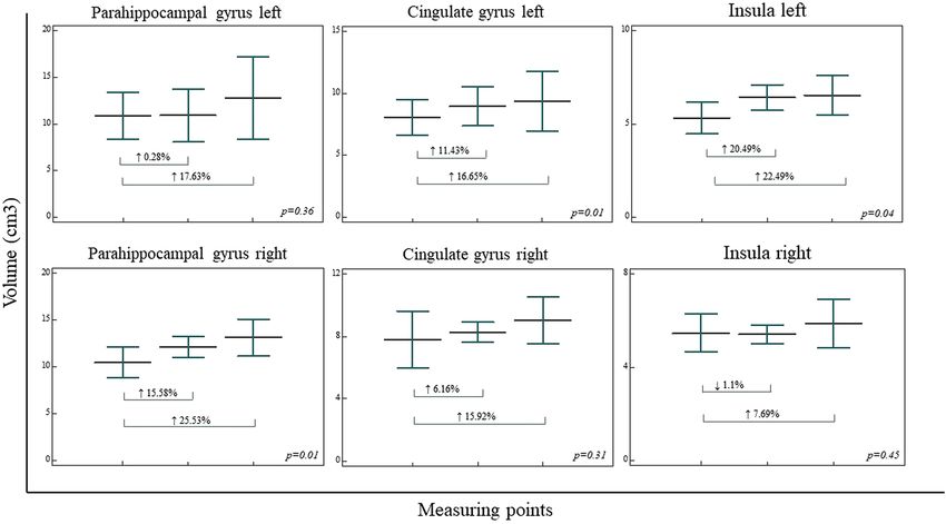
www.nature.com/scientificreports/
Level of awareness (RDR,
C/NC scale)
Time from initial Follow up duration
Patient no. Sex Etiology Age at Injury (years) injury to DBS (month) Before DBS After DBS (month)
1 ♂ CA 17 2 2.0/1, MCS 0, aware 111
2 ♂ CA 23 2 1.8/1, MCS 0, aware 57
3 ♀ TBI 15 11 1.6/1, MCS 0, aware 102
4 ♀ CA 16 4 2.6/2, UWS 0, aware 88
5 ♀ TBI 18 14 2.6/2, UWS 0, aware 23
Table 1. Demographic and clinical characteristics of consciousness improved patients after DBS. CA cardiac
arrest, TBI traumatic brain injury, RDR rappaport disability rating scale, C/NC coma/near coma scale, UWS
unresponsive wakefulness syndrome, MCS minimally conscious state.
it is characterized by variable cycles of sleeping and wakefulness, with preserved spontaneous respiration, diges-
tion, and t hermoregulation1,2,10–13. MCS is on the other hand characterized by inconsistent, but visible evidence
of consciousness1,14 with the patient being awake but unaware. A patient in MCS can follow instructions occa-
sionally, give simple gestural or non-verbal yes/no responses, and show a certain level of purposeful movement.
The incidence of UWS is roughly 5 to 25 per million, while the prevalence in the adult population in the USA
ranges from 40 to 168 per million15. Additionally, various neurological impairments occur, including seizures,
movement disorders, myoclonus, focal motor, and sensorimotor deficits, as well as emotional, behavioral, and
cognitive disturbances16,17.
Despite advances in diagnosis, improved classification, use of modern technology, and well-known pharma-
cological treatments alongside non-invasive brain stimulation, the surgical therapeutic approaches are still not
significantly advanced18,19. Several studies used deep brain stimulation (DBS) of certain nuclei, such as centro-
median parafascicle (CM-pf) complex and brain stem reticular formation, to regain consciousness in UWS and
MCS patients5,6,19–23. Research thus often focused on the thalamus as a functionally important thalamocortical
link, where stimulation through key thalamocortical and thalamus-forebrain circuitry was shown to be impor-
tant and caused widespread e ffect24. The use of the CM-pf complex as a target was based on literature overview
and clinical recommendation, as well as due to the connectivity and prospective effect on systems involved in
consciousness5,6,19–23,25. Previous studies were done on a small cohort, mainly as case reports, and did not yield a
definite answer whether this highly specific and refined method can be therapeutically successful5,6,19–23,26–30. Even
though some studies claimed that there was no definitive evidence of the efficacy of this method for treatment
of DOC patients, the usefulness of DBS was shown using controlled studies, as well as our previous s tudy6,22,23.
Virtually all of the studies mentioned are missing an explanation of the detailed structural and functional
cerebral changes induced by DBS. Recently, renewed interest in the grading of DOC and different trials of
therapeutic approaches have been noted. Previous studies observed short-term volume changes in the human
brain structure following DBS, potentially indicating restorative possibilities in patients with neurodegenerative
diseases31. Modern magnetic resonance imaging (MRI) techniques play an important role in diagnostic clas-
sification and evaluation of cerebral damage in order to improve the gradation of different types and levels of
DOC32. Moreover, MRI has been shown to be a potential tool for morphometric measurements and changes in
a restructured, “reorganized” brain in DOC p atients33,34. Modern neuroimaging thus uses different methods to
objectively evaluate both structural and functional changes as an effect of different treatments.
Previously, it has been documented that DBS changes function in such structures as the striatum, hippocam-
pus, amygdala, etc. It would thus arguably follow that if electrical stimulation changes the brain’s structure and
its function, stimulation of damaged circuitry could then induce both functional and structural changes with
an eventual improvement of f unction35–37.
To determine the impact of CM-pf stimulation on a possible brain structural reorganization after functional
recovery, we performed a quantitative volumetric MRI analysis in DOC patients who underwent CM-pf DBS.
Our study aimed to reveal if CM-pf DBS could induce an impact on brain structural reorganization in DOC
patients and lead to a changed volume of specific cortical and subcortical structures.
Methods
Patients. This retrospective study included five patients who underwent CM-pf DBS due to DOC after which
they achieved functional recovery (Table 1). Out of the five patients, three were female, average age 16 ± 1.53.
The cause of injury in one patient was anoxic due to cardiac arrest, while other two patients experienced trau-
matic injuries. Two patients were male with an average age of 20 ± 4.24 years. In both of these patients the cause
of injury was anoxia due to cardiac arrest. The length between the injury and time to DBS was 2 months in
males and 11 ± 5.13 months (range 4–14) in females. The average follow-up duration in female patients was
88 ± 38.17 months (range 30–102), while in male patients it was 84 ± 38.18 months (range 57–111).
In the present study we included patients previously described in Chudy et al.22,23. Forty-nine patients were
enrolled in the previous study. Fourteen patients (10 UWS and 4 MCS) fulfilled neurophysiologic, clinical, and
neuroimaging criteria and underwent DBS implantation. Four patients (3 MCS and 1 UWS) achieved functional
recovery, while other patients were further followed by an attending palliative physician22,23. In this study we
included one additional UWS patient who underwent DBS implantation after fulfilling the aforementioned
criteria and achieved functional recovery.
Scientific Reports | (2021) 11:4401 | https://doi.org/10.1038/s41598-021-83873-y 2
Vol:.(1234567890)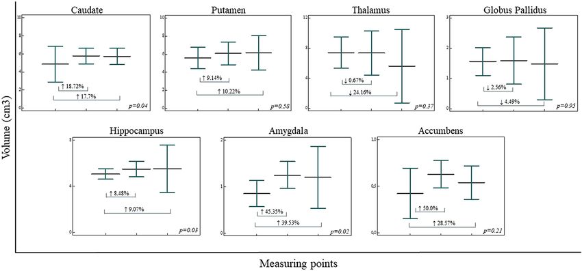
www.nature.com/scientificreports/
Figure 1. Initial structural MRI scans of included patients showing an absence of any large brain lesion
(patients 1–5, from left to right).
Patients selected for DBS were evaluated utilizing the standard Rappaport Disability Rating (RDR) scale and
the Coma/Near Coma (C/NC) s cale22,23,38. Inconsistently responsive to simple commands patients were classified
as MCS patients (C/NC Level 1), while patients who were not able to respond to any command were classified
as UWS (C/NC Level 2–4). DOC patients were selected based on three main factors: their neurophysiologic
evaluation, 12/24 h electroencephalography (EEG) and neuroimaging (MRI). The neurophysiological criteria
we investigated were recordable somatosensory evoked potential (SEP), motor evoked potentials, and brainstem
auditory evoked potentials, even with pathological parameters such as prolonged latencies or central conduc-
tion time. The entry criterion for SEPs was recordability via stimulation of median nerves, with or without SEPs
elicited by tibial nerve stimulation. The second main factor we looked at was EEG. The entry criterion for this
method was the presence of periods of desynchronized EEG activity during 12/24 h of monitoring processed
EEG22,23. Obtained neuroimaging (MRI) showed absence of a structural defect. After passing extensive selec-
tion criteria, the patients underwent DBS electrode implantation into the CM-pf complex of the left thalamic
intralaminar nuclei, while in patients with posttraumatic lesions, the electrode was placed in the better-preserved
hemisphere22,23. On the third postoperative day, monopolar stimulation was initiated using the contact eliciting
the strongest arousal response with minimal current (25 Hz frequency, 90 µs pulse duration, voltage 2.5–3.5 V).
Stimulation was applied for a 30-min period every two hours during the daytime. Stimulation duration was up
to 10 months after DBS electrode implantation. The informed parental/caregivers consent was obtained for all
patients in accordance with the Declaration of Helsinki and ethical approval was obtained from the Institutional
Review Board of the Dubrava University Hospital, School of Medicine, University of Zagreb, Croatia.
MRI acquisition. MRI scans were obtained on a 1.5 T MRI scanner (MAGNETOM Aera, Siemens Health-
ineers, Erlangen) using 24 channeled head coils. Standard clinical sequences were used, as well as high-res-
olution 3D T1-weighted magnetization-prepared rapid acquisition gradient echo (MPRAGE) sequence with
the following scanning parameters: TR = 2300 ms, TE = 3 ms, flip angle = 15°, matrix size = 256 × 256, field of
view = 256 mm, voxel size = 1.0 × 1.0 × 1.0 mm. Obtained MRI scans were closely examined showing the absence
of any large brain lesion (Fig. 1). MRI was obtained three times; the first one prior to DBS implantation, the
second one between 8 and 12 months after surgery (approximately 1 year), and the third time between 1 and
7 years after DBS implementation.
Quantitative volumetric methods. Volumetric analysis was conducted using an automated CIVET
processing pipeline (version 2.1.0., http://www.bic.mni.mcgill.ca/ServicesSoftware/CIVET) of different inter-
dependent algorithms, on a web-based portal, CBRAIN (https://portal.cbrain.mcgill.ca), providing tools for
observer-independent corticometric analysis39–41. During analysis, the pipeline included the following steps:
correction of radiofrequency intensity and nonuniformity artifacts corrected using the N3 a lgorithm42, linear
registration using the 9-parameter affine process of translation, scaling, and rotation, and the 3D T1 volume
registration to the ICBM 152 stereotactic s pace43,44. The brain was masked, including scull stripping and tissue
classification as grey matter, cerebrospinal fluid, and white matter, using the discrete classifier with advanced
neural network m ethods45,46. Cortical, grey matter and white matter surfaces extraction using the Laplacian map,
partial volume classification47,48 and thickness calculation49 were final steps. The surfaces were smoothed and
registered in order to calculate the regional cortical volume and the average regional thickness of the cerebral
cortex of automatically parcellated lobes50–52. Quantification of regional cortical thickness averages where lobe
borders were determined by sulcal landmarks and detected by the CIVET pipeline and regional cortical volume
estimates were made for parietal, occipital, frontal, temporal lobes, parahippocampal gyrus, cingulate gyrus,
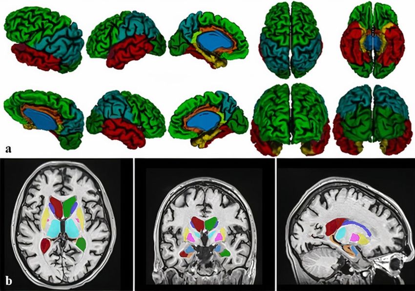
isthmus of the cingulate gyrus, and insula (Fig. 2a). Subcortical structure segmentation (putamen, caudate, thal-
amus, globus pallidus, hippocampus, amygdala, nucleus accumbens) was performed using an automated MRI
brain volumetry system, volBrain53 (Fig. 2b). Volumetric analysis was performed at three measuring points,
according to MRI scans: prior to DBS, approximately 1 year after, and 7 years after DBS implementation.
Statistical analysis. Data analysis was performed using the MedCalc Statistical Software version 12.5.0
(MedCalc Software, Ostend, Belgium; https://www.medcalc.org). Data were plotted as individual values, hori-
zontal lines and markers represent mean ± SD. Comparison of the regional cortical volumes and thicknesses
of parcellated lobes and volumes of subcortical structures were obtained. Depending on the distribution and
Scientific Reports | (2021) 11:4401 | https://doi.org/10.1038/s41598-021-83873-y 3
Vol.:(0123456789)www.nature.com/scientificreports/
Figure 2. Volumetric analysis was conducted using the CIVET pipeline measuring regional cortical volume
and thickness for parietal, occipital, frontal, temporal lobes, isthmus of the cingulate gyrus, parahippocampal,
and cingulate gyrus and insula (a), and volBrain software, for subcortical structures segmentation (b). Single-
subject volumetric analysis is presented.
type of variables, volumetric data in all measuring points were analyzed using one-way ANOVA, with the Stu-
dent–Newman–Keuls post hoc test, or Kruskal–Wallis test with pairwise comparisons according to C onover54.
In order to reduce intersubject variability we have performed the regional volumes normalization by head size.
Normalization was done using the subject with lowest head size value as a calibrator value to calculate the coef-
ficient (subject value/calibrated value). Regional volumes were multiplied with the calculated coefficient for
each subject and further analyzed using previously mentioned statistical test. We did not correct for multiple
comparisons in the present small patient sample. The statistical significance was set at p < 0.05.
Ethics approval and consent to participate. This study was carried out in accordance with the recom-
mendations of the Ethical Board of the Dubrava University Hospital and University of Zagreb, School of Medi-
cine with written informed parents/caregivers consent from all subjects in accordance with the Declaration of
Helsinki. The protocol was approved by the Institutional Review Board of the University Hospital Dubrava and
University of Zagreb, School of Medicine.
Results
Analysis of segregated volumes of the lobes of the cerebral cortex in three measuring points revealed statistically
significant volume increase in right parahippocampal gyrus (↑25.53%) (F(2, 9) = 6.658, p = 0.01), left cingulate
gyrus (↑16.65%) (F(2, 9) = 6.679, p = 0.01) and left insula (↑22.74%) (F(2,9) = 6.202, p = 0.04). However, volume
increase was observed for left parahippocampal gyrus volume (↑17.63%) (F(2,9) = 1.142, p = 0.36), right cingulate
gyrus volume (↑15.92%) (F(2,9) = 1.311, p = 0.31) and right insula volume (↑7.69%) (F(2,9) = 0.871, p = 0.45)
(Fig. 3). Volumetric analysis of frontal (L ↓7.57%, R ↑3.22%), parietal (L ↑0.05%, R ↓2.54%), occipital (L ↓9.99%,
R ↓15.08%,), and temporal (L ↓3.38%, R ↑2.14%) lobe, as well as isthmus of the cingulate gyrus (L ↑1.53%, R
↑1.52%), bilaterally, did not revealed statistically significant results.
Subcortical structures volumetric analysis revealed statistically significant volume increase in caudate (↑17.7%)
(F(2,9) = 4.964, p = 0.04), hippocampus (↑9.07%) (F(2,9) = 4.052, p = 0.02) and amygdala(↑39.53%) (F(2,9) = 4.163,
p = 0.02). While putamen (↑10.22%) (F(2,9) = 1.576, p = 0.58) and accumbens (↑28.57%) (F(2,9) = 1.915, p = 0.21)
volumes were observed increased, both globus pallidus (↓4.49%) (F(2,9) = 0.054, p = 0.95) and thalamus (↓24.16%)
(F(2,9) = 1.104, p = 0.37) volume revealed volume decrease over time, especially prominent in thalamus (Fig. 4).
While right parahippocampal gyrus regional cortical thickness increase significantly (↑16.9%) (F(2,9) = 1.937,
p = 0.02), regional cortical thickness increase was observed in both right cingulate gyrus and insula bilaterally,
as well as in the left parahippocampal gyrus, although these values were not statistically significant. Regional
Scientific Reports | (2021) 11:4401 | https://doi.org/10.1038/s41598-021-83873-y 4
Vol:.(1234567890)www.nature.com/scientificreports/
Figure 3. Regional cortical volumetric analysis in three measuring points (prior to DBS, 1 year after DBS,
and 7 years after DBS). The regional cortical volumetric analysis revealed significant volume increase in right
parahippocampal gyrus, left cingulate gyrus, and left insula, while the trend of volume increase was presented
in left parahippocampal gyrus volume, right cingulate gyrus volume, and right insula volume. Vertical bars,
standard deviation.
Figure 4. Subcortical structures volumetric analysis in three measuring points (prior to DBS, 1 year after DBS,
and 7 years after DBS). Significant volume increase of caudate, hippocampus, and amygdala were presented,
while putamen and accumbens volumes presented a trend of volume increase during three measuring points.
Both globus pallidus and thalamus volume decreased over time. Vertical bars, standard deviation.
cortical thickness is observed to decrease over time in the parietal, frontal, temporal, and occipital lobe, as well
as the isthmus of the cingulate gyrus.
Statistical analysis of normalized segregated volumes revealed significantly higher volume of the left insula
(F(2,9) = 6.804, p = 0.01). Volumes of the right insula (F(2,9) = 1.407, p = 0.29), the left parahippocampal gyrus
(F(2,9) = 1.142, p = 0.36), the right parahippocampal gyrus (F(2,9) = 1.694, p = 0.23), the left cingulate gyrus
Scientific Reports | (2021) 11:4401 | https://doi.org/10.1038/s41598-021-83873-y 5
Vol.:(0123456789)www.nature.com/scientificreports/
Patient no. Sex Etiology DOC Verbal communication Non-verbal communication Observed physical difficulties
1 ♂ CA MCS + + Stuttering, problems with concentration
Slowness in movements and talk; difficulties in fine motor skills; problems
2 ♂ CA MCS + +
with concentration
motor difficulties (spasticity, contractures, inability to move limbs and to
3 ♀ TBI MCS +* +
articulate words)
motor difficulties (spasticity, contractures, inability to move limbs and to
4 ♀ CA UWS +* +
articulate words)
motor difficulties (spasticity, contractures, inability to move limbs and to
5 ♀ TBI UWS +* +
articulate words)
Table 2. Neurocognitive and behavioral assessment of consciousness improved patients after DBS. CA cardiac
arrest, TBI traumatic brain injury, UWS unresponsive wakefulness syndrome, MCS minimally conscious state.
*Under intensive speech therapy at the moment, able to pronounce simple words.
(F(2,9) = 3.064, p = 0.09) and the right cingulate gyrus (F(2,9) = 2.423, p = 0.14), as well as volumetric analysis
of frontal, parietal, occipital, and temporal lobe, and isthmus of the cingulate gyrus, did not significantly differ
among groups. Subcortical normalized volumes of the amygdala (F(2,9) = 4.451, p = 0.04) were significantly
higher. Normalized volumes of the caudate (F(2,9) = 2.952, p = 0.22), hippocampus (F(2,9) = 3.390, p = 0.08),
putamen (F(2,9) = 1.015, p = 0.40), accumbens (F(2,9) = 2.436, p = 0.14), globus pallidus (F(2,9) = 0.032, p = 0.96)
and thalamus (F(2,9) = 0.747, p = 0.50) did not significantly differ.
All five patients included in the study raised to full awareness and regained the ability to communicate and
interact. Two male patients (Patient 1 and 2) were graded as MCS using RDR and C/NC scale (Table 1). Both
of them are currently able to live independently to a large extent22,23, although both of them are experiencing
problems with concentration and memory (Table 2). Out of the three female patients, one was graded as MCS
(Patient 3), while two of them were graded as UWS (Patient 4 and 5) using RDR and C/NC scale (Table 1). All
female patients regained consciousness, although they are currently still unable to live independently22,23. The
main obstacle for further progress is related to motor difficulties, such as spasticity, contractures, inability to
move their limbs, to articulate words, etc. For several years, their communication was in a form of non-verbal
signs, predominantly as facial gestures and/or limb movement. Today, these patients are able to form simple
words (Table 2).
Discussion
Previous studies reported that DBS in DOC is still in its exploratory phase5,6,19–23,26–30. Keeping in mind the ethi-
cal criteria and seriousness of DOC, our previous results showed limited but encouraging success22,23. Although
brain atrophy was initially observed on MRI prior to DBS, it was difficult and challenging to explain the volume
increase of several brain structures and areas after DBS, which was in accordance with previous studies31. Two
main aspects are going to be discussed in the following paragraphs. Firstly, a brief overview of consciousness,
the role of the thalamus in conscious networks, as well as the general functional effects of stimulation. Secondly,
we will discuss the structural changes that occurred after functional improvement, as a result of processes of
repair, plasticity, and reorganization.
General effects of deep brain stimulation. All major factors and phenomena of consciousness are not
fully understood at the moment. Despite numerous different study explanations and well-elaborated hypoth-
eses, the neurobiological mechanism of consciousness is still largely unexplored37,55–58. What is known how-
ever is that various components of the central nervous systems participate simultaneously in this intriguing
phenomenon56–61. Alongside the reticular ascending systems projecting from the upper reticular formation
of the brainstem, other structures participate significantly in forming consciousness. These structures include
monoaminergic pathways, the locus coeruleus, serotonergic and dopaminergic systems, the forebrain choliner-
gic system, as well as the nucleus b asalis62–66. In addition, a special system of neurons connecting the brainstem
to the interstitial neurons may also p articipate67 and form important networks for maintaining and regulating
cortical functions68,69. These neurons project towards a number of subcortical structures, in which projections
from thalamus nuclei are also received62–66. We propose that the main functional system responsible for the
observed structural changes is widespread thalamocortical and thalamosubcortical (feedback) circuitry, modu-
lated by several systems: modulatory monoaminergic systems, the reticular ascending system from the upper
brainstem, basal forebrain cholinergic systems, etc.
The thalamus is a crucial relay of the cortico-striatal-thalamo-cortical circuits and its role in arousal, attention
to salient stimuli, and processing of information has been documented by both animal models as well as func-
tional connectivity studies in h umans24,25,70–74. The CM-pf complex as the main part of the intralaminar n uclei24
provides particularly strong connections to several subcortical s tructures74. It also provides strong connections to
the basal g anglia75, primarily the caudate n ucleus76, putamen24, nucleus a ccumbens77, and pallidum, all of which
was demonstrated using both tracing and tractographic studies78. A relatively weaker subcortical connectivity
was noticed between CM-pf and the a mygdala79, and the h ippocampus80,81. On the other hand, excitatory pro-
jections from the CM-pf complex toward cortical areas have been reported as much weaker and non-specific24.
The cortical projections seem to be limited to the agranular cortex with the only exception being the anterior
Scientific Reports | (2021) 11:4401 | https://doi.org/10.1038/s41598-021-83873-y 6
Vol:.(1234567890)www.nature.com/scientificreports/
insular/frontal operculum78. Additionally, the CM-pf complex probably interacts with various cortical areas via
the corticostriatal pathways, through strong retrograde p rojections36.
The selection of the thalamus as a stimulation target thus seems justified because of its potent effect in the
thalamocortical circuitry. Our preliminary results showed the thalamic volume decrease less than 1% at the sec-
ond measuring point, while at the third measuring point thalamic volume decrease of almost 25% was observed
(Fig. 4). Since the thalamic volume was slightly decreased at the period when functional recovery was observed
in patients, we presume that the constant volume contributed to functional connectivity of the thalamus and
previously mentioned structures. Results of positive stimulation effects point to the thalamus and reorganization
of functional circuitry as important factors in recovery after DOC. The DBS electrode, positioned deep inside
the brain, provides permanent stimulation. This in turn provides new possibilities for integrating function and
structure for consciousness recovery. However, despite the evidence that thalamic intralaminar nuclei have
an influence on cerebral function and connectivity, it is unlikely that it can explain such major changes in the
brain structure volume. In our patients, a larger volume decrease was observed several years after the functional
recovery, thus we can only assume it occurred as a further adaptation of the thalamus to the brain reorganization
due to the initial brain injury, either trauma or anoxic lesion. Additionally, the thalamus volume could decrease
in response to the lack of adequate stimulus. Once the DBS is removed, the stimulus that maintained thalamic
volume by providing sufficient stimulation may cease. The variability of structural changes observed after DBS
may be partly explained by the great variety of cortical, subcortical, and brainstem functional systems involved.
This variability suggests that there is no single functional system affected. Therefore, besides the thalamus and
indirectly affected systems, other factors play a possible role and will be discussed below.
Structural effects observed on MRI. The exact underlying mechanisms contributing to volume changes
are still unknown. There are several possible explanations about how long functional stimulation could have
led to a volume increase. These include synaptogenesis, gliogenesis, axonal remodeling, micro vascularization,
neuronal size increase, and extracellular matrix changes. A combination of all these factors is also possible. Of
note is that adult neurogenesis has also been described in certain brain regions (i.e. the hippocampus)31,82. It is
well known that after various lesions, the brain volume could be changed by different underlying processes of
brain tissue repair (apoptosis, microstructural glial reactivity, changes in the extracellular matrix, etc.), plas-
ticity (sprouting, myelination, dendritic plasticity), and reorganization83–87. Therefore, we could expect similar
events occurring and leading to the increased brain volume in our patients. Additionally, processes of repair and
plasticity probably overlap. The reorganization of pathways is thus expected to be primarily functional (since
various pathways transduce electrical information when the brain is stimulated), rather than structural, which is
possible primarily during d evelopment86,87. Nevertheless, considering how the most voluminous components of
the brain tissue are cell bodies of neurons, glia, dendrites, and myelinated a xons88–90, it is unlikely that these cells
experience a significant increase in their size receiving more excitatory input. Moreover, an increase of dendritic
branching postsynaptic spines of super excited neurons is possible. The data on glia are less consistent, but it
is generally expected that astroglia follows a metabolic increase of its function and become hypertrophic when
stimulated91. Volume increase could therefore occur during an increase in synapsis number and size which are
more activate during prolonged stimulation of important excitatory glutamatergic n eurons92. However, synapsis
90
makes only a small proportion of the brain volume .
Several examples of increased brain volume during development in abnormal conditions have been previously
reported. In such cases, hyper-connectivity within the cortex was explained by the changes in the extracellular
substance, which makes approximately 70% of the developing brain, while being scarce in the normal adult
brain93–95.
The aforementioned structural changes (repair, plasticity, and reorganization) are impossible to follow in
patients, even if postmortem material is available. Therefore, MRI is so far the only available method providing
general information that something is structurally different. Additionally, it is especially useful to combine differ-
ent MRI techniques such as tractography with easily performed volumetric measurements, a valuable indicator
that structural processes are taking place in the brain after stimulation. Only a few volumetric MRI analyses in
patients with DOC were performed so-far33,34. Grey matter volume of the parahippocampal gyrus, thalamus,
and caudate were previously presented to be the key features differing healthy subjects and patients with DOC.
Additionally, white matter volumes of the parahippocampal gyrus, isthmus of cingulate gyrus, and brainstem
were previously described as the most affected white matter r egions34. Analysis of the subcortical structures
was previously performed only for the thalamus; slower atrophy of the thalamus was observed in MCS than in
UWS patients33. These results suggest that patients with better-preserved levels of consciousness are more likely
to preserve an increased brain volume over time. Our results indicate that the most informative region is the
parahippocampal area. This area is known to be important for the formation of episodic memories as well as
permitting rapid processing which in turn enables contextualization of outside events. It is therefore well located
to bind associations from other cortical streams, suggesting an important function in consciousness96. Our
findings are in the accordance with the results of previous preclinical studies of DBS in rodents which observed
structural neuroplasticity (hippocampal neurogenesis97,98), as well as an increased complexity of apical dendrites
and the length of basal dendritic trees of pyramidal neurons of the hippocampus99. Furthermore, our findings
are in accordance with results showing short-term brain volume increase, namely the hippocampal area, in
Alzheimer’s dementia patients following D BS31.
Several limitations of the presented study should be mentioned, such as a small number of patients, their
mixed gender, different age and etiology of injury as well as different times for MRI scans from the initial injury
and volumetric analysis in included patients. Still, we believe the presented results are valuable and can be a valid
starting point for future research.
Scientific Reports | (2021) 11:4401 | https://doi.org/10.1038/s41598-021-83873-y 7
Vol.:(0123456789)www.nature.com/scientificreports/
Conclusions
Even on a small sample size included in the present study, we tried to emphasize the importance of morphometric
MRI quantification which can expand our understanding of the relationship between brain structures and con-
sciousness recovery. Our results are in accordance with the results of very few human studies connecting DBS
and brain volume changes. They also support the idea that DBS in the CM-pf complex may have a widespread
effect on cerebral function and structure.
Structures with altered volumes belong to cortico-thalamo-cortico-basal ganglia circuitry, limbic circuitry
and at the same time have strong modulatory monoaminergic brainstem input. The nature of the initial lesion of
neural networks, neurons, and glia, is difficult to determine with the current criteria and methodology applied.
Which mechanisms are behind the observed brain changes and whether the observed structural changes are
caused by consciousness recovery and/or DBS in DOC patients is yet to be elucidated. In future studies, differ-
ent MRI volumetric analysis of cortical areas or deep brain structures could help to identify cell types and more
details contributing to the regional volume change.
Data availability
The datasets generated for this study are available on request to the corresponding author.
Received: 4 November 2020; Accepted: 9 February 2021
References
1. Plum, F. & Posner, J. B. The Diagnosis of Stupor and Coma 3rd edn. (F A Davis Co, Philadelphia, 1980).
2. Bernat, J. L. Chronic disorders of consciousness. Lancet 367(9517), 1181–1192. https://doi.org/10.1016/S0140-6736(06)68508-5
(2006).
3. Schnakers, C. & Laureys, S. Coma and Disorders of Consciousness (Springer, Switzerland, 2017). https://doi.org/10.1007/978-3-
319-55964-3.
4. Zeman, A. Consciousness. Brain 124(Pt 7), 1263–1289. https://doi.org/10.1093/brain/124.7.1263 (2001).
5. Yamamoto, T. et al. DBS therapy for the vegetative state and minimally conscious state. Acta Neurochir. Suppl. 93, 101–104. https
://doi.org/10.1007/3-211-27577-0_17 (2005).
6. Yamamoto, T. et al. Deep brain stimulation for the treatment of vegetative state. Eur. J. Neurosci. 32, 1145–1151. https://doi.org/1
0.1111/j.1460-9568.2010.07412.x (2010).
7. Vanhoecke, J. & Hariz, M. Deep brain stimulation for disorders of consciousness: Systematic review of cases and ethics. Brain
Stimul. 10(6), 1013–1023. https://doi.org/10.1016/j.brs.2017.08.006 (2017).
8. Schiff, N. D., Giacino, J. T. & Fins, J. J. Deep brain stimulation, neuroethics, and the minimally conscious state: Moving beyond
proof of principle. Arch. Neurol. 66(6), 697–702. https://doi.org/10.1001/archneurol.2009.79 (2009).
9. Johnson, L. S. The right to die in the minimally conscious state. J. Med. Ethics 37(3), 175–178. https://doi.org/10.1136/
jme.2010.038877 (2011).
10. Laureys, S. et al. Unresponsive wakefulness syndrome: A new name for the vegetative state or apallic syndrome. BMC Med. 1(8),
68. https://doi.org/10.1186/1741-7015-8-68 (2010).
11. Calvet, J. & Coll, J. Meningitis of sinusoid origin with the form of coma vigil. Rev. Otoneuroophtalmol. 31, 443–445 (1959).
12. Kretschmer, E. Das apallische Syndrom. Z. Ges. Neurol. Psychiat. 169, 292–296 (1940).
13. Jennett, B. & Plum, F. Persistent vegetative state after brain damage. A syndrome in search of a name. Lancet 1(7753), 734–737.
https://doi.org/10.1016/s0140-6736(72)90242-5 (1972).
14. Giacino, J. T. et al. The minimally conscious state: Definition and diagnostic criteria. Neurology 58(3), 349–353. https://doi.
org/10.1212/wnl.58.3.349 (2002).
15. Beaumont, J. G. & Kenealy, P. M. Incidence and prevalence of the vegetative and minimally conscious states. Neuropsychol. Rehabil.
15(3–4), 184–189. https://doi.org/10.1080/09602010443000489 (2005).
16. Anderson, C. A. & Arciniegas, D. B. Cognitive sequelae of hypoxic-ischemic brain injury: A review. NeuroRehabilitation 26(1),
47–63. https://doi.org/10.3233/NRE-2010-0535 (2010).
17. Lu-Emerson, C. & Khot, S. Neurological sequelae of hypoxic-ischemic brain injury. NeuroRehabilitation 26(1), 35–45. https://doi.
org/10.3233/NRE-2010-0534 (2010).
18. Thibaut, A., Schiff, N., Giacino, J., Laureys, S. & Gosseries, O. Therapeutic interventions in patients with prolonged disorders of
consciousness. Lancet Neurol. 18(6), 600–614. https://doi.org/10.1016/S1474-4422(19)30031-6 (2019).
19. Tsubokawa, T. et al. Deep-brain stimulation in a persistent vegetative state: Follow-up results and criteria for selection of candidates.
Brain Inj. 4(4), 315–327. https://doi.org/10.3109/02699059009026185 (1990).
20. Cohadon, F. & Richer, E. Deep cerebral stimulation in patients with post-traumatic vegetative state 25 cases. Neurochirurgie 39(5),
281–292 (1993).
21. Yamamoto, T. et al. Deep brain stimulation and spinal cord stimulation for vegetative state and minimally conscious state. World
Neurosurg. 80(3–4), S30.e1-9. https://doi.org/10.1016/j.wneu.2012.04.010 (2013).
22. Chudy, D. et al. Deep brain stimulation for the early treatment of the minimally conscious state and vegetative state: Experience
in 14 patients. J. Neurosurg. 128(4), 1189–1198. https://doi.org/10.3171/2016.10.JNS161071 (2018).
23. Chudy, D., Raguž, M. & Deletis, V. Deep brain stimulation for treatment patients in vegetative state and minimally conscious state.
In Neurophysiology in Neurosurgery: A Modern Approach 2nd edn (eds Deletis, V. et al.) 515–521 (Academic Press, New York,
2020).
24. Van der Werf, Y. D., Witter, M. P. & Groenewegen, H. J. The intralaminar and midline nuclei of the thalamus. Anatomical and
functional evidence for participation in processes of arousal and awareness. Brain Res. Brain Res. Rev. 39(2–3), 107–140. https://
doi.org/10.1016/s0165-0173(02)00181-9 (2002).
25. Metzger, C. D., van der Werf, Y. D. & Walter, M. Functional mapping of thalamic nuclei and their integration into cortico-striatal-
thalamo-cortical loops via ultra-high resolution imaging-from animal anatomy to in vivo imaging in humans. Front. Neurosci.
8(7), 24. https://doi.org/10.3389/fnins.2013.00024 (2013).
26. Hassler, R., Ore, G. D., Dieckmann, G., Bricolo, A. & Dolce, G. Behavioural and EEG arousal induced by stimulation of unspecific
projection systems in a patient with post-traumatic apallic syndrome. Electroencephalogr. Clin. Neurophysiol. 27(3), 306–310. https
://doi.org/10.1016/0013-4694(69)90060-1 (1969).
27. McLardy, T., Mark, V., Scoville, W. & Sweet, W. Pathology in diffuse projection system preventing brainstem-electrode arousal
from traumatic coma. Confin. Neurol. 31(4), 219–225. https://doi.org/10.1159/000103482 (1969).
Scientific Reports | (2021) 11:4401 | https://doi.org/10.1038/s41598-021-83873-y 8
Vol:.(1234567890)www.nature.com/scientificreports/
28. Sturm, V., Kühner, A., Schmitt, H. P., Assmus, H. & Stock, G. Chronic electrical stimulation of the thalamic unspecific activating
system in a patient with coma due to midbrain and upper brain stem infarction. Acta Neurochir. (Wien) 47(3–4), 235–244. https
://doi.org/10.1007/bf01406406 (1979).
29. Schiff, N. D. et al. Behavioural improvements with thalamic stimulation after severe traumatic brain injury. Nature 448(7153),
600–603. https://doi.org/10.1038/nature06041 (2007).
30. Magrassi, L. et al. Results of a prospective study (CATS) on the effects of thalamic stimulation in minimally conscious and vegeta-
tive state patients. J. Neurosurg. 125(4), 972–981. https://doi.org/10.3171/2015.7.JNS15700 (2016).
31. Sankar, T. et al. Deep brain stimulation influences brain structure in Alzheimer’s disease. Brain Stimul. 8(3), 645–654. https://doi.
org/10.1016/j.brs.2014.11.020 (2015).
32. Gosseries, O., Di, H., Laureys, S. & Boly, M. Measuring consciousness in severely damaged brains. Annu. Rev. Neurosci. 37, 457–478.
https://doi.org/10.1146/annurev-neuro-062012-170339 (2014).
33. Rubeaux, M., Jayashri Mahalingam, J., Gomez, F., Nelson, M., & Vanhaudenhuyse, A. et al. Thalamic volume as a biomarker for
disorders of consciousness. I Proceedings Volume 9287, 10th International Symposium on Medical Information Processing and
Analysis. 2015;9287:92870R. https://doi.org/10.1117/12.2071146.
34. Annen, J. et al. Regional brain volumetry and brain function in severely brain-injured patients. Ann. Neurol. 83(4), 842–853. https
://doi.org/10.1002/ana.25214(2018).
35. Dandy, W. E. The location of the conscious center in the brain: The corpus striatum. Bull. Johns Hopkins Hosp. 79, 34–58 (1946).
36. Halgren, E., Walter, R. D., Cherlow, D. G. & Crandall, P. H. Mental phenomena evoked by electrical stimulation of the human
hippocampal formation and amygdala. Brain 101(1), 83–117. https://doi.org/10.1093/brain/101.1.83 (1978).
37. Crick, F., Koch, C., Kreiman, G. & Fried, I. Consciousness and neurosurgery. Neurosurgery 55(2), 273–281. https://doi.
org/10.1227/01.neu.0000129279.26534.76 (2004).
38. Rappaport, M. The disability rating and coma/near-coma scales in evaluating severe head injury. Neuropsychol. Rehabil. 15(3–4),
442–453. https://doi.org/10.1080/09602010443000335 (2005).
39. Ad-Dab’bagh, Y., Einarson, D., & Lyttelton, O., et al. The CIVET image-processing environment: A fully automated comprehensive
pipeline for anatomical neuroimaging research. In: Proceedings of the 12th Annual Meeting of the Organization for Human Brain
Mapping (Corbetta, M. eds). Florence: NeuroImage; 2006.
40. Sherif, T. et al. CBRAIN: A web-based, distributed computing platform for collaborative neuroimaging research. Front. Neuroin-
form. 21(8), 54. https://doi.org/10.3389/fninf.2014.00054 (2014).
41. Bregant, T. et al. Region-specific reduction in brain volume in young adults with perinatal hypoxic-ischaemic encephalopathy.
Eur. J. Paediatr. Neurol. 17(6), 608–614. https://doi.org/10.1016/j.ejpn.2013.05.005 (2013).
42. Sled, J. G., Zijdenbos, A. P. & Evans, A. C. A nonparametric method for automatic correction of intensity nonuniformity in MRI
data. IEEE Trans. Med. Imaging 17(1), 87–97. https://doi.org/10.1109/42.668698 (1998).
43. Collin, D. L., Neelin, P., Peters, T. M. & Evans, A. C. Automatic 3D intersubject registration of MR volumetric data in standardized
talairach space. J. Comput. Assist. Tomogr. 18(2), 192–205. https://doi.org/10.1097/00004728-199403000-00005 (1994).
44. Mazziotta, J. et al. A probabilistic atlas and reference system for the human brain: International Consortium for Brain Mapping
(ICBM). Philos. Trans. R. Soc. Lond. B Biol. Sci. 356(1412), 1293–1322. https://doi.org/10.1098/rstb.2001.0915 (2001).
45. Smith, S. M. Fast robust automated brain extraction. Hum. Brain Mapp. 17(3), 143–155. https: //doi.org/10.1002/hbm.10062- (2002).
46. Zijdenbos A, Forghani R, Evans A. Automatic quantification of MS lesions in 3D MRI brain data sets: Validation of INSECT. In
Medical Image Computing and Computer-Assisted Intervention (MICCAI98). Lecture Notes in Computer Science (Wells, W. M.,
Colchester, A., & Delp, S. eds.) Boston (1998).
47. Kim, J. S. et al. Automated 3-D extraction and evaluation of the inner and outer cortical surfaces using a Laplacian map and partial
volume effect classification. Neuroimage 27(1), 210–221. https://doi.org/10.1016/j.neuroimage.2005.03.036 (2005).
48. MacDonald, D., Kabani, N., Avis, D. & Evans, A. C. Automated 3-D extraction of inner and outer surfaces of cerebral cortex from
MRI. Neuroimage 12(3), 340–356. https://doi.org/10.1006/nimg.1999.0534 (2000).
49. Ad-Dab’bagh Y, Singh V, Robbins S, Lerch P, Lyttelton O, Fombonne E. et al. Native space cortical thickness measurement and the
absence of correlation to cerebral volume. In Proceedings of the 11th Annual Meeting of the Organization for Human Brain Mapping
(Zilles, K., eds). Toronto: NeuroImage (2005).
50. Robbins, S. M. Anatomical standardization of the human brain in Euclidean 3-D space and on the cortical 2-Manifold. Ph.D.
thesis. School of Computer Science. Montreal, McGill University (2004).
51. Lyttelton, O., Boucher, M., Robbins, S. & Evans, A. An unbiased iterative group registration template for cortical surface analysis.
Neuroimage 34(4), 1535–1544. https://doi.org/10.1016/j.neuroimage.2006.10.041 (2007).
52. Boucher, M., Whitesides, S. & Evans, A. Depth potential function for folding pattern representation, registration and analysis.
Med. Image Anal. 13(2), 203–214. https://doi.org/10.1016/j.media.2008.09.001 (2009).
53. Manjón, J. V. & Coupé, P. volBrain: An online MRI brain volumetry system. Front. Neuroinform. 27(10), 30. https: //doi.org/10.3389/
fninf.2016.00030 (2016).
54. Conover, W. J. Practical Nonparametric Statistics (Wiley, New York, 1999).
55. Crick, F. & Koch, C. A framework for consciousness. Nat. Neurosci. 6(2), 119–126. https://doi.org/10.1038/nn0203-119 (2003).
56. Mogensen, J. & Overgaard, M. Reorganization of the connectivity between elementary functions—a model relating conscious
states to neural connections. Front Psychol. 20(8), 625. https://doi.org/10.3389/fpsyg.2017.00625 (2017).
57. Overgaard, M. & Mogensen, J. Visual perception from the perspective of a representational, non-reductionistic, level-depend-
ent account of perception and conscious awareness. Philos. Trans. R. Soc. Lond. B Biol. Sci. 369(1641), 20130209. https://doi.
org/10.1098/rstb.2013.0209 (2014).
58. Overgaard, M. The status and future of consciousness research. Front. Psychol. 10(8), 1719. https: //doi.org/10.3389/fpsyg. 2017.01719
(2017).
59. Delacour, J. Neurobiology of consciousness: An overview. Behav. Brain Res. 85(2), 127–141. https://doi.org/10.1016/s0166
-4328(96)00161-1 (1997).
60. Block, N. Consciousness, accessibility, and the mesh between psychology and neuroscience. Behav. Brain Sci. 30(5–6), 481–499.
https://doi.org/10.1017/S0140525X07002786 (2007).
61. Bayne, T., Hohwy, J. & Owen, A. M. Are there levels of consciousness?. Trends Cogn. Sci. 20(6), 405–413. https://doi.org/10.1016/j.
tics.2016.03.009 (2016).
62. Moruzzi, G. & Magoun, H. W. Brain stem reticular formation and activation of the EEG. Electroencephalogr. Clin. Neurophysiol.
1(4), 455–473. https://doi.org/10.1016/0013-4694(49)90219-9 (1949).
63. Ramón-Moliner, E. & Nauta, W. J. The isodendritic core of the brain stem. J. Comput. Neurol. 126(3), 311–335. https://doi.
org/10.1002/cne.901260301 (1966).
64. Young, G. B. & Pigott, S. E. Neurobiological basis of consciousness. Arch. Neurol. 56(2), 153–157. https://doi.org/10.1001/archn
eur.56.2.153 (1999).
65. Parvizi, J. & Damasio, A. R. Neuroanatomical correlates of brainstem coma. Brain 126(Pt 7), 1524–1536. https://doi.org/10.1093/
brain/awg166 (2003).
66. Samuels, E. R. & Szabadi, E. Functional neuroanatomy of the noradrenergic locus coeruleus: Its roles in the regulation of
arousal and autonomic function part I: Principles of functional organization. Curr. Neuropharmacol. 6(3), 235–253. https://doi.
org/10.2174/157015908785777229 (2008).
Scientific Reports | (2021) 11:4401 | https://doi.org/10.1038/s41598-021-83873-y 9
Vol.:(0123456789)www.nature.com/scientificreports/
67. Leontovich, T. A. & Zhukova, G. P. The specificity of the neuronal structure and topography of the reticular formation in the brain
and spinal cord of carnivora. J. Comp. Neurol. 121, 347–379. https://doi.org/10.1002/cne.901210305 (1963).
68. Das, G. D. & Kreutzberg, G. W. Evaluation of interstitial nerve cells in the central nervous system. A correlative study using ace-
tylcholinesterase and Golgi techniques. Ergeb. Anat. Entwicklungsgesch 41(1), 3–58 (1969).
69. Kostovic, I. & Rakic, P. Cytology and time of origin of interstitial neurons in the white matter in infant and adult human and
monkey telencephalon. J. Neurocytol. 9(2), 219–242. https://doi.org/10.1007/bf01205159 (1980).
70. Newman, J. Thalamic contributions to attention and consciousness. Conscious Cogn. 4(2), 172–193. https://doi.org/10.1006/
ccog.1995.1024 (1995).
71. Kinomura, S., Larsson, J., Gulyás, B. & Roland, P. E. Activation by attention of the human reticular formation and thalamic intrala-
minar nuclei. Science 271(5248), 512–515. https://doi.org/10.1126/science.271.5248.512 (1996).
72. Minamimoto, T. & Kimura, M. Participation of the thalamic CM-Pf complex in attentional orienting. J. Neurophysiol. 87(6),
3090–3101. https://doi.org/10.1152/jn.2002.87.6.3090 (2002).
73. Schiff, N. D. Central thalamic contributions to arousal regulation and neurological disorders of consciousness. Ann. N. Y. Acad.
Sci. 1129, 105–118. https://doi.org/10.1196/annals.1417.029 (2008).
74. Haber, S. N. & Calzavara, R. The cortico-basal ganglia integrative network: The role of the thalamus. Brain Res. Bull. 78(2–3),
69–74. https://doi.org/10.1016/j.brainresbull.2008.09.013 (2009).
75. Jones, E. G. & Leavitt, R. Y. Retrograde axonal transport and the demonstration of non-specific projections to the cerebral cor-
tex and striatum from thalamic intralaminar nuclei in the rat, cat and monkey. J. Comp. Neurol. 154(4), 349–377. https://doi.
org/10.1002/cne.901540402 (1974).
76. Macchi, G., Bentivoglio, M., Molinari, M. & Minciacchi, D. The thalamo-caudate versus thalamo-cortical projections as studied in
the cat with fluorescent retrograde double labeling. Exp. Brain. Res. 54(2), 225–239. https://doi.org/10.1007/bf00236222 (1984).
77. Jayaraman, A. Organization of thalamic projections in the nucleus accumbens and the caudate nucleus in cats and its relation with
hippocampal and other subcortical afferents. J. Comp. Neurol. 231(3), 396–420. https://doi.org/10.1002/cne.902310309 (1985).
78. Eckert, U. et al. Preferential networks of the mediodorsal nucleus and centromedian-parafascicular complex of the thalamus—a
DTI tractography study. Hum. Brain Mapp. 33(11), 2627–2637. https://doi.org/10.1002/hbm.21389 (2012).
79. Su, H. S. & Bentivoglio, M. Thalamic midline cell populations projecting to the nucleus accumbens, amygdala, and hippocampus
in the rat. J. Comp. Neurol. 297(4), 582–593. https://doi.org/10.1002/cne.902970410 (1990).
80. Green, J. D. & Adey, W. R. Electrophysiological studies of hippocampal connections and excitability. Electroencephalogr. Clin.
Neurophysiol. 8(2), 245–263. https://doi.org/10.1016/0013-4694(56)90117-1 (1956).
81. Cavdar, S. et al. The pathways connecting the hippocampal formation, the thalamic reuniens nucleus and the thalamic reticular
nucleus in the rat. J. Anat. 212(3), 249–256. https://doi.org/10.1111/j.1469-7580.2008.00858.x (2008).
82. Fotuhi, M., Do, D. & Jack, C. Modifiable factors that alter the size of the hippocampus with ageing. Nat. Rev. Neurol. 8(4), 189–202.
https://doi.org/10.1038/nrneurol.2012.27 (2012).
83. Jessberger, S. Neural repair in the adult brain. F1000Res 12, 5. https://doi.org/10.12688/f1000research.7459.1 (2016).
84. Mogensen, J. Reorganization of the injured brain: Implications for studies of the neural substrate of cognition. Front. Psychol.
26(2), 7. https://doi.org/10.3389/fpsyg.2011.00007 (2011).
85. Chang, Y. Reorganization and plastic changes of the human brain associated with skill learning and expertise. Front. Hum. Neurosci.
4(8), 35. https://doi.org/10.3389/fnhum.2014.00035 (2014).
86. Kerr, A. L., Cheng, S. Y. & Jones, T. A. Experience-dependent neural plasticity in the adult damaged brain. J. Commun. Disord.
44(5), 538–548. https://doi.org/10.1016/j.jcomdis.2011.04.011 (2011).
87. Sharma, N., Classen, J. & Cohen, L. G. Neural plasticity and its contribution to functional recovery. Handb. Clin. Neurol. 110, 3–12.
https://doi.org/10.1016/B978-0-444-52901-5.00001-0 (2013).
88. Leise, E. M. Modular construction of nervous systems: A basic principle of design for invertebrates and vertebrates. Brain Res.
Brain Res. Rev. 15(1), 1–23. https://doi.org/10.1016/0165-0173(90)90009-d (1990).
89. Mota, B. & Herculano-Houzel, S. All brains are made of this: A fundamental building block of brain matter with matching neuronal
and glial masses. Front. Neuroanat. 8, 127. https://doi.org/10.3389/fnana.2014.00127 (2014).
90. Santuy, A., Rodríguez, J. R., DeFelipe, J. & Merchán-Pérez, A. Study of the size and shape of synapses in the Juvenile Rat soma-
tosensory cortex with 3D electron microscopy. eNeuro 5, 1. https://doi.org/10.1523/ENEURO.0377-17.2017 (2018).
91. Turner, D. A. & Adamson, D. C. Neuronal-astrocyte metabolic interactions: Understanding the transition into abnormal astrocy-
toma metabolism. J. Neuropathol. Exp. Neurol. 70(3), 167–176. https://doi.org/10.1097/NEN.0b013e31820e1152 (2011).
92. Okubo, Y. & Iino, M. Visualization of glutamate as a volume transmitter. J. Physiol. 589(Pt 3), 481–488. https://doi.org/10.1113/
jphysiol.2010.199539 (2011).
93. Levitt, J. G. et al. Cortical sulcal maps in autism. Cereb. Cortex 13(7), 728–735. https://doi.org/10.1093/cercor/13.7.728 (2003).
94. Casanova, M. F., Buxhoeveden, D., Switala, A. & Roy, E. Neuronal density and architecture (gray level index) in the brains of
autistic patients. J. Child. Neurol. 17(7), 515–521. https://doi.org/10.1177/088307380201700708 (2002).
95. Casanova, M. F. et al. Reduced gyral window and corpus callosum size in autism: Possible macroscopic correlates of a minicolu-
minopathy. J. Autism Dev. Disord. 39(5), 751–764. https://doi.org/10.1007/s10803-008-0681-4 (2009).
96. Behrendt, R. Hippocampus and consciousness. Rev. Neurosci. 24(3), 239–266. https: //doi.org/10.1515/revneu ro-2012-0088 (2013).
97. Hamani, C., Stone, S. S., Garten, A., Lozano, A. M. & Winocur, G. Memory rescue and enhanced neurogenesis following electrical
stimulation of the anterior thalamus in rats treated with corticosterone. Exp. Neurol. 232(1), 100–104. https://doi.org/10.1016/j.
expneurol.2011.08.023 (2011).
98. Stone, S. S. et al. Stimulation of entorhinal cortex promotes adult neurogenesis and facilitates spatial memory. J. Neurosci. 31(38),
13469–13484. https://doi.org/10.1523/JNEUROSCI.3100-11.2011 (2011).
99. Bezchlibnyk, Y. B., Stone, S. S. D., Hamani, C. & Lozano, A. M. High frequency stimulation of the infralimbic cortex induces mor-
phological changes in rat hippocampal neurons. Brain Stimul. 10(2), 315–323. https://doi.org/10.1016/j.brs.2016.11.013 (2017).
Author contributions
M.R. designed the study, conducted the volumetric analysis, and wrote the first version of the manuscript. N.P.
designed the study and contributed to the statistical data analysis and interpretation of results. D.D., D.O. and
A.R. conducted literature research, contributed to the data analysis, and designed the figures and tables. D.R.,
F.A. and P.M. contributed to the volumetric analysis and performed a neurological assessment of included
patients. V.D., I.K., and D.C. contributed to the study concept and design, interpreted the results and revised the
manuscript. All authors read and approved the final version of the manuscript as submitted.
Funding
This research was supported by the Croatian Science Fundation project CSF-IP-2020-02-4308, the European
Union through the European Regional Development Fund, Operational Program Competitiveness and Cohesion,
Grant agreement No. KK.01.1.1.01.0007, CoRE-Neuro. In addition, this research was supported by the Adris
Scientific Reports | (2021) 11:4401 | https://doi.org/10.1038/s41598-021-83873-y 10
Vol:.(1234567890)You can also read