Comparison of plasma and cerebrospinal fluid proteomes identifies gene products guiding adult neurogenesis and neural differentiation in birds
←
→
Page content transcription
If your browser does not render page correctly, please read the page content below
www.nature.com/scientificreports
OPEN Comparison of plasma
and cerebrospinal fluid proteomes
identifies gene products guiding
adult neurogenesis and neural
differentiation in birds
Eleni Voukali*, Nithya Kuttiyarthu Veetil, Pavel Němec, Pavel Stopka & Michal Vinkler*
Cerebrospinal fluid (CSF) proteins regulate neurogenesis, brain homeostasis and participate in
signalling during neuroinflammation. Even though birds represent valuable models for constitutive
adult neurogenesis, current proteomic studies of the avian CSF are limited to chicken embryos. Here
we use liquid chromatography–tandem mass spectrometry (nLC-MS/MS) to explore the proteomic
composition of CSF and plasma in adult chickens (Gallus gallus) and evolutionarily derived parrots:
budgerigar (Melopsittacus undulatus) and cockatiel (Nymphicus hollandicus). Because cockatiel lacks
a complete genome information, we compared the cross-species protein identifications using the
reference proteomes of three model avian species: chicken, budgerigar and zebra finch (Taeniopygia
guttata) and found the highest identification rates when mapping against the phylogenetically
closest species, the budgerigar. In total, we identified 483, 641 and 458 unique proteins consistently
represented in the CSF and plasma of all chicken, budgerigar and cockatiel conspecifics, respectively.
Comparative pathways analyses of CSF and blood plasma then indicated clusters of proteins involved
in neurogenesis, neural development and neural differentiation overrepresented in CSF in each
species. This study provides the first insight into the proteomics of adult avian CSF and plasma and
brings novel evidence supporting the adult neurogenesis in birds.
The central nervous system (CNS) function is regulated by a vivid communication with other tissues. This is also
achieved through the cerebrospinal fluid (CSF) that supports the brain cell f unctions1. Being from ~ 80% plasma
(PL)-derived and ~ 20% brain-cell-derived2, CSF dynamically reflects both peripheral and CNS physiological
processes. Despite the paramount importance of the relationship between blood and CSF, little comparative
effort has been made in non-mammalian vertebrates to characterise the two vital fluids integrating processes
in and out of the CNS.
There is widespread evidence that the birth and growth of neurons also occurs in adult invertebrate and
vertebrate CNS and that these cells add onto existing neuronal networks or replace apoptotic n eurons3. In addi-
tion, several in vitro and in vivo studies have provided evidence that, CSF can promote neurogenesis even in
rain4–9. In particular, spatial gradients of CSF proteins induced the migration of neuroblasts from the
the adult b
subventricular zone to the olfactory bulb in the adult mouse b rain8. Also, when rat CSF was applied as a culture
medium to neural stem cells and cortical explants, the cells survived without additional supplements, particularly
for age-matched CSF samples and t issues9. Therefore, increased interest has been raised to study and describe
the molecular composition of CSF, especially in h umans10–16.
Although CSF proteomic composition shares many similarities across species such as rat, human and
chicken6, 10–12, species differ in physiological requirements of their tissues and different species show different
neurogenesis rates as well as anatomical f eatures11, 17. Birds have been proposed as suitable models to investigate
the molecular basis of the regulation of adult constitutive n eurogenesis18. Nonetheless, the available avian CSF
proteomic analyses have addressed only embryonic chicken CSF. Based on two-dimensional polyacrylamide gel
electrophoresis (2D-PAGE) followed by mass spectrometry (MS) or tandem mass spectrometry (MS/MS) meth-
ods, the chicken embryonic CSF (eCSF) consists of hundreds of proteins of the extracellular matrix, regulators of
Department of Zoology, Faculty of Science, Charles University, Viničná 7, 128 44 Prague, Czech Republic. *email:
voukalie@natur.cuni.cz; michal.vinkler@natur.cuni.cz
Scientific Reports | (2021) 11:5312 | https://doi.org/10.1038/s41598-021-84274-x 1
Vol.:(0123456789)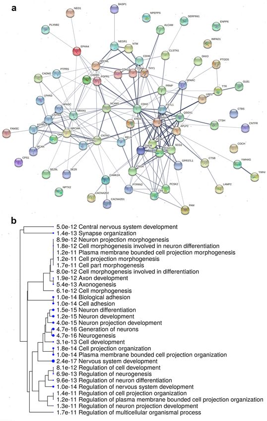
www.nature.com/scientificreports/
osmotic pressure, ion carriers, hormone-binding proteins, regulators of lipid metabolism, and various enzymes
and their regulators6, 10, 11. However, the advent of methods such as gel-free-LC/MS for comparative protein
analysis has resulted in several advantages over the label-based methods used by the aforementioned studies,
including the greater coverage of the p roteome19.
While the domestic chicken (Gallus gallus, GG, order Galliformes) serves as a basic avian model organism
in developmental biology and neuroscience20, 21, current evidence shows that its neural densities are much lower
than in some other, evolutionarily derived birds22, 23. Importantly, recently, parrots (order Psittaciformes) have
been proposed as models for neurobiological research24. Similarly to humans, parrots have large brains rela-
tive to their body size25, high density of neurons in the forebrain22, exhibit outstanding cognitive skills such as
object permanence26, 27 and tool use26, 28, cooperative problem solving29, complex social organization30, learning
of vocalizations through cultural transmission31, 32, and some species are on par with great apes in number of
cognitive domains33–35. Furthermore, despite their high metabolism, they show extended developmental periods36
and longevity24, 37.
In the present study, we focus for the first time on avian adult CSF and PL proteomes, comparing the domes-
tic chicken to two parrot species: the budgerigar (Melopsittacus undulatus, MU) and the cockatiel (Nymphicus
hollandicus, NH), representing two distinct parrot clades (Psittacoidea and Cacatuoidea, respectively38). Since
the cockatiel genome is not presently available, a condition that may affect the peptide/protein identification,
we first test the consistency of the proteomic annotation by comparing the MS/MS identification success rates
after mapping all spectra against the reference proteomes of three model avian species: chicken, budgerigar
and zebra finch (Taeniopygia guttata). Second, using the quality-filtered and consistent data, we describe the
proteomic profiles of PL and CSF in the three bird species, searching for the pathways common to differentiate
the avian PL and CSF proteomes.
Results
Reference selection for proteome mapping. We mapped each chicken, budgerigar and cockatiel CSF
and PL sample spectra against the chicken, zebra finch and budgerigar reference proteomes (Fig. 1a). The total
numbers of spectra, numbers of unique spectra and numbers of the identified peptide sequences obtained for
each mapping combination are shown in Supplementary Table S1, along with the total numbers of unique pro-
teins identified for each data set before and after filtering. The list of proteins represented in individual samples
together with their abundances and the ortholog gene names corresponding to each identified protein ID is
also provided in Supplementary Table S1. We found that a large proportion (25.1%, 35% and 41.1% for chicken,
budgerigar and cockatiel, respectively) of orthologous gene IDs overlapped when mapped against chicken, zebra
finch and budgerigar reference proteomes in either CSF or PL, being consistent for all avian species examined
in this study (Fig. 1b). However, part of the proteins remained unidentified when the CSF and PL spectra were
mapped against either the same species proteome or the budgerigar proteome in the case of cockatiel. Regarding
chicken spectra, about 7.2% (37) out of the total proteins identified for chicken were not identified when mapped
against the chicken reference proteome but found when mapped against zebra finch and budgerigar references;
for the budgerigar, 18% (125) of identities were missed when mapped against budgerigar genome, but detected
when mapped against the zebra finch or chicken; and for the cockatiel, 13.8% (70) of identities were missed when
mapped against the budgerigar proteome, but detected when mapped against the zebra finch and/or chicken.
The list of these genes is included in Supplementary Table S1 and contains, among others, members of multigene
families such as histones, and myoglobins. A Gene Ontology (GO) classification of the biological functions for
each subset of these missing proteins is shown in Fig. S1 in Supplement. The most represented biological func-
tions of these missing proteins were: (1) Cellular processes (61.1, 47.7, 53.3% of the missing proteins in GG,
MU and NH, respectively) including the cell communication, cellular component organization, developmental
process, homeostasis, metabolic process, response to stimulus, export from cell, movement of cell or subcellu-
lar component, protein folding and signal transduction; (2) Biological regulation of molecular functions (27.8,
19.3, 28.9% of the missing proteins in GG, MU and NH, respectively); and (3) Metabolic processes (27.8, 6.4,
35.6% of the missing proteins in GG, MU and NH respectively) including ATP metabolic process, biosynthetic
process, catabolic process, cellular metabolic process, nitrogen compound metabolic process, organic substance
metabolic process, oxidation–reduction process, primary metabolic process and small molecule metabolic pro-
cess. Since mappings from multiple annotations could not be combined and a higher proportion of gene prod-
ucts were exclusively identified when the proteomes were mapped against the same species reference proteome
(chicken and budgerigar) or a closely related one (cockatiel), we selected only these datasets for further analysis
and focused mainly on consistently represented proteins.
Blood PL and CSF proteomes description. In total, after filtering, we identified 464 proteins repre-
sented in the chicken CSF proteome, 586 proteins in the budgerigar CSF proteome and 437 proteins in the cock-
atiel CSF proteome. The PL proteome consisted of 304 proteins in the chicken, 478 in the budgerigar and 310 in
the cockatiel. From these, 109 proteins (11.3%) were common to all species (GG, MU, NH) and all sample types
(PL or CSF) (Fig. 2a), while a large number of proteins varied even between individual samples of the same tissue
and species (Supplementary Fig. S2). The detailed protein coverage of each dataset is shown on Supplementary
Fig. S3. We then defined the core proteomes, that is, proteins represented in all conspecific individuals for each
fluid. The complete list of the proteins and abundances of the CSF and PL core proteomes for the three species
is tabulated in the Supplementary Table S2. Considering the list of proteins detected across all individuals across
all species in the particular sample types, we identified 115 common proteins in the CSF (22.3%, Fig. 2b), 53
common proteins in the PL (15.8%, Fig. 2c), and 40 proteins overlapping between CSF and PL (Fig. 2d). The CSF
core proteome consisted of 344 proteins (71.22% of the total proteins detected across all individuals) in chicken,
Scientific Reports | (2021) 11:5312 | https://doi.org/10.1038/s41598-021-84274-x 2
Vol:.(1234567890)
www.nature.com/scientificreports/
Figure 1. Comparison of the nLC-MS/MS cross-species protein identification success in both cerebrospinal
fluid (CSF) and plasma (PL) samples of chickens (GG), budgerigars (MU) and cockatiels (NH), after mapping to
three selected avian reference proteomes GG, MU and Taeniopygia guttata (TG). (a) The overview of the study
design and mapping approach is schematically illustrated. (b) Venn diagrams show the proportions of proteins
identified based on the different reference proteomes of GG (grey), TG (red) and MU (green) for GG, MU and
NH (BioVenn, http://www.biovenn.nl/).
Scientific Reports | (2021) 11:5312 | https://doi.org/10.1038/s41598-021-84274-x 3
Vol.:(0123456789)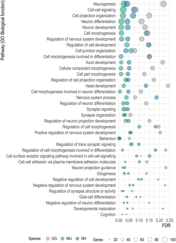
www.nature.com/scientificreports/
Figure 2. Overview of the proteomic coverage across species and body fluids. The identified plasma (PL)
and cerebrospinal fluid (CSF) proteins of chicken (GG), budgerigar (MU) and cockatiel (NH) and the shared
identifications between the groups are demonstrated in (a) (Venn Diagrams, http://bioinformatics.psb.ugent.be/
webtools/Venn/). The Venn diagrams are showing the overlapping numbers and percentages of core proteomes
of CSF (b) and PL (c) for GG, MU and NH, and (d) the numbers and percentages of shared proteins across all
the three species for CSF and PL (BioVenn, http://www.biovenn.nl/).
269 proteins (41.97%) in budgerigar and 274 (56.92%) proteins in cockatiel. The PL core proteome consisted of
173 proteins (55.27%) in chicken, 227 proteins (45.31%) in budgerigar and 124 proteins (38.87%) in cockatiel.
We ranked the shared identified proteins according to their normalized logarithmic abundance means. There
was a considerable inter-individual variability in the expression of the lowly abundant proteins in both PL and
CSF. In contrast, among the highly abundant proteins, the protein composition appears relatively consistent in
both fluids between the three species, parrot species being more similar (Fig. 3). Among the 2% most abundant
proteins identified in both the fluids and across all the species belongs namely albumin (ALB), ovotransferrin
(TF), alpha 2 macroglobulin (A2M), transthyretin (TTR), vitronectin (VTN) and apolipoproteins (APOA1,
APOA4).
In general, the proteins commonly represented in CSF and blood PL were involved in metabolic and signal-
ling pathways and most of their functions are common to CSF and plasma (Fig. 4). As expected, CSF proteins
are mostly extracellular components expressed into the extracellular space. The complete results of the pathway
enrichment analysis are shown in Supplementary Table S3.
Comparison of the plasma and CSF proteomes. Raw and normalised data were inspected by their
log2 distributions and correlation of their principal component 1 of the shared genes. After data normalization,
the distributions showed almost no variation between the samples and their first principal component positively
correlated with the first principal component of raw data (Supplementary Fig. S4). Principal component analyses
(PCA) of CSF and PL protein abundances showed that the two fluids form separate clusters for all three studied
species (Supplementary Fig. S5). For chicken, the first two principal components related to fluid type explained
84.76% of the total variance; for budgerigar 70.22% and cockatiel 74.21%, indicating that CSF and PL proteomes
are distinguishable in these species. The exploration of the differentially represented proteins in CSF and PL
proteomes in GG identified 413 significantly differentially abundant proteins, including 49 under-represented
proteins and 289 over-represented proteins in CSF compared to PL with false discovery rate (FDR) adjusted p
value < 0.05 and fold change cut-off ≥ 2. Similarly, 332 proteins were significantly differentially represented in
MU CSF compared to PL proteome, including 99 under-represented and 224 over-represented proteins (FDR
adjusted p value < 0.05, fold change cut-off ≥ 2) and, for NH, the analysis revealed 289 differentially represented
Scientific Reports | (2021) 11:5312 | https://doi.org/10.1038/s41598-021-84274-x 4
Vol:.(1234567890)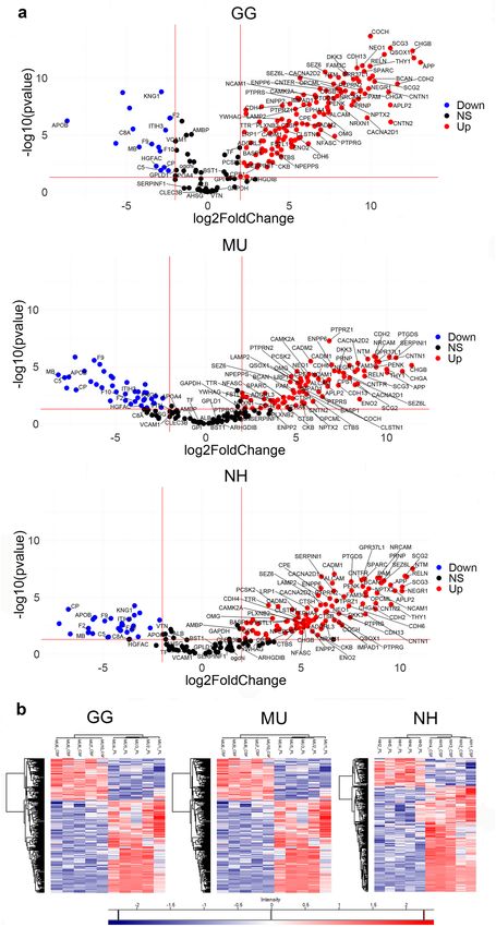
www.nature.com/scientificreports/
Figure 3. The top 2% most abundant cerebrospinal fluid (CSF) and plasma (PL) proteins of chicken (GG),
budgerigar (MU) and cockatiel (NH) are presented using their gene code: GG—grey, MU—green and
NH—blue. The logarithmic normalised abundances are shown as a range (minimum–maximum) across all
individuals (GG n = 7, MU n = 5, NH n = 5) with a mean highlighted as a dot (R version 4.0.0, www.r-proje
ct.org).
proteins, 46 under-represented and 206 over-represented in CSF compared to PL (FDR adjusted p value, fold
change cut-off ≥ 2). The fold change differences in the common proteins across species are shown in volcano
plots at Fig. 5a. The fold change differences in all differentially represented proteins for each species are shown in
the Supplementary Fig. S6. Detailed information on these differentially represented proteins is provided in the
Supplementary Table S4. The differentiation between protein content of CSF and PL was further indicated based
on hierarchical clustering that we used to visualise the groups of differentially represented proteins in either CSF
or PL in all the three studied avian species (Fig. 5b).
We found that 72 proteins over-represented in CSF compared to PL overlap between GG, MU or NH, belong-
ing to various functional modules such as cell-adhesion proteins, growth factors, subunits of calcium chan-
nels etc. Interestingly, four commonly shared CSF proteins (LCAT, phosphatidylcholine-sterol acyltransferase;
MCFD2, multiple coagulation factor deficiency protein 2; PRDX1, peroxiredoxin-1) are under-represented in the
CSF of parrots but over-represented in chicken. We did not identify any proteins over-represented in the CSF of
chicken but under-represented in parrots. STRING analysis of the 72 commonly over-represented proteins in CSF
of all species indicates a network of protein interactions, containing 72 nodes and 199 edges (Fig. 6a). The GO
Biological function terms related to this protein–protein interaction (PPI) network indicate developmental and
neogenesis processes. The same gene set was visualised as a hierarchical clustering tree using ShinyGO (Fig. 6b).
This is consistent with the result of the gene set enrichment analysis (GSEA) based on GO Biological functions
classification and pathway enrichment in CSF compared to plasma that revealed differential representation in
proteins involved in the neural function. Enriched pathways in plasma were mostly related to immune functions.
Supplementary Table S5 shows the complete list of the significantly enriched pathways at nominal p value < 0.05
and FDR < 0.25 in CSF and PL, and Supplementary Figs. S7–S10 the graphical representation of their gene over-
lap. The GSEA analysis revealed several gene sets linked to neurogenesis, neural development and differentiation
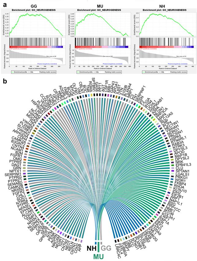
which were preferentially expressed into CSF and shared across the species (Fig. 7). Figure 8 shows the nominal
enrichment scores for GO term Neurogenesis and the genes implicated for each species.
Discussion
CSF connects the bloodstream with the CNS and reflects the physiology of the brain. Here we have described
the adult CSF and PL protein composition in three avian species (chicken, budgerigar, and cockatiel) using gel-
free nLC-MS/MS. First, we have confirmed that the phylogenetically closest avian reference proteome yields the
highest efficacy in protein identification success rates, although a proportion of proteins (7.2%, 18% and 13.8%
for GG, MU and NH, respectively) was mapped only using other references. In total, following the data filtering,
we identified in the CSF and PL 483 proteins in the chicken, 641 proteins in the budgerigar and 458 proteins in
the cockatiel. GO pathway analyses of the core proteomes revealed components of several signalling and meta-
bolic processes such as regulation of peptidases, synapse organisation, regulation of IGF transport and uptake
by IGFBPs in CSF and blood coagulation, complement activation and glycolysis in PL. Comparative analysis
of the proteomes derived from the two biological fluids showed that the CSF and PL are clearly distinguishable
in terms of their protein content. In CSF, the pathways involved in neurogenesis, and neural development and
differentiation are over-represented when compared to blood plasma.
Current research increasingly adopts investigation in diversified taxa lacking complete and annotated
genomes and proteomes. In this study, we investigated also the cockatiel, a species currently lacking a genome
Scientific Reports | (2021) 11:5312 | https://doi.org/10.1038/s41598-021-84274-x 5
Vol.:(0123456789)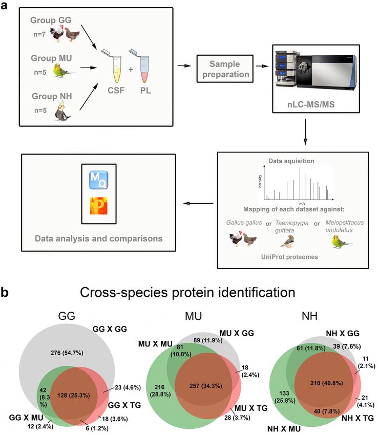
www.nature.com/scientificreports/
Figure 4. Top significant over-represented pathways revealed after functional enrichment analysis of the
cerebrospinal fluid (CSF) and blood plasma (PL). Enriched pathways in CSF are shown on the left-hand side
and in PL on the right-hand side for (a) chicken (GG), (b) budgerigar (MU) and (c) cockatiel (NH). The x-axis
indicates the numbers of genes represented in each pathway, the y-axis indicates the GO (Molecular function,
MF; Biological Process, BP; Cellular component, CC) or Pathways terms (KEGG, REACT) involved, and the
colours represent the size of the negative log10 of the adjusted p value (adjusted p < 0.05). IGF Insulin-like
Growth Factor; IGFBPs Insulin-like Growth Factor Binding Proteins (g:Profiler version e102_eg49_p15_
b486be1, https://biit.cs.ut.ee/gprofi ler/gost; R version 4.0.0, www.r-project.org).
sequence. We found that by cross-species protein identification using the sequence database from its evolution-
arily closest relative, the budgerigar, we could identify 458 proteins represented in the CSF and PL. This is more
than if other reference genomes were used (chicken, 327; zebra finch, 291). This trend was consistent with our
findings in other species included in this study. Interestingly, however, some proteins were identified only when
mapped against the other mapping combinations. Quantitative proteomics of unsequenced organisms is still a
challenge and, when de novo sequencing of the unidentified proteins is unavailable, the reference proteome of
a close-related phylogenetically species is utilised to identify the proteome in question. An analogous approach
has been used to characterise the proteome of unsequenced micro-organisms and Xenopus laevis microtubule-
associated proteome39, 40, but this is the first study reporting a paradigm of cross-species quantitative proteomics
in diverse avian taxa.
We found more proteins in CSF compared to plasma, a finding which was consistently found in all avian
species in this study and similar to what has been previously reported for humans in paired s amples41, 42. In a
cohort of more than 100 individuals, Dayon et al. identified 790 and 422 proteins in parallel CSF and PL samples
respectively with an overlap of 255 proteins shared by both fluids41. In a previous study, a maximal protein set of
3,081 and 1,050 CSF and PL proteins was identified respectively with 877 proteins common in both fluids from a
group of 21 healthy i ndividuals42. The reasons for the differences in the number of protein identifications between
these two studies could be attributed in different sample preparation approaches and detection techniques. Thus,
although the protein concentration is likely higher in PL compared to C SF43, the variety of proteins is greater
in the CSF compared to PL, being enriched from ca. 20% with proteins expressed in the choroid plexus and
brain44. Blood and serum comprise receptor ligands, immunoglobulins, tissue leakage products, aberrant secre-
tions and proteins that are extraneous to the animal. Many blood proteins are synthesized in liver45. Although
quality of CSF collection can influence the protein content observed, the CSF concentration of CNS-derived
proteins remains unaffected following contamination of the fluid with blood46. In the present dataset, we found
the presence of haemoglobin subunits and carbonic anhydrase that can be attributed to some minor blood
contamination47. Yet, like in Smith et al.13, although haemoglobin subunits were found amongst the most CSF
Scientific Reports | (2021) 11:5312 | https://doi.org/10.1038/s41598-021-84274-x 6
Vol:.(1234567890)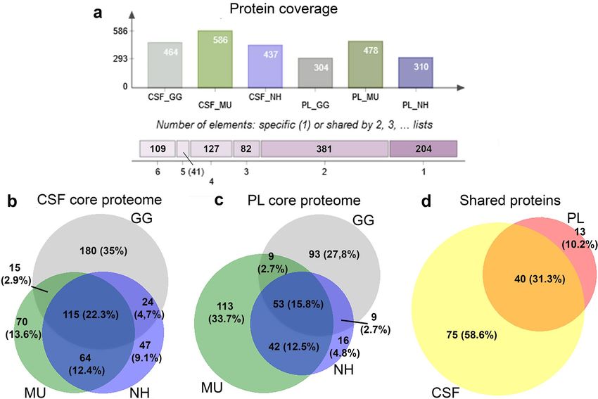
www.nature.com/scientificreports/
Figure 5. Comparative analyses of cerebrospinal fluid (CSF) and plasma (PL) proteomes. The distribution of the shared proteins
across species and clustering of over-represented and down-represented proteins between the two fluids is shown in for chicken (GG),
budgerigar (MU) and cockatiel (NH). (a) The volcano plots graphically demonstrate the fold changes on p values between CSF and PL
proteomes that are shared across species, following Student t-tests corrected by the Permutation Based false-discovery rate (FDR) (FDR-
adjusted p value < 0.05). The fold differences of the proteins (x-axis) dependent on their p values (y-axis) are shown in dots, colour-
coded in blue, red and black, demonstrating the significantly under-represented proteins (Down), over-represented proteins (Up), and
non-significantly represented proteins (NS, p < 0.05, fold change cut-off ≥ 2) respectively in CSF compared to PL for GG, MU and NH.
The proteins that are commonly differentiated are indicated and labelled using their gene codes (R version 4.0.0, www.r-project.org). (b)
Hierarchical clustering heatmaps show differences in protein expression between CSF and PL in GG, MU and NH, respectively (p < 0.05,
after multiple testing). Proteins with high (red) and low (blue) expression form two clusters differentiating the fluids (Perseus version
1.6.10.50, http://www.perseus-framework.org).
Scientific Reports | (2021) 11:5312 | https://doi.org/10.1038/s41598-021-84274-x 7
Vol.:(0123456789)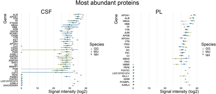
www.nature.com/scientificreports/
Figure 6. Protein–protein interactions (PPIs) and enriched pathways of 72 over-represented proteins in CSF
compared to PL commonly in chicken, budgerigar and cockatiel. (a) STRING analysis of the protein interaction
network showing associations of the proteins. Line thickness indicates the strength of data support (STRING
version 11.0, http://string-db.org). (b) Hierarchical clustering tree summarizing the correlation among the
significant pathways enriched in the 72 commonly over-represented CSF proteins across the three species. GO
terms of biological functions revealed pathways related to neurogenesis and neuron differentiation. Pathways
with many shared genes are clustered together and dot sizes indicate the degree of statistical significance
(ShinyGO version 0.61, http://bioinformatics.sdstate.edu/go/).
Scientific Reports | (2021) 11:5312 | https://doi.org/10.1038/s41598-021-84274-x 8
Vol:.(1234567890)www.nature.com/scientificreports/
Figure 7. Significant gene sets related to neural function commonly enriched in all species. The dot plot shows
the identified enriched terms by GSEA with nominal p value < 0.05. P values were adjusted for multiplicity of
testing. The x-axis shows the range of the false discovery rate (FDR) values, the y-axis lists the GO Biological
function terms, the colours indicate the involved species and the dot size shows the number of genes in each set
(GSEA version 4.1.0, http://www.gsea-msigdb.org; R version 4.0.0, www.r-project.org).
Scientific Reports | (2021) 11:5312 | https://doi.org/10.1038/s41598-021-84274-x 9
Vol.:(0123456789)www.nature.com/scientificreports/
Figure 8. Gene set enrichment analysis (GSEA) and comparison of cerebrospinal fluid and plasma proteomes
reveals neurogenesis in all studied avian species. (a) GSEA plots of the GO term Neurogenesis for chicken
(GG), budgerigar (MU) and cockatiel (NH) in cerebrospinal fluid compared to plasma (GSEA version 4.1.0,
http://www.gsea-msigdb.org) (b) Circular plot representation of proteins identified by GSEA with the GO term
Neurogenesis for GG, MU and NH. The proteins are labelled by their gene codes (R version 4.0.0, www.r-proje
ct.org).
Scientific Reports | (2021) 11:5312 | https://doi.org/10.1038/s41598-021-84274-x 10
Vol:.(1234567890)www.nature.com/scientificreports/
abundant proteins, our clustering analysis clearly differentiated CSF and plasma samples in all studied individuals
and 58.5% of the shared proteins across species, such as gamma-enolase, neuronal growth regulator 1 or neural
cell adhesion molecule 1, could be identified selectively in CSF.
Using gel-based techniques and mass spectrometry, the proteomes of eCSF from rat, chicken and human
have been previously described to elucidate the molecular composition of CSF and its biological role during
embryonic neurogenesis10–12, 48. Human eCSF contained 188 proteins, 44% of those also present in rat eCSF.
Functional analysis revealed protease inhibitors, extracellular matrix proteins, and transport proteins as well as
signalling and intracellular proteins12. Comparing the data available for human eCSF with our results in birds,
39%, 50% and 47% of the proteins overlap with adult CSF from chicken, budgerigar and cockatiel, respectively.
In chicken embryos investigated at the developmental stage HH24, using 2D-PAGE, 26 proteins were identified,
related to the extracellular matrix, regulation of the osmotic pressure and metal transport, cell survival, transport
of retinol and vitamin D as well as mitogen-activated protein kinase activators, antioxidant and antimicrobial
proteins, intracellular proteins and some unknown proteins. We found that 50% of these proteins were included
also in our data. Many of these proteins regulate the development in systems other than CNS, while others are
altered during n eurodegeneration10, 11.
LC-based methods allowed the identification of a dramatically increased number of proteins in CSF. Most
recently, using LC–MS/MS after abundant protein depletion, Macron et al. identified 3,174 proteins in a com-
mercial pool of “normal” human CSF s amples15. However, expectedly, interindividual variation is accountable
for identification of many of the rare proteins. For example, out of 2,615 CSF proteins, only 20% of the identified
proteins were shared by all six healthy subjects16. In a combined dataset from three studies the full human CSF
proteome of 4814 proteins was revealed, but only about 40% of these proteins were common. We found that about
a half of the proteins identified in either plasma or CSF in each species showed a high degree of inter-individual
variability, while the other half was relatively consistently represented across all individuals. In our study we have
shown that 71.22 and 55.27% (GG), 41.97 and 45.31% (MU), and 56.92 and 38.87% (NH) proteins identified
for CSF and PL respectively were common to all individuals within species and only 193 proteins were common
across all three species. Thus, the number of proteins that are individually specific is high. The reasons for this
variation, however, remain u nclear42, 49, 50. The molecular variations in CSF composition can be possibly increased
by post-translational modifications, age, blood c ontamination47 and circadian r hythm51.
We also found proteins of intracellular origin in the CSF, unlike in PL. These were consistently observed in
human, rat and chick CSF/eCSF and classified mainly as “binding proteins”10, 12, 52–54. It has been suggested that
these proteins are waste products eliminated from the brain extracellular space by the CSF. A small proportion
could also indicate cellular contamination of the samples54. Post-mortem samples have been previously reported
to contain also cytoskeletal proteins, glycolysis and antioxidant enzymes released into the CSF following cell
necrosis55.
Our comparative analysis shows that CSF and plasma were distinguishable based on their proteome compo-
sitions. From the proteins over-represented in CSF compared to plasma, such as homologs of neuronal growth
regulator 1 (NEGR1) and reelin (RELN), 72 were shared across all the three avian species. A PPI network
constructed based on these proteins, as well as the enrichment analysis of biological functions revealed namely
representation of pathways associated with cell adhesion, nervous system development and neurogenesis. In
addition, using GSEA, we indicated in CSF enrichment of pathways involved in neural functions across the
studied species. In addition to those revealed by differential analysis, the CSF samples shared several gene prod-
ucts related to neurogenesis for example neogenin 1 (NEO1), neurofascin (NFASC), neuromodulin (GAP43),
or pigment epithelium-derived factor (SERPINF1). Our results suggest that adult neurogenesis occurs in all the
avian species studied. Previous reports have stressed the importance of various growth factors present in CSF
which bind to receptors located on the apical membrane of cortical progenitor cells and promote the develop-
ment and growth of neural stem cells and cortical e xplants9, 56. In this study, we identified proteins such as 14–3-3
proteins (YWHAG , YWHAZ) and fibroblast growth factor receptor 1 (FGFR1-4) participating in the fibroblast
growth factor pathway57, molecules interacting with the Wtn9 pathway such as the Wtn inhibitor Dickkopf-
related protein 3 (DKK3) and cadherins (CDH13, CDH2, CDH6, CDH4), amyloid beta A4 protein precursor
(APP)12, insulin-like growth factor II (IGF2) and insulin-like growth factor-binding proteins (IGFALS, IGFBP2,
IGFBP7)9, 58. However, how these proteins are regulated in the context of systems level, their lifetime in CSF, as
well as the contribution of interindividual variability requires further investigation.
In vitro studies of adult human CSF treatment in neural stem cells showed that adult human CSF promoted
gliogenesis, but not neurogenesis59, 60. Despite ontogenetic differences in CSF protein composition in its ability
to induce neurogenesis, the neurogenic potential is preserved in the adult mammalian brain and can be activated
only by external stimuli including exercise, signalling molecules following injury or e CSF6, 61. In contrast, in birds,
neuronal cell proliferation occurs in the subventricular zone of the lateral ventricle, and migration to widespread
regions of telencephalon follows62. Immunohistochemical studies have demonstrated adult neurogenesis in the
telencephalon and other brain regions of canaries63, marsh tits64, ring doves65, chicken66, pigeons67, 68, parrots69,
ostrich and emu70. While interspecific differences in adult neurogenesis likely exist, it is well established that
factors such as age, environmental complexity, seasonal variation, hormones, stress, rearing conditions, physical
activity, social isolation and social complexity influence its outcome18. For example, the protein expression of vari-
ous neurotrophic factors such as nerve growth factor, insulin-like growth factor 1, vascular endothelial growth
factor, and brain-derived neurotrophic factor may alter upon increased physical activity or dietary r estriction71.
It is difficult to analyse the effects of the above-mentioned factors across species in this study, because the species
examined have different developmental modes (precocial and nidifugous chickens vs. altricial and nidicolous
parrots). The functional role of adult neurogenesis in birds remains unknown. It has been proposed to be asso-
ciated with continuous learning. In adult mammals, the proliferative hot spots may disappear a ltogether62, but
comparative approaches between birds and mammals have elucidated similarities on the origin of neurons, the
Scientific Reports | (2021) 11:5312 | https://doi.org/10.1038/s41598-021-84274-x 11
Vol.:(0123456789)www.nature.com/scientificreports/
phenotype and proliferative mechanisms of stem cells and the migration and differentiation of neurons as well
as differences such as the spatial distribution of adult-born neurons, their mode of migration and phenotypic
diversity3, 69, 72. We found that chicken and parrots share common proteins and pathways associated with the
modulation of neuronal cell proliferation and plasticity in CSF. Being based on a single methodological approach,
our identification of these gene sets and pathways in birds serves for an initial insight and subsequent experimen-
tal verification is required. Nevertheless, our study forms an essential reference for further molecular analysis.
Conclusion
The advent of the proteomic techniques has allowed new insights into molecular processes driving the CNS func-
tion, with research in CSF forming its cornerstone. For the first time, we concurrently explored in birds the pro-
teomes of two main biological fluids, PL and CSF. Importantly, we focused on multiple species, the chicken and
two parrot species, the budgerigar and cockatiel, investigating adult individuals. The proteins over-represented
in CSF were mainly classified into functional pathways involved in neural function, providing a novel molecular
evidence for adult neurogenesis in birds. Consistently with histological evidence for adult neurogenesis in birds,
understanding the molecular components involved in this process is essential for designing future experimental
work in this system.
Methods
Sample collection. CSF and PL samples were collected from a total of seven chicken, five budgerigars and
five cockatiels. The animals were originated from legal imports for the pet trade (Prague, Czech Republic) who
confirmed their status as healthy, adult birds and were kept in our animal facility (accreditation No. 37428/2019-
MZE-18134). The animals were euthanised by CO2 and mounted on a stereotaxic instrument (Stoelting Co.,
Illinois, USA). After incision in the dorsal neck region and exposure of the skull occipital region, a needle of a
0.5 ml sterile insulin syringe was inserted into cisterna magna73. Approximately 3–10 µl of clear, colourless CSF
was collected per animal and stored at −80 °C until analysis preparation. Blood was extracted from the carotid
artery and PL obtained by centrifugation (14,000 g) was stored at −80 °C, until further use. Experimental design
involving animals was performed by accredited researchers (EV: CZ03814, MV: CZ02568). The research was
approved by the Ethical Committee of Charles University, Faculty of Science (permits MSMT-1373/2016-4 and
MSMT-30397/2019-5) and was carried out in accordance with the current laws of the Czech Republic and Euro-
pean Union and in compliance with the ARRIVE guidelines.
Protein extraction and precipitation. The ProteoSpin detergent-free total protein isolation kit (Norgen
Biotek, Thorold, ON, Canada) was used for isolation and purification of the total protein content from CSF and
PL according to the manufacturer’s instructions and then frozen at −20 °C. Protein precipitation was performed
using acetone (1/5, acetone/protein) at −20 °C for 1 h. All precipitated samples were centrifuged at 14,000 g and
4 °C for 15 min. After centrifugation, the supernatant was discarded, the samples dried for 30 min at 37 °C and
were cleaved with trypsin (i.e., 1/50, trypsin/protein) at 37 °C overnight before mass spectrometry analysis.
nLC − MS/MS analysis. Nano reversed-phase columns were used to elute peptide cations using a previously
described method74. The eluting peptide cations were converted to gas-phase ions by electrospray ionization
and analysed on a Thermo Orbitrap Fusion mass spectrometer (Q-OT-qIT, Thermo). Survey scans of peptide
precursors from 350 to 1400 m/z were performed at 120 K resolution (at 200 m/z) with a 5 × 10⁵ ion count target.
Tandem MS/MS was performed by isolation at 1.5 Th with the quadrupole, high-energy collision dissociation
fragmentation with a normalized collision energy of 30 and rapid scan MS analysis in the ion trap. The MS/MS
ion count target was set to 104 and the max injection time was 35 ms. Only those precursors with a charge state
of 2–6 were sampled for MS/MS. The dynamic exclusion duration was set to 45 s with a 10-ppm tolerance around
the selected pre-cursor and its isotopes. Monoisotopic precursor selection was turned on and the instrument was
run at top speed with 2 s cycles.
Proteome data analysis. All data were collected and quantified using MaxQuant software version
1.6.10.4375. False discovery rate (FDR) was set to 1% for identification of all peptides and proteins. We set a
minimum peptide length of seven amino acids. The Andromeda search engine was used for the MS/MS spectra
search against the chicken Gallus gallus UniProt reference proteome (downloaded on March 2020, contain-
ing 27,540 entries), budgerigar Melopsittacus undulatus UniProt reference proteome (downloaded on Febru-
ary 2020, containing 23,704 entries) and zebra finch Taeniopygia guttata UniProt reference proteome (down-
loaded on February 2017, containing 23,455 entries), with all duplicates removed. Enzyme specificity was set
as C-terminal to Arg and Lys, also allowing cleavage at proline bonds76 and a maximum of two missed cleav-
ages. Dithiomethylation of cysteine was selected as a fixed modification and N-terminal protein acetylation and
methionine oxidation as variable modifications. Quantifications were performed with the label-free quantifica-
tion algorithms using a combination of unique and razor peptides75. The mass spectrometry proteomics data
have been deposited to the ProteomeXchange Consortium via the P RIDE77 partner repository with the dataset
identifier PXD021633.
Mapping approaches. Annotated reference proteomes are presently available for the c hicken78 and the
budgerigar79, but not for the cockatiel. Thus, mapping cockatiel reads on its own reference proteome was not
possible. To estimate the effect of this condition, we compared the MS/MS identification success rates of chicken,
budgerigar and cockatiel proteomes, each mapped against three applicable avian reference proteomes, the
Scientific Reports | (2021) 11:5312 | https://doi.org/10.1038/s41598-021-84274-x 12
Vol:.(1234567890)www.nature.com/scientificreports/
chicken, budgerigar and zebra finch. We performed the following data filtering to eliminate low-scoring spec-
tra: from further analysis we excluded all the unlabelled peptides, peptides only identified by site or reverse, all
contaminants, proteins identified by < 2 peptides and all proteins occurring in less than 1/3 of the samples for
each group. For the purpose of comparison, chicken ortholog gene codes were assigned to all identified proteins
using the database OrthoDB (v10.1)80. When the gene code was not possible to retrieve, the UniProt protein ID
was used.
For protein classification, we used the online Protein Analysis Through Evolutionary Relationships (PAN-
THER) library81. To find overrepresented Gene Ontologies (GOs)82 in the core proteomes of CSF and PL, we
launched the g:profiler against the chicken reference list (all genes in the database) and the annotation data sets of
the Gene Ontology and biological pathways (KEGG, REACTOME)83–85. The Benjamini–Hochberg FDR threshold
was set to 0.05 of all identified proteins in PL and CSF of chicken, budgerigar and cockatiel.
Statistical analysis. The statistical and bioinformatic analysis conducted in the R software86, Microsoft
Excel and Perseus software platform (http://www.perseus-framework.org; Max Planck Institute of Biochemistry,
Martinsried, Germany). The Venn diagrams were prepared using Biovenn87 and Jvenn88. In order to remove
minor technical variation between samples, the abundances of the identified proteins were previously normal-
ised using the Variance stabilization normalization m ethod89, 90 and DEP 1.12.0 R package. Missing data were
imputed using random draws from a manually defined left-shifted Gaussian distribution applying the default
settings of Perseus software. PCA was performed to examine the variation between the PL and CSF fluids in these
species using Perseus and R. Significantly differentially represented proteins were identified by a Student’s t-test
corrected by the Permutation Based FDR (FDR-adjusted p value < 0.05). In order to visualise biological protein
groups clustered together in either CSF or PL, hierarchical cluster analysis was performed in Perseus using
default parameters (Euclidean distance with average linkage, 300 clusters, 10 iterations, 1 restart and k-means
pre-processing for both row and column tree). To indicate the proteins significantly differentially represented in
CSF and PL in the three studied species, a Volcano Plot analysis was performed in Perseus (unpaired Student’s
T-test, S0 = 2, permutation-based FDR) and R. In the volcano plot, protein intensity fold-change between the
fluids is represented as log2. The Retrieval of Interacting Genes (STRING 11.0; http://string-db.org)91 web-tool
was used to analyse and construct the PPI network of the common group of proteins across the three species
that were over-represented in CSF compared to PL (adjusted p value cut off (FDR) < 0.05, identified by the t-test
analysis described above) . The interaction sources included experimental data and significant PPIs were consid-
ered those with a combined score > 0.4 and annotations from human (Homo sapiens). For the network visualiza-
tion, also using annotations from Homo sapiens, we used ShinyGO (v0.61)92 online tool to determine the GO
terms of biological functions which are significantly overrepresented in CSF compared to blood PL in the same
set of common gene products over-represented in the CSF compared to PL. GSEA was also performed. All pro-
teins were ranked based on the association between their abundance and the class distinction (CSF or PL), using
annotations from Homo sapiens (Human Gene Symbol with Remapping MSigDB.v7.2). We used the GOs analy-
sis of biological processes, nominal p value < 0.05 and adjusted p value < 0.25, as previously suggested. An FDR of
< 25% is recommended for discovering candidate gene sets to be further validated as a result of future research93.
Data availability
The mass spectrometry proteomics data have been deposited to the ProteomeXchange Consortium via the
PRIDE77 partner repository with the dataset identifier PXD021633.
Received: 30 December 2020; Accepted: 10 February 2021
References
1. Illes, S. Chapter 3 - More than a drainage fluid: the role of CSF in signaling in the brain and other effects on brain tissue. in
Handbook of Clinical Neurology (eds. Deisenhammer, F., Teunissen, C. E. & Tumani, H.) vol. 146 33–46, https://doi.org/10.1016/
B978-0-12-804279-3.00003-4 (Elsevier, 2018).
2. Reiber, H. Dynamics of brain-derived proteins in cerebrospinal fluid. Clin. Chim. Acta 310, 173–186, https://doi.org/10.1016/
S0009-8981(01)00573-3 (2001).
3. Lindsey, B. W. & Tropepe, V. A comparative framework for understanding the biological principles of adult neurogenesis. Prog.
Neurobiol. 80, 281–307, https://doi.org/10.1016/j.pneurobio.2006.11.007 (2006).
4. Villeda, S. A. et al. The ageing systemic milieu negatively regulates neurogenesis and cognitive function. Nature 477, 90–94, https
://doi.org/10.1038/nature10357 (2011).
5. Bachy, I., Kozyraki, R. & Wassef, M. The particles of the embryonic cerebrospinal fluid: How could they influence brain develop-
ment? Brain Res. Bull. 75, 289–294, https://doi.org/10.1016/j.brainresbull.2007.10.010 (2008).
6. Zappaterra, M. W. & Lehtinen, M. K. The cerebrospinal fluid: regulator of neurogenesis, behavior, and beyond. Cell. Mol. Life Sci.
69, 2863–2878, https://doi.org/10.1007/s00018-012-0957-x (2012).
7. Fame, R. M. & Lehtinen, M. K. Emergence and developmental roles of the cerebrospinal fluid system. Dev. Cell 52, 261–275, https
://doi.org/10.1016/j.devcel.2020.01.027 (2020).
8. Sawamoto, K. et al. New neurons follow the flow of cerebrospinal fluid in the adult brain. Science 311, 629–632, https://doi.
org/10.1126/science.1119133 (2006).
9. Lehtinen, M. K. et al. The cerebrospinal fluid provides a proliferative niche for neural progenitor cells. Neuron 69, 893–905, https
://doi.org/10.1016/j.neuron.2011.01.023 (2011).
10. Parada, C., Gato, A., Aparicio, M. & Bueno, D. Proteome analysis of chick embryonic cerebrospinal fluid. Proteomics 6, 312–320,
https://doi.org/10.1002/pmic.200500085 (2006).
11. Parada, C., Gato, Á. & Bueno, D. Mammalian embryonic cerebrospinal fluid proteome has greater apolipoprotein and enzyme
pattern complexity than the avian proteome. J. Proteome Res. 4, 2420–2428, https://doi.org/10.1021/pr050213t (2005).
12. Zappaterra, M. D. et al. A comparative proteomic analysis of human and rat embryonic cerebrospinal fluid. J. Proteome Res. 6,
3537–3548, https://doi.org/10.1021/pr070247w (2007).
Scientific Reports | (2021) 11:5312 | https://doi.org/10.1038/s41598-021-84274-x 13
Vol.:(0123456789)www.nature.com/scientificreports/
13. Smith, J. S. et al. Characterization of individual mouse cerebrospinal fluid proteomes. Proteomics 14, 1102–1106, https://doi.
org/10.1002/pmic.201300241 (2014).
14. Macron, C., Lane, L., Núñez Galindo, A. & Dayon, L. Deep Dive on the Proteome of human cerebrospinal fluid: a valuable data
resource for biomarker discovery and missing protein identification. J. Proteome Res. 17, 4113–4126, https://doi.org/10.1021/acs.
jproteome.8b00300 (2018).
15. Macron, C. et al. Exploration of human cerebrospinal fluid: A large proteome dataset revealed by trapped ion mobility time-of-
flight mass spectrometry. Data Brief 31, 105704, https://doi.org/10.1016/j.dib.2020.105704 (2020).
16. Begcevic, I., Brinc, D., Drabovich, A. P., Batruch, I. & Diamandis, E. P. Identification of brain-enriched proteins in the cerebrospinal
fluid proteome by LC-MS/MS profiling and mining of the Human Protein Atlas. Clin. Proteomics 13, 11, https://doi.org/10.1186/
s12014-016-9111-3 (2016).
17. Barker, J. M., Boonstra, R. & Wojtowicz, J. M. From pattern to purpose: how comparative studies contribute to understanding the
function of adult neurogenesis. Eur. J. Neurosci. 34, 963–977, https://doi.org/10.1111/j.1460-9568.2011.07823.x (2011).
18. Barnea, A. & Pravosudov, V. Birds as a model to study adult neurogenesis: bridging evolutionary, comparative and neuroethological
approaches. Eur. J. Neurosci. 34, 884–907, https://doi.org/10.1111/j.1460-9568.2011.07851.x (2011).
19. Patel, V. J. et al. A comparison of labeling and label-free mass spectrometry-based proteomics approaches. J. Proteome Res. 8,
3752–3759, https://doi.org/10.1021/pr900080y (2009).
20. Zhang, G. et al. Comparative genomics reveals insights into avian genome evolution and adaptation. Science 346, 1311–1320, https
://doi.org/10.1126/science.1251385 (2014).
21. Clayton, N. S. & Emery, N. J. Avian models for human cognitive neuroscience: a proposal. Neuron 86, 1330–1342, https://doi.
org/10.1016/j.neuron.2015.04.024 (2015).
22. Olkowicz, S. et al. Birds have primate-like numbers of neurons in the forebrain. Proc. Natl. Acad. Sci. 113, 7255–7260, https://doi.
org/10.1073/pnas.1517131113 (2016).
23. Němec, P. & Osten, P. The evolution of brain structure captured in stereotyped cell count and cell type distributions. Curr. Opin.
Neurobiol. 60, 176–183, https://doi.org/10.1016/j.conb.2019.12.005 (2020).
24. Wirthlin, M. et al. Parrot genomes and the evolution of heightened longevity and cognition. Curr. Biol. 28, 4001-4008.e7, https://
doi.org/10.1016/j.cub.2018.10.050 (2018).
25. Iwaniuk, A. N., Dean, K. M. & Nelson, J. E. Interspecific allometry of the brain and brain regions in parrots (Psittaciformes):
comparisons with other birds and primates. Brain. Behav. Evol. 65, 40–59, https://doi.org/10.1159/000081110 (2005).
26. Auersperg, A. M. I., Szabo, B., von Bayern, A. M. P. & Bugnyar, T. Object permanence in the Goffin cockatoo (Cacatua goffini). J.
Comp. Psychol. 128, 88–98, https://doi.org/10.1037/a0033272 (2014).
27. Pepperberg, I. M., Willner, M. R. & Gravitz, L. B. Development of Piagetian object permanence in grey parrot (Psittacus erithacus).
J. Comp. Psychol. 111, 63–75, https://doi.org/10.1037/0735-7036.111.1.63 (1997).
28. Emery, N. J. & Clayton, N. S. Evolution of the avian brain and intelligence. Curr. Biol. 15, R946–R950, https://doi.org/10.1016/j.
cub.2005.11.029 (2005).
29. Péron, F., Rat-Fischer, L., Lalot, M., Nagle, L. & Bovet, D. Cooperative problem solving in African grey parrots (Psittacus erithacus).
Anim. Cognit. 14, 545–553, https://doi.org/10.1007/s10071-011-0389-2 (2011).
30. Hobson, E. A., Avery, M. L. & Wright, T. F. The socioecology of Monk Parakeets: Insights into parrot social complexity. Socioec-
ología de Myiopsitta monachus: Revelaciones de la complejidad social de los loros Monk Parakeet socioecology. Auk Ornithol.
Adv. 131, 756–775, https://doi.org/10.1642/AUK-14-14.1 (2014).
31. Brauth, S. E., Heaton, J. T., Shea, S. D., Durand, S. E. & Hall, W. S. Functional Anatomy of Forebrain Vocal Control Pathways in
the Budgerigar (Melopsittacus undulatus)a. Ann. N. Y. Acad. Sci. 807, 368–385, https://doi.org/10.1111/j.1749-6632.1997.tb519
33.x (1997).
32. Pepperberg, I. M. The Alex Studies: cognitive and communicative abilities of grey parrots, (Harvard University Press, 2009).
33. Emery, N. J. Cognitive ornithology: the evolution of avian intelligence. Philos. Trans. R. Soc. B Biol. Sci. 361, 23–43, https://doi.
org/10.1098/rstb.2005.1736 (2006).
34. Güntürkün, O. & Bugnyar, T. Cognition without Cortex. Trends Cognit. Sci. 20, 291–303, https: //doi.org/10.1016/j.tics.2016.02.001
(2016).
35. Lambert, M. L., Jacobs, I., Osvath, M. & Bayern, A. M. P. von. Birds of a feather? Parrot and corvid cognition compared. Behaviour
156, 505–594, https://doi.org/10.1163/1568539X-00003527 (2019).
36. Iwaniuk, A. N. & Nelson, J. E. Developmental differences are correlated with relative brain size in birds: a comparative analysis.
Can. J. Zool. 81, 1913–1928, https://doi.org/10.1139/z03-190 (2003).
37. Munshi-South, J. & Wilkinson, G. S. Diet Influences Life Span in Parrots (Psittaciformes). Auk 123, 108–118, https://doi.
org/10.1093/auk/123.1.108 (2006).
38. Provost, K. L., Joseph, L. & Smith, B. T. Resolving a phylogenetic hypothesis for parrots: implications from systematics to conserva-
tion. Emu - Austral Ornithol. 118, 7–21, https://doi.org/10.1080/01584197.2017.1387030 (2018).
39. Wright, J. C., Beynon, R. J. & Hubbard, S. J. Cross Species Proteomics. in Proteome Bioinformatics (eds. Hubbard, S. J. & Jones, A.
R.) 123–135, https://doi.org/10.1007/978-1-60761-444-9_9 (Humana Press, 2010).
40. Liska, A. J. & Shevchenko, A. Expanding the organismal scope of proteomics: Cross-species protein identification by mass spec-
trometry and its implications. Proteomics 3, 19–28, https://doi.org/10.1002/pmic.200390004 (2003).
41. Dayon, L. et al. Proteomes of paired human cerebrospinal fluid and plasma: relation to blood–brain barrier permeability in older
adults. J. Proteome Res. 18, 1162–1174, https://doi.org/10.1021/acs.jproteome.8b00809 (2019).
42. Guldbrandsen, A. et al. In-depth characterization of the cerebrospinal fluid (CSF) proteome displayed through the CSF proteome
resource (CSF-PR). Mol. Cell. Proteomics MCP 13, 3152–3163, https://doi.org/10.1074/mcp.M114.038554 (2014).
43. Thompson, E. J. CHAPTER 4 - Differences between proteins in CSF and serum. in Proteins of the Cerebrospinal Fluid (ed. Thomp-
son, E. J.) 33–41, https://doi.org/10.1016/B978-012369369-3/50007-2 (Academic Press, 2005).
44. Reiber, H. Proteins in cerebrospinal fluid and blood: barriers, CSF flow rate and source-related dynamics. Restor. Neurol. Neurosci.
21, 79–96 (2003).
45. Anderson, N. L. & Anderson, N. G. The human plasma proteome: history, character, and diagnostic prospects. Mol. Cell. Proteomics
1, 845–867, https://doi.org/10.1074/mcp.R200007-MCP200 (2002).
46. Aasebø, E. et al. Effects of blood contamination and the rostro-caudal gradient on the human cerebrospinal fluid proteome. PLoS
ONE 9, e90429, https://doi.org/10.1371/journal.pone.0090429 (2014).
47. You, J.-S. et al. The impact of blood contamination on the proteome of cerebrospinal fluid. Proteomics 5, 290–296, https://doi.
org/10.1002/pmic.200400889 (2005).
48. Zhang, C. Proteomic studies on the development of the central nervous system and beyond. Neurochem. Res. 35, 1487–1500, https
://doi.org/10.1007/s11064-010-0218-z (2010).
49. Macron, C., Lane, L., Núñez Galindo, A. & Dayon, L. Identification of missing proteins in normal human cerebrospinal fluid. J.
Proteome Res. 17, 4315–4319, https://doi.org/10.1021/acs.jproteome.8b00194 (2018).
50. Kroksveen, A. C. et al. In-depth cerebrospinal fluid quantitative proteome and deglycoproteome analysis: presenting a compre-
hensive picture of pathways and processes affected by multiple sclerosis. J. Proteome Res. 16, 179–194, https://doi.org/10.1021/acs.
jproteome.6b00659 (2017).
Scientific Reports | (2021) 11:5312 | https://doi.org/10.1038/s41598-021-84274-x 14
Vol:.(1234567890)You can also read