In vitro antagonistic inhibitory effects of palm seed crude oils and their main constituent, lauric acid, with oxacillin in Staphylococcus aureus
←
→
Page content transcription
If your browser does not render page correctly, please read the page content below
www.nature.com/scientificreports
OPEN In vitro antagonistic inhibitory
effects of palm seed crude oils
and their main constituent,
lauric acid, with oxacillin
in Staphylococcus aureus
Klara Lalouckova1,2, Eva Skrivanova1,2, Johana Rondevaldova3, Adela Frankova4,
Josef Soukup1 & Ladislav Kokoska3*
Infections caused by Staphylococcus aureus are a serious global threat, and with the emergence
of antibiotic resistance, even more difficult to treat. One of the possible complications in
antistaphylococcal therapy represents negative interactions of antibiotics with food. In this study,
the in vitro interaction between oxacillin and crude palm seed oil from Astrocaryum vulgare,
Cocos nucifera, and Elaeis guineensis against nine strains of S. aureus was determined using the
checkerboard method. Lauric acid was identified as a major constituent of all tested oils by gas
chromatography. The results showed strong concentration dependent antagonistic interactions
between palm oils and oxacillin with values of fractional inhibitory concentrations indices ranging
from 4.02 to 8.56 at concentrations equal or higher than 1024 µg/mL of the tested oils. Similarly,
lauric acid in combination with oxacillin produced antagonistic action with fractional inhibitory
concentration indices ranging from 4.01 to 4.28 at 1024 µg/mL. These findings suggest that
interference between oxacillin and palm oils and their constituents can negatively affect the
treatment of staphylococcal infections in humans and other animals.
Staphylococcus aureus, a gram-positive bacterium, occurs naturally on the skin and mucus membranes of healthy
individuals and is a common cause of pneumonia, skin infections, and systemic infections in humans and other
animals1. Its ability to resist a broad range of antibacterials in a short period makes it one of the most dangerous
microorganisms influencing the global population, with strains resistant to beta-lactams, such as methicillin-
resistant S. aureus (MRSA) being considered a high priority pathogen2. However, currently used antistaphylococ-
cal antibiotics (e.g., vancomycin, daptomycin, and linezolid) are struggling against rising resistance, serious side
effects, and relatively high c osts3. Hence, steps toward elimination of bacteremia caused by multi-drug resistant
strains are still needed. Among other possible options, combinatory treatment is valuable for eradicating evolu-
tion of resistance by targeting different sites of the bacterial cell4,5. For example, the combination of vancomycin
and a beta-lactam antibiotic (oxacillin) is currently being used effectively in the treatment of M RSA6. A combi-
nation of beta-lactam antibiotic and beta-lactamase inhibitors, such as co-amoxiclav consisting of amoxicillin
and clavulanic acid, has also been used for MRSA therapy7. However, combinations of certain antibiotics can
produce negative interactions and undesirable side effects. Antagonistic action generally occurs in the treatment
of infection when mixing bactericidal and bacteriostatic d rugs8, as seen when using a combination of the slow
acting bactericidal vancomycin and bacteriostatic clindamycin against S. aureus9. Generally faster acting agents,
such as clindamycin or oxacillin, inhibit the function of vancomycin, which has a gradual onset of action and
exhibits antibacterial properties only on replicating c ells10.
1
Department of Microbiology, Nutrition and Dietetics, Faculty of Agrobiology, Food and Natural Resources, Czech
University of Life Sciences Prague, 165 00 Praha 6‑Suchdol, Czech Republic. 2Department of Nutritional Physiology
and Animal Product Quality, Institute of Animal Science, 104 00 Praha‑Uhrineves, Czech Republic. 3Department
of Crop Science and Agroforestry, Faculty of Tropical AgriSciences, Czech University of Life Sciences Prague, 165
00 Praha 6‑Suchdol, Czech Republic. 4Department of Food Science, Faculty of Agrobiology, Food and Natural
Resources, Czech University of Life Sciences Prague, 165 00 Praha 6‑Suchdol, Czech Republic. *email: kokoska@
ftz.czu.cz
Scientific Reports | (2021) 11:177 | https://doi.org/10.1038/s41598-020-80481-0 1
Vol.:(0123456789)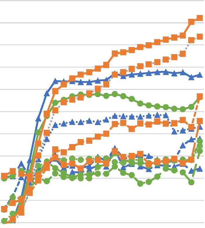
www.nature.com/scientificreports/
Combining various drugs is the prevalent praxis used to obtain an increase of the desired effects, such as in
anesthesia and pain management. However, enhancement of the undesired effects might also occur, limiting
the therapeutic v alue11. Such interactions are also known to occur when combining drugs with various foods
and food products. Drug-food interactions are defined as changes in efficacy and/or toxicity of pharmaceutical
drugs induced by the consumption of any food product, including functional foods and dietary s upplements12.
Various drug–food interactions (e.g., drug interaction with the fat content of the meal), drug–nutrient interac-
tions (e.g., with grapefruit juice or soy) and herb–drug interactions (e.g., with ginkgo or St John’s wort) have
been described and r eviewed13–16. The most well-known example of drug–food interaction is that of grapefruit
juice that can inhibit the intestinal metabolism of more than 85 drugs by altering cytochrome P450 (CYP3A4)
isoforms17. Grapefruit juice, combined with erythromycin, increases the bioavailability of the antibiotic in the
small intestine18, thereby increasing the possibility of adverse effects, such as cardiac d ysrhythmias19. In contrast,
a combination of ampicillin and pomegranate methanol extract acts synergistically in vitro against M RSA20.
Similarly, fatty acid methyl esters obtained from soybean, corn, and sunflower crude oils potentiate the antifungal
effect of itraconazole in vitro21.
Vegetable oils are essential components of animal nutrition and contain various biologically active constitu-
ents such as carotenoids, tocopherols22, coenzyme Q1023, and fatty acids24. These are carboxylic acids with long,
unbranched carbon chains, some of which may contain double bonds25. In general, fatty acids are believed to
be responsible for the antibacterial activity of palm oils. They usually occur in the form of triglycerides; their
hydrolysis into free fatty acids is necessary for their antibacterial effect to be exerted26–28. The seeds of tropi-
cal palms such as tucuma (Astrocaryum vulgare), coconut (Cocos nucifera), and African oil (Elaeis guineensis)
palm are one of the most economically important sources of plant oils, and are known to contain mainly fatty
acids of medium-chain length (MCFAs), with a prevalence of lauric acid ( C12:0; LA). Apart from various physi-
ological functions, the LA produces a growth-inhibitory effect against algae29, fungi30, protozoa31, and both
gram-negative32–34 and gram-positive bacteria35. It also inhibits the production of bacterial virulence factors
such as beta-lactamases, a group of exoproteins inducing beta-lactams inactivation and moreover enhancing
toxin production by S. aureus at subinhibitory antibiotic c oncentrations36; toxic shock syndrome t oxins37; and
hemolysins38. In addition to LA, tropical palms contain caproic ( C6:0), caprylic ( C8:0), and capric ( C10:0) acids that
also possess various antimicrobial properties39–41. In addition, MCFAs are known to be effective against S. aureus.
Batovska et al.42 concluded that when these fatty acids are esterified with glycerol and create e.g., monolaurin,
their direct antistaphylococcal effect is intensified. Ubgogu et al.43 observed a noticeable in vitro inhibitory effect
of E. guineensis palm kernel oil against S. aureus. The mechanism of antimicrobial action of fatty acids is not
fully known, the prime target seems to be the bacterial cell membrane together with various essential processes
that occur within and at the membrane, including nutrient uptake or enzyme inhibition44. The amphiphilic
nature of fatty acids enables them to act as detergents at high concentrations and aid the solubilization of the
lipids in the membranes45. It has been experimentally proven that doses of LA equal to or higher than 100 µg/
mL (≥ 500 µM) induce reversible morphological changes of lipid bilayers46, cause partial solubilization of the
cell membrane, and interfere with metabolic regulation, leading to the inhibition of bacterial growth44. It is well
known that bactericidal drugs are most potent with actively growing cells and that inhibition of growth, induced
by a combination with a bacteriostatic drug, can result in reduction of drug e fficacy8.
Besides the antimicrobial properties, LA is known to exhibit synergistic antistaphylococcal activity in com-
bination with m onolaurin42, lactic acid26, and gentamicin47. However, limited information is available regarding
the possible negative interactions of palm oils with antimicrobial agents. Therefore, we decided to perform a
screening test, focused on determining the combined effect of the chosen palm oils and free MCFAs ( C6:0–C12:0)
with representatives of all major antibiotic groups, namely beta-lactams (amoxicillin, ampicillin, and oxacillin),
tetracyclines (tetracycline), glycopeptides (vancomycin), and aminoglycosides (gentamicin), against three refer-
ence strains of S. aureus. Among the free MCFAs, only LA showed above mentioned synergism with gentamicin
against chosen S. aureus strains; the interactions of all free MCFAs with tetracycline and vancomycin were indif-
ferent, but beta-lactams, namely amoxicillin, ampicillin and especially oxacillin, showed results that were war-
ranted further investigation with LA showing the strongest antagonistic interactions (Lalouckova and Kokoska,
unpublished data). In the present study, we evaluated in vitro combinatory effect of A. vulgare, C. nucifera, and
E. guineensis seed crude oils and their main constituent LA with oxacillin, using the checkerboard microdilution
method, against different strains of S. aureus.
Results
Fatty acid composition of crude oils. In the first part of the study, fatty acid composition of the tested
oils was identified using GC-FID. As shown in Table 1, oils from A. vulgare, C. nucifera, and E. guineensis con-
sisted mainly of 58.2, 53.88 and 52.24 mg/g of MCFAs, respectively. LA was a major constituent of the oils, pre-
sent at a concentrations of 53.37 mg/g in A. vulgare, 45.24 mg/g in E. guineensis, and 41.31 mg/g in C. nucifera
oil. This profile corresponded with the total saturated fatty acid composition, where A. vulgare is followed by
C. nucifera and E. guineensis oil with values of 90.32, 82.35, and 80.21 mg/g, respectively. The contents of other
MCFAs, namely caprylic, capric, and caproic acids, were 6.73, 5.29, and 0.55 mg/g in C. nucifera, 3.48, 3.27, and
0.25 mg/g in E. guineensis, and 2.47, 2.15, and 0.21 mg/g in A. vulgare oil, respectively. In addition to MCFAs,
saturated fatty acids consisted of myristic, palmitic, and stearic acids. Their respective contents were 24.82, 5.41,
and 1.89 mg/g in A. vulgare; 16.5, 9.05, and 2.92 mg/g in C. nucifera; and 15.85, 9.46, and 2.66 mg/g in E. guineen-
sis oil.
Although saturated fatty acids were the major constituents of all oils analysed, the composition of monoun-
saturated (MUFAs) and polyunsaturated (PUFAs) fatty acids was also determined. MUFAs, consisting of oleic,
eicosenoic, and palmitoleic acids, were the most abundant in E. guineensis oil (16.67 mg/g), and were present at
Scientific Reports | (2021) 11:177 | https://doi.org/10.1038/s41598-020-80481-0 2
Vol:.(1234567890)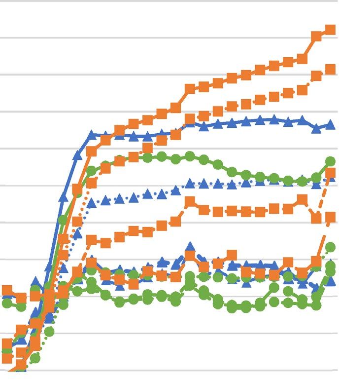
www.nature.com/scientificreports/
Fatty acids content (mg/g)/plant species
Astrocaryum vulgare Cocos nucifera Elaeis guineensis
Saturated fatty acids (SFA)
Caproic (C6:0) 0.21 0.55 0.25
Caprylic (C8:0) 2.47 6.73 3.48
Capric (C10:0) 2.15 5.29 3.27
Lauric (C12:0) 53.37 41.31 45.24
Myristic (C14:0) 24.82 16.5 15.85
Palmitic (C16:0) 5.41 9.05 9.46
Stearic (C18:0) 1.89 2.92 2.66
Total SFA 90.32 82.35 80.21
Total MCFA 58.2 53.88 52.24
Monounsaturated fatty acids (MUFA)
Palmitoleic (C16:1) 0.02 – 0.03
Oleic (C18:1) 6.47 11.72 16.54
Eicosenoic (C20:1) 0.05 0.15 0.10
Total MUFA 6.55 11.87 16.67
Polyunsaturated fatty acids (PUFA)
Linoleic (C18:2) 2.77 4.79 2.68
α-Linoleic (C18:3) 0.06 0.88 0.03
Arachidonic (C20:4) 0.07 0.13 0.13
Total PUFA 2.9 5.8 2.84
Table 1. Fatty acid profile of crude palm seed oils. Data are presented as average of two analyses, each
performed in triplicate.
relatively lower concentrations in C. nucifera and A. vulgare oil (11.87 and 6.55 mg/g, respectively). The oleic acid
content was the highest in all samples, ranging from 6.47 to 16.54 mg/g. In contrast, eicosenoic (0.05–0.15 mg/g)
and palmitoleic acid (≤ 0.03 mg/g) were present in minor amounts.
PUFA content in C. nucifera oil (5.8 mg/g) was double that of A. vulgare (2.9 mg/g) and E. guineensis
(2.84 mg/g) oil. In C. nucifera oil, linoleic acid was the most abundant PUFA, similar to A. vulgare and E.
guineensis oil at 4.79, 2.77, and 2.68 mg/g, respectively. The second most abundant PUFA in C. nucifera oil was
α-linoleic acid (1.88 mg/g), which was also present in lower amounts in A. vulgare (0.06 mg/g) and E. guineensis
(0.03 mg/g) oil. Compared to C. nucifera and A. vulgare oil (both 0.13 mg/g), arachidonic acid was the least
abundant PUFA in E. guineensis oil (0.07 mg/g).
Antistaphylococcal antagonistic effect of crude oils and oxacillin. In the first step, the suscep-
tibility of the three tested S. aureus strains to oxacillin and hydrolyzed seed oils of A. vulgare, C. nucifera, and
E. guineensis was determined using broth microdilution method to evaluate the suitable starting concentrations
for combined effect (MIC—minimum inhibitory concentration—values for all tested oils in non-hydrolyzed
forms > 8192 µg/mL; data not shown). A. vulgare oil induced the strongest antistaphylococcal effect, with MIC
values ranging from 240 to 356 µg/mL, followed by that of C. nucifera (MIC values from 241 to 512 µg/mL) and
E. guineensis (MIC values from 427 to 512 µg/mL). The MIC values of oxacillin ranged from 0.72 to 56.89 µg/mL.
Further, analysis of the combined effect of tested seed crude oils and oxacillin was performed by the check-
erboard method. The results showed some degree of antagonism in all tested strains at certain concentrations
of combinations, with indices of fractional inhibitory concentration (FICI) ranging between 8.56 and 4.02. In
addition, a consistently antagonistic, concentration-dependent effect of the selected palm oils at concentration of
2048 µg/mL, and in some cases 1024 µg/mL, was seen, when combined with oxacillin against all tested S. aureus
strains. All other combinations showed an indifferent relationship between tested agents, as shown in Table 2.
A strong antagonistic effect, manifested by very high FIC indices of 8.56 (clinical isolate SA1) and 8.51 (MSSA
ATCC 29213), was observed for A. vulgare oil at a concentration 2048 µg/mL, when combined with oxacillin. At
the same concentration, A. vulgare oil caused an adverse interaction in MRSA ATCC 43300 with a FICI of 5.78.
This antagonistic effect was also observed at a concentration of 1024 µg/mL with the MSSA clinical isolate, SA1
(FICI 4.30), and the reference strain, ATCC 29213 (FICI 4.27). Indifferent relationships were displayed in all
other combinations for both agents. Interestingly, a significant increase in MIC of oxacillin was observed when
combined with A. vulgare oil at two lowest concentrations tested (32 and 16 µg/mL). In these cases, the MIC
values rose by approximately 1/3 and 3/4, respectively, when compared to the MIC of the antibiotic alone, against
MSSA ATTC 29213. In combination with oxacillin, C. nucifera and E. guineensis oils also exerted antagonistic
properties in all tested strains, at a concentration of 2048 µg/mL (FICI 4.02–6.28). C. nucifera oil showed the
strongest antagonistic effect for MRSA ATCC 43300 strain, followed by MRSA clinical isolate SA1 and MSSA
ATCC 29213 strain (FICI values were 6.28, 6.03, and 4.09, respectively). E. guineensis oil exerted similar antago-
nistic activity in MSSA ATCC 29213, MRSA clinical isolate SA1, and MRSA ATCC 43300 strain, with FICI values
Scientific Reports | (2021) 11:177 | https://doi.org/10.1038/s41598-020-80481-0 3
Vol.:(0123456789)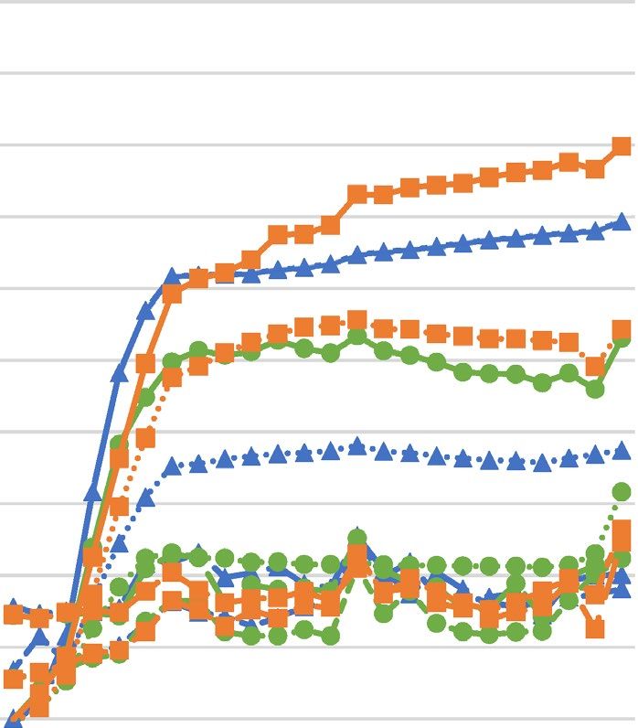
www.nature.com/scientificreports/

                     MIC of          OXA with Astrocaryum vulgare oil at concentration (µg/mL)
                     compounds alone
                     (µg/mL)         2048            1024               512               256                    128                 64              32             16
           S. aureus MIC             MIC                  MIC               MIC              MIC                 MIC                 MIC             MIC            MIC
           strain    OXA     MIC OIL OXA           FICI   OXA      FICI     OXA      FICI    OXA      FICI       OXA         FICI    OXA      FICI   OXA     FICI   OXA     FICI
           29213     1.67    242         0.06      8.51   0.06     4.27     0.06     2.16    0.23     1.20       0.44        0.80    1.11     0.93   2.22    1.47   2.89    1.80
           43300     56.89   356         1         5.78   1        2.90     6        1.55    7        0.84       21.67       0.74    30.22    0.71   43.56   0.86   53.33   0.98
           SA1       2       240         0.06      8.56   0.06     4.30     0.07     2.17    0.21     1.17       0.41        0.74    0.63     0.58   1.06    0.66   1.88    1
                     MIC of          OXA with Cocos nucifera oil at concentration (µg/mL)
                     compounds alone
                     (µg/mL)         2048            1024                512              256                    128                 64              32             16
           S. aureus MIC             MIC                  MIC               MIC              MIC                 MIC                 MIC             MIC            MIC
           strain    OXA     MIC OIL OXA           FICI   OXA      FICI     OXA      FICI    OXA      FICI       OXA         FICI    OXA      FICI   OXA     FICI   OXA     FICI
           29213     0.72    512         0.06      4.09   0.71     2.98     0.32     1.44    0.38     1.02       0.71        1.23    0.94     1.43   2.39    3.37   2       2.80
           43300     49.78   327         1         6.28   1        3.15     1        1.59    6.89     0.92       14.22       0.68    21.33    0.62   28.44   0.67   32      0.69
           SA1       2       241         0.06      6.03   0.06     3.03     0.08     1.54    0.28     0.89       0.58        0.67    1        0.69   1.33    0.76   2       1.05
                     MIC of          OXA with Elaeis guineensis oil at concentration (µg/mL)
                     compounds alone
                     (µg/mL)         2048             1024                512              256                   128                 64              32             16
           S. aureus MIC     MIC         MIC              MIC               MIC              MIC                 MIC                 MIC             MIC            MIC
           strain    OXA     OIL         OXA       FICI   OXA      FICI     OXA      FICI    OXA      FICI       OXA         FICI    OXA      FICI   OXA     FICI   OXA     FICI
           29213     0.75    427         0.06      4.88   0.06     2.48     0.09     1.32    0.31     1.02       0.53        1       0.69     1.08   0.78    1.11   0.56    0.78
           43300     56.89   512         1         4.02   1        2.02     7        1.12    15.78    0.78       19.56       0.59    32       0.69   37.33   0.72   42.67   0.78
           SA1       2       427         0.06      4.83   0.07     2.43     0.21     1.30    0.40     0.80       0.89        0.74    1.53     0.93   2       1.08   2       1.04

                                                Table 2.  Combinatory effect of crude palm seed oils and oxacillin against Staphylococcus aureus determined
                                                by checkerboard method. Data are presented as average of three analyses, each performed in triplicate.
                                                S. aureus Staphylococcus aureus, MIC minimum inhibitory concentration, FICI fractional inhibitory
                                                concentration index, OXA oxacillin. Bold values: antagonism (ƩFIC > 4).

                     MIC of             OXA with LA at concentration (µg/mL)
                     compounds
                     alone (µg/mL)      1024              512              256              128              64                     32               16             8
           S. aureus MIC                MIC               MIC              MIC              MIC              MIC                    MIC              MIC            MIC
           strain    OXA     MIC LA     OXA       FICI    OXA     FICI     OXA      FICI    OXA      FICI    OXA         FICI       OXA      FICI    OXA     FICI   OXA     FICI
           29213     0.53    256        0.01      4.01    0.01    2.01     0.01     1.02    0.58     1.61    0.67        1.51       0.78       1.6   0.89    1.75   0.78    1.5
           33591     683     256        8         4.01    8       2.01     8        1.01    319.56   0.97    853         1.5        1024       1.63 1024     1.5    1024    1.53
           33592     569     256        8         4.01    8       2.01     8        1.01    112.89   0.7     512         1.15       682.49     1.33 568.89   1.06   512     0.97
           43300     46.22   242        2         4.28    2.22    2.17     2.67     1.12    9.11     0.73    33.78       1          103.11     2.36 110.22   2.45   67.56   1.49
           EMRSA
                     99.56   256        1         4.01    1       2.01     1        1.01    8.22     0.58    112         1.38       > 128     > 1.41 128     1.35   128     1.32
           15
           BAA 976   32      256        0.83      4.03    0.83    2.03     0.83     1.03    19.56    1.11    85.33       2.92       128        4.13 118.86   3.78   64      2.03
           SA1       1.44    256        0.13      4.09    0.13    2.09     0.13     1.09    0.39     0.77    1           0.94       1.78       1.36 1.89     1.37   2.22    1.57
           SA2       67.56   256        2         4.03    2       2.03     7.11     1.11    24       0.86    74.64       1.36       92.44      1.49 85.33    1.33   74.67   1.14
           SA3       0.47    256        0.06      4.13    0.06    2.13     0.06     1.13    0.5      1.56    0.83        2.01       0.61       1.24 1.17     2.53   0.69    1.49

                                                Table 3.  Combinatory effect of lauric acid and oxacillin against Staphylococcus aureus determined by
                                                checkerboard method. Data are presented as average of three analyses, each performed in triplicate.
                                                S. aureus Staphylococcus aureus, MIC minimum inhibitory concentration, FICI fractional inhibitory
                                                concentration index, OXA oxacillin, LA lauric acid. Bold values: antagonism (ƩFIC > 4).

                                                of 4.88, 4.83, and 4.02, respectively. The rise of oxacillin MIC, when combined with the lowest concentrations
                                                (16–64 µg/mL) of the tested oils, was even more pronounced with C. nucifera. It induced an increase of MIC in
                                                MSSA ATCC 29213 strain up to more than two times. A slight increase of oxacillin MIC was also detected in
                                                ATCC 29213 strain at a concentration of 0.32 µg/mL of E. guineensis oil.

                                                Antagonistic growth‑inhibitory effect of lauric acid with oxacillin against S. aureus. As men-
                                                tioned above, GC-FID analysis revealed LA to be the predominant fatty acid in all tested oils. Based on this
                                                result, the susceptibility of nine S. aureus strains to oxacillin, LA, and a combination of oxacillin and LA was
                                                investigated using the same methodology as that used for palm oils. As shown in Table 3, MIC values of oxacillin
                                                ranged from 0.53 to 683 µg/mL. MIC values of LA were the same for all S. aureus strains at 256 µg/mL, except
                                                for MRSA ATCC 43300, where the MIC of LA decreased to 242 µg/mL.

          Scientific Reports |       (2021) 11:177 |                     https://doi.org/10.1038/s41598-020-80481-0                                                                4

Vol:.(1234567890)
www.nature.com/scientificreports/
A) B) 5
5
4.8 4.8
4.6 4.6
4.4 4.4
ln(OD*100) 4.2 4.2
ln(OD*100)
4 4
3.8 3.8
3.6 3.6
3.4 3.4
3.2 3.2
3 3
10.5
11.5
12.5
13.5
23.5
0
2
4
6
7
8
9
10.5
11.5
12.5
13.5
23.5
0
2
4
6
7
8
9
hour hour
C) 5
D) 5
4.8 4.8
4.6 4.6
4.4 ln(OD*100) 4.4
4.2 4.2
ln(OD*100)
4 4
3.8 3.8
3.6 3.6
3.4 3.4
3.2 3.2
3 3
0
2
4
6
7
8
9
10.5
11.5
12.5
13.5
23.5
10.5
11.5
12.5
13.5
23.5
0
2
4
6
7
8
9
hour hour
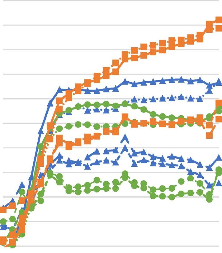
Figure 1. Growth curves of Staphylococcus aureus strains upon different concentrations of hydrolyzed palm
oils or lauric acid ((A)—Astrocaryum vulgare oil; (B)—Cocos nucifera oil; (C)—Elaeis guineensis oil; (D)—
lauric acid) determined spectrophotometrically. SA Staphylococcus aureus, OD optical density.
Subsequently, FICI values were calculated and the effect of combinations was analysed. Antagonistic mode
of action was observed for LA at a concentration of 1024 µg/mL, when combined with oxacillin (FICI values
were 4.01–4.28) in all tested S. aureus strains; the strongest antagonistic interaction was exhibited for the MRSA
ATCC 43300 strain. Other combinations of concentrations revealed indifferent relationships, except for a com-
bination of LA at 32 µg/mL with oxacillin against the S. aureus ATCC BAA 976 strain. In this case, the MIC of
oxacillin increased four times, to 128 µg/mL, and the FICI value was 4.13. In all tested bacterial strains, rise of
MIC values of tested antibiotics occurred when combined with LA at certain concentrations (16–256 µg/mL),
up to two times with the average increase by nearly two-thirds.
To determine whether the hydrolyzed seed oils of A. vulgare, C. nucifera, and E. guineensis and LA influence
the growth of S. aureus strains, the analysis of growth curves of three S. aureus strains, namely ATCC 29213,
ATCC 43300, and clinical isolate SA1, was performed. According to Fig. 1, the graphical evaluation of bacterial
Scientific Reports | (2021) 11:177 | https://doi.org/10.1038/s41598-020-80481-0 5
Vol.:(0123456789)www.nature.com/scientificreports/
growth upon increasing concentrations of hydrolyzed oils and LA revealed decreasing growth rate and increase
in generation time for all tested S. aureus strains.
Discussion
In our study, crude seed oils rich in MCFAs, hydrolyzed by porcine pancreatic lipase, and LA showed in vitro
growth-inhibitory effect against reference strains and clinical isolates of S. aureus. As per the results of GC
analysis, the oils used for antibacterial testing were of standard composition, as reported by other groups, who
also identified LA as the major constituent of A. vulgare48, C. nucifera49, and E. guineensis oils50. In this study,
the MIC values of LA against the tested S. aureus strains (242–256 µg/mL) were comparable to those reported
by other authors. For example, Batovska et al.42 reported an MIC values for LA, measured by the macrodilution
method against S. aureus strains at ≥ 125 µg/mL. Moreover, Nitbani et al.51 reported the antibacterial activity of
LA isolated from C. nucifera oil against S. aureus. In contrast, Parsons et al.52 determined an MIC value of 50 µg/
mL (250 µM) for LA against S. aureus, using the broth microdilution method. Similarly, Kelsey et al.53 observed
MIC values equal to 50 µg/mL of LA against three different S. aureus strains, using turbidimetry with visual
evaluation and ethanol as a solvent; however, these variations could be caused by the different methodologies
and S. aureus strains used. To the best of our knowledge, there is only limited information on the antibacterial
properties of palm oils rich in MCFAs. Free-fatty acids are known to have antibacterial activity in contrast with
those bound to t riglycerides27. As described by various authors p reviously46,52, the antistaphylococcal effect of
fatty acids is induced by disruption of bacterial cell membrane resulting in its destabilization by inducing tubule
formation on the lipid bilayer and cell lysis; thus, the antimicrobial activity of oils containing fatty acids is facili-
tated only upon hydrolysis of triglycerides26–28. In contrast, Ubgogu43 showed that E. guineensis oil exerted slight
antibacterial effect on S. aureus, without getting hydrolyzed, using the disc diffusion technique. This result is the
opposite of that observed in this study, where the oils acted as an antibacterial only after being hydrolyzed by
the porcine pancreatic lipase. Rossato et al.54 also observed no antistaphylococcal activity of unhydrolyzed A.
vulgare oil. Results of in vitro screening of C. nucifera oil and other MCFA-containing fats, with lipolytic enzymes
that simulated gastric conditions in piglets, showed a significant change in suppression of gut microbiota (total
anaerobes and E. coli)55. This finding suggests that MCFA-rich oils exert their antibacterial effects after enzymatic
hydrolysis by lipases synthesized in the gastrointestinal tracts of humans and other animals.
Studies on the combinatory effect of MCFAs and their esters with different organic acids, inorganic com-
pounds, and antibiotics against various bacteria, including S. aureus, can be found in literature26,42,47,56. However,
the findings of these studies markedly vary, depending on the class of antimicrobial agent used. For example,
Kitahara et al.47 observed synergistic interaction of LA at 50 µg/mL with gentamicin (FICI values were 0.25–0.31)
and imipenem (0.13–0.25) and indifferent interactions of LA (50 µg/mL) with ampicillin and oxacillin, against
MRSA clinical isolates. Similarly, Hess et al.57 observed indifferent interactions of the LA-ampicillin or vanco-
mycin combination against S. aureus biofilms and a synergistic interaction between LA and streptomycin. We
observed indifferent antistaphylococcal action of oxacillin and LA (50 µg/mL); however, increasing the amount
(≥ 1024 µg/mL) of LA, produced strong antagonism in the presence of the antibiotic. The reason for the lack of
antagonism in Kitahara et al.47 and Hess et al.57 could be the concentration of LA used in the study, which prob-
ably did not reach a sufficient value for the antagonism to be exerted. There is no information on the interaction
of palm oils rich in MCFAs as their antibacterial activity was not confirmed in the study54,58 because hydrolysis
of oils was not performed26.
From the results presented in this study, it is not possible to infer by which mechanism the antagonism
between LA, tested oils and oxacillin occurs. Nevertheless, the detected prolongation of the generation time
resulting in the decrease of specific growth rate arising with increasing concentrations of LA and LA-rich tested
palm oils indicates that a possible mechanism underlying the antagonistic interactions between tested compounds
might lie in the halting of cell division caused by LA/A. vulgare/C. nucifera/E. guineensis oil. It can be assumed
that while the LA temporarily prevents the growth of bacterial cells, oxacillin is not able to properly exert its activ-
ity. However, it is not just the membrane lipid structure that can change under the influence of exogenous lipids
but the protein structure as well. Other factors including membrane strain may account for the organization of
membrane proteins59. It can be hypothesized that under increased membrane strain, such as after the treatment
of lipid bilayers with high concentrations of LA46, membrane protein function is a ltered60. Such a circumstance
can affect the antibacterial activity of oxacillin, which strongly depends on its ability to inhibit bacterial cell wall
synthesis by preferentially binding to penicillin-binding proteins (PBPs) that are located inside the bacterial
cell wall61. Therefore, oxacillin probably becomes ineffective after change in PBP function, induced by LA. As
membrane protein stability also depends on membrane energetics, LA can reduce membrane fluidity and dis-
rupt the electron transport system, perhaps by restricting the movement of carriers within the membrane44. The
eventual impairment of membrane electron carriers can lead to a change in the intracellular and extracellular
pH, which can cause the precipitation of P BPs62 and make them to lose the ability to interact with oxacillin. In
addition, the change in extracellular pH can affect chemical structure of oxacillin as this drug is highly unstable
in acidic e nvironments63. Moreover, S. aureus is known to produce persisters, which are representing a fraction
of the bacterial population that exhibits tolerance to antibiotics in response to various s tresses64. According to
the finding of Peyrusson et al.65, S. aureus in the presence of high concentration of various antibiotics including
oxacillin showed a biphasic killing manner, meaning that a bulk of the bacterial population was susceptible and
rapidly killed while a subpopulation with a slower killing rate was persisting for a much longer period of time,
in addition showing the reversibility of the phenotype after antibiotic removal. Therefore, another possible
explanation of antagonism between oxacillin and LA/LA-rich palm oils, can be in inducing persister cells of S.
aureus in the presence of high concentrations of tested compounds.
Scientific Reports | (2021) 11:177 | https://doi.org/10.1038/s41598-020-80481-0 6
Vol:.(1234567890)www.nature.com/scientificreports/
Food-drug interactions are a major threat to safe and effective oral pharmacotherapy and can result in
decreased bioavailability of a drug, which predisposes the patient to treatment failure, increases the risk of adverse
events, and may even precipitate toxicities19,66. For this reason, coadministration of a drug with specific foods
is noted in medical leaflets. Generally, food intake can influence the effectiveness of an a ntibiotic67. Ingestion
of food, dietary fiber, or milk reduces the bioavailability of most antibiotics, including some penicillins68. For
example, minerals in milk and cheese create complexes with antibiotics that decrease their absorption69, and as
seen in the case of isoxazolyl penicillins, when administered shortly before or after a meal, delayed gastric empty-
ing and increased acidity interfere with their absorption70. The consumption of coconut oil and related products
is currently growing among certain populations, for the claimed health benefits associated with cardiovascular
disease and weight loss71. On the other hand, recommendations of lowering intake of saturated fatty acids and
replacing them with unsaturated fatty acids exist in order to reduce risk of atherosclerosis and type-2 diabetes72,73.
The average concentration of LA in human serum of healthy adult male and female blood donors, ages ranging
from 18 to 55 years, was found to be < 10 µg/mL74. It has, however, been proven that higher (14.2–140 g/day)
intakes of MCFAs in diet may result in higher concentrations of high density lipoprotein cholesterol than found
with long-chain fatty acids, highlighting the importance of considering chain length when measuring the effect
of dietary saturated fatty acids on lipid p rofile75. Moreover, in a high-carbohydrate, high-fat diet, the increases
in systolic blood pressure and diastolic stiffness in the heart were inhibited in mice with diet enriched by vir-
gin coconut oil, composed predominantly of LA, at concentrations of 200 g/kg76. Our results showing strong
in vitro antagonistic effect of oxacillin with LA or LA-rich palm oils at concentrations ≥ 1024 µg/mL (meaning
approximately ≥ 1.113 g/kg; 1 g/kg of body mass) suggest that simultaneous administration of these agents can
negatively affect their pharmacological properties. The recommended intake of fats in the diet of men is estimated
to be around 65 g/day (daily energy intake 2000 kcal with fats representing 30% of it)77. According to FAO78,
average energy requirement of an adult female is 2410 kcal/day and of an adult men is 3100 kcal/day. Counting
daily intake of fats as 30% of total energy requirement, the daily intake of fats can be estimated to be 80 g/day
for women and 103 g/day for men. Studies on coconut oil supplementation in diet usually focus on the addition
of the oil in range 20–50 g/day79–81. Nevertheless, there have been reports on even higher daily consumption
of coconut oil reaching up to 80 g/day82–84. Therefore, the observed antagonistic action between LA-rich oils/
LA may be important especially in high-fat diets. Thus, our results showing strong in vitro antagonistic effect
of oxacillin with LA or LA-rich palm oils suggest that simultaneous administration of these agents in high,
but still reachable concentrations can negatively affect their pharmacological properties in the treatment of S.
aureus. The risk of antagonistic interactions between oxacillin and LA-rich oils might be primarily important
to systemic application, as their antibacterial effect is attributed to fatty acids unleashed from triglycerides only,
therefore the topical application of LA-rich oils such as A. vulgare/C. nucifera/E. guineensis should not influence
the antibacterial activity of oxacillin. But, according to Verallo-Rowell et al.85, 5 mL of extra virgin coconut oil
applied two times a day on the affected areas that include the test sites is able to decolonise skin from S. aureus
in adults with atopic dermatitis. This discrepancies between the theoretical background and practice can be
debited to the lipolytic activity of skin microbiota, including s taphylococci86, highlighting the importance of
possible negative effect of LA-rich oils on topical treatment of S. aureus. Moreover, LA was previously tested
in vitro at concentration of 0.24–500 μg/mL to evaluate its antibacterial properties against various bacteria
causing inflammatory acne vulgaris, including S. aureus, proposing it as an promising remedy in the treatment
of staphylococcal skin i nfections87. However, the mentioned tested concentrations of LA were not high enough
for the antagonism with oxacillin to be exerted. Nevertheless, these hypotheses must be confirmed by further
in vitro and in vivo tests and clinical trials because physiological processes can also induce changes in antibacte-
rial activity of tested compounds.
In summary, this in vitro study revealed a concentration-dependent antagonistic effect between A. vulgare,
C. nucifera, and E. guineensis oils when combined with oxacillin in higher amounts against various strains of
S. aureus. The strongest antagonism was observed for A. vulgare oil, which contains the highest amount of LA.
This compound was identified as the main agent responsible for antagonistic antistaphylococcal action of all
oils assayed. To the best of our knowledge, this is the first study to report the antagonistic interactions between
these agents. The mechanism underlying the antagonistic action of tested agents probably acts at the cellular level
and is linked to the cell membranes. These findings suggest that interference between oxacillin and palm oils
and their constituents can negatively affect the treatment of staphylococcal infections in humans and animals.
However, these assumptions are based on in vitro tests and the negative interactions of the above-mentioned
combinations should be confirmed by in vivo trials.
Methods
Chemicals and samples preparation. LA, C. nucifera and E. guineensis oils, and oxacillin sodium mono-
hydrate were obtained from Sigma-Aldrich (Prague, CZ). A. vulgare oil was purchased from Sweet Natural
Botanicals (Panama City, FL, USA). LA and A. vulgare, C. nucifera, and E. guineensis oils were dispersed in dime-
thyl sulfoxide, which was previously proposed for sparingly soluble substances88, including the LA89 and palm
oil90; and emulsified using Tween 80 (Sigma-Aldrich, Prague, CZ), that is a common procedure of hydrophobic
sample preparation91, not influencing the antibacterial properties of tested compounds when used in recom-
mended concentrations92,93. Complete dissolution of LA was achieved by heating (70 °C for 10 min) it in an
ultrasonic-bath. Analysed plant oils were selected based on the high content of LA as described in l iterature94–96.
Hydrolysis of oils, to induce antimicrobial properties, was achieved by adding 5 × 10⁻³ mg/mL of porcine pancre-
atic lipase (Sigma-Aldrich, Prague, CZ) and shaking for 1 h in a water-bath heated to 37 °C. Degree of hydrolysis
of oils was dependent on the lipolytic activity of the enzyme. One unit of porcine pancreatic lipase hydrolyzes
1.0 microequivalent of fatty acid from triacetin in 1 h at pH 7.4, at 37 °C.
Scientific Reports | (2021) 11:177 | https://doi.org/10.1038/s41598-020-80481-0 7
Vol.:(0123456789)www.nature.com/scientificreports/
Bacterial strains and media. In this study, nine S. aureus strains were tested. Five reference strains includ-
ing methicillin sensitive (MSSA) ATCC 29213, MRSA ATCC 33591, ATCC 33592, ATCC 43300 and ATCC
BAA 976 were purchased from Oxoid (Basingstoke, UK). Three clinical isolates of drug resistant S. aureus (SA1,
SA2, and SA3) and one epidemic MRSA strain (EMRSA-15) were obtained from the Motol University Hospital
(Prague, CZ). Oxacillin, gentamicin, tetracycline, and penicillin were used as markers of the antibiotic resistance
etermined97–99. Based on the MIC values, clinical isolates were identified to be resistant
as it has been previously d
to: SA1—resistant to gentamicin (MIC 8 µg/mL) and tetracycline (MIC 8 µg/mL); SA2—resistant to oxacillin
(MIC 68 µg/mL), gentamicin (MIC 16 µg/mL) and tetracycline (MIC 8 µg/mL); SA3—resistant to gentamicin
(MIC 8 µg/mL) and penicillin (MIC 18.67 µg/mL); EMRSA-15—resistant to oxacillin (MIC 99.56 µg/mL) and
penicillin (MIC 16 µg/mL). The clinical isolates were identified using matrix-assisted laser desorption/ionization
time-of-flight mass spectrometry (MALDI-TOF MS), as described previously by Rondevaldova et al.98. Bacterial
stocks were stored at 4 °C on Müller–Hinton agar (Oxoid, Basingstoke, UK). Working cultures were maintained
in Müller-Hinton broth at 37 °C for 24 h before testing.
Determination of fatty acid composition. To evaluate fatty acid composition of oils obtained from A.
vulgare, C. nucifera, and E. guineensis, alkaline trans-methylation of fatty acids was carried out as described by
Raes et al.100. Analysis of methyl esters was performed using gas chromatography (GC) with the HP 6890 chro-
matograph (Agilent Technologies, Inc., Santa Clara, CA, USA), equipped with a 60 m DB-23 capillary column
(60 m × 0.25 mm × 0.25 μm) and a flame-ionization detector (FID); split injections were performed using an Agi-
lent autosampler. A total of 1 µL of standards in hexane were injected in split mode (1:40 ratio) into the injector,
heated to 230 °C. The column temperature was initially set at 120 °C for 6 min then programmed to 170 °C at a
rate of 15 °C/min. The temperate gradient was further configured to 210 °C at the rate of 3 °C/min and held for
13.5 min. Finally, the temperature was programmed to 230 °C at the rate of 40 °C/min and held for 7 min. Nitro-
gen was used as the carrier gas, at a flow rate of 0.8 mL/min. Supelco 37 Component FAME Mix, PUFA 1, PUFA
2, PUFA 3, trans-vaccenic acid, and a mixture of conjugated isomers of linoleic acid (Sigma-Aldrich, Prague, CZ)
were used as standards. Fatty acids were identified based on retention times with respect to standards.
Evaluation of minimum inhibitory concentrations and antagonistic combinatory effect. Using
guidelines of the Clinical and Laboratory Standards Institute101, the antibacterial activities of oxacillin; oils
extracted from A. vulgare, C. nucifera, and E. guineensis; and LA were evaluated in vitro by the broth microdilu-
tion method, modified as per the recommendations of Cos et al.102, for effectively assessing the anti-infective
potential of the natural products. Antistaphylococcal effect of a combination of oxacillin/LA or oxacillin/palm
oil was tested in vitro using the microdilution broth checkerboard method, as described in the Clinical Microbi-
ology Procedures Handbook103. The determination of MIC of oxacillin, palm oils and LA, as well as oxacillin/LA
or oxacillin/A. vulgare oil/C. nucifera oil/E. guineensis oil combinatory effect evaluation by FICI was performed
in 96-well microtiter plates. For the testing of combinatory effects, eight twofold serial dilutions of oxacillin were
placed in the horizontal rows of the plate and were subsequently cross-diluted vertically by eight twofold serial
dilutions of the test compound (palm oil or LA), resulting in 64 different combinations of concentrations. The
initial concentration for palm oils was 4096 µg/mL and for LA 2048 µg/mL; the starting concentration of anti-
biotic was adjusted according to the tested strain. The microtiter plate assay was performed using the automatic
pipetting platform, Freedom EVO 100 equipped with a four-channel liquid handling arm (Tecan, Männedorf,
CH). Cation-adjusted Müller-Hinton broth, equilibrated to a final pH of 7.6 with Trizma base (Sigma-Aldrich,
Prague, CZ) was used as growth medium. Buffering the culture media was performed to ensure the stability of
oxacillin that is known to decrease under the low pH conditions63. Inoculation of the plates was carried out using
bacterial suspensions, at a final density of 5 × 10⁵ CFU/mL, standardized using Densi-La-Meter II by adjust-
ing turbidity of the microorganism suspension to 0.5 McFarland standard. Next, incubation at 37 °C for 24 h
was performed. Evaluation of bacterial growth was performed spectrophotometrically using multimode reader
Cytation 3 (BioTek Instruments, Winooski, VT) at 405 nm. MIC values were expressed as the lowest compound
concentrations that resulted in ≥ 80% growth reduction compared to that of the agent-free growth control. The
lipase added to the Müller-Hinton broth, in concentrations ranging from 0.005 to 9.77 × 10⁻⁶ mg/mL did not
affect the growth of any strain of S. aureus tested when assayed as a negative control. FICI values were deter-
mined as ΣFIC, derived from the equation,
FIC = FICA + FICB
MICA (in the presence of B)
where, FICA = MICA (alone) and FICB = MICB (inMIC
the presence of A)
B (alone)
.
According to the value of FIC, three different effects could be observed according to O dds104—synergy
(ΣFIC ≤ 0.5), indifference (ΣFIC > 0.5–4), and antagonism (ΣFIC > 4). All compounds and their combinations
were tested in three independent experiments, each carried out in triplicate; MIC values and FICs presented in
this paper are average values.
Growth rates determination. To evaluate the influence of tested palm oils and LA concentrations on
parameters of the S. aureus growth, standardised microdilution assay was used105. Briefly, the determination
in 96-well microtiter plates where eight two-fold serial dilutions of tested compound (LA or A. vulgare/C.
nucifera/E. guineensis oil emulsion cleaved by porcine pancreatic lipase as described previously) starting at con-
centration 4096 µg/mL in cation-adjusted Müller-Hinton broth was performed. Next, the plates were inocu-
lated with bacterial suspensions at a final density of 5 × 10⁵ CFU/mL, standardized using Densi-La-Meter II by
adjusting turbidity of the microorganism suspension to 0.5 McFarland standard, same as in case of MIC and
FIC determination. Subsequently, incubation at 37 °C was performed, measuring the absorbance of each well
Scientific Reports | (2021) 11:177 | https://doi.org/10.1038/s41598-020-80481-0 8
Vol:.(1234567890)www.nature.com/scientificreports/
spectrophotometrically by multimode reader Cytation 3 (BioTek Instruments, Winooski, VT) at 405 nm every
half-to-one hour for 14 h and after 24 h.
Data availability
The datasets generated during and/or analysed during the current study are available from the corresponding
author on reasonable request.
Received: 5 May 2020; Accepted: 22 December 2020
References
1. Vos, P. et al. Bergey’s Manual of Systematic Bacteriology: The Firmicutes (Springer, Berlin, 2011).
2. WHO. Global Priority List of Antibiotic-Resistant Bacteria to Guide Research, Discovery, and Development of New Antibiotics
(WHO Press, Geneva, 2017).
3. Rodvold, K. A. & McConeghy, K. W. Methicillin-resistant Staphylococcus aureus therapy: Past, present, and future. Clin. Infect.
Dis. 58, S20–S27. https://doi.org/10.1093/cid/cit614 (2014).
4. Aldeyab, M. A. et al. Modelling the impact of antibiotic use and infection control practices on the incidence of hospitalac-
quired methicillin-resistant Staphylococcus aureus: A time-series analysis. J. Antimicrob. Chemother. 62, 593–600. https://doi.
org/10.1093/jac/dkn198 (2008).
5. Qin, X. et al. A randomised, double-blind, phase 3 study comparing the efficacy and safety of ceftazidime/avibactam plus
metronidazole versus meropenem for complicated intra-abdominal infections in hospitalised adults in Asia. Int. J. Antimicrob.
Agents 49, 579–588. https://doi.org/10.1016/j.ijantimicag.2017.01.010 (2017).
6. Tabuchi, F. et al. Synergistic effects of vancomycin and β-lactams against vancomycin highly resistant Staphylococcus aureus. J.
Antibiot. 70, 771–774. https://doi.org/10.1038/ja.2017.7 (2017).
7. Ba, X. et al. Old drugs to treat resistant bugs: Methicillin-resistant Staphylococcus aureus isolates with mecc are susceptible to a
combination of penicillin and clavulanic acid. Antimicrob. Agents Chemother. 59, 7396–7404. https: //doi.org/10.1128/AAC.01469
-15 (2015).
8. Ocampo, P. S. et al. Antagonism between bacteriostatic and bactericidal antibiotics is prevalent. Antimicrob. Agents Chemother.
58, 4573–4582. https://doi.org/10.1128/AAC.02463-14 (2014).
9. Booker, B. M., Stahl, L. & Smith, P. F. In vitro antagonism with the combination of vancomycin and clindamycin against Staphy-
lococcus aureus. J. Appl. Res. Clin. Exp. Ther. 4, 385–395 (2004).
10. Ho, J. L. & Klempner, M. S. In vitro evaluation of clindamycin in combination with oxacillin, rifampin, or vancomycin against
Staphylococcus aureus. Diagn. Microbiol. Infect. Dis. 4, 133–138. https://doi.org/10.1016/0732-8893(86)90147-1 (1986).
11. Zanderigo, E. et al. The well-being model a new drug interaction model for positive and negative effects. Anesthesiol. J. Am. Soc.
Anesthesiol. 104, 742–753. https://doi.org/10.1097/00000542-200604000-00019 (2006).
12. Mouly, S., Lloret-Linares, C., Sellier, P.-O., Sene, D. & Bergmann, J.-F. Is the clinical relevance of drug-food and drug-herb inter-
actions limited to grapefruit juice and saint-john’s wort?. Pharmacol. Res. 118, 82–92. https: //doi.org/10.1016/j.phrs.2016.09.038
(2017).
13. Boullata, J. I. & Hudson, L. M. Drug–nutrient interactions: A broad view with implications for practice. J. Acad. Nutr. Diet. 112,
506–517. https://doi.org/10.1016/j.jada.2011.09.002 (2012).
14. Cheng, T. O. Food-drug interactions. Int. J. Cardiol. 106, 392–393. https://doi.org/10.1016/j.ijcard.2004.12.083 (2006).
15. Fugh-Berman, A. Herb-drug interactions. Lancet 355, 134–138. https://doi.org/10.1016/S0140-6736(99)06457-0%20 (2000).
16. Pirmohamed, M. Drug-grapefruit juice interactions. BMJ 346, f1. https://doi.org/10.1136/bmj.f1 (2013).
17. Bailey, D. G., Dresser, G. & Arnold, J. M. O. Grapefruit–medication interactions: Forbidden fruit or avoidable consequences?.
CMAJ 185, 309–316. https://doi.org/10.1503/cmaj.120951 (2013).
18. Kanazawa, S., Ohkubo, T. & Sugawara, K. The effects of grapefruit juice on the pharmacokinetics of erythromycin. Eur. J. Clin.
Pharmacol. 56, 799–803. https://doi.org/10.1007/s002280000229 (2001).
19. Genser, D. Food and drug interaction: Consequences for the nutrition/health status. Ann. Nutr. Metab. 52, 29–32. https://doi.
org/10.1159/000115345 (2008).
20. Braga, L. et al. Synergic interaction between pomegranate extract and antibiotics against Staphylococcus aureus. Can. J. Microbiol.
51, 541–547. https://doi.org/10.1139/w05-022 (2005).
21. Pinto, M. E. et al. Antifungal and antioxidant activity of fatty acid methyl esters from vegetable oils. An. Acad. Bras. Cienc. 89,
1671–1681. https://doi.org/10.1590/0001-3765201720160908 (2017).
22. Chiu, M. C., de Morais Coutinho, C. & Gonçalves, L. A. G. Carotenoids concentration of palm oil using membrane technology.
Desalination 245, 783–786. https://doi.org/10.1016/j.desal.2009.03.002 (2009).
23. de Souza Guedes, L. et al. Study of the effect of the operating parameters on the separation of bioactive compounds of palm oil
by ultra-high performance supercritical fluid chromatography using a design of experiments approach. Can. J. Chem. Eng. 95,
2306–2314. https://doi.org/10.1002/cjce.22969 (2017).
24. Anushree, S., André, M., Guillaume, D. & Frédéric, F. Stearic sunflower oil as a sustainable and healthy alternative to palm oil.
A review. Agron. Sustain. Dev. 37, 18. https://doi.org/10.1007/s13593-017-0426-x (2017).
25. McGaw, L., Jäger, A. & Van Staden, J. Antibacterial effects of fatty acids and related compounds from plants. S. Afr. J. Bot. 68,
417–423. https://doi.org/10.1016/S0254-6299(15)30367-7 (2002).
26. Tangwatcharin, P. & Khopaibool, P. Activity of virgin coconut oil, lauric acid or monolaurin in combination with lactic acid
against Staphylococcus aureus. Southeast Asian J. Trop. Med. Public Health 43, 969–985 (2012).
27. Lee, S., Ariffin, A., Son, R. & Ghazali, H. Effect of lipase hydrolysis on the antibacterial activity of coconut oil, palm mesocarp
oil and selected seed oils against several pathogenic bacteria. Int. Food Res. J. 22, 46 (2015).
28. Hovorková, P., Laloučková, K. & Skřrivanová, E. Determination of in vitro antibacterial activity of plant oils containing medium-
chain fatty acids against gram-positive pathogenic and gut commensal bacteria. Czech J. Anim. Sci. 63, 119–125. https://doi.
org/10.17221/70/2017-CJAS (2018).
29. McGrattan, C. J., Sullivan, J. D. Jr. & Ikawa, M. Inhibition of chlorella (chlorophyceae) growth by fatty acids, using the paper
disc method. J. Phycol. 12, 129–131. https://doi.org/10.1111/j.1529-8817.1976.tb02839.x (1976).
30. Era, M. et al. Antifungal activity of fatty acid salts against Penicillium pinophilum. Jpn. J. Food Eng. 16, 99–108. https://doi.
org/10.11301/jsfe.16.99 (2015).
31. Dohme, F., Machmüller, A., Sutter, F. & Kreuzer, M. Digestive and metabolic utilization of lauric, myristic and stearic acid in
cows, and associated effects on milk fat quality. Arch. Anim. Nutr. 58, 99–116. https://doi.org/10.1080/00039420410001667485
(2004).
32. Sun, C. Q., O’Connor, C. J. & Roberton, A. M. Antibacterial actions of fatty acids and monoglycerides against Helicobacter pylori.
FEMS Immunol. Med. Microbiol. 36, 9–17. https://doi.org/10.1016/S0928-8244(03)00008-7 (2003).
Scientific Reports | (2021) 11:177 | https://doi.org/10.1038/s41598-020-80481-0 9
Vol.:(0123456789)www.nature.com/scientificreports/
33. Skrivanova, E., Marounek, M., Benda, V. & Brezina, P. Susceptibility of Escherichia coli, Salmonella sp. and Clostridium perfringens
to organic acids and monolaurin. Vet. Med. Praha 51, 81 (2006).
34. Thormar, H., Hilmarsson, H. & Bergsson, G. Stable concentrated emulsions of the 1-monoglyceride of capric acid (monocaprin)
with microbicidal activities against the food-borne bacteria Campylobacter jejuni, Salmonella spp., and Escherichia coli. Appl.
Environ. Microbiol. 72, 522–526. https://doi.org/10.1128/AEM.72.1.522-526.2006 (2006).
35. Bergsson, G., Arnfinnsson, J., Steingrímsson, Ó. & Thormar, H. Killing of gram-positive cocci by fatty acids and monoglycerides
note. Apmis 109, 670–678. https://doi.org/10.1034/j.1600-0463.2001.d01-131.x (2001).
36. Projan, S. J., Brown-Skrobot, S., Schlievert, P. M., Vandenesch, F. & Novick, R. P. Glycerol monolaurate inhibits the production
of beta-lactamase, toxic shock toxin-1, and other staphylococcal exoproteins by interfering with signal transduction. J. Bacteriol.
176, 4204–4209. https://doi.org/10.1128/jb.176.14.4204-4209.1994 (1994).
37. Ruzin, A. & Novick, R. P. Equivalence of lauric acid and glycerol monolaurate as inhibitors of signal transduction in Staphylococ-
cus aureus. J. Bacteriol. 182, 2668–2671. https://doi.org/10.1128/JB.182.9.26z68-2671.2000 (2000).
38. Liaw, S.-J., Lai, H.-C. & Wang, W.-B. Modulation of swarming and virulence by fatty acids through the rsba protein in Proteus
mirabilis. Infect. Immunity 72, 6836–6845. https://doi.org/10.1128/IAI.72.12.6836-6845.2004 (2004).
39. Hanczakowska, E. et al. Caprylic, capric and/or fumaric acids as antibiotic replacements in piglet feed. Ann. Anim. Sci. 11,
115–124 (2011).
40. Huang, C. B., Alimova, Y., Myers, T. M. & Ebersole, J. L. Short-and medium-chain fatty acids exhibit antimicrobial activity for
oral microorganisms. Arch. Oral Biol. 56, 650–654. https://doi.org/10.1016/j.archoralbio.2011.01.011 (2011).
41. Kollanoor-Johny, A. et al. Caprylic acid reduces Salmonella enteritidis populations in various segments of digestive tract and
internal organs of 3-and 6-week-old broiler chickens, therapeutically. Poult. Sci. 91, 1686–1694. https: //doi.org/10.3382/ps.2011-
01716(2012).
42. Batovska, D. I., Todorova, T., Tsvetkova, V. & Najdenski, H. M. Antibacterial study of the medium chain fatty acids and their
1-monoglycerides: Individual effects and synergistic relationships. Pol. J. Microbiol. 58, 43–47 (2009).
43. Ubgogu, O., Onyeagba, R. & Chigbu, O. Lauric acid content and inhibitory effect of palm kernel oil on two bacterial isolates
and Candida albicans. Afr. J. Biotechnol. 5, 1045 (2006).
44. Desbois, A. P. & Smith, V. J. Antibacterial free fatty acids: Activities, mechanisms of action and biotechnological potential. Appl.
Microbiol. Biotechnol. 85, 1629–1642. https://doi.org/10.1007/s00253-009-2355-3 (2010).
45. Churchward, C. P., Alany, R. G. & Snyder, L. A. Alternative antimicrobials: The properties of fatty acids and monoglycerides.
Crit. Rev. Microbiol. 44, 561–570. https://doi.org/10.1080/1040841X.2018.1467875 (2018).
46. Yoon, B. K., Jackman, J. A., Kim, M. C. & Cho, N.-J. Spectrum of membrane morphological responses to antibacterial fatty acids
and related surfactants. Langmuir 31, 10223–10232. https://doi.org/10.1021/acs.langmuir.5b02088 (2015).
47. Kitahara, T. et al. In vitro activity of lauric acid or myristylamine in combination with six antimicrobial agents against methicillin-
resistant Staphylococcus aureus (MRSA). Int. J. Antimicrob. Agents 27, 51–57. https://doi.org/10.1016/j.ijantimicag.2005.08.020
(2006).
48. Mambrin, M. & Arellano, D. B. Characterization of palm tree fruit oils from brazilian amazonia region. Grasas Aceites 48,
154–158. https://doi.org/10.3989/gya.1997.v48.i3.783 (1997).
49. Marina, A., Che Man, Y., Nazimah, S. & Amin, I. Chemical properties of virgin coconut oil. J. Am. Oil Chem. Soc. 86, 301–307.
https://doi.org/10.1007/s11746-009-1351-1 (2009).
50. Edem, D. Palm oil: Biochemical, physiological, nutritional, hematological and toxicological aspects: A review. Plant Foods Hum.
Nutr. 57, 319–341. https://doi.org/10.1023/A:1021828132707 (2002).
51. Nitbani, F. O., Jumina, S. D. & Solikhah, E. N. Isolation and antibacterial activity test of lauric acid from crude coconut oil.
Procedia Chem. 18, 132–140. https://doi.org/10.1016/j.proche.2016.01.021 (2016).
52. Parsons, J. B., Yao, J., Frank, M. W., Jackson, P. & Rock, C. O. Membrane disruption by antimicrobial fatty acids releases low-
molecular-weight proteins from Staphylococcus aureus. J. Bacteriol. 194, 5294–5304. https: //doi.org/10.1128/JB.00743- 12 (2012).
53. Kelsey, J., Bayles, K. W., Shafii, B. & McGuire, M. Fatty acids and monoacylglycerols inhibit growth of Staphylococcus aureus.
Lipids 41, 951–961. https://doi.org/10.1007/s11745-006-5048-z (2006).
54. Rossato, A. et al. Evaluation in vitro of antimicrobial activity of tucumã oil (Astrocaryum vulgare). Arch. Biosci. Health 1, 99–112.
https://doi.org/10.18593/abh.19701 (2019).
55. Dierick, N., Decuypere, J., Molly, K., Van Beek, E. & Vanderbeke, E. The combined use of triacylglycerols containing medium-
chain fatty acids (MCFAs) and exogenous lipolytic enzymes as an alternative for nutritional antibiotics in piglet nutrition: I.
In vitro screening of the release of MCFAs from selected fat sources by selected exogenous lipolytic enzymes under simulated
pig gastric conditions and their effects on the gut flora of piglets. Livest. Prod. Sci. 75, 129–142. https://doi.org/10.1016/s0301
-6226(01)00303-7 (2002).
56. Rosenblatt, J., Reitzel, R. A. & Raad, I. Caprylic acid and glyceryl trinitrate combination for eradication of biofilm. Antimicrob.
Agents Chemother. 59, 1786–1788. https://doi.org/10.1128/AAC.04561-14 (2015).
57. Hess, D. J., Henry-Stanley, M. J. & Wells, C. L. Antibacterial synergy of glycerol monolaurate and aminoglycosides in Staphylo-
coccus aureus biofilms. Antimicrob. Agents Chemother. 58, 6970–6973. https://doi.org/10.1128/AAC.03672-14 (2014).
58. Ferreira, B. S. et al. Comparative properties of amazonian oils obtained by different extraction methods. Molecules 16, 5875–5885.
https://doi.org/10.1128/10.3390/molecules16075875 (2011).
59. Robertson, J. L. The lipid bilayer membrane and its protein constituents. J. Gen. Physiol. 150, 1472–1483. https: //doi.org/10.1085/
jgp.201812153 (2018).
60. Zhang, X. C. & Li, H. Interplay between the electrostatic membrane potential and conformational changes in membrane proteins.
Protein Sci. 28, 502–512. https://doi.org/10.1002/pro.3563 (2019).
61. Papich, M. G. Saunders Handbook of Veterinary Drugs (Elsevier, Hoboken, 2007).
62. Roch, M. et al. Thermosensitive pbp2a requires extracellular folding factors prsa and htra1 for Staphylococcus aureus MRSA
β-lactam resistance. Commun. Biol. 2, 1–11. https://doi.org/10.1038/s42003-019-0667-0 (2019).
63. Rad, I., Croitoru, M. & Gyéresi, Á. Improvements of oxacillin stability in a ph = 1.2 acidic environment. Acta Med. Marisiensis
57, 328–330 (2011).
64. Kubistova, L., Dvoracek, L., Tkadlec, J., Melter, O. & Licha, I. Environmental stress affects the formation of Staphylococcus aureus
persisters tolerant to antibiotics. Microb. Drug Resist. 24, 547–555. https://doi.org/10.1089/mdr.2017.0064 (2018).
65. Peyrusson, F. et al. Intracellular Staphylococcus aureus persisters upon antibiotic exposure. Nat. Commun. 11, 1–14. https://doi.
org/10.1038/s41467-020-15966-7 (2020).
66. Koziolek, M. et al. The mechanisms of pharmacokinetic food-drug interactions—A perspective from the ungap group. Eur. J.
Pharm. Sci. 134, 31–59. https://doi.org/10.1016/j.ejps.2019.04.003 (2019).
67. Hodel, M. & Genné, D. Antibiotics: Drug and food interactions. Rev. Med. Suisse 5, 1979–1984 (2009).
68. Schmidt, L. & Dalhoff, K. Food–drug interactions. Drugs 62, 1481–1502. https://doi.org/10.2165/00003495-200262100-00005
(2002).
69. Bushra, R., Aslam, N. & Khan, A. Y. Food-drug interactions. Oman Med. J. 26, 77–83. https://doi.org/10.5001/omj.2011.21
(2011).
70. Marcy, S. M. & Klein, J. O. The isoxazolyl penicillins: Oxacillin, cloxacillin, and dicloxacillin. Med. Clin. N. Am. 54, 1127–1143.
https://doi.org/10.1016/S0025-7125(16)32582-2 (1970).
Scientific Reports | (2021) 11:177 | https://doi.org/10.1038/s41598-020-80481-0 10
Vol:.(1234567890)You can also read