Hypoxic Hypoxia and Brain Function in Military Aviation: Basic Physiology and Applied Perspectives - Frontiers
←
→
Page content transcription
If your browser does not render page correctly, please read the page content below
REVIEW
                                                                                                                                                   published: 17 May 2021
                                                                                                                                         doi: 10.3389/fphys.2021.665821

                                                Hypoxic Hypoxia and Brain Function
                                                in Military Aviation: Basic Physiology
                                                and Applied Perspectives
                                                David M. Shaw 1,2, Gus Cabre 1 and Nicholas Gant 3*
                                                1
                                                  Aviation Medicine Unit, Royal New Zealand Air Force Base Auckland, Auckland, New Zealand, 2 School of Sport, Exercise
                                                and Nutrition, Massey University, Auckland, New Zealand, 3 Department of Exercise Sciences, University of Auckland,
                                                Auckland, New Zealand

                                                Acute hypobaric hypoxia (HH) is a major physiological threat during high-altitude flight
                                                and operations. In military aviation, although hypoxia-related fatalities are rare, incidences
                                                are common and are likely underreported. Hypoxia is a reduction in oxygen availability,
                                                which can impair brain function and performance of operational and safety-critical tasks.
                                                HH occurs at high altitude, due to the reduction in atmospheric oxygen pressure. This
                           Edited by:
                          Ginés Viscor,
                                                physiological state is also partially simulated in normobaric environments for training and
        University of Barcelona, Spain          research, by reducing the fraction of inspired oxygen to achieve comparable tissue oxygen
                       Reviewed by:             saturation [normobaric hypoxia (NH)]. Hypoxia can occur in susceptible individuals below
                      Peter Kochunov,           10,000 ft (3,048 m) in unpressurised aircrafts and at higher altitudes in pressurised
    University of Maryland School of
              Medicine, United States           environments when life support systems malfunction or due to improper equipment use.
                Mathias Roland Aebi,            Between 10,000 ft and 15,000 ft (4,572 m), brain function is mildly impaired and hypoxic
    University of Lausanne, Belgium
                        Anna Gerega,
                                                symptoms are common, although both are often difficult to accurately quantify, which
      Institute of Biocybernetics and           may partly be due to the effects of hypocapnia. Above 15,000 ft, brain function exponentially
     Biomedical Engineering, Polish             deteriorates with increasing altitude until loss of consciousness. The period of effective
 Academy of Sciences (PAN), Poland
                                                and safe performance of operational tasks following exposure to hypoxia is termed the
                  *Correspondence:
                       Nicholas Gant            time-of-useful-consciousness (TUC). Recovery of brain function following hypoxia may
               n.gant@auckland.ac.nz            also lag beyond arterial reoxygenation and could be exacerbated by repeated hypoxic
                                                exposures or hyperoxic recovery. This review provides an overview of the basic physiology
                   Specialty section:
         This article was submitted to          and implications of hypoxia for military aviation and discusses the utility of hypoxia
   Environmental, Aviation and Space            recognition training.
                           Physiology,
               a section of the journal         Keywords: oxygen, hypoxaemia, cognitive function, performance, safety
               Frontiers in Physiology

        Received: 09 February 2021
           Accepted: 22 April 2021
                                                INTRODUCTION
           Published: 17 May 2021

                             Citation:          Acute hypoxia is a major physiological threat during high-altitude flight and operations in
            Shaw DM, Cabre G and                military aviation. The human brain requires a continuous oxygen supply to function effectively.
 Gant N (2021) Hypoxic Hypoxia and
                                                It is, therefore, vulnerable to environments with low atmospheric oxygen availability. At high-
   Brain Function in Military Aviation:
               Basic Physiology and
                                                altitude, the reduced barometric pressure (hypobaria) lowers the partial pressure of inspired
               Applied Perspectives.            oxygen (PiO2) causing hypoxic hypoxia; henceforth referred to as hypoxia. The resulting
          Front. Physiol. 12:665821.            hypoxaemia elicits a metabolic insult that impairs brain function and, with increasing severity,
   doi: 10.3389/fphys.2021.665821               will cause loss of consciousness and eventually death. High-altitude experiments from balloon

Frontiers in Physiology | www.frontiersin.org                                          1                                             May 2021 | Volume 12 | Article 665821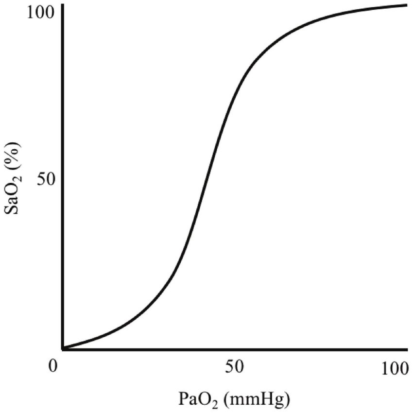
Shaw et al.                                                                                                                                  Hypoxia and Brain Function in Aviation

ascents in the 1800s were the first to describe the disastrous                                The threat of hypoxia also extends to all aircrew, including
events of acute hypoxia (West, 2016). Then, during the Second                                 rear crew, such as air warfare specialists, loadmasters, and
World War in the 1930s and 1940s, it became apparent that                                     medics. Whilst it is acknowledged that some military aircraft
the limitations of military aircraft were not necessarily due to                              can elicit loading in the +Gz axis (i.e., commonly referred to
mechanical or engineering failures, but the lack of oxygen to                                 as pulling Gs) to impair cerebral perfusion and cause stagnant
the brain of aircrew during flight. More recently, with the                                   hypoxia, such as high-performance jets, the physiological effects
advent of pressurised environments and oxygen supply systems,                                 and mitigating factors markedly differ from hypoxic hypoxia
pursuit into hypoxic environments is common. Acute hypoxia                                    and are considered outside the scope of the current review.
is the primary risk when life support systems malfunction in                                  Therefore, the aim of this review is to summarise the basic
these environments and have been a focal area of research in                                  physiology of hypoxic hypoxia on brain function and recovery
military aviation for several decades.                                                        and to discuss the implications for military aviation, including
   Compared with civilian aviation, military aircrews have to                                 the utility of hypoxia recognition training (HRT) for improving
navigate greater safety risks during flight, which increases the                              emergency responses to hypoxic incidences.
likelihood of being exposed to hypoxia. The onset of acute
hypoxia can be rapid and pronounced (i.e., seconds) or slow
and insidious (i.e., minutes-to-hours) depending on the type                                  HYPOXIA IN MILITARY AVIATION
of equipment malfunction and magnitude of the hypoxic dose.
Hypoxia may not present with clear physiological responses                                    Military aviation has the largest stake in hypoxia-related risks
or perceptible signs and symptoms (Table 1), which is a major                                 (Gradwell and Rainford, 2016). Since the early 1940s, aircraft
operational concern as unanticipated severe hypoxia will prevent                              have relied upon pressurised environments for safe, comfortable,
recognition of hypoxia and implementation of emergency                                        and efficient flight at high altitudes as the atmospheric oxygen
recovery procedures prior to loss of consciousness. For example,                              partial pressure (PO2) exponentially declines with increasing
in a hypoxic emergency, pilots are required to immediately                                    altitude [e.g., PO2 is 149 mmHg at sea-level and 49 mmHg
don oxygen mask, then to declare an in-flight emergency,                                      at 25,000 ft (7,620 m)]. Although hypoxia-related aviation
descend below 10,000 ft, and land as soon as possible.                                        fatalities are rare, incidences are common, particularly in fighter

TABLE 1 | Functional impairment and clinical status during hypoxia and hyperventilation-induced hypocapnia whilst sedentary at altitude.

                   Altitude                               PaO2                    PaCO2                                         Signs and symptoms

ft                                m                      mmHg                     mmHg              Hypoxia                                             Hypocapnia

                                                                                                    No symptoms and normal function                 No symptoms and normal
0–5,000                        0–1,524                   80–95                       40
                                                                                                                                                    function
                                                                                                    Impaired performance of novel or highly         Minor hyperventilation and
5,000–10,000              1,524–3,048                    80–60                    35–40             complex tasks; and impaired night and colour hypocapnia
                                                                                                    vision
                                                                                                    Impaired performance of some simple tasks; Mild dizziness; light-
                                                                                                    further impairment of novel and complex         headedness; and feelings
10,000–15,000             3,048–4,572                    40–60                    30–35             tasks; mild hyperventilation; reduced physical of unreality
                                                                                                    capacity; and headache if exposure is
                                                                                                    prolonged
                                                                                                    Moderate-to-severe cognitive impairment;        Moderate-to-severe
                                                                                                    confusion; task fixation; impaired critical     dizziness; light-
                                                                                                    judgement; reduced willpower; impaired          headedness;
                                                                                                    neuromuscular control; personality and mood apprehension;
                                                                                                    changes (e.g., euphoria, pugnacious, morose, neuromuscular irritability;
                                                                                                    and aggressiveness); hyperventilation; visual   paraesthesia of limbs and
15,000–20,000             4,572–6,096                    30–40                    25–30             impairments (including reduced peripheral       lips; tetany with
                                                                                                    vision, reduced light and colour intensity, and carpopedal; and facial
                                                                                                    visual acuity); hot or cold flushes; sweating;  spasms
                                                                                                    central and peripheral cyanosis; impaired
                                                                                                    sense of touch and fine motor skills; sensory
                                                                                                    loss; nausea; fatigue; lethargy; and possible
                                                                                                    loss of consciousness
                                                                                                    Myoclonic (muscle) twitches and convulsions; Loss of consciousness
Above 20,000              Above 6,096
Shaw et al. Hypoxia and Brain Function in Aviation
and training aircraft (Cable, 2003; Files et al., 2005). For
example, in 1055 aircraft depressurisation incidences between
1981 and 2003 within the United States Air Force, a reported
221 (21%) involved hypoxia, with three of these resulting in
(preventable) death (Files et al., 2005). It is probable that
hypoxia incidences are underreported, particularly when the
onset of hypoxia is slow or gradual (i.e., insidious), suggesting
the issue is greater than what is published.
The insidious onset of hypoxia may occur following an
inboard leak within a pressurised cabin or when ascending
in an unpressurised aircraft above 10,000 ft (3,048 m;
PO2 < 100 mmHg). In such situations, hypoxia may not
be identified as a causal factor for in-flight incidences and
accidents. In contrast, rapid onset hypoxia may occur following
a rapid depressurisation above 20,000 ft (6,096 m;
PO2 < 63 mmHg), such as following an explosion or loss
of the aircraft’s canopy. In these situations, environmental
cues, physiological responses, and brain dysfunction are more
evident and perceptible. Additionally, equipment malfunctions
with oxygen supply systems, such as liquid oxygen systems
or on board oxygen generating systems, are commonly
reported, with the latter a concern in newer generation
aircraft, such as the Hornet (The National Interest, 2016). FIGURE 1 | Illustration demonstrating the sigmoidal relationship between
Some individuals may also elicit mild symptoms and arterial blood oxygen-haemoglobin saturation (SaO2) and oxygen partial
pressure (PaO2).
performance impairments at low altitudes (below 10,000 ft;
Cable, 2003; Smith, 2005) and rapid loss of consciousness
at moderate altitudes (e.g., 18,000 ft or 5,486 m; PO2 when PaO2 declines below ~50 mmHg (i.e., ~85% SaO2) by
70 mmHg; Chiang et al., 2012). Therefore, susceptibility to 0.5–2.5% per 1% reduction in SaO2 (during isocapnic-hypoxia);
hypoxia may limit the operational capability for some however, this is not uniform for all brain regions (Ainslie
military aircrew. et al., 2016; Hoiland et al., 2016). Ultimately, these
compensatory mechanisms are insufficient and brain function
Hypoxia deteriorates markedly during severe hypoxaemia with SaO2
Generalised hypoxia is a state of insufficient oxygen availability declining below 50% prior to loss of consciousness in some
throughout the body that is caused by exposure to a reduced individuals (Ernsting, 1963).
atmospheric PO2, thus lowering PiO2 and disrupting the Signs and symptoms of hypoxia are common in most
ventilation-perfusion equilibrium. A hypoxic cellular environment individuals following exposure to altitude, particularly above
is caused by hypoxaemia, which is a reduction in arterial blood 10,000 ft (Table 1). These can largely be classified into five
oxygen partial pressure (PaO2) and haemoglobin-bound oxygen categories: cognition, vision, psychomotor, psychological (e.g.,
saturation (SaO2) that results in inadequate oxygen delivery mood), and non-specific (Smith, 2008). The onset and intensity
to tissues. Hypoxaemia is characterised by a sigmoidal relationship of hypoxic symptoms and compensatory responses depend
between PaO2 and SaO2, which occurs when breathing on a variety of factors, including the altitude attained and
atmospheric PO2 below 149 mmHg (Adair, 1925; Lambertsen the rate of ascent, PO2 of the breathing gas (if oxygen supply
et al., 1952; Figure 1). In a resting, healthy individual at systems are in use), and duration of exposure. This hypoxic
sea-level, an SaO2 is ~97–99% and remains relatively stable dose can be simulated in laboratory settings by manipulating
until PaO2 declines below ~80 mmHg (Collins et al., 2015). the fraction of inspired oxygen (FiO2), barometric pressure,
Nevertheless, humans can function, albeit impaired, with an and duration of exposure (see Simulating hypoxic
SaO2 of 80–90% for hours-to-days as demonstrated in high- environments). However, hypoxic doses comprising different
altitude, mountaineering studies. contributions of each factor do not necessarily elicit identical
The body’s initial compensatory responses to hypoxaemia physiological effects (Conkin and Wessel, 2008; Conkin,
involve an increase in cardiac output (Richardson et al., 1966) 2016). The severity of hypoxia can be based on the level
and stimulation of the ventilatory chemoreflex by the carotid of blood or tissue oxygenation, or hypoxic signs and
bodies (Richard and Koehle, 2012). This cardiorespiratory symptomology. There is large inter-individual variation in
upregulation aims to correct the ventilation-perfusion mismatch hypoxia tolerance, which may, in part, be attributable to
and increase arterial blood oxygenation (López-Barneo et al., the magnitude of the hypoxic ventilatory response and
2016). Compensatory responses to hypoxaemia support cerebral cardiovascular reflex (Virués-Ortega et al., 2004). These factors
oxygen delivery, including increased cerebral blood flow (CBF) make comparisons between studies and interpreting their
to protect brain function (Friend et al., 2019). CBF increases relevance to military aviation difficult.
Frontiers in Physiology | www.frontiersin.org 3 May 2021 | Volume 12 | Article 665821Shaw et al. Hypoxia and Brain Function in Aviation Hypocapnia “equivalent-air-altitude” (EAA). However, if PH2O is not Hypocapnia tends to manifest following an increased ventilatory accounted for in NH, the altitude would be underestimated, response to hypoxaemia and can elicit similar signs and symptoms which increases in magnitude with ascent (Conkin, 2011). as hypoxia (Bresseleers et al., 2010; Table 1). The interaction For example, dry NH equivalent to 25,000 ft (7,620 m) is between PaO2 and arterial blood carbon dioxide partial pressure actually 22,999 ft (7,010 m) once PH2O is accommodated. (PaCO2) are principle determinants of CBF (Hoiland et al., The EAA is employed to elicit a hypoxic dose to a specific 2016), but not cardiac output (Richardson et al., 1966). altitude in order to induce isohypoxia (i.e., identical Hypocapnia increases cerebral vasoconstriction to reduce CBF; physiological responses, signs, and symptoms). whereas, both hypercapnia and hypoxia increase cerebral It should be emphasised that the physiological responses vasodilation and CBF (Willie et al., 2014). The brain is more to HH and NH appear to differ, despite exposure to an identical sensitive to changes in PaCO2 than PaO2 (Kety and Schmidt, PiO2, suggesting an independent effect of barometric pressure 1948; Willie et al., 2014), with CBF declining by ~3–4% per (Coppel et al., 2015; Millet and Debevec, 2021). This may 1 mmHg reduction in PaCO2 (Brugniaux et al., 2007; Ainslie be underpinned by duration of hypoxic exposure and increased and Duffin, 2009; Willie et al., 2012). However, during severe physiological deadspace (i.e., the volume of inhaled air not hypoxaemia (i.e.,
Shaw et al.                                                                                                     Hypoxia and Brain Function in Aviation

QUANTIFYING HYPOXIA                                                        ascertaining reliable measurements, which may be exacerbated
                                                                           by additional factors pertinent to aviation (Phillips et al., 2012),
Measuring Hypoxaemia                                                       such as changes in barometric pressure, gravitational forces,
Arterial blood oxygen partial pressure and SaO2 can be quantified          human movement, and perspiration.
directly using arterial blood gas co-oximetry. Whereas, peripheral
blood oxygen saturation (SpO2) is an estimate of SaO2 that is
measured indirectly using pulse oximetry. Pulse oximetry is                HYPOXIA, BRAIN FUNCTION, AND
based on photoplethysmography; an optical technique which
illuminates the skin of the finger-tip, earlobe, or other tissue
                                                                           PERFORMANCE
to measure changes in haemoglobin light absorption. Pulse
                                                                           Metabolic Vulnerability of the Brain
oximetry is a non-invasive, immediate, and a convenient
                                                                           The brain’s obligatory demand for oxygen and reliance on
alternative to the gold standard, yet invasive, blood gas
                                                                           oxidative energy metabolism makes it vulnerable to oxygen
measurements (Mannheimer, 2007). A bias of below 3–4%
                                                                           deficit. Despite weighing ~2% of body mass, the brain requires
between SaO2 and SpO2 is generally considered negligible for
                                                                           20–25% of the body’s resting energy requirements, resulting
measurements under normoxic conditions (Nitzan et al., 2014),
                                                                           in an oxygen consumption per unit of mass greater than all
but when SpO2 is below 70–80%, the agreement with direct
                                                                           other tissues (Bailey, 2018). The majority of the brain’s energy
measures is reduced and the validity of SpO2 is compromised
                                                                           requirement supports neuronal signalling, involving networks
(Severinghaus et al., 1989). Under these conditions, there can
                                                                           composed of billions of neurons, with 40–60% of the energy
be a systematic underestimation of SpO2 (Severinghaus et al.,
                                                                           contributing toward driving ions up gradients (Bailey, 2018).
1989; Ottestad et al., 2018); however, because pulse oximeters
                                                                           During hypoxia, cerebral oxygen consumption appears to
are typically not calibrated at these levels (Nitzan et al., 2014),
                                                                           marginally increase, or at least remain similar to normoxic
the direction and magnitude of error are uncertain. Skin
                                                                           conditions (Ainslie et al., 2016), to maintain adequate rates
pigmentation, sex, and pulse oximeter design also increase
                                                                           of oxidative energy metabolism. This compensatory effect suggests
SpO2 variability (Feiner et al., 2007).
                                                                           energy production is not always impaired, at least when hypoxia
                                                                           is not severe. Rather, under these circumstances, hypoxia may
Measuring Cerebral Oxygenation                                             impair the metabolism of neurotransmitters (Gibson et al.,
Measures of brain tissue oxygenation, such as ScO2, can provide            1981), although impairment to other metabolic factors is likely.
more relevant and localised indices of oxygen deficit compared             These derangements in cerebral metabolism can be detected
to systemic arterial blood gas measurements (i.e., SaO2 or                 by electrophysiological markers, such as EEG (Kraaier et al.,
SpO2). ScO2 can be measured directly using cerebral vessel                 1988; Malle et al., 2016; Altbäcker et al., 2019; Rice et al.,
blood sampling (Ernsting, 1963) and estimated non-invasively               2019), particularly at a SaO2 of ≤75% or PaO2 of ≤40 mmHg
using near infrared spectroscopy (NIRS; Scheeren et al., 2012;             (Goodall et al., 2014). Nevertheless, simultaneous performance
Bickler et al., 2013). ScO2 measurements may be expressed                  of cognitive tasks may negate reductions in EEG power (Malle
relative to baseline or as absolute tissue saturation (MacLeod             et al., 2016), which would make it difficult to evaluate the
et al., 2012), which rely on proprietary algorithms (based on              magnitude of impairment to hypoxia-induced cerebral
arterial and cerebral mixed venous haemoglobin-oxygen                      metabolism in operational environments, such as when piloting
saturations) for their estimation, and can vary markedly.                  an aircraft.
Moreover, skin pigmentation, sex, and NIRS design increase
ScO2 variability (Bickler et al., 2013). This may underpin the
                                                                           Brain Injury
inconsistent findings compared with arterial blood oxygenation
                                                                           Humans appear remarkably tolerant to hypoxia (Bailey et al.,
following hypoxic exposure as ScO2 has been shown to decline
                                                                           2017; Bailey, 2019); however, the harmful effect of repeated
to a similar (Ottestad et al., 2018), lower (Williams et al., 2019),
                                                                           exposures remains uncertain. In fact, some researchers suggest
and greater (Phillips et al., 2009) extent to SpO2.
                                                                           there are no long-lasting detriments following hypoxia, unless
                                                                           perfusion is impaired (i.e., ischaemia; Bickler et al., 2017), as
Field-Based Oximetry in Aviation                                           hypoxia-ischaemia produces more severe effects on the brain
Currently, the prevalence of hypoxia incidences in aviation is             (Lee et al., 2000). For example, in a population of breath-hold
based on self-reports due to an absence of biomonitoring.                  divers regularly experiencing hypoxaemia below an SpO2 of 60%,
This makes it difficult to reliably state the contribution of              cognitive performance appeared normal (Ridgway and McFarland,
hypoxia to flight safety events and to differentiate hypoxia               2006), which was interpreted as the absence of hypoxic brain
from the effects hypocapnia or hypobaria (Ainslie et al., 2016;            injury in a recent review (Bickler et al., 2017). Nevertheless, if
Hoiland et al., 2016). Whilst measurements of ScO2 have                    hypoxia becomes sufficiently severe, ischaemia may result.
occurred within field studies of F-15 fighter pilots (Kobayashi            Neuronal tolerance to hypoxia may also be greater than initially
et al., 2002), integration of NIRS, and other forms of oximetry,           thought (Bailey, 2019); for example, bioenergenic reserves may
within aviation environments does not appear to be common                  be sufficient for ~3–4 min following withdrawal from lifesaving
practice. This is possibly due to difficulty integrating oximetry          therapy in brain injured patients (Dreier et al., 2018). Further
devices into aircrew flight clothing and equipment and                     research is required to better understand the impact of hypoxia

Frontiers in Physiology | www.frontiersin.org                          5                                       May 2021 | Volume 12 | Article 665821
Shaw et al. Hypoxia and Brain Function in Aviation
and hypoxia-induced ischaemia on brain injury and its operational (Malle et al., 2016), potentially by eliciting a compensatory
significance for exposure envelopes experienced by aircrew. It cerebral autoregulatory response. Simple cognitive tasks, such
should also be noted here that non-hypoxic dysbaric neurological as simple and choice reaction speed, may also be impaired
injuries can result from hypobaria per se. The most common (Friend et al., 2019) or maintained (Williams et al., 2019)
acute condition being neurological decompression sickness (Jersey during hypoxia (75–80% SpO2). These inconsistencies may have
et al., 2010; Vann et al., 2011; Hundemer et al., 2012). It is been due to underlying physiological differences between studies,
also emerging that hypobaria may independently influence such as regional CBF and ScO2. It should also be noted that
neuroinflammatory responses (Tchantchou et al., 2021) and the preservation of cognitive performance, such as speed, may
induce symptoms associated with acute mountain sickness or be at the expense of accuracy, or vice versa (Friend et al.,
high-altitude cerebral oedema (Basnyat and Murdoch, 2003), 2019; Williams et al., 2019).
and during chronic/career exposure alter white matter integrity Most studies examining the effect of hypoxia on cognitive
(McGuire et al., 2014, 2019). The pathophysiology of these performance have employed single bouts of hypoxia at a fixed
conditions is poorly understood. altitude or EAA. This approach may not accurately reflect
hypoxia doses encountered in real-world scenarios. For example,
Cognition a pilot may experience moderate-to-severe hypoxia at a high
Hypoxia impairs a spectrum of cognitive domains as previously altitude followed by mild hypoxia once they descend to a
described in narrative (Virués-Ortega et al., 2004; Petrassi et al., lower altitude. In a recent study, flight performance deteriorated
2012; Yan, 2014; Taylor et al., 2016) and systematic reviews during exposure to simulated 10,000 ft preceded by exposure
(McMorris et al., 2017). Both simple (e.g., simple and choice to 25,000 ft (Robinson et al., 2018), which suggests a lagging
reaction speed) and complex (e.g., processing speed, working effect or an interaction of the two hypoxic exposures, despite
memory, short-term memory, attention, executive function, and the absence of hypoxaemia. This effect of sequential hypoxic
novel task learning) tasks are negatively affected by hypoxia; exposures with varying recovery times on cognitive performance
the degree of which can vary greatly between individuals is yet to be fully elucidated (discussed below), but is critical
(McMorris et al., 2017). Given the dynamic environment of if real-world operations are to continue following recovery
military aviation, even small impairments to cognition may from hypoxia. Existing research also does not adequately address
result in a serious or fatal accident. Despite the physiological the interaction of additional real-world scenarios on cognition,
differences induced by NH and HH, only slight differences in such as reduced cerebral perfusion following the onset +Gz
cognitive impairment may be attributed to dysbaria (Aebi et al., forces and or rapid changes in barometric pressure (e.g., rapid
2020b). Moreover, whilst it is possible that repeated exposure or explosive depressurisation).
to hypobaria resulting in loss of white matter integrity can Arterial blood carbon dioxide partial pressure and/or acid-
impair cognition (McGuire et al., 2014), hypoxia itself is largely base status (i.e., alkalosis) not only influences cerebrovascular
regarded as a greater acute threat to cognition. haemodynamics, but also cognitive performance (Leacy et al.,
Previous research has aimed to categorise altitudes that impair 2019). For example, a recent study demonstrated hyperventilation-
specific domains of cognitive function (Fowler et al., 1987). induced hypocapnia (~60–80 min) slowed simple and choice
Generally, at high-altitudes, particularly above 15,000 ft (4,472 m; reaction time during both normoxia (end-tidal CO2 of
Petrassi et al., 2012), or with lower arterial blood oxygenation ~33 mmHg) and hypoxia (end-tidal CO2 of ~38 mmHg), with
(Ochi et al., 2018; Williams et al., 2019) or cerebral oxygenation no differences between conditions (Friend et al., 2019), suggesting
(Williams et al., 2019), there is greater and more predictable an independent effect of hypocapnia on hypoxia-induced
impairment to cognition. Complex and novel cognitive task cognitive dysfunction. This may partly explained by lower CBF
performance may be impaired between 6,500 and 12,000 ft, in the poikilocapnic compared with isocapnic hypoxia condition;
which typically invoke an SpO2 of 70–90% (Legg et al., 2012, however, the increased CBF had no effect on ScO2 (Friend
2014; Petrassi et al., 2012; Pilmanis et al., 2016). Whereas, simple et al., 2019). Overall, this corroborates previous research
cognitive task performance (e.g., card naming and/or sorting) demonstrating that supplementing with CO2 during hypoxia
may not deteriorate until below an SpO2 of 65% (Hoffman (80% SpO2) can mitigate performance impairments of complex
et al., 1946; Mitchell et al., 2019), which typically occurs following cognitive tasks (Dorp et al., 2007). Therefore, it is important
exposure above 18,000–25,000 ft. Although the relevance of these to distinguish the influence of hypoxia and hypocapnia, including
cognitive deficits to military aviation is difficult to interpret, differences in regional brain blood flow and oxygenation, on
operational tasks have been impaired by hypoxia, such as simulated cognitive impairment and its implications for military aviation.
flight performance (Temme et al., 2010; Robinson et al., 2018). Recognising hypoxia before profound cognitive impairment
The severity of hypoxia at which meaningful cognitive is critical for implementing emergency recovery procedures.
impairment begins is uncertain. Complex, compared with simple, Since hypoxia impairs the ability to identify cognitive impairment
cognitive tasks appear more sensitive to hypoxia, such as central within oneself (Mitchell et al., 2019), the capacity to recognise
executive function (McMorris et al., 2017), presumably due to hypoxic symptoms is also compromised (Asmaro et al., 2013;
increased oxygen demand of greater neural activation (Raichle Rice et al., 2019). Moreover, hypoxia can be insidious and
and Gusnard, 2002). However, complex tasks vary in sensitivity include pleasant sensations, such as euphoria, decreased
(Williams et al., 2019). It is also possible that more complex inhibitions, and a strong sense of wellbeing, which will attenuate
tasks protect against the detrimental effects of hypoxia any perception of urgency. For example, in a recent study,
Frontiers in Physiology | www.frontiersin.org 6 May 2021 | Volume 12 | Article 665821Shaw et al. Hypoxia and Brain Function in Aviation
more than 20% of participants did not action emergency Hoffman et al., 1946; Hall, 1949). The validity of the TUC
procedures during hypoxia and 17% actioned emergency criterion has been debated since its inception (Hoffman et al.,
procedures without being hypoxic, meaning 37% of participants 1946; Izraeli et al., 1988) as TUC endpoints differ between
either misidentified or failed to recognise they were hypoxic studies and often fail to reflect the demands of operational
(Rice et al., 2019). Whilst it is possible to perceive and recognise environments. These endpoint tasks have included: card sorting
hypoxic symptoms prior to cognitive impairment (Turner et al., (Hoffman et al., 1946), card recognition (Mitchell et al., 2019),
2015; Pilmanis et al., 2016), this may not occur for all individuals. single and choice reaction speed (Hall, 1949), two-digit number
Measuring lapses in cognitive performance, rather than average addition (Izraeli et al., 1988), sequential numeric writing (Yoneda
performance, could also increase the sensitivity of tests as et al., 2000), handwriting (Yoneda and Watanabe, 1997),
increased effort may mask potential decrements (Phillips et al., behavioural disturbances (Malle et al., 2016), and the magnitude
2016). It should also be noted that increased mental effort of hypoxaemia (Hoffman et al., 1946; Malle et al., 2016).
and task-fixation, a common sign of hypoxia, to maintain Whether these provide an accurate estimate of time to recognise
cognitive performance of operational tasks may detract from hypoxia and implement emergency recovery procedures is
recognizing hypoxic symptoms. uncertain. The reduced reliability of pulse oximeters in very
low SpO2 ranges (Severinghaus et al., 1989) also has potential
to confound the estimation of hypoxaemia during TUC protocols.
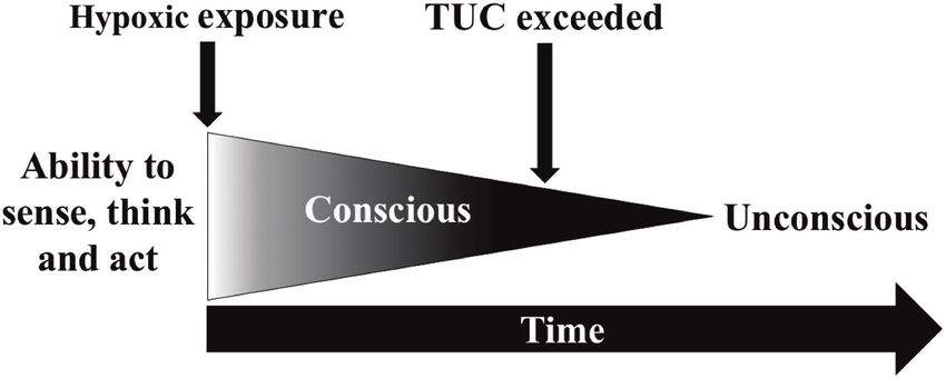
TIME-OF-USEFUL-CONSCIOUSNESS Table 2 summarises the estimated TUC ranges at various
altitudes and EAAs. For example, TUC is 3–5 min at 25,000 ft
If arterial and tissue deoxygenation does not stabilise, brain (PO2 49 mmHg) and declines to less than 15 s at 50,000 ft
function progressively declines, which occur exponentially at (PO2 8 mmHg). With more complex tasks, such as self-directed
a very low PiO2. The initial phase is referred to as the recovery, TUC may be shorter than current estimates, particularly
time-of-useful-consciousness (TUC) and is the duration of for altitudes below 35,000 ft (PO2Shaw et al. Hypoxia and Brain Function in Aviation
Time-of-useful-consciousness estimates for altitudes below can cause brain injury (Shimabuku et al., 2005; Koch et al.,
35,000 ft are characterised by large ranges due to inter-individual 2008; Chalkias and Xanthos, 2012). Some individuals may also
variability in hypoxia (and hypocapnia) tolerance. Some of the experience a transient (15–60 s) worsening of hypoxic symptoms
lowest tolerable levels of hypoxaemia also appear to be from and brain dysfunction during sudden reoxygenation of arterial
opposite ends of the atmospheric-biospheric pressure system blood, which is referred to as the Oxygen Paradox (Latham,
(Bailey et al., 2017): PaO2 of 19 mmHg (SaO2 34%; PaCO2 1951). This may be underpinned by hypoxia-induced hypocapnia
16 mmHg) in an altitude acclimatised mountaineer on descent and reduction in peripheral vasoconstriction, causing cerebral
from the summit of Mount Everest (Grocott et al., 2009); vasoconstriction and hypoperfusion (Gradwell and Rainford,
PaO2 of 22.5 mmHg (SaO2 48%; PaCO2 29 mmHg) during 2016). For example, although 100% oxygen breathing following
simulated descent from 30,000 ft in a high-altitude parachutist acute NH increased arterial blood reoxygenation faster than
(Ottestad et al., 2017); and PaO2 of 23 mmHg (SaO2 38%; room air (i.e., 21% oxygen), recovery was associated with a
PaCO2 61 mmHg) in a free-diver following static apnoea (Bailey robust EEG slowing and impaired working memory (Malle
et al., 2017). TUC may also be extended by: (1) oxygen et al., 2016). This suggests breathing hyperoxic air following
pre-breathing (Malle et al., 2016), which increases oxygen stores hypoxia may be more hazardous than normoxic recovery, which
in the lungs (Tanoubi et al., 2009); (2) greater haemoglobin may impact performance of operational and safety-critical tasks.
oxygen carrying capacity of the blood (Hall, 1949); and (3) Further research investigating the effects of hypoxic recovery
avoiding physical activity during exposure to hypoxia by breathing varying PO2 levels and how this differs with
(Busby et al., 1976). Nevertheless, TUC does not appear to hypoxia severity is required. Moreover, the effects of CO2
be extended by previous hypoxia exposures, suggesting it is inclusion in recovery gases should also be explored.
not trainable (Izraeli et al., 1988; Mitchell et al., 2019).
TRAINING AND PREPARING FOR
RECOVERY OF BRAIN FUNCTION HYPOXIC INCIDENTS
FOLLOWING HYPOXIA
Hypoxia recognition training is a critical component of military
Cognitive impairment may persist for several minutes-to-hours aviation training of aircrew (Neuhaus and Hinkelbein, 2014)
following arterial blood reoxygenation (Phillips et al., 2009, and could have implications in other operational environments.
2015; Beer et al., 2017; Varis et al., 2019). For example, after Currently, the North Atlantic Treaty Organisation Standardisation
10 min normobaric hypoxia (simulated 20,000 ft), reaction Agreement (STANAG) and Air Force Interoperability Council
times were impaired during a 10-min recovery (normoxic) recommend refresher training a maximum of every 5 years;
period, despite resolution of hypoxaemia within ~1 min (Phillips however, some countries may require more frequent trainings
et al., 2009). This was suggested to be due to poor cerebral for at-risk aircrew. The primary rationale for HRT is the
reoxygenation (Phillips et al., 2009), which was also demonstrated intentional induction of hypoxia within a safe and controlled
in follow-up study for up to 24 h following 30 min normobaric environment to: (1) familiarise individuals with their constellation
hypoxia (simulated 18,000 ft), which occurred alongside impaired of personal hypoxia symptoms, including order of appearance
simple and choice reaction speed (Phillips et al., 2015); therefore, and intensity; (2) experience the speed of onset and insidious
the brain may reoxygenate at a slower rate to peripheral tissue. nature of hypoxia; (3) observe hypoxia-induced cognitive and
This means that performance of operational tasks or psychomotor impairment in others; and (4) practice using
implementation of emergency recovery procedures may continue equipment and implementing emergency recovery procedures.
to be compromised following apparent recovery from hypoxia. An individual’s most prominent symptoms are reported to
This “hypoxia hangover” was demonstrated in a group of be consistent for up to 4–5 years for a given hypoxic dose
experienced Hawk pilots, demonstrating impaired (simulated) (Woodrow et al., 2011; Johnston et al., 2012; Tu et al., 2020),
flight performance 10 min after recovery with 100% oxygen which is referred to as their Hypoxic Signature (Smith, 2008).
following a hypoxic exposure (~75% SpO2; Varis et al., 2019), However, not all individuals accurately remember symptoms
which emphasies the need to land as soon as possible following following hypoxia exposures in training (Smith, 2008; Woodrow
hypoxic recovery. Nevertheless, not all studies demonstrate et al., 2011; Tu et al., 2020) and operational (Files et al., 2005)
delayed cerebral reoxygenation (Uchida et al., 2020). Further environments. Reported hypoxic symptoms during training may
research is required to determine whether there are operationally also be different to operational environments, which could be
relevant temporal effects on brain function and cognitive due to a reduced capacity for memory recall, as well as differences
performance following recovery from hypoxia. in hypoxic dose, environmental conditions and biological variation.
Currently, there is a scarcity of research evaluating the efficacy
Hyperoxic Recovery of HRT and how it translates to hypoxia recognition in operational
Breathing air comprising more than 21% oxygen (i.e., hyperoxia) environments. Nevertheless, numerous anecdotal reports highlight
to accelerate recovery from hypoxia is a common practice in the importance of HRT for improving operational safety (Cable,
military aviation. There is cause for enquiry whether hypoxia 2003; Files et al., 2005). Mask-on normobaric HRT was reported
proceeded by hyperoxia breathing is harmful to the brain given to reduce the time to recognise hypoxia in 64% of participants
recovery from ischaemic-hypoxia using more than 21% oxygen (Leinonen et al., 2020); however, there was no control group
Frontiers in Physiology | www.frontiersin.org 8 May 2021 | Volume 12 | Article 665821Shaw et al. Hypoxia and Brain Function in Aviation
and the operational experience of participants between HRT such as ear popping. If task saturation occurs using either the
sessions (i.e., ~2.4 years) could have interfered with the effect hypobaric chamber or ROBD, the subtle signs and symptoms
of HRT. Further, it would seem prudent to isolate how HRT are less likely to be perceived; therefore, depending on the
could benefit a greater proportion of individuals to recognise training aim, this can either compromise or enhance HRT.
hypoxia, which may warrant individualised approaches to HRT. A targeted variety of HRT methods and hypoxic doses, or
The threshold at which hypoxaemia should reach during HRT customised approach, will best prepare individuals to recognise
should also be evaluated due to impairments on learning and the hypoxia symptoms they are likely to experience in
memory (Nation et al., 2017), which would be counterintuitive operational environments.
to the aim of the training. Generally, pulse oximetry appears
to be the preferred method determining hypoxia during HRT,
with profiles being terminated when SpO2 declines below CONCLUSIONS AND FUTURE
~65–70%. The interaction of other physiological stressors, such DIRECTIONS
as fatigue, temperature, and dehydration, on hypoxia recognition
is also unknown, which is relevant given their prevalence within Hypoxia is a major physiological threat during high-altitude
military aviation. flight and operations in military aviation. The extent of the
Although hypobaric chambers provided the initial tools to issue is probably underestimated due to a lack of rigorous
induce hypoxia, reduced oxygen breathing devices (ROBD) biomonitoring of military aircrew (i.e., pilots and rear crew).
providing mask-on NH (Sausen et al., 2001) and a combination Reducing the risk of hypoxic-related incidents and accidents
of HH and mask-on NH (i.e., CADO; Singh et al., 2010) have requires oxygen supply systems, pressurised environments, HRT,
more recently been incorporated into HRT to prevent potential implementation of emergency recovery procedures, and adherence
adverse effects of barometric pressure reduction, such as to safety regulations (e.g., mask wearing); however, there is a
decompression sickness (Webb and Pilmanis, 2011), white matter risk of malfunction for all life support systems, improper use
hyperintensities (McGuire et al., 2014, 2019; Sherman and of equipment, and failure to adhere to safety regulations. The
Sladky, 2018) and barotrauma. Additional advantages of ROBDs ensuing hypoxia can present within seconds-to-minutes, such
are their simplicity, ease of transport, reduced expense, and as a sudden abrogation of oxygen supply (e.g., failure of oxygen
lower maintenance, and they can be the preferred mode of supply system), or develop gradually over minutes-to-hours,
HRT for some individuals, particularly fighter pilots (Artino such as a slow decompression within the aircraft. Even minor
et al., 2006). NH using the ROBD is purported to closely impairments to brain function resulting from hypoxaemia,
replicate symptoms experienced within hypobaric chambers for cerebral hypoxia, or hyperventilation-induced hypocapnia can
brief exposures (Self et al., 2011); however, this remains be catastrophic in military aviation due to the dynamic and
controversial and the ROBD may not necessarily mirror hypoxic demanding environment aircrew must operate within. However,
symptoms experienced by aircrew in operational environments the relevance of hypoxia-induced brain dysfunction for military
(Deussing et al., 2011). Moreover, issues with breathing-gas aviaition can be difficult to accurately quantify, particularly
flow rates when using the ROBD may alter hypoxia symptoms, due to the large inter-individual variation in hypoxia tolerance
particularly air hunger (Artino et al., 2009). Physiological and concurrent effects of hypoxia-induced hypocapnia.
differences between HH, NH, and CADO are suggested to Wearable biomonitoring can be used to signal the early
be irrelevant to symptomology (Singh et al., 2010) and, thus, stages of hypoxia or hypocapnia. Measures of SpO2, SaO2,
provide equivalent training value. However, this does not SaCO2, and ScO2 could warn aircrew prior to brain function
necessarily hold true (Aebi et al., 2020b) because hypocapnia diminishing below recoverable levels. Numerous technological
recognition (hyperventilation-induced) may not be accounted devices are available that continuously monitor oxygenation
for, and can be the primary indication of a hypoxic environment (e.g., pulse oximetry and NIRS); however, the measurement
at moderate altitudes (Petrassi et al., 2012). of carbon dioxide within the body is less common. Potentially,
Hypoxia recognition training should include a high level inbuilt breath-by-breath gas analysers within breathing masks
of fidelity, with signs and symptoms of hypoxia reflecting what may provide insight into arterial blood gas levels, via end-tidal
is likely to be experienced within operational environments. PO2 and PCO2 measurement. Moreover, validated EEG techniques
Hypoxic exposures should, therefore, require individuals to measuring real-time brain wave activity may also be able
perform cognitive tasks specific to the aims of the training indicate hypoxia-induced brain dysfunction. These devices appear
session. The ROBD allows individuals to engage in a variety to be rarely used within military aviation, which could be due
of operational-specific tasks without restriction from the confines to difficulty incorporating them into the life support equipment
of the chamber and changes in pressure. For example, tactical and acquiring accurate measurements within extreme
flight simulation enables decision-making training, environments (e.g., high gravitational forces during fighter jet
implementation of actual emergency recovery procedures, and manoeuvres or hypobaria). If successful, monitoring of
the continuation of the hypoxia training mission until simulated physiological status would mean that hypoxia and hypocapnia
landing. Alternatively, the hypobaric chamber provides a group are reported more often and accurately, thus improving the
environment for hypoxia to be viewed in others and barometric surveillance of operational hypoxic and hypocapnic events. It
pressure changes (i.e., gradual and rapid decompression), which would also provide reassurance on proper treatment procedures
can elicit important signs and symptoms for recognising hypoxia, as hypoxia and hypocapnia can present similarly.
Frontiers in Physiology | www.frontiersin.org 9 May 2021 | Volume 12 | Article 665821Shaw et al. Hypoxia and Brain Function in Aviation
Hypoxia tolerance varies markedly between individuals. accurate thresholds that impair brain function to prevent
Arterial and cerebral oxygenation, CBF, and ventilatory responses compromising training aims. Implementing more realistic training
can all vary greatly to a specific hypoxic dose, which may approaches to better simulate the operational environment and
underpin differences in simple and complex cognitive outcomes. provide immediate objective feedback should also be prioritised;
Importantly, simple cognitive tasks are unlikely to correlate however, any cognitive tasks integrated within the training should
well with the requirements of real-world emergencies, but may not supersede training objectives. The use of HRT modalities
provide a reliable surrogate for automated operational tasks. (e.g., normobaric vs. hypobaric, mask on vs. mask off) and
It is, therefore, recommended to use a range of cognitive tests hypoxic doses should align with training objectives since
when examining the effects of hypoxia on cognition, particularly generalised training profiles may not be translatable to real-world
complex tasks requiring executive, innovative, creative, and events. If not, training may be misleading and cause additional
flexible thinking. These domains are necessary for comprehending safety risks if exact replication of hypoxic symptoms is expected.
and functioning within real-world, novel, and dangerous scenarios HRT will never be an exact replication of real-world hypoxia
that demand situational awareness, complex multi-tasking, self- events, particularly in normobaric modalities that do not simulate
reflection, effective communication, managing behaviours and the hypoxic dysbaric physiological state experienced in a
emotions, evaluating evolving situations, and decision making. depressurised aircraft. Hence, if aircrews feel abnormal during
Further, if complex tasks have increased oxygen demand, the high-altitude flight, then hypoxia should always be suspected.
requirement for supplemental oxygen should not solely be based
on altitude or hypoxaemia, but also operational tasks. Recovery
of brain function following hypoxia should also be assessed AUTHOR CONTRIBUTIONS
as there appears to be a lagging effect, despite resolution of
hypoxaemia, which may differ based on the level of oxygen DS wrote the first draft of the manuscript. DS, NG, and GC
administered and inclusion of carbon dioxide. wrote sections of the manuscript. All authors contributed to
Hypoxia recognition training appears to be an important the article and approved the submitted version.
safety precaution to prevent hypoxic fatalities by enhancing the
response to unanticipated hypoxia. Although the efficacy of HRT
is yet to be systematically evaluated, particularly in operational ACKNOWLEDGMENTS
environments, it is a training requirement for military aircrew.
Similar to biomonitoring in operational environments, The authors would like to thank David Barber, Baz Belzile,
measurement of ScO2, in addition to SpO2, may provide more and Colin Edie for their feedback on the present manuscript.
REFERENCES Bailey, D. M. (2018). Oxygen, evolution and redox signalling in the human brain;
quantum in the quotidian. J. Physiol. Lond. 597, 15–28. doi: 10.1113/jp276814
Adair, G. S. (1925). The hemoglobin system VI. The oxygen dissociation curve Bailey, D. M. (2019). Oxygen and brain death; back from the brink. Q. J. Exp.
of hemoglobin. J. Biol. Chem. 63, 529–545. doi: 10.1016/S0021-9258(18)85018-9 Physiol. 104, 1769–1779. doi: 10.1113/EP088005
Aebi, M. R., Bourdillon, N., Kunz, A., Bron, D., and Millet, G. P. (2020a). Bailey, D. M., Willie, C. K., Hoiland, R. L., Bain, A. R., MacLeod, D. B.,
Specific effect of hypobaria on cerebrovascular hypercapnic responses in Santoro, M. A., et al. (2017). Surviving without oxygen: how low can the
hypoxia. Phys. Rep. 8:e14372. doi: 10.14814/phy2.14372 human brain go? High Alt. Med. Biol. 18, 73–79. doi: 10.1089/ham.2016.0081
Aebi, M. R., Bourdillon, N., Noser, P., Millet, G. P., and Bron, D. (2020b). Basnyat, B., and Murdoch, D. R. (2003). High-altitude illness. Lancet 361,
Cognitive impairment during combined normobaric vs. hypobaric and 1967–1974. doi: 10.1016/S0140-6736(03)13591-X
normoxic vs. hypoxic acute exposure. Aerosp. Med. Hum. Perform. 91, Beer, J. M. A., Shender, B. S., Chauvin, D., Dart, T. S., and Fischer, J. (2017).
845–851. doi: 10.3357/AMHP.5616.2020 Cognitive deterioration in moderate and severe hypobaric hypoxia conditions.
Ainslie, P. N., and Duffin, J. (2009). Integration of cerebrovascular CO2 reactivity Aerosp. Med. Hum. Perform. 88, 617–626. doi: 10.3357/AMHP.4709.2017
and chemoreflex control of breathing: mechanisms of regulation, measurement, Bickler, P. E., Feiner, J. R., Lipnick, M. S., Batchelder, P., MacLeod, D. B., and
and interpretation. Am. J. Phys. Regul. Integr. Comp. Phys. 296, R1473–R1495. Severinghaus, J. W. (2017). Effects of acute, profound hypoxia on healthy
doi: 10.1152/ajpregu.91008.2008 humans. Anesth. Analg. 124, 146–153. doi: 10.1213/ANE.0000000000001421
Ainslie, P. N., Hoiland, R. L., and Bailey, D. M. (2016). Lessons from the Bickler, P. E., Feiner, J. R., and Rollins, M. D. (2013). Factors affecting the
laboratory; integrated regulation of cerebral blood flow during hypoxia. performance of 5 cerebral oximeters during hypoxia in healthy volunteers.
Exp. Physiol. 101, 1160–1166. doi: 10.1113/EP085671 Anesth. Analg. 117, 813–823. doi: 10.1213/ANE.0b013e318297d763
Altbäcker, A., Takács, E., Barkaszi, I., Kormos, T., Czigler, I., and Balázs, L. Bresseleers, J., Diest, I. V., Peuter, S. D., Verhamme, P., and den Bergh, O. V..
(2019). Differential impact of acute hypoxia on event related potentials: (2010). Feeling lightheaded: the role of cerebral blood flow. Psychosom.
impaired task-irrelevant, but preserved task-relevant processing and response Med., 72, 672–680. doi: 10.1097/PSY.0b013e3181e68e94
inhibition. Physiol. Behav. 206, 28–36. doi: 10.1016/j.physbeh.2019.03.022 Brugniaux, J. V., Hodges, A. N. H., Hanly, P. J., and Poulin, M. J. (2007).
Artino, A. R., Folga, R. V., and Swan, B. (2006). Mask-on hypoxia training Cerebrovascular responses to altitude. Respir. Physiol. Neurobiol. 158, 212–223.
for tactical jet aviators: evaluation of an alternate instructional paradigm. doi: 10.1016/j.resp.2007.04.008
Aviat. Space Environ. Med. 77, 857–863. Busby, D. E., Higgins, E. A., and Funkhouser, G. E. (1976). Effect of physical
Artino, A. R., Folga, R. V., and Vacchiano, C. (2009). Normobaric hypoxia activity of airline flight attendants on their time of useful consciousness in
training: the effects of breathing-gas flow rate on symptoms. Aviat. Space a rapid decompression. Aviat. Space Environ. Med. 47, 117–120.
Environ. Med. 80, 547–552. doi: 10.3357/ASEM.2464.2009 Cable, G. G. (2003). In-flight hypoxia incidents in military aircraft: causes and
Asmaro, D., Mayall, J., and Ferguson, S. (2013). Cognition at altitude: impairment implications for training. Aviat. Space Environ. Med. 74, 169–172.
in executive and memory processes under hypoxic conditions. Aviat. Space Chalkias, A., and Xanthos, T. (2012). Post-cardiac arrest brain injury: pathophysiology
Environ. Med. 84, 1159–1165. doi: 10.3357/ASEM.3661.2013 and treatment. J. Neurol. Sci. 315, 1–8. doi: 10.1016/j.jns.2011.12.007
Frontiers in Physiology | www.frontiersin.org 10 May 2021 | Volume 12 | Article 665821Shaw et al. Hypoxia and Brain Function in Aviation
Chiang, K.-T., Yang, C.-S., Chiou, W.-Y., and Chu, H. (2012). Repeated hypoxic flow. Am. J. Physiol. Regul. Integr. Comp. Physiol. 310, R398–R413. doi:
syncope in a helicopter pilot at a simulated altitude of 18,000 feet. Aviat. 10.1152/ajpregu.00270.2015
Space Environ. Med. 83, 609–613. doi: 10.3357/ASEM.3273.2012 Hundemer, G. L., Jersey, S. L., Stuart, R. P., Butler, W. P., and Pilmanis, A. A.
Cohen, P. J., Alexander, S. C., Smith, T. C., Reivich, M., and Wollman, H. (2012). Altitude decompression sickness incidence among U-2 pilots: 1994-2010.
(1967). Effects of hypoxia and normocarbia on cerebral blood flow and Aviat. Space Environ. Med. 83, 968–974. doi: 10.3357/ASEM.3201.2012
metabolism in conscious man. J. Appl. Physiol. 23, 183–189. doi: 10.1152/ Izraeli, S., Avgar, D., Glikson, M., Shochat, I., Glovinsky, Y., and Ribak, J.
jappl.1967.23.2.183 (1988). Determination of the “time of useful consciousness” (TUC) in
Collins, J.-A., Rudenski, A., Gibson, J., Howard, L., and O’Driscoll, R. (2015). repeated exposures to simulated altitude of 25, 000 ft (7620 m). Aviat.
Relating oxygen partial pressure, saturation and content: the haemoglobin– Space Environ. Med. 59, 1103–1105.
oxygen dissociation curve. Breathe 11, 194–201. doi: 10.1183/20734735.001415 Jersey, S. L., Baril, R. T., McCarty, R. D., and Millhouse, C. M. (2010). Severe
Conkin, J. (2011). PH2O and simulated hypobaric hypoxia. Aviat. Space Environ. neurological decompression sickness in a U-2 pilot. Aviat. Space Environ.
Med. 82, 1157–1158. doi: 10.3357/ASEM.3145.2011 Med. 81, 64–68. doi: 10.3357/ASEM.2303.2010
Conkin, J. (2016). Equivalent air altitude and the alveolar gas equation. Aerosp. Johnston, B. J., Iremonger, G. S., Hunt, S., and Beattie, E. (2012). Hypoxia
Med. Hum. Perform. 87, 61–64. doi: 10.3357/AMHP.4421.2016 training: symptom replication in experienced military aircrew. Aviat. Space
Conkin, J., and Wessel, J. H. (2008). Critique of the equivalent air altitude Environ. Med. 83, 962–967. doi: 10.3357/ASEM.3172.2012
model. Aviat. Space Environ. Med. 79, 975–982. doi: 10.3357/ASEM.2331.2008 Kety, S. S., and Schmidt, C. F. (1948). The effects of altered arterial tensions
Coppel, J., Hennis, P., Gilbert-Kawai, E., and Grocott, M. P. (2015). The of carbon dioxide and oxygen on cerebral oxygen consumption of normal
physiological effects of hypobaric hypoxia versus normobaric hypoxia: a young men. J. Clin. Investig. 27, 484–492. doi: 10.1172/JCI101995
systematic review of crossover trials. Extreme Physiol. Med. 4:2. doi: 10.1186/ Kobayashi, A., Tong, A., and Kikukawa, A. (2002). Pilot cerebral oxygen status
s13728-014-0021-6 during air-to-air combat maneuvering. Aviat. Space Environ. Med. 73, 919–924.
Debevec, T., and Millet, G. P. (2014). Discerning normobaric and hypobaric Koch, J. D., Miles, D. K., Gilley, J. A., Yang, C.-P., and Kernie, S. G. (2008).
hypoxia: significance of exposure duration. J. Appl. Physiol. 116:1255. doi: Brief exposure to hyperoxia depletes the glial progenitor pool and impairs
10.1152/japplphysiol.00873.2013 functional recovery after hypoxic-ischemic brain injury. J. Cereb. Blood Flow
Deussing, E. C., Artino, A. R., and Folga, R. V. (2011). In-flight hypoxia events Metab. 28, 1294–1306. doi: 10.1038/jcbfm.2008.15
in tactical jet aviation: characteristics compared to Normobaric training. Kraaier, V., Huffelen, A. C. V., and Wieneke, G. H. (1988). Quantitative EEG
Aviat. Space Environ. Med. 82, 775–781. doi: 10.3357/ASEM.2941.2011 changes due to hypobaric hypoxia in normal subjects. Electroencephalogr.
Dorp, E. V., Los, M., Dirven, P., Sarton, E., Valk, P., Teppema, L., et al. (2007). Clin. Neurophysiol. 69, 303–312. doi: 10.1016/0013-4694(88)90002-8
Inspired carbon dioxide during hypoxia: effects on task performance and Lambertsen, C. J., Bunce, P. L., Drabkin, D. L., and Schmidt, C. F. (1952).
cerebral oxygen saturation. Aviat. Space Environ. Med. 78, 666–672. Relationship of oxygen tension to hemoglobin oxygen saturation in the
Dreier, J. P., Major, S., Foreman, B., Winkler, M. K. L., Kang, E., Milakara, D., arterial blood of normal men. J. Appl. Physiol. 4, 873–875. doi: 10.1152/
et al. (2018). Terminal spreading depolarization and electrical silence in jappl.1952.4.12.873
death of human cerebral cortex. Ann. Neurol. 83, 295–310. doi: 10.1002/ Latham, F. (1951). The oxygen paradox. experiments on the effects of oxygen
ana.25147 in human anoxia. Lancet 1, 77–81. doi: 10.1016/s0140-6736(51)91165-8
Ernsting, J. (1963). The effect of brief profound hypoxia upon the arterial and Leacy, J. K., Day, T. A., and O’Halloran, K. D. (2019). Is alkalosis the dominant
venous oxygen tensions in man. J. Physiol. 169, 292–311. doi: 10.1113/ factor in hypoxia-induced cognitive dysfunction? Q. J. Exp. Physiol. 104,
jphysiol.1963.sp007257 1443–1444. doi: 10.1113/EP087967
Feiner, J. R., Severinghaus, J. W., and Bickler, P. E. (2007). Dark skin decreases Lee, J.-M., Grabb, M. C., Zipfel, G. J., and Choi, D. W. (2000). Brain tissue
the accuracy of pulse oximeters at low oxygen saturation: The effects of responses to ischemia. J. Clin. Investig. 106, 723–731. doi: 10.1172/JCI11003
oximeter probe type and gender. Anesth. Analg. 105, S18–S23. doi: 10.1213/01. Legg, S., Hill, S., Gilbey, A., Raman, A., Schlader, Z., and Mündel, T. (2014).
ane.0000285988.35174.d9 Effect of mild hypoxia on working memory, complex logical reasoning,
Files, D. S., Webb, J. T., and Pilmanis, A. A. (2005). Depressurization in military and risk judgment. Int. J. Aviat. Psychol. 24, 126–140. doi: 10.1080/
aircraft: rates, rapidity, and health effects for 1055 incidents. Aviat. Space 10508414.2014.892751
Environ. Med. 76, 523–529. Legg, S., Hill, S., Mundel, T., Gilbey, A., Schlader, Z., and Raman, A. (2012).
Fowler, B., Elcombe, D. D., Kelso, B., and Porlier, G. (1987). The threshold Could mild hypoxia impair pilot decision making in emergencies? Work
for hypoxia effects on perceptual-motor performance. Hum. Factors 29, 41, 198–203. doi: 10.3233/WOR-2012-0156-198
61–66. doi: 10.1177/001872088702900106 Leinonen, A., Varis, N., Kokki, H., and Leino, T. K. (2020). Normobaric hypoxia
Friend, A. T., Balanos, G. M., and Lucas, S. J. E. (2019). Isolating the independent training in military aviation and subsequent hypoxia symptom recognition.
effects of hypoxia and hyperventilation-induced hypocapnia on cerebral Ergonomics 64, 545–552. doi: 10.1080/00140139.2020.1842514
haemodynamics and cognitive function. Q. J. Exp. Physiol. 104, 1482–1493. Loeppky, J. A., Icenogle, M., Scotto, P., Robergs, R., Hinghofer-Szalkay, H.,
doi: 10.1113/EP087602 and Roach, R. C. (1997). Ventilation during simulated altitude, normobaric
Gibson, G. E., Pulsinelli, W., Blass, J. P., and Duffy, T. E. (1981). Brain dysfunction hypoxia and normoxic hypobaria. Respir. Physiol. 107, 231–239. doi: 10.1016/
in mild to moderate hypoxia. Am. J. Med. 70, 1247–1254. doi: 10.1016/ S0034-5687(97)02523-1
0002-9343(81)90834-2 López-Barneo, J., González-Rodríguez, P., Gao, L., Fernández-Agüera, M. C.,
Goodall, S., Twomey, R., and Amann, M. (2014). Acute and chronic hypoxia: Pardal, R., and Ortega-Sáenz, P. (2016). Oxygen sensing by the carotid
implications for cerebral function and exercise tolerance. Fatigue 2, 73–92. body: mechanisms and role in adaptation to hypoxia. Am. J. Phys. Cell
doi: 10.1080/21641846.2014.909963 Phys. 310, C629–C642. doi: 10.1152/ajpcell.00265.2015
Gradwell, D., and Rainford, D. (2016). Ernsting’s Aviation and Space Medicine MacLeod, D. B., Ikeda, K., Vacchiano, C., Lobbestael, A., Wahr, J. A., and
5E. On CRC Press. CRC Press. Shaw, A. D. (2012). Development and validation of a cerebral oximeter
Grocott, M. P. W., Martin, D. S., Levett, D. Z. H., McMorrow, R., Windsor, J., capable of absolute accuracy. J. Cardiothorac. Vasc. Anesth. 26, 1007–1014.
and Montgomery, H. E. (2009). Arterial blood gases and oxygen content doi: 10.1053/j.jvca.2012.06.010
in climbers on Mount Everest. N. Engl. J. Med. 360, 140–149. doi: 10.1056/ Malle, C., Bourrilhon, C., Quinette, P., Laisney, M., Eustache, F., and Piérard, C.
NEJMoa0801581 (2016). Physiological and cognitive effects of acute normobaric hypoxia and
Hall, F. G. (1949). Interval of useful consciousness at various altitudes. J. Appl. modulations from oxygen breathing. Aerosp. Med. Hum. Perform. 87, 3–12.
Physiol. 1, 490–495. doi: 10.1152/jappl.1949.1.7.490 doi: 10.3357/AMHP.4335.2016
Hoffman, C. E., Clark, R. T. Jr., and Brown, E. B. Jr. (1946). Blood oxygen Mannheimer, P. D. (2007). The light–tissue interaction of pulse oximetry. Anesth.
saturations and duration of consciousness in anoxia at high altitudes. Am. Analg. 105, S10–S17. doi: 10.1213/01.ane.0000269522.84942.54
J. Phys. 145, 685–692. McGuire, S. A., Ryan, M. C., Sherman, P. M., Sladky, J. H., Rowland, L. M.,
Hoiland, R. L., Bain, A. R., Rieger, M. G., Bailey, D. M., and Ainslie, P. N. Wijtenburg, S. A., et al. (2019). White matter and hypoxic hypobaria in
(2016). Hypoxemia, oxygen content, and the regulation of cerebral blood humans. Hum. Brain Mapp. 40, 3165–3173. doi: 10.1002/hbm.24587
Frontiers in Physiology | www.frontiersin.org 11 May 2021 | Volume 12 | Article 665821You can also read