Characterizing the Retinal Phenotype of the Thy1-h A30P α-syn Mouse Model of Parkinson's Disease - Frontiers
←
→
Page content transcription
If your browser does not render page correctly, please read the page content below
ORIGINAL RESEARCH
                                                                                                                                         published: 07 September 2021
                                                                                                                                        doi: 10.3389/fnins.2021.726476

                                            Characterizing the Retinal Phenotype
                                            of the Thy1-h[A30P]α-syn Mouse
                                            Model of Parkinson’s Disease
                                            Lien Veys 1,2 , Joyce Devroye 1,2 , Evy Lefevere 1,2 , Lien Cools 1,2 , Marjan Vandenabeele 1,2 and
                                            Lies De Groef 1,2*
                                            1
                                                Research Group of Neural Circuit Development and Regeneration, Department of Biology, KU Leuven, Leuven, Belgium,
                                            2
                                                Department of Biomedical Sciences, Leuven Brain Institute, Leuven, Belgium

                                            Despite decades of research, disease-modifying treatments of Parkinson’s disease
                                            (PD), the second most common neurodegenerative disease worldwide, remain out of
                                            reach. One of the reasons for this treatment gap is the incomplete understanding
                                            of how misfolded alpha-synuclein (α-syn) contributes to PD pathology. The retina,
                                            as an integral part of the central nervous system, recapitulates the PD disease
                                            processes that are typically seen in the brain, and retinal manifestations have emerged
                          Edited by:        as prodromal symptoms of the disease. The timeline of PD manifestations in the
                             Yuyi You,
                                            visual system, however, is not fully elucidated and the underlying mechanisms are
        Macquarie University, Australia
                                            obscure. This highlights the need for new studies investigating retinal pathology,
                          Reviewed by:
                           Thierry Baron,   in order to propel its use as PD biomarker, and to develop validated research
          Laboratoire de Lyon, Agence       models to investigate PD pathogenesis. The present study pioneers in characterizing
        Nationale de Sécurité Sanitaire
 de l’Alimentation, de l’Environnement      the retina of the Thy1-h[A30P]α-syn PD transgenic mouse model. We demonstrate
          et du Travail (ANSES), France     widespread α-syn accumulation in the inner retina of these mice, of which a proportion
                          Jürgen Winkler,
                                            is phosphorylated yet not aggregated. This α-syn expression coincides with inner
   University of Erlangen Nuremberg,
                                Germany     retinal atrophy due to postsynaptic degeneration. We also reveal abnormal retinal
                   *Correspondence:         electrophysiological responses. Absence of selective loss of melanopsin retinal ganglion
                         Lies De Groef      cells or dopaminergic amacrine cells and inflammation indicates that the retinal
             lies.degroef@kuleuven.be
                                            manifestations in these transgenic mice diverge from their brain phenotype, and
                    Specialty section:      questions the specific cellular or molecular alterations that underlie retinal pathology
          This article was submitted to     in this PD mouse model. Nevertheless, the observed α-syn accumulation, synapse loss
                    Neurodegeneration,
                a section of the journal    and functional deficits suggest that the Thy1-h[A30P]α-syn retina mimics some of the
             Frontiers in Neuroscience      features of prodromal PD, and thus may provide a window to monitor and study the
           Received: 16 June 2021           preclinical/prodromal stages of PD, PD-associated retinal disease processes, as well as
        Accepted: 19 August 2021
     Published: 07 September 2021
                                            aid in retinal biomarker discovery and validation.
                             Citation:      Keywords: retina, visual system, alpha-synuclein, transgenic mouse model, Parkinson’s disease
       Veys L, Devroye J, Lefevere E,
       Cools L, Vandenabeele M and
     De Groef L (2021) Characterizing
                                            Abbreviations: α-syn, Alpha-synuclein; α-syn mice, Thy1-h[A30P]α-syn mice; AQP4, Aquaporin 4; ChAT, Choline
          the Retinal Phenotype of the
                                            acetyltransferase; CNS, Central nervous system; ERG, Electroretinography; DAPI, 40 ,6-diamidino-2-phenylindole; GCL,
    Thy1-h[A30P]α-syn Mouse Model           Ganglion cell layer; GFAP, Glial fibrillary acidic protein; Iba-1, Ionized calcium-binding adapter molecule 1; INL, Inner
               of Parkinson’s Disease.      nuclear layer; IPL, Inner plexiform layer; NFL, Nerve fiber layer; OP, Oscillatory potential; p-α-syn, Phosphorylated serine-
          Front. Neurosci. 15:726476.       129 α-syn; pSTR, Positive scotopic threshold response; RGC, Retinal ganglion cell; TH, Tyrosine hydroxylase; ThioS,
     doi: 10.3389/fnins.2021.726476         Thioflavin S; WT, Wild type.

Frontiers in Neuroscience | www.frontiersin.org                                    1                                      September 2021 | Volume 15 | Article 726476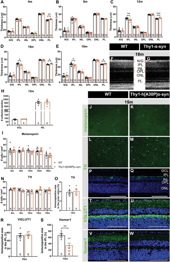
Veys et al.                                                                                                 Thy1-h[A30P]α-syn Retinal Characterization

INTRODUCTION                                                                have revealed pathological manifestations that may underlie
                                                                            these changes in in vivo measures, including a reduction in
Despite decades of research, disease-modifying treatments                   dopamine levels in the retina (Nguyen-Legros, 1988; Harnois
of Parkinson’s disease (PD), the second most common                         and Di Paolo, 1990; Chorostecki et al., 2015), reduced density
neurodegenerative disease worldwide, remain out of reach (Guo               and complexity of dopaminergic neurons (Ortuño-Lizarán et al.,
et al., 2018; Veys et al., 2019). It has been suggested that one of         2020) and melanopsin-positive retinal ganglion cells (RGCs;
the principal reasons for this treatment gap is the lack of accurate        Ortuno-Lizaran et al., 2018b), and, finally, the presence of alpha-
and timely diagnosis. Traditionally, diagnosis is based on the              synuclein (α-syn) and phosphorylated (S129) α-syn (p-α-syn)
cardinal motor symptoms of PD (tremor, rigidity, bradykinesia,              inclusions in the retina (Beach et al., 2014; Ho et al., 2014; Bodis-
and postural instability), which only arise years after a long              Wollner et al., 2014a; Ortuno-Lizaran et al., 2018a; Veys et al.,
non-symptomatic phase during which a large proportion of the                2019). Importantly, p-α-syn deposits in the retina accumulate
dopaminergic cells in the substantia nigra are lost (Jankovic,              in parallel with the brain, already during the prodromal stage
2008). In order to preserve brain function, therapies -and hence            of PD, and are associated with PD severity (Ortuno-Lizaran
diagnosis- should be focused on the preclinical (asymptomatic)              et al., 2018a). This reinforces that retinal biomarkers have a high
and prodromal (early symptomatic) stages (Forsaa et al., 2010;              potential for PD diagnosis and disease monitoring.
Mahlknecht et al., 2015; Hustad and Aasly, 2020). In 2017, new                  Further research into the (temporal) relationship between
diagnostic criteria for PD have been defined by the International           retinal biomarker alterations and neurodegenerative changes in
Parkinson Disease and Movement Disorders Society (Postuma                   the brain is needed, however, for retinal biomarkers to be adopted
and Berg, 2017; Marsili et al., 2018), whereby the probability of           in the clinic. Longitudinal and prospective studies in PD patients
an individual to develop PD is now calculated based on several              and patients at risk of developing PD will be essential to assess the
predictors, such as age, environmental predictors, prodromal                value of retinal biomarkers for PD (Kashani et al., 2021). Animal
signs, genetic risk variables, and biomarker testing (Postuma               models of PD, on the other hand, can support these studies,
et al., 2016). Constant updating of these diagnostic criteria is            by providing a framework in which the correlation between
required as more insights into early stage PD emerge (Postuma               retinal biomarkers and disease manifestations can be explored
and Berg, 2017).                                                            and novel insights into the molecular and cellular changes
   The retina has become a target organ in the search for                   underlying the retinal manifestations of PD can be obtained
early biomarkers, relevant diagnostic criteria and techniques               (Santano et al., 2011; Normando et al., 2016; Price et al., 2016;
that are amenable to population-wide patient screening and                  Mammadova et al., 2018, 2021; Veys et al., 2019). Altogether, the
disease monitoring. As an integral part of the central nervous              wide availability of technologies for non-invasive high-resolution
system (CNS), the eye can be considered a window to the                     ocular imaging, such as OCT, is a clear advantage over current
brain. The visual pathway has shown to be an excellent model                brain imaging techniques (De Groef and Cordeiro, 2018) and,
system to gain insight into classical neurodegenerative diseases,           collectively, visual function measures, ERG, and retinal imaging
as both retina and brain are often affected by these diseases and           could offer a multimodal biomarker approach for PD diagnosis,
share disease processes (e.g., neurodegeneration, inflammation,             stratification, and monitoring (Guo et al., 2018; Turcano et al.,
aggregation of misfolded proteins, mitochondrial dysfunction;               2018; Veys et al., 2019).
Armstrong, 2009; Martínez-Lapiscina et al., 2014; Rahimi et al.,                In this study, we aim to fill the need for well-characterized
2015; Veys et al., 2019; Kashani et al., 2021). Therefore, it               preclinical models to study retinal alternations in PD.
is not surprising that in many PD patients, one or more                     We characterized the retinal phenotype of the Thy1-
visual symptoms are described, such as decreased visual acuity,             h[A30P]α-syn mouse model, by studying α-syn accumulation,
spatial contrast sensitivity, and color vision (Price et al., 1992;         neurodegeneration, inflammation, synaptic integrity, and retinal
Archibald et al., 2011; Armstrong, 2011; Bodis-Wollner, 2013;               function. The brain phenotype of this mouse model has been
Guo et al., 2018). Retinal dysfunction at least partially contributes       studied before, yet the retinal phenotype remains untouched
to these deficits (Bertrand et al., 2012; Mazzarella and Cole,              (Kahle et al., 2000; Neumann et al., 2002; Freichel et al., 2007;
2016). This is corroborated by retinal imaging via optical                  Ekmark-Lewen et al., 2018). Here, we used in vivo retinal
coherence tomography (OCT) and with electroretinography                     imaging and electrophysiology measurements with high clinical
(ERG) measurements, which revealed, respectively, retinal nerve             translatability, combined with post mortem histological studies, to
fiber layer (NFL), ganglion cell layer (GCL), inner plexiform               map the timeline of retinal disease manifestations in these mice.
layer (IPL), and inner nuclear layer (INL) thinning (Shrier et al.,
2012; Adam et al., 2013; London et al., 2013; Spund et al., 2013;
Lee et al., 2014; Bodis-Wollner et al., 2014b; Boeke et al., 2016;          MATERIALS AND METHODS
Aydin et al., 2018; Matlach et al., 2018); and abnormalities of
the photopic b-wave, scotopic oscillatory potentials (OPs), and             Animals
P50 component of the pattern ERG in PD patients (Nightingale                Thy1-h[A30P]α-syn        mice     (C57BL/6      background,
et al., 1986; Gottlob et al., 1987; Burguera et al., 1990; Ikeda            RRID:MGI:2652214) and corresponding wild type (WT)
et al., 1994; Peppe et al., 1992, 1995, 1998; Langheinrich et al.,          controls, were bred under standard laboratory conditions
2000; Sartucci et al., 2006; Garcia-Martin et al., 2014; Nowacka            (Kahle et al., 2000). Both female and male mice were used
et al., 2015; Kashani et al., 2021). Histopathological studies              at 4, 8, 12, 15, and 18 months of age. All experiments were

Frontiers in Neuroscience | www.frontiersin.org                         2                                 September 2021 | Volume 15 | Article 726476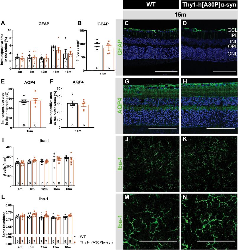
Veys et al.                                                                                          Thy1-h[A30P]α-syn Retinal Characterization

performed according to the European directive 2010/63/EU              Image Analysis
and in compliance with protocols approved by the KU Leuven            Imaging was performed using a FV1000 confocal or FV1000-
institutional ethical committee.                                      M multiphoton microscope (Olympus) or a conventional
                                                                      epifluorescence microscope (DM6, Leica).
(Immuno)histochemistry                                                   Image analyses were performed with Fiji software (Schindelin
Prior to eye dissection, mice were euthanized by an                   et al., 2012). For retinal wholemounts, the entire perimeter of
intraperitoneal injection of 60 mg/kg sodium pentobarbital            the wholemount was outlined and its area measured prior to
(Dolethal, Vetoquinol) followed by transcardial perfusion with        analysis. For sections, five sections per mouse were investigated,
saline and 4% paraformaldehyde (PFA). Next, eyes were either          including the central section containing the optic nerve head,
fixed in 1% PFA for 4 h at 4◦ C and embedded in paraffin, or in       and the sections located 210 and 420 µm anterior/posterior.
4% PFA for 1 h at RT for wholemount preparations. The latter          On each section, analysis was done over a distance of 300 µm
were post-fixed for 1 h in 4% PFA for another hour.                   at four locations per section. For α-syn, TH and GFAP, the
    Seven-micrometer       sagittal paraffin    sections  were        immunopositive area was measured in the inner retina (from
deparaffinized and stained with hematoxylin (Sigma) and               the retinal NFL until the INL included), while for AQP4 both
eosin (Sigma) and mounted with Distyrene Plasticizer Xylene           the outer retina (from OPL to ONL) and inner retina were
mounting medium (Sigma). For Thioflavin S histological                measured and for VGLUT1 and Homer1, only the IPL was
staining, sections were stained for 5 min with Thioflavin S           included (Van Hove et al., 2020). For cell counting, both
(Sigma, 1/200 in 1:1 distilled water and ethanol) and mounted         on wholemounts and sections, Fiji “Cell Counter” plugin was
with mowiol (Sigma). For immunohistochemistry, sections were          used. Microglia density and morphology were quantified as
incubated overnight with one or two of the following primary          described in Davis et al. (2017) on projection images of z-stack
antibodies: human specific α-syn (1/5000; Millipore, clone            (step size 1.5 µm) pictures of Iba-1 stained wholemounts
Syn211 [36-008] RRID:AB_310817), α-syn (1/1000; produced              (Davis et al., 2017).
and kindly provided by V. Baekelandt, KU Leuven, for double
staining with p-α-syn), p-α-syn (1/5000; Elan Pharmaceuticals),
                                                                      Optical Coherence Tomography
p62 (1/200; Proteintech [#55274-1-AP], RRID:AB_11182278),
                                                                      Optical coherence tomography imaging was performed as
Brn3a (1/750; Santa Cruz Biotechnology, c-20 [#sc-31984],
                                                                      described before (Sergeys et al., 2019; Vandenabeele et al., 2021).
RRID:AB_2167511), tyrosine hydroxylase (TH; 1/1000; Millipore
                                                                      Briefly, after pupil dilatation with tropicamide (0.5%, Tropicol,
[#AB152], RRID:AB_390204), choline acetyltransferase (ChAT;
                                                                      Théa), the retina of anesthetized animals was imaged (1000
1/100; Millipore [#AB144P], RRID:AB_2079751), VGLUT1
                                                                      A-scans, 100 B-scans, 1.4 × 1.4 mm, Bioptigen Envisu R2200).
(1/1000, Synaptic Systems [#135 302], RRID:AB_887877),
                                                                      Retinal layer thickness was measured using InVivoVue Diver (v
Prox1 (1/500; Biolegend [PCB-238C]), Homer1 (1/500; Synaptic
                                                                      3.0.8, Bioptigen) software, at 16 locations in the central retina
Systems [#160 003], RRID:AB_887730), glial fibrillary acidic
                                                                      spaced around the optic nerve head, and averaged per mouse.
protein (GFAP; 1/1000; Dako [#Z0334], RRID:AB_10013382),
or aquaporin 4 (AQP4; 1/10000; Alomone labs [AQP-004],
RRID:AB_2039734). For α-syn, Brn3a, TH, ChAT, Prox1,                  Electroretinography
and Homer1, antigen retrieval with heated citrate buffer              Electroretinography was performed as described before (Sergeys
(20 min, 95◦ C) was used, while no antigen retrieval treatment        et al., 2019; Vandenabeele et al., 2021). Full-field flash dark-
was used for VGLUT1 and proteinase K (5 min, 20 µg/ml,                adapted electroretinograms were measured at increasing
Qiagen) antigen retrieval was used for GFAP stainings.                flash intensities of 0.003, 0.01, 0.1, 1, 2.5, and 7.5 cd∗ s/m2 .
Fluorescent labeling was performed using an Alexa-488                 Electroretinograms were analyzed using Espion software
labeled secondary antibody (Invitrogen) for Brn3a, TH, ChAT,          (v6.59.9, Diagnosys), as shown in Supplementary Figure 1. To
Prox1, VGLUT1, and GFAP staining, or with a fluorescein               analyze the OPs on the rising part of the b-wave, a band pass
or cyanine 3 tyramid signal amplification kit (PerkinElmer)           filter (75–300 Hz) was used. The positive scotopic threshold
for p-α-syn, α-syn, and Homer1 stainings. Finally, slides             response (pSTR) was measured at 1 × 10⁻⁴ cd∗ s/m2 . 1 week after
were counterstained with 40 ,6-diamidino-2-phenylindole and           baseline ERG or pSTR measurement, mice were intraperitoneally
mounted with mowiol.                                                  injected with benserazide hydrochloride (12.5 g/kg, Sigma)
    For     wholemount          immunohistochemistry,    tissue       and L-DOPA (25 g/kg, Sigma) 500 and 300 prior to ERG/pSTR
permeabilization was achieved by a freeze-thaw step                   measurement, respectively.
(15 min, −80◦ C), followed by overnight incubation with
one of the following primary antibodies: p-α-syn (1/5000;             Statistical Analysis
Elan Pharmaceuticals), TH (1/1000; Millipore [#AB152],                Statistical analyses were performed using GraphPad Prism
RRID:AB_390204), melanopsin (1/5000; Advanced Targeting               (v8.4.3, GraphPad, RRID:SCR_002798). The number of animals
Systems [#AB-N38], RRID:AB_1608077), or ionized calcium-              (n) used is depicted on the figures and the statistical analyses
binding adapter molecule 1 (Iba-1; 1/1000; Wako [#019-19741],         are indicated in the figure legends. Data are presented as
RRID:AB_839504). Subsequently, fluorescent labeling was               mean ± SEM. Differences were considered statistically significant
performed using an Alexa-488 labeled secondary antibody               for two-sided p-values < 0.05 (∗ p < 0.05; ∗∗ p < 0.01;
(Invitrogen) and wholemounts were mounted with mowiol.                ∗∗∗ p < 0.001; and ∗∗∗∗ p < 0.0001).

Frontiers in Neuroscience | www.frontiersin.org                   3                                September 2021 | Volume 15 | Article 726476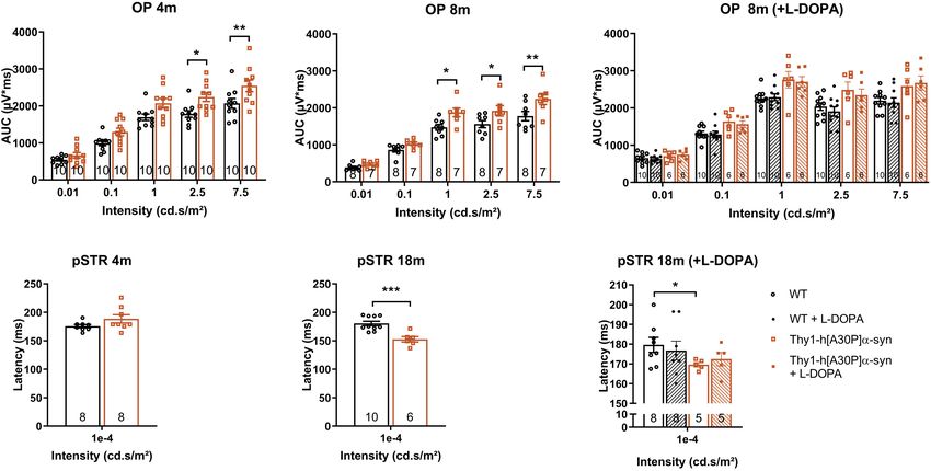
Veys et al. Thy1-h[A30P]α-syn Retinal Characterization
RESULTS difference in retinal thickness persisted at 12, 15, and 18 months
(p = 0.0009, p = 0.0130, and p = 0.0122; Figures 2C–G).
Retinal Accumulation of Furthermore, at 12 months, α-syn mice also displayed thinning
of the IPL, which persisted at 15 and 18 months (p = 0.0034 at
(Phosphorylated) α-syn in
12 months, p = 0.0336 at 15 months, p = 0.0444 at 18 months;
Thy1-h[A30P]α-syn Mice Figures 2C–G).
α-syn expression, phosphorylation, and aggregation was studied As retinal thinning is typically a sign of neurodegeneration,
in the retina of WT and Thy1-h[A30P]α-syn mice (α-syn mice) of we next performed a more in-depth analysis of different
various ages, using (immuno)stainings for transgenic human α- subpopulations of inner retinal neurons at 15 months of age to
syn, phosphorylated (serine-129) α-syn (p-α-syn; detecting both clarify the origin of the observed IPL thinning. Given that the IPL
human and rodent α-syn), thioflavin S (ThioS) and p62. Conform consists of neurites emerging from cell bodies in both the GCL
with previously published data of Veys et al. (2019), hα-syn and INL, cell density was assessed in these layers on hematoxylin
expression was observed in neuronal cell bodies in the GCL, and eosin-stained sections. No overt neurodegeneration was seen
in neurites in the retinal NFL and IPL and in dispersed cell in α-syn mice (Figure 2H). Additionally, a detailed analysis
bodies in the INL of 4-, 8-, 12-, 15-, and 18-month-old Thy1- of disease-relevant neuronal subtypes, also at 15 months of
h[A30P]α-syn mice (Figures 1A–F,T; Veys et al., 2019). The age, revealed that cell numbers of intrinsically photosensitive
hα-syn positive cell types in the inner retina comprise RGCs, as RGCs (melanopsin positive) in the GCL and of dopaminergic
shown by double staining with Brn3a (Figure 1U), and amacrine (TH positive) amacrine cells in the INL (Figures 2I–N) were
cells, based on their morphology and location (Figures 1V– not affected. However, IPL thinning may also occur due to
X). Furthermore, the accumulation of hα-syn in dopaminergic, dendrite or synapse loss, a pathological process that is known
(nor)adrenergic, cholinergic, or AII amacrine cells was ruled out to precede loss of neuronal cell bodies. In line with the
based on the lack of colocalization with TH, ChAT, and Prox1 preservation of dopaminergic cell bodies (cfr. above), we found
positive cells, respectively (Figures 1V–X; Müller et al., 2017). that the dopaminergic plexus of the retina, measured as the
Quantitative analysis of the hα-syn fluorescent area did not reveal TH-immunopositive area in the inner retina, was unaltered in
any progressive changes in hα-syn expression in the inner retina α-syn mice of 15 months of age (Figures 2O–Q). However,
of α-syn mice with aging (Figure 1M). Next, a fraction of α-syn taking a closer look at the synaptic integrity of the IPL, via
was phosphorylated, most prominently in cell bodies and neurites immunostainings with the established pre- and postsynaptic
in the GCL (Figures 1G–L,S,T), and this did not change with age markers VGLUT1 and Homer1, we revealed loss of postsynaptic
(Figure 1N), not even in end-stage diseased animals with severe contacts yet preservation of the presynaptic terminals in 15-
signs of hind limb paralysis (data not shown). At 18 months of month-old transgenic mice (Figures 2R–W). Altogether, these
age, only 34 ± 8% of strongly α-syn positive cells in the GCL also findings suggest that synaptic degeneration in the retina underlies
contained p-α-syn. Finally, we assessed p62 and ThioS labeling to the observed IPL thinning.
investigate α-syn ubiquitination and aggregation, respectively. At
18 months of age, no p62 accumulation nor relocalization were
observed in the retina of α-syn mice as compared to WT mice Electrophysiological Changes in the
(Figures 1Q,R), and no ThioS positive aggregates were found Retina of Thy1-h[A30P]α-syn Mice With
in the retina of transgenic nor WT animals (Figures 1O,P). Of Aging
note, although no accumulation of ThioS-positive or p62-positive In a next series of experiments, we sought to further identify the
cellular inclusions was detted in the Thy1-h[A30P]α-syn PD neuronal cell types that are affected in the α-syn mouse and to
mouse model, we cannot exclude that oligomeric, prefibrillar, or establish whether neuronal dysfunction can be detected already
non-fibril α-syn conformers contribute to the retinal phenotype at younger ages compared to the OCT thinning that only become
observed in these mice (Lashuel et al., 2013; Roberts and Brown, apparent at 12 months. Indeed, neuronal death is often preceded
2015; Cascella et al., 2021). This needs to be explored in follow- by functional changes, and these prodromal manifestations of
up studies. the disease are of particular interest for biomarker development
Altogether, these data show that, while both α-syn (Nowacka et al., 2015; Barber et al., 2017; Turcano et al., 2018;
overexpression and phosphorylation are present in the retina Hustad and Aasly, 2020). First, OPs as a read-out for amacrine
of Thy1-h[A30P]α-syn mice already at a young age, α-syn cell function, were assessed. Already at 4 months, the area
aggregation and ubiquitination do not manifest. under the curve was larger in α-syn mice as compared to WT
animals for high intensity light stimuli (2.5 cd∗ s/m2 : p = 0.0137;
Synaptic Degeneration in the Retina of 7.5 cd∗ s/m2 : p = 0.0094), and this effect persisted in older
Old Thy1-h[A30P]α-syn Mice transgenic animals of 8 (1 cd∗ s/m2 : p = 0.0191; 2.5 cd∗ s/m2 :
Spectral domain OCT was applied in a longitudinal in vivo p = 0.0452; 7.5 cd∗ s/m2 : p = 0.0050), 12 (1 cd∗ s/m2 : p = 0.0034;
experiment to measure the thickness of the retinal layers in α-syn 2.5 cd∗ s/m2 : p = 0.0039; 7.5 cd∗ s/m2 : p = 0.0023), and 18 months
mice and WT controls, early in their life (4 and 8 months) and at of age (1 cd∗ s/m2 : p = 0.0001; Figures 3A,B and Supplementary
12, 15, and 18 months of age (Figures 2A,B,D–F). At 4 months of Figure 1F). Second, we measured RGC function via the pSTR.
age, a minor yet significant thickening of the photoreceptor layer Not yet at 4 months, but at 8, 12, and 18 months, the pSTR latency
(PL) was found in the α-syn mice (p = 0.0023; Figure 2A). This time was shorter in α-syn mice as compared to WT controls
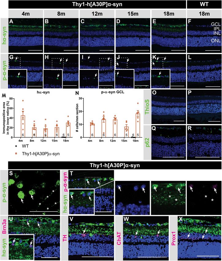
Frontiers in Neuroscience | www.frontiersin.org 4 September 2021 | Volume 15 | Article 726476Veys et al. Thy1-h[A30P]α-syn Retinal Characterization FIGURE 1 | Inner retinal hα-syn expression is accompanied by α-syn phosphorylation, yet no ThioS positive aggregation or p62 accumulation, in the retina of Thy1-h[A30P]α-syn mice. Representative images of hα-syn immunostainings (A–E); p-α-syn immunostainings (G–K); and ThioS staining (O) on retinal sections of α-syn mice at 4, 8, 12, 15 and 18 months of age. (F,L,P) No staining was observed in the WT controls, at any age (only 18 months shown here). (M,N) Quantitative analysis of the hα-syn fluorescent area and counting of the p-α-syn positive cells did not reveal an increase of hα-syn expression in the inner retina or p-α-syn cell density in α-syn mice with age. (O,P) No ThioS positive inclusions were found in the retina of transgenic nor wild type animals in any of the age groups. (Q,R) No difference in retinal p62 accumulation or localization was detected between transgenic and wild type animals at 18 months of age. (S) p-α-syn immunostaining on a retinal wholemount of an α-syn mouse showed p-α-syn localization in cell bodies (arrows) and neurites (asterisks). (T) Double staining of hα-syn with p-α-syn revealed clear colocalization. (U–X) Double staining of hα-syn with Brn3a, TH, ChAT and Prox1 revealed expression of Brn3a in hα-syn positive cells, yet no colocalization in dopaminergic and cholinergic cells. Scale bar: 100 µm (A–R, V–X) or 50 µm (S–U); GCL, ganglion cell layer; INL, inner nuclear layer; IPL, inner plexiform layer; and ONL, outer nuclear layer. Frontiers in Neuroscience | www.frontiersin.org 5 September 2021 | Volume 15 | Article 726476
Veys et al.                                             Thy1-h[A30P]α-syn Retinal Characterization

  FIGURE 2 | Continued

Frontiers in Neuroscience | www.frontiersin.org   6   September 2021 | Volume 15 | Article 726476
Veys et al.                                                                                                                    Thy1-h[A30P]α-syn Retinal Characterization

  FIGURE 2 | Outer retinal thickening and inner retinal thinning, associated with loss of postsynaptic labeling, in Thy1-h[A30P]α-syn mice. (A–E) Longitudinal OCT
  measurements in 4- (A), 8- (B), 12- (C), 15- (D), and 18-month-old (E–G) mice, revealed significant differences in retinal layer thickness between α-syn and WT mice
  of 4 months (PL thickening), 15 months (PL thickening and IPL thinning), and 12 and 18 months of age (PL thickening and IPL thinning). (H) Cell counts on
  hematoxylin and eosin-stained sections in the GCL and in the INL did not reveal significant differences between transgenic animals and WT controls at 15 months of
  age. (I–W) Representative images of retinal wholemounts stained for melanopsin (J,K) and TH (L,M), and of retinal sections stained for TH (P,Q), VGLUT1 (T,U), and
  Homer-1 (V,W), of 15-month-old α-syn and WT mice. Counting the number of melanopsin- (I) and TH- (N) positive cells on retinal wholemounts revealed no
  significant differences between transgenic and WT animals. No significant differences were uncovered in TH plexus (O) and VGLUT1 (R) immunopositive area, yet a
  strong decrease of the Homer1 (S) signal was seen. Scale bar: 100 µm; Two-Way ANOVA with Tukey multiple comparisons post hoc test (I–N). Unpaired t-test (per
  retinal layer; A–F,O,R,S): *p < 0.05; **p < 0.01; and ***p < 0.001. N/G, retinal nerve fiber layer + GCL; GCL, ganglion cell layer; INL, inner nuclear layer; IPL, inner
  plexiform layer; ONL, outer nuclear layer; OPL, outer plexiform layer; and PL, photoreceptor layer.

(p = 0.0082 at 8 months, p = 0.0119 at 12 months, and p = 0.0006                         by the Müller glia, of which differences in expression levels
at 18 months; Figures 3D–F and Supplementary Figure 1G).                                 and cellular localization have been linked to retinal edema and
a- and b-wave measurements were unaltered, indicating normal                             neuroinflammation (Amann et al., 2016). In AD patients, it
functioning of the photoreceptors, bipolar cells, and Müller glia                        was found to be overexpressed in the brain and associated with
(Supplementary Figures 1B–E).                                                            blood-brain barrier disruption (Foglio and Luigi Fabrizio, 2010;
    In PD patients, visual defects have been attributed to                               Fukuda and Badaut, 2012). However, no genotypic difference
malfunctioning of the dopaminergic retinal neurons -which                                in immunofluorescent area nor localization in the inner versus
constitute a subtype of amacrine cells-, which is supported by                           outer retinal layers was revealed in mice of 15 months old
the fact that ERG abnormalities can be alleviated by L-DOPA                              (Figures 4E,F). Third, microgliosis was investigated on retinal
treatment (Ikeda et al., 1994; Djamgoz et al., 1997; Peppe                               wholemounts stained for Iba-1 (Figures 4J,K,M,N). Cell density
et al., 1998; Turcano et al., 2018). Hence, we assessed the                              did not differ in transgenic versus WT mice at any of the selected
effect of systemic L-DOPA treatment 30 min prior to the                                  ages (Figure 4I). Furthermore, we investigated cell morphology,
ERG measurement in a second, independent study. We found                                 to probe for changes in soma roundness as a sign of microglia
that L-DOPA did not fully reverse the effects of genotype on                             reactivity (Davis et al., 2017). However, no difference in cell
the OPs in 8-month-old mice, nor the pSTR latency in 18-                                 body roundness of Iba-1+ cells was observed between the two
month-old mice (Figures 3C,F). These findings are in line with                           genotypes (Figure 4L). In conclusion, this data suggests that
the absence of dopaminergic degeneration as observed in the                              retinal inflammation nor edema underlie the OCT and ERG
immunohistological studies (cfr. above). Overall, ERG changes                            abnormalities that we observed in the α-syn mice.
in the α-syn mice suggest that amacrine cells and RGCs become
dysfunctional with age, yet TH immunostainings showed that
it is unlikely that a selective loss of dopaminergic neurons                             DISCUSSION
underlies this phenotype.
                                                                                         In recent years, neurodegenerative disease research is
                                                                                         increasingly focusing on the pre- and early symptomatic
No Signs of Neuroinflammation in the                                                     stages of disease, when the cascade of neurodegenerative events
Retina of Thy1-h[A30P]α-syn Mice                                                         has only just started and a sufficiently large pool of neurons still
Previous studies demonstrated that α-syn triggers                                        remains that can be rescued with disease-modifying treatments
neuroinflammation, and that, in turn, inflammation increases                             to preserve brain function. To identify and take opportunity
α-syn phosphorylation and pathology in synucleinopathies                                 of this early time window for treatment, however, novel
(Lee et al., 2010; Tansey and Goldberg, 2010; Ramirez et al.,                            biomarkers and inexpensive, minimally invasive, and widely
2017; Ferreira and Romero-Ramos, 2018). Furthermore, retinal                             available screening and diagnostic tests are needed. These may
inflammation has been linked to both swelling of the outer                               be found in the retina. As an integral part of the CNS, the retina
retina and ERG deviations, and may therefore underlie -at least                          recapitulates many of the PD-related neurodegenerative process
in part- the OCT and ERG abnormalities that we observed                                  in the brain. Indeed, a multitude of OCT and ERG studies
in the Thy1-h[A30P]α-syn mice (Mirza and Jampol, 2013;                                   has shown that neuronal dysfunction and degeneration affects
Petzold, 2016; Pisa et al., 2021; Xia et al., 2021). Hence, we                           the retina of PD patients (Garcia-Martin et al., 2014; Boeke
next investigated macroglia and microglia reactivity and water                           et al., 2016; Aydin et al., 2018; Veys et al., 2019). Furthermore,
homeostasis in the retina. First, Müller glia and astrocytes                             accumulating evidence of retinal dopamine deficits and α-syn
were investigated. Analysis of GFAP immunostainings on                                   misfolding suggest that this is the result of the same disease
retinal cross-sections of α-syn versus WT mice did not reveal                            processes that also drive neurodegeneration in the brain (Guo
differences in immunofluorescent area at 4, 8, 12, 15, and                               et al., 2018; Ortuno-Lizaran et al., 2018a; Veys et al., 2019;
18 months of age and radial fiber density at 15 months of age                            Ortuño-Lizarán et al., 2020). It remains to be explored, however,
between the two genotypes, although an expected aging effect                             what the correlation between the PD manifestations in the
was present (Figures 4A–D). Second, the cause of outer retinal                           brain and retina is, and whether the mechanisms behind these
swelling was further investigated by measuring the expression                            manifestations are the same. A deeper understanding of this
of AQP4 (Figures 4G,H). AQP4 is a water channel expressed                                will be essential for the rational use of retinal biomarkers for

Frontiers in Neuroscience | www.frontiersin.org                                      7                                       September 2021 | Volume 15 | Article 726476
Veys et al. Thy1-h[A30P]α-syn Retinal Characterization FIGURE 3 | Electrophysiological changes in the retina of older Thy1-h[A30P]α-syn mice cannot be alleviated by L-DOPA treatment. ERG was used to measure the electrophysiological responses of different retinal cell types. (A,B) Quantification of the OPs, measured as the area under the curve (AUC), revealed larger OPs in 4- and 8-month-old α-syn mice as compared to WT controls for light stimuli with high intensity. (D,E) Quantification of pSTR response did not reveal any differences at 4 months of age, yet a shorter pSTR latency time was observed in 18-month-old transgenic mice as compared to WTs. (C,F) L-DOPA treatment did not have an overt rescue effect on observed OP (C) and pSTR (F) differences in α-syn mice. Repeated measures Two-Way ANOVA (A–C) with Bonferroni’s multiple comparisons post hoc test or unpaired t-test (D–F): *p < 0.05; **p < 0.01; and ***p < 0.001. Full ERG data is shown in Supplementary Figure 1. PD diagnosis, monitoring and/or stratification, and will also disease. Of note, this is in line with findings in the brain, where aid research into novel retinal biomarkers. Animal research will a lack of progressive neurodegeneration has been reported for remain an essential complement to the extensive clinical studies several rodent PD models (Lim and Ng, 2009; Dawson et al., that are obviously needed, offering flexibility in study subjects 2010; Kin et al., 2019). Furthermore, the diverging retinal and read-outs to dig into the cellular and molecular changes manifestations observed in these two mouse models might that characterize the PD retina and dictate the retinal biomarker result from the use of distinct promoters (Thy1 versus Prp) results. Up till now, multiple studies have investigated the brain and/or different mutated forms of α-syn (A30P versus A53T), phenotype of PD animal models, yet retinal manifestations have which might influence the aggregation process (Flagmeier et al., received little attention (Santano et al., 2011; Normando et al., 2016). By examining the retina of the Thy1-h[A30P]α-syn PD 2016; Price et al., 2016; Veys et al., 2019). Mammadova et al. mouse model, we aim to establish a research model with a investigated the retinal phenotype of the TgM83 mouse model. retinal α-syn expression pattern that more closely resembles This transgenic mouse is characterized by α-syn accumulation α-synucleinopathy in PD patients. We believe that such as mainly in the outer retina and p-α-syn pathology in both outer model is valuable to investigate the retina-brain connection and inner retina, and thereby only partially mimics the inner in PD and thereby propel retinal biomarker discovery and retina pathology seen in PD patients (Mammadova et al., 2018, validation research and fundamental studies of the role of α-syn 2021). In addition, and in contrast to the Thy1-h[A30P]α-syn in health and disease. model, neuroinflammation, and photoreceptor cell loss were seen We revealed that, from a young age onward, α-syn in the TgM83 model, again partially reflecting human disease – overexpression can be observed in the inner retina of α-syn mice, where also microglia reactivity was seen (Tansey and Goldberg, alongside a fraction of phosphorylated α-syn in RGC neurites and 2010; Ferreira and Romero-Ramos, 2018; Mammadova et al., somata; an observation that complies with previously described 2018). Both in the Thy1-h[A30P]α-syn and TgM83 mice, and (p)-α-syn localization in the retina of PD patients (Table 1; in contrast to reports on the human PD retina (Archibald et al., Ortuno-Lizaran et al., 2018a; Veys et al., 2019). Despite the 2009; Mammadova et al., 2018; Ortuño-Lizarán et al., 2020), lack of ThioS positive protein aggregates and accumulation of TH immunoreactivity was unaltered (Table 1). The lack of the Lewy body marker p62, α-syn overexpression did result dopaminergic degeneration, even in end-stage animals (data not in thinning of the inner retina in α-syn mice from the age shown), highlights the limitations of the available transgenic of 12 months, similar to the inner retinal remodeling seen in mouse models in recapitulating the full complexity of human PD patients (Table 1; Shrier et al., 2012; Adam et al., 2013; Frontiers in Neuroscience | www.frontiersin.org 8 September 2021 | Volume 15 | Article 726476
Veys et al.                                                                                                                 Thy1-h[A30P]α-syn Retinal Characterization

  FIGURE 4 | Macroglia and microglia reactivity and water homeostasis appear normal in Thy1-h[A30P]α-syn mice. Representative images of retinal cross-sections
  stained for GFAP (C,D) and wholemounts stained for Iba-1 (J,K,M,N) and cross-sections stained for AQP4 (G,H) in 15-month-old α-syn and WT mice. (A,B) When
  measuring the GFAP immunopositive area and the number of radial fibers in the inner retina, no differences in macroglia reactivity were uncovered between
  transgenic and WT animals in any of the age groups. (I,L) No differences in Iba-1+ cell density and cell soma roundness, indicative of microgliosis, were observed.
  (E,F) AQP4 immunopositive area or localization in the inner versus outer retina of α-syn mice versus age-matched WT animals was similar. Two-Way ANOVA with
  Sidak’s multiple comparisons post hoc test (A,I,L) or unpaired t-test (B,E,F). Scale bar: 100 µm.

Spund et al., 2013; Lee et al., 2014; Bodis-Wollner et al., 2014b).                    changes in the density of Homer1+ postsynaptic -yet not
Our data revealed that neurodegeneration of dopaminergic                               VGLUT1+ presynaptic- terminals in the IPL underscore the
amacrine cells or melanopsin positive RGCs cannot account                              OCT alterations. Postsynaptic terminals in the IPL come from
for this IPL thinning uncovered with OCT imaging. Instead,                             RGCs and amacrine cells, neurons for which we also observed hα-
synapse loss may underlie this retinal atrophy. Indeed, significant                    syn overexpression and abnormal ERG responses (Connaughton,

Frontiers in Neuroscience | www.frontiersin.org                                    9                                      September 2021 | Volume 15 | Article 726476
Veys et al.                                                                                                                           Thy1-h[A30P]α-syn Retinal Characterization

TABLE 1 | Summary of the phenotypical alterations observed in the retina of PD patients, Thy1-h[A30P]α -syn mice.

PD patients                                        References                                    Thy1-h[A30P]α -syn mice                     TgM83 mice

α-syn in GCL, IPL, and INL                         Beach et al., 2014; Ho et al., 2014;          α-syn in GCL, IPL, and INL                  α-syn in ONL and INL
                                                   Bodis-Wollner et al., 2014a
p-α-syn positive cell bodies and neurites in       Beach et al., 2014; Ortuno-Lizaran            p-α-syn positive cell bodies and            p-α-syn labeling in outer and inner
GCL                                                et al., 2018a                                 neurites in GCL                             retina
                                                                                                                                             p-Tau (Thr231) in OPL and GCL
Thinning of NFL, GCL, IPL, and INL (OCT)           Shrier et al., 2012; Adam et al.,             Thinning of IPL (OCT)                       Thinning of ONL (histology)
                                                   2013; Spund et al., 2013; Lee
                                                   et al., 2014; Bodis-Wollner et al.,
                                                   2014b; Matlach et al., 2018                   Thickening of PL (OCT)
Decreased TH levels and TH-positive cell           Nguyen-Legros, 1988; Harnois and              Preserved TH-positive cell density          Preserved TH levels in INL
density in INL Decreased TH + plexus               Di Paolo, 1990; Chorostecki et al.,           in INL
complexity                                         2015; Ortuño-Lizarán et al., 2020             Preserved TH + plexus size in IPL
Decreased melanopsin-positive cell density         Ortuno-Lizaran et al., 2018b                  Preserved melanopsin-positive cell
in GCL and dendritic tree complexity                                                             density in GCL
Increased microglial reactivity (Iba-1)            Doorn et al., 2014; Ferreira and              No microglial reactivity (Iba-1)            Increased microglial reactivity
                                                   Romero-Ramos, 2018                                                                        (CD11b, CD68)
No macroglial reactivity (GFAP)                    Mirza et al., 1999                            No macroglial reactivity (GFAP)             Macroglial reactivity (GFAP)
RGC, bipolar and amacrine cell dysfunction         Nightingale et al., 1986; Gottlob             RGC and amacrine cell dysfunction
(ERG):                                             et al., 1987; Burguera et al., 1990;          (ERG):
− diminished responses of the photopic             Ikeda et al., 1994; Peppe et al.,             − supernormal responses of the
b-wave, scotopic oscillatory potentials and        1992, 1995, 1998; Langheinrich                scotopic oscillatory potentials and
P50 component of the pattern ERG                   et al., 2000; Sartucci et al., 2006;          pSTR (starting at 4 and 8 months,
reversed by L-DOPA                                 Garcia-Martin et al., 2014;                   respectively)
− reversed by L-DOPA                               Nowacka et al., 2015; Kashani                 − no overt response to L-DOPA
                                                   et al., 2021

Retinal manifestations on the TgM83 mouse model were described in Mammadova et al. (2018). OCT, optical coherence tomography; NFL, nerve fiber layer; GCL, ganglion
cell layer; IPL, inner plexiform layer; INL, inner nuclear layer; ONL, outer nuclear layer; OPL, outer plexiform layer; PL, photoreceptor layer; ERG, electroretinography; OPs,
oscillatory potentials; pSTR, positive scotopic threshold response; and TH: tyrosine hydroxylase.

1995). Furthermore, synapse loss has been shown to occur early                                (Normando et al., 2016). Outer retinal thickening has also been
in the neurodegenerative process, for example in the retina                                   observed to co-occur with supernormal ERG measurements in
of glaucoma models and patients, or in the brain of AD or                                     the retina of the 3×Tg-AD Alzheimer’s (Chiquita et al., 2019a).
PD models and patients (Selkoe, 2002; Della Santina et al.,                                   Furthermore, both supernormal scotopic ERG measurements
2013; Purro et al., 2014; Bellucci et al., 2016; Subramanian and                              and PL layer thickening have been related to a mild inflammatory
Tremblay, 2021). More specifically, a decrease in synaptic volume                             phenotype in the early stages of retinal pathology linked to
in of pre- and post-synapses has been reported in the striatum                                multiple sclerosis (Mirza and Jampol, 2013; Petzold, 2016). Yet,
of PD patients (Bellucci et al., 2016; Reeve et al., 2018; Gcwensa                            with the measurements used in this study, no abnormalities
et al., 2021). Of note, an age-related decrease of postsynaptic                               in AQP4 water channels and no inflammatory response of
retinal proteins was also observed in the plexiform layers of                                 the macro- and microglia was detected. Second, an equally
Octodon degus, the only rodent with naturally occurring AD                                    striking observation in this study is the increased conduction
(Chang et al., 2020).                                                                         velocity of RGC electrophysiological responses in older animals,
    The retinal atrophy and synapse loss observed in α-syn                                    reminiscent of the RGC hyperactivity in early AD disease
mice is accompanied by functional alterations, which were                                     stages of 5×FAD mice (Araya et al., 2021). In AD models,
uncovered using ERG. These were striking for several reasons.                                 amyloid-beta overproduction can lead to neuronal network
First, amacrine cell responses were supernormal in α-syn mice.                                hyperexcitability (Kazim et al., 2021). As AD and PD are both
Although abnormal OPs are also typically seen in PD patients,                                 neurodegenerative proteinopathies and amyloid-beta and α-syn
these ERG alterations tend to decrease rather than increase in                                biology show many parallels, one could hypothesize that similar
human patients (Table 1; Gottlob et al., 1987; Burguera et al.,                               neuronal network hyperexcitability events might occur in PD
1990; Ikeda et al., 1994; Nowacka et al., 2015). Remarkably,                                  too (Goedert, 2015). This hypothesis is supported by our data
these supernormal ERG responses in α-syn mice coincide with                                   on synaptic integrity, which show preservation of presynaptic
a thickening of the PL, which might be caused by local edema                                  integrity yet loss of postsynaptic density. The postsynaptic
or swelling of the photoreceptors (Devos et al., 2005; Archibald                              density Homer1 proteins link metabotropic glutamate receptors
et al., 2009). Interestingly, this outer retinal swelling was also seen                       to intracellular effectors, mediating the glutamate inducible
in the early disease stages of a rotenone-induced PD rat model,                               effects in postsynaptic RGCs and amacrine cells (Connaughton,
where it was suggested to be linked to increased mitochondrial                                1995). Dysregulation of extracellular glutamate concentrations
biogenesis in the highly energy demanding photoreceptor cells                                 at the synapse can lead to excess release of glutamate, which

Frontiers in Neuroscience | www.frontiersin.org                                          10                                         September 2021 | Volume 15 | Article 726476
Veys et al. Thy1-h[A30P]α-syn Retinal Characterization
is known to induce hyperexcitability in postsynaptic neurons (Table 1; Archibald et al., 2009). Along with the observed lack
(Gasparini and Griffiths, 2013). An alternative explanation for of dopaminergic cell loss in the retina and the absence of hα-
the supernormal ERG responses by RGCs might relate to syn in dopaminergic amacrine cells in the α-syn mice, this
the physiological role of α-syn at the synapse, where it is suggests a dopamine-independent mechanism underlying the
suggested to associate with synaptic vessels and to influence ERG alterations. Which neuronal subtype(s) account for the
neurotransmitter release (Sulzer and Edwards, 2019). Since observed electrophysiological abnormalities should be elucidated
α-syn overexpression inhibits synaptic vesicle exocytosis, one in future research via more advanced electrophysiology studies,
could hypothesize that decreased exocytosis might disturb the e.g., using patch clamping or microelectrode arrays (Obien et al.,
tightly maintained balance that is involved in synaptic regulation 2015; Chiquita et al., 2019b).
(Sulzer and Edwards, 2019). Finally, the electrophysiological Besides generating insights into the (patho)physiological role
alterations observed in this study were, in contrast to ERG of α-syn and the disease processes that lead to the retinal PD
changes in PD patients, not reversed by L-DOPA treatment phenotype, we postulate that the α-syn mouse may also aid the
TABLE 2 | Overview of the reported phenotypical alterations in the brain and spinal cord of Thy1-h[A30P]α-syn mice, in relation to observations in the retina.
Observations in the brain and spinal cord Time point of first References Own observations in the Time point of first
observation retina observation
Functional read-outs
Decreased fine motor performance (beam transversal 2 months, worsens Ekmark-Lewen et al.,
test) with age 2018
Lower general activity and more risk-taking (multivariate 8 months Ekmark-Lewen et al.,
concentric square field test) 2018
Impaired spatial learning and memory (Morris water 12 months Freichel et al., 2007
maze)
Impaired fear conditioning (freezing behavior after foot 12 months Freichel et al., 2007
shock)
Higher locomotor activity 12 months Freichel et al., 2007
Impaired motor behavior (rotarod test) 17 months Freichel et al., 2007
(Hind limb) paralysis 18 months Freichel et al., 2007
Premature death 18 months Freichel et al., 2007
Decreased frequency of spontaneous excitatory 1 month Chesselet et al., 2012 OP alterations (ERG) 4 months
postsynaptic currents (electrophysiology)
pSTR latency alterations 18 months
(ERG)
Histopathology
α-syn overexpression in neuronal cell bodies and 6 month Kahle et al., 2000 α-syn overexpression in 4 months
neurites in the brain and spinal cord neuronal cell bodies and
neurites in the inner retina
p-α-syn positive neurons in spinal cord and brainstem 1 months Freichel et al., 2007 p-α-syn positive neurons in 4 months
GCL
Oligomeric α-syn in brainstem, midbrain and 8 months Ekmark-Lewen et al.,
hippocampus 2018
PK-resistant α-syn in brain 9 months Neumann et al., 2002;
Freichel et al., 2007
Ubiquitin-positive inclusions in pontine reticular nuclei 12 months Neumann et al., 2002
and ventral horn of the spinal cord
ThioS reactive species in brainstem 16 months Schell et al., 2009 No ThioS reactivity
detected
Decreased TH immunoreactivity in central midbrain 8 months Ekmark-Lewen et al., No changes in TH
regions 2018 immunoreactivity
Increased GFAP immunoreactivity in brainstem 8 months Neumann et al., 2002; No changes in GFAP
Ekmark-Lewen et al., immunoreactivity
2018
Limited inflammatory response (increase in Mac2+ 8 months Ekmark-Lewen et al., No changes in Iba-1
immune cells) 2018 immunoreactivity
No reports of neurodegeneration PL thickening (OCT) 4 months
IPL thinning (OCT) 15 months
OCT, optical coherence tomography; PL, photoreceptor layer; IPL, inner plexiform layer; INL, inner nuclear layer; ERG, electroretinography; OPs, oscillatory potentials;
pSTR, positive scotopic threshold response; TH, tyrosine hydroxylase; PK, proteinase K; and ThioS, thioflavin S.
Frontiers in Neuroscience | www.frontiersin.org 11 September 2021 | Volume 15 | Article 726476Veys et al. Thy1-h[A30P]α-syn Retinal Characterization
understanding of the retina-brain connection. Indeed, the α-syn in this study thus constitute a toolbox for research of the early,
mouse is characterized by hα-syn overexpression in neuronal cell preclinical/prodromal stages of PD, and may aid fundamental
bodies and neurites in the brain and spinal cord (Table 2; Kahle research of PD-associated retinal disease processes, such as α-
et al., 2000; Freichel et al., 2007) and p-α-syn and oligomeric α- syn mediated synaptic dysfunction, as well as retinal biomarker
syn were detected in brainstem, midbrain, and hippocampus of 8- discovery and validation.
month-old transgenic mice. In addition, older mice also develop
proteinase K-resistant α-syn deposits, ubiquitin-positive neuritic
and cell body inclusions, and ThioS reactive α-syn species in DATA AVAILABILITY STATEMENT
various CNS regions (Table 2; Neumann et al., 2002; Schell
et al., 2009). This synucleinopathy in the brain is accompanied The raw data supporting the conclusions of this article will be
by astrogliosis and dopaminergic neurodegeneration (Ekmark- made available by the authors, without undue reservation.
Lewen et al., 2018), and led to a variety of behavioral changes in
fine motor performance, learning, and memory, finally leading
to paralysis and premature death around the age of 18 months ETHICS STATEMENT
(Table 2; Freichel et al., 2007; Ekmark-Lewen et al., 2018).
We conclude that the rather subtle retinal phenotype stands in The animal study was reviewed and approved by KU Leuven
marked contrast to findings in the brain of these mice, exposing institutional ethical committee.
the organotypic heterogeneity of the retina compared to other
brain structures. Notably, this heterogeneity may be exploited
as a strength in future research, and aid the understanding AUTHOR CONTRIBUTIONS
of disease mechanisms and selective vulnerability in different
LV and LD contributed to the conception of the study, elaborated
locations in the CNS.
on the study design, and wrote the manuscript. LV, JD, EL,
Irrespective of the differences in the retina versus brain
LC, and MV performed the experimental work. LC edited the
phenotype of the α-syn mice, this study highlights the potential
manuscript. All authors have read and approved the manuscript.
of the retina for in vivo imaging and electrophysiology
measurements with non-invasive techniques, such as OCT
and ERG. Especially OCT, which detected retinal thinning in
the inner retina in our transgenic mice similar to what has
FUNDING
been described in the human PD retina, has the potential LV, MV, and LD are supported by the Research Foundation
to become a low-cost, non-invasive tool for diagnosis and Flanders (fellowships 1S51718N, 1190320N, and 12I3817N). The
follow-up of PD disease progression (Shrier et al., 2012; Adam funders had no role in study design, data collection and analysis,
et al., 2013; Spund et al., 2013; Lee et al., 2014; Bodis-Wollner decision to publish, or preparation of the manuscript.
et al., 2014b). Importantly, these techniques have the advantage
of being suitable for both patient and preclinical research,
thereby providing relevant endpoint measures and enhancing the ACKNOWLEDGMENTS
translatability of this research to the clinic.
In conclusion, this study uncovered morphological and We acknowledge support from Research Foundation Flanders
electrophysiological abnormalities in the α-syn mouse retina. (fellowships to LV, MV, and LD). We like to express our sincere
While this mouse model does not display dopaminergic thanks to Philipp J. Kahle for donating the Thy1-h[A30P]α-syn
neurodegeneration or neuroinflammation, its retina is mice, and to Isabelle Etienne and Tine Van Bergen (Oxurion NV,
characterized by a decreased density of postsynaptic terminals Leuven, Belgium) for assistance with tissue processing.
that may reflect neurotransmitter dysregulation and as such
is linked to the observed ERG changes and IPL thinning.
These pathological changes resemble the loss of synapses and SUPPLEMENTARY MATERIAL
neuronal dysfunction that are typically observed during the
earliest stages of neurodegenerative diseases and are in line with The Supplementary Material for this article can be found
a multitude of OCT and ERG studies in PD patients and animal online at: https://www.frontiersin.org/articles/10.3389/fnins.
models. The methodologies and the α-syn mouse model used 2021.726476/full#supplementary-material
REFERENCES 11 in retinas of 15 different species. Int. J. Mol. Sci. 17:1145. doi: 10.3390/
ijms17071145
Adam, C. R., Shrier, E., Ding, Y., Glazman, S., and Bodis-Wollner, I. (2013). Araya, J., Bello, F., Shivashankar, G., Neira, D., Durán-Aniotz, C., Acosta, M. L.,
Correlation of inner retinal thickness evaluated by spectral-domain optical et al. (2021). Retinal ganglion cells functional changes in a mouse model of
coherence tomography and contrast sensitivity in Parkinson disease. J. Neuro Alzheimer’s disease are linked with neurotransmitter alterations. J. Alzheimers
Ophthalmol. 33, 137–142. doi: 10.1097/WNO.0b013e31828c4e1a Dis. 82, S5–S18. doi: 10.3233/jad-201195
Amann, B., Kleinwort, K. J. H., Hirmer, S., Sekundo, W., Kremmer, E., Hauck, Archibald, N. K., Clarke, M. P., Mosimann, U. P., and Burn, D. J. (2009). The retina
S. M., et al. (2016). Expression and distribution pattern of aquaporin 4, 5 and in Parkinson’s disease. Brain 132, 1128–1145. doi: 10.1093/brain/awp068
Frontiers in Neuroscience | www.frontiersin.org 12 September 2021 | Volume 15 | Article 726476Veys et al. Thy1-h[A30P]α-syn Retinal Characterization Archibald, N. K., Clarke, M. P., Mosimann, U. P., and Burn, D. J. (2011). De Groef, L., and Cordeiro, M. F. (2018). Is the eye an extension of the brain Visual symptoms in Parkinson’s disease and Parkinson’s disease dementia. Mov. in central nervous system disease? J. Ocul. Pharmacol. Ther. 34, 129–133. doi: Disord. 26, 2387–2395. doi: 10.1002/mds.23891 10.1089/jop.2016.0180 Armstrong, R. A. (2009). Alzheimer’s disease and the eye. J. Optom. 2, 103–111. Della Santina, L., Inman, D. M., Lupien, C. B., Horner, P. J., and Wong, R. O. L. doi: 10.3921/joptom.2009.103 (2013). Differential progression of structural and functional alterations in Armstrong, R. A. (2011). Visual symptoms in Parkinson’s disease. Parkinson Dis. distinct retinal ganglion cell types in a mouse model of glaucoma. J. Neurosci. 2011:908306. doi: 10.4061/2011/908306 33, 17444–17457. doi: 10.1523/JNEUROSCI.5461-12.2013 Aydin, T. S., Umit, D., Nur, O. M., Fatih, U., Asena, K., Nefise, O. Y., et al. (2018). Devos, D., Tir, M., Maurage, C. A., Waucquier, N., Defebvre, L., Defoort- Optical coherence tomography findings in Parkinson’s disease. Kaohsiung J. Dhellemmes, S., et al. (2005). ERG and anatomical abnormalities suggesting Med. Sci. 34, 166–171. doi: 10.1016/j.kjms.2017.11.006 retinopathy in dementia with Lewy bodies. Neurology 65, 1107–1110. doi: 10. Barber, T. R., Klein, J. C., Mackay, C. E., and Hu, M. T. M. (2017). Neuroimaging 1212/01.wnl.0000178896.44905.33 in pre-motor Parkinson’s disease. NeuroImage Clin. 15, 215–227. doi: 10.1016/ Djamgoz, M. B., Hankins, M. W., Hirano, J., and Archer, S. N. (1997). Neurobiology j.nicl.2017.04.011 of retinal dopamine in relation to degenerative states of the tissue. Vision Res. Beach, T. G., Carew, J., Serrano, G., Adler, C. H., Shill, H. A., Sue, L. I., et al. (2014). 37, 3509–3529. doi: 10.1016/s0042-6989(97)00129-6 Phosphorylated alpha-synuclein-immunoreactive retinal neuronal elements in Doorn, K. J., Moors, T., Drukarch, B., van de Berg, W. D. J., Lucassen, P. J., Parkinson’s disease subjects. Neurosci. Lett. 571, 34–38. doi: 10.1016/j.neulet. and van Dam, A. M. (2014). Microglial phenotypes and toll-like receptor 2 2014.04.027 in the substantia nigra and hippocampus of incidental Lewy body disease Bellucci, A., Mercuri, N. B., Venneri, A., Faustini, G., Longhena, F., Pizzi, M., cases and Parkinson’s disease patients. Acta Neuropathol. Commun. 2, 1–17. et al. (2016). Parkinson’s disease: from synaptic loss to connectome dysfunction. doi: 10.1186/s40478-014-0090-1 Neuropathol. Appl. Neurobiol. 42, 77–94. doi: 10.1111/nan.12297 Ekmark-Lewen, S., Lindstrom, V., Gumucio, A., Ihse, E., Behere, A., Kahle, P. J., Bertrand, J. A., Bedetti, C., Postuma, R. B., Monchi, O., Genier Marchand, D., et al. (2018). Early fine motor impairment and behavioral dysfunction in (Thy- Jubault, T., et al. (2012). Color discrimination deficits in Parkinson’s disease are 1)-h[A30P] alpha-synuclein mice. Brain Behav. 8:e00915. doi: 10.1002/brb3. related to cognitive impairment and white-matter alterations. Mov. Disord. 27, 915 1781–1788. doi: 10.1002/mds.25272 Ferreira, S. A., and Romero-Ramos, M. (2018). Microglia response during Bodis-Wollner, I. (2013). Foveal vision is impaired in Parkinson’s disease. Parkinson’s disease: alpha-synuclein intervention. Front. Cell. Neurosci. 12:247. Parkinsonism Relat. Disord. 19, 1–14. doi: 10.1016/j.parkreldis.2012.07.012 doi: 10.3389/fncel.2018.00247 Bodis-Wollner, I., Kozlowski, P. B., Glazman, S., and Miri, S. (2014a). alpha- Flagmeier, P., Meisl, G., Vendruscolo, M., Knowles, T. P. J., Dobson, C. M., Buell, synuclein in the inner retina in Parkinson disease. Ann. Neurol. 75, 964–966. A. K., et al. (2016). Mutations associated with familial Parkinson’s disease alter doi: 10.1002/ana.24182 the initiation and amplification steps of α-synuclein aggregation. Proc. Natl. Bodis-Wollner, I., Miri, S., and Glazman, S. (2014b). Venturing into the no-man’s Acad. Sci. U.S.A. 113, 10328–10333. doi: 10.1073/PNAS.1604645113 land of the retina in Parkinson’s disease. Mov. Disord. 29, 15–22. doi: 10.1002/ Foglio, E., and Luigi Fabrizio, R. (2010). Aquaporins and neurodegenerative mds.25741 diseases. Curr. Neuropharmacol. 8, 112–121. doi: 10.2174/15701591079123 Boeke, A., Rosen, D., Mastrianni, J., Xie, T., and Bernard, J. (2016). Optical 3150 coherence tomography as potential biomarker in Parkinson’s disease and Forsaa, E. B., Larsen, J. P., Wentzel-Larsen, T., and Alves, G. (2010). What predicts Alzheimer’s disease (P5.177). Neurology 86(16 Suppl.):P5.177. mortality in Parkinson disease? A prospective population-based long-term Burguera, J. A., Vilela, C., Traba, A., Ameave, Y., and Vallet, M. (1990). [The study. Neurology 75, 1270–1276. doi: 10.1212/WNL.0b013e3181f61311 electroretinogram and visual evoked potentials in patients with Parkinson’s Freichel, C., Neumann, M., Ballard, T., Muller, V., Woolley, M., Ozmen, L., et al. disease]. Arch. Neurobiol. 53, 1–7. (2007). Age-dependent cognitive decline and amygdala pathology in alpha- Cascella, R., Chen, S. W., Bigi, A., Camino, J. D., Xu, C. K., Dobson, C. M., synuclein transgenic mice. Neurobiol. Aging 28, 1421–1435. doi: 10.1016/j. et al. (2021). The release of toxic oligomers from α-synuclein fibrils induces neurobiolaging.2006.06.013 dysfunction in neuronal cells. Nat. Commun. 12:1814. doi: 10.1038/s41467-021- Fukuda, A. M., and Badaut, J. (2012). Aquaporin 4: a player in cerebral edema 21937-3 and neuroinflammation. J. Neuroinflammation 9:279. doi: 10.1186/1742-2094- Chang, L. Y. L., Ardiles, A. O., Tapia-Rojas, C., Araya, J., Inestrosa, N. C., Palacios, 9-279 A. G., et al. (2020). Evidence of synaptic and neurochemical remodeling in the Garcia-Martin, E., Rodriguez-Mena, D., Satue, M., Almarcegui, C., Dolz, I., Alarcia, retina of aging degus. Front. Neurosci. 14:161. doi: 10.3389/fnins.2020.00161 R., et al. (2014). Electrophysiology and optical coherence tomography to Chesselet, M. F., Richter, F., Zhu, C., Magen, I., Watson, M. B., and Subramaniam, evaluate Parkinson disease severity. Investig. Ophthalmol. Vis. Sci. 55, 696–705. S. R. (2012). A progressive mouse model of Parkinson’s disease: the Thy1- doi: 10.1167/iovs.13-13062 aSyn (“Line 61”) mice. Neurotherapeutics 9, 297–314. doi: 10.1007/s13311-012- Gasparini, C. F., and Griffiths, L. R. (2013). The biology of the glutamatergic system 0104-2 and potential role in migraine. Int. J. Biomed. Sci. 9, 1–8. Chiquita, S., Campos, E. J., Castelhano, J., Ribeiro, M., Sereno, J., Moreira, P. I., Gcwensa, N. Z., Russell, D. L., Cowell, R. M., and Volpicelli-Daley, L. A. et al. (2019a). Retinal thinning of inner sub-layers is associated with cortical (2021). Molecular mechanisms underlying synaptic and axon degeneration in atrophy in a mouse model of Alzheimer’s disease: a longitudinal multimodal Parkinson’s disease. Front. Cell. Neurosci. 15:44. doi: 10.3389/fncel.2021.626128 in vivo study. Alzheimers Res. Ther. 11:90. doi: 10.1186/s13195-019-0542-8 Goedert, M. (2015). NEURODEGENERATION. Alzheimer’s and Parkinson’s Chiquita, S., Rodrigues-Neves, A. C., Baptista, F. I., Carecho, R., Moreira, P. I., diseases: the prion concept in relation to assembled Abeta, tau, and alpha- Castelo-Branco, M., et al. (2019b). The retina as a window or mirror of the synuclein. Science 349:1255555. doi: 10.1126/science.1255555 brain changes detected in Alzheimer’s disease: critical aspects to unravel. Mol. Gottlob, I., Schneider, E., Heider, W., and Skrandies, W. (1987). Alteration Neurobiol. 56, 5416–5435. doi: 10.1007/s12035-018-1461-6 of visual evoked potentials and electroretinograms in Parkinson’s disease. Chorostecki, J., Seraji-Bozorgzad, N., Shah, A., Bao, F., Bao, G., George, E., et al. Electroencephalogr. Clin. Neurophysiol. 66, 349–357. doi: 10.1016/0013- (2015). Characterization of retinal architecture in Parkinson’s disease. J. Neurol. 4694(87)90032-0 Sci. 355, 44–48. doi: 10.1016/j.jns.2015.05.007 Guo, L., Normando, E. M., Shah, P. A., De Groef, L., and Cordeiro, M. F. (2018). Connaughton, V. (1995). Glutamate and Glutamate Receptors in the Vertebrate Oculo-visual abnormalities in Parkinson’s disease: possible value as biomarkers. Retina. Salt Lake City, UT: University of Utah Health Sciences Center. Mov. Disord. 33, 1390–1406. doi: 10.1002/mds.27454 Davis, B. M., Salinas-Navarro, M., Cordeiro, M. F., Moons, L., and De Groef, Harnois, C., and Di Paolo, T. (1990). Decreased dopamine in the retinas of patients L. (2017). Characterizing microglia activation: a spatial statistics approach to with Parkinson’s disease. Investig. Ophthalmol. Vis. Sci. 31, 2473–2475. maximize information extraction. Sci. Rep. 7:1576. doi: 10.1038/s41598-017- Ho, C. Y., Troncoso, J. C., Knox, D., Stark, W., and Eberhart, C. G. (2014). Beta- 01747-8 amyloid, phospho-tau and alpha-synuclein deposits similar to those in the brain Dawson, T. M., Ko, H. S., and Dawson, V. L. (2010). Genetic animal models of are not identified in the eyes of Alzheimer’s and Parkinson’s disease patients. Parkinson’s disease. Neuron 66:646. doi: 10.1016/J.NEURON.2010.04.034 Brain Pathol. 24, 25–32. doi: 10.1111/bpa.12070 Frontiers in Neuroscience | www.frontiersin.org 13 September 2021 | Volume 15 | Article 726476
You can also read