Predicted B Cell Epitopes Highlight the Potential for COVID-19 to Drive Self-Reactive Immunity
←
→
Page content transcription
If your browser does not render page correctly, please read the page content below
ORIGINAL RESEARCH
                                                                                                                                              published: 19 August 2021
                                                                                                                                         doi: 10.3389/fbinf.2021.709533

                                             Predicted B Cell Epitopes Highlight the
                                             Potential for COVID-19 to Drive
                                             Self-Reactive Immunity
                                             Rhiane Moody 1, Kirsty L. Wilson 1, Jennifer C. Boer 1, Jessica K. Holien 2,
                                             Katie L. Flanagan 1,3,4,5, Anthony Jaworowski 1 and Magdalena Plebanski 1*
                                             1
                                               School of Health and Biomedical Science, STEM College, RMIT University, Bundoora, VIC, Australia, 2School of Science, STEM
                                             College, RMIT University, Bundoora, VIC, Australia, 3Tasmanian Vaccine Trial Centre, Clifford Craig Foundation, Launceston
                                             General Hospital, Launceston, TAS, Australia, 4School of Medicine, University of Tasmania, Launceston, TAS, Australia,
                                             5
                                               Department of Immunology and Pathology, Monash University, Melbourne, VIC, Australia

                                             COVID-19, caused by the Severe Acute Respiratory Syndrome Coronavirus 2 (SARS-
                                             CoV-2), whilst commonly characterised as a respiratory disease, is reported to have
                                             extrapulmonary manifestations in multiple organs. Extrapulmonary involvement in COVID-
                                             19 includes autoimmune-like diseases such as Guillain-Barré syndrome and Kawasaki
                          Edited by:
            Sandeep Kumar Dhanda,            disease, as well as the presence of various autoantibodies including those associated with
St. Jude Children’s Research Hospital,       autoimmune diseases such a systemic lupus erythematosus (e.g. ANA, anti-La). Multiple
                        United States
                                             strains of SARS-CoV-2 have emerged globally, some of which are found to be associated
                        Reviewed by:
                                             with increased transmissibility and severe disease. We performed an unbiased
                        Sajjad Ahmad,
         Abasyn University, Pakistan         comprehensive mapping of the potential for cross-reactivity with self-antigens across
                          Lipi Thukral,      multiple SARS-CoV-2 proteins and compared identified immunogenic regions across
    Council of Scientific and Industrial
               Research (CSIR), India        multiples strains. Using the Immune Epitope Database (IEDB) B cell epitope prediction tool,
                 *Correspondence:            regions predicted as antibody epitopes with high prediction scores were selected. Epitope
               Magdalena Plebanski           sequences were then blasted to eight other global strains to identify mutations within these
    magdalena.plebanski@rmit.edu.au
                                             regions. Of the 15 sequences compared, eight had a mutation in at least one other global
                                             strain. Predicted epitopes were then compared to human proteins using the NCBI blast
                   Specialty section:
        This article was submitted to        tool. In contrast to studies focusing on short sequences of peptide identity, we have taken
    Drug Discovery in Bioinformatics,        an immunological approach to selection criteria for further analysis and have identified 136
              a section of the journal
           Frontiers in Bioinformatics       alignments of 6–23 amino acids (aa) in 129 human proteins that are immunologically likely
           Received: 14 May 2021             to be cross-reactive with SARS-CoV-2. Additionally, to identify regions with significant
         Accepted: 03 August 2021            potential to interfere with host cell function-or promote immunopathology, we identified
         Published: 19 August 2021
                                             epitope regions more likely to be accessible to pathogenic autoantibodies in the host,
                              Citation:
                                             selected using a novel combination of sequence similarity, and modelling protein and
       Moody R, Wilson KL, Boer JC,
Holien JK, Flanagan KL, Jaworowski A         alignment localization with a focus on extracellular regions. Our analysis identified 11 new
    and Plebanski M (2021) Predicted         predicted B-cell epitopes in host proteins, potentially capable of explaining key aspects of
B Cell Epitopes Highlight the Potential
           for COVID-19 to Drive Self-       COVID-19 extrapulmonary pathology, and which were missed in other in silico studies
                    Reactive Immunity.       which used direct identity rather than immunologically related functional criteria.
           Front. Bioinform. 1:709533.
      doi: 10.3389/fbinf.2021.709533         Keywords: peptides, epitope mapping, COVID-19, self-reactivity, molecular-mimicry, autoimmunity

Frontiers in Bioinformatics | www.frontiersin.org                                   1                                           August 2021 | Volume 1 | Article 709533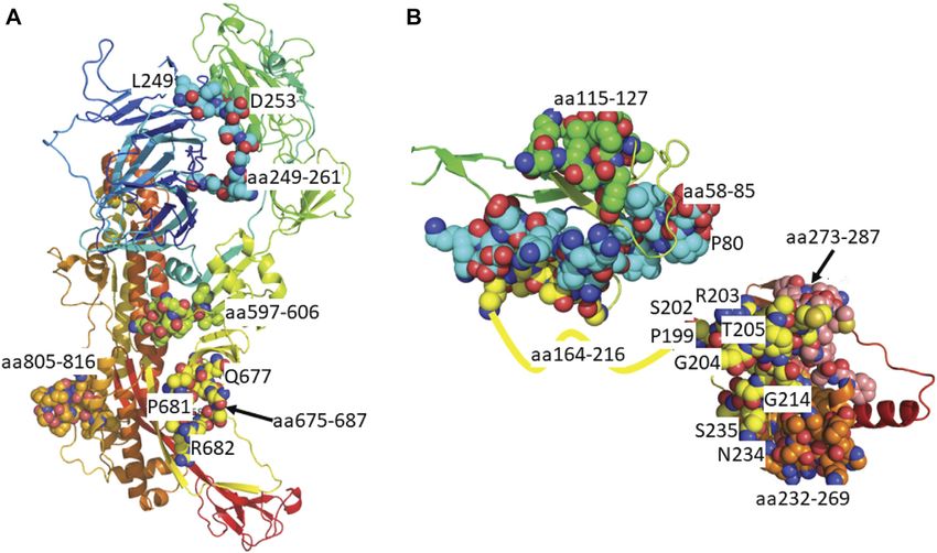
Moody et al.                                                                                              COVID-19 Epitopes and Self-Reactive Immunity

INTRODUCTION                                                                 Smatti et al., 2019). Given that a range of extrapulmonary
                                                                             pathologies have been reported in COVID-19 (Cheng et al.,
In March 2020, the World Health Organization (WHO) declared                  2020; Cheung et al., 2020; Han et al., 2020; Hundt et al., 2020;
the disease known as COVID-19, caused by the Severe Acute                    Nalleballe et al., 2020; Oxley et al., 2020; Poyiadji et al., 2020; Xie
Respiratory Syndrome Coronavirus (SARS-CoV-2), as a global                   et al., 2020; Zheng et al., 2020), molecular mimicry has been
pandemic. As of the 2nd of August 2021, there have been over                 hypothesized to be playing a role (Angileri et al., 2020a; Cappello
198.2 million confirmed cases and over 4.3 million deaths                     et al., 2020; Marino Gammazza et al., 2020). Additionally,
reported (WHO, 2020a). COVID-19 patients typically present                   conditions similar to the autoimmune diseases Guillain-Barré
with symptoms such as fever, dry cough, tiredness, as well as                Syndrome (Ameer et al., 2020; Korem et al., 2020) and Kawasaki
others such as myalgia, sore throat, loss of taste or smell, red eyes,       disease (Bordet et al., 2020; Jones et al., 2020) have been reported
diarrhoea and skin rash (Huang et al., 2020; Meng et al., 2020;              in COVID-19 patients. Within this Kawasaki-like disease, termed
WHO, 2020b). The SARS-CoV-2 genome encodes a series of                       Multisystem Inflammatory Syndrome in children (MIS-C),
structural and non-structural proteins (Chan et al., 2020; Wu                autoantibodies targeting a range of antigens including, but not
et al., 2020). There are four structural proteins consisting of the          limited to, anti-La, anti-Jo-1, anti-MUC15, anti-P2RX4, anti-
surface glycoprotein (spike, SP), nucleocapsid phosphoprotein                MAP2K2 and anti-CSNK1A1 have been reported (Consiglio
(nucleoprotein, NP), membrane (M) and envelope (E). There are                et al., 2020; Gruber et al., 2020). Additionally, autoantibodies
additionally eight open reading frames (Orf) encoding the non-               targeting other antigens including type I interferons (IFNs)
structural proteins: Orf1ab, Orf3a, Orf3b, Orf6, Orf7a, Orf7b,               (Bastard et al., 2020) have been identified in COVID-19
Orf8 and Orf10. Multiple studies have used serological assays to             positive patients (Zhou et al., 2020a; Bastard et al., 2020;
measure antibody responses to the SARS-CoV-2 virus (Long                     Vlachoyiannopoulos et al., 2020) alongside key autoimmune
et al., 2020; Zhao et al., 2020; Post et al., 2021). Antibodies are          associated antigens which are often associated with tissue
usually evaluated for reactivity against the spike and the                   damage, for example, anti-nuclear antigen (ANA) (Zhou et al.,
nucleoprotein with the aim of understanding seroconversion,                  2020a; Vlachoyiannopoulos et al., 2020). It is therefore highly
as well as correlating disease severity with neutralizing antibody           likely autoantibodies that recognize a range of other self-proteins
titres (systematically reviewed in Post et al. (2021)). Additional           are induced by infection with SARS-CoV-2.
reports have identified increases in antibodies to other SARS-                    Computational biology methods such as immune epitope
CoV-2 proteins, including Orf proteins, after infection (Hachim              mapping and Basic Local Alignment Search Tool (BLAST)
et al., 2020). Since the beginning of the pandemic, variants of              allow for relatively quick predictive screening analysis. They
SARS-CoV-2 have been emerging and circulating worldwide (van                 can help narrow findings and hypotheses, leading to more
Dorp et al., 2020). Some mutations, such as K417N and E484K in               targeted laboratory-based validation work. In the context of
the spike protein, are immune evasion mutations (Altmann et al.,             COVID-19, understanding immune epitopes, the sequences
2021). Since the second half of 2020 variants of SARS-CoV-2,                 that are recognized by antibodies, have been used to predict
containing mutations such as these, have arisen with increased               potential peptide-based vaccine candidates or to increase
transmissibility, associated with more severe disease and a                  sensitivity in serological assays (Ahmed et al., 2020; Amrun
reduction in neutralizing antibodies. These variants have been               et al., 2020; Poh et al., 2020). In the initial stages of the
called variants of concern (VOC) or variants of interest (VOI). As           COVID-19 outbreak, B-cell epitope mapping was performed
of July 2021, there are four VOCs and four VOIs classified by the             in SARS-CoV-2 to check for potential cross-reactivity of
Centers for Disease Control and Prevention (CDC) (SARS-,                     immune responses with other coronaviruses (Ahmed et al.,
2021). The VOCs consist of the strains originating in the                    2020; Grifoni et al., 2020). Recently, there have been a small
United Kingdom (Alpha strain), Brazil (detected in Japan,                    number of studies reporting sequence similarities between SARS-
Gamma strain), South Africa (Beta strain), and India (Delta                  CoV-2 proteins and human proteins, in support of the molecular
strain). Whereas VOIs consist of strains originating in Canada               mimicry hypothesis (Angileri et al., 2020a; Angileri et al., 2020b;
(Eta strain), United States (Iota strain), India (Kappa strain) and          Ehrenfeld et al., 2020; Kanduc, 2020; Kanduc and Shoenfeld,
Peru (Lambda strain).                                                        2020; Lucchese and Flöel, 2020; Lyons-Weiler, 2020; Marino
    Viral infections generally can play a role in promoting or               Gammazza et al., 2020). With the exception of one study,
exacerbating autoimmune diseases (reviewed in Smatti et al.                  which used longer epitopes (Lyons-Weiler, 2020), most of
(2019)). Proposed mechanisms for this association include                    these studies sought to find short regions (5-6aa) of exact
molecular mimicry and bystander activation (Fujinami et al.,                 identity between the SARS-CoV-2 proteins and human
2006; Smatti et al., 2019). Molecular mimicry refers to the                  proteins (Angileri et al., 2020a; Angileri et al., 2020b; Kanduc,
phenomenon of immune cross-reactivity between a foreign                      2020; Kanduc and Shoenfeld, 2020; Lucchese and Flöel, 2020;
pathogen and self-protein, where an immune cell recognizes                   Marino Gammazza et al., 2020). Whilst these studies suggested a
both due to their sequence similarity (Fujinami et al., 2006;                number of regions, most of the identical sequences identified
Smatti et al., 2019). Bystander activation refers to the                     would be unlikely to sustain a pathogenic autoreactive response
activation of self-reacting immune cells driven by the                       (Angileri et al., 2020a; Kanduc, 2020; Kanduc and Shoenfeld,
liberation of self-antigens (the targets which stimulate immune              2020; Lucchese and Flöel, 2020; Marino Gammazza et al., 2020).
responses) from virus-lysed cells which otherwise would not be               Specifically, while assessing its potential accessibility for antibody
typically exposed to the immune system (Fujinami et al., 2006;               binding, these studies did not discriminate the localization of the

Frontiers in Bioinformatics | www.frontiersin.org                        2                                      August 2021 | Volume 1 | Article 709533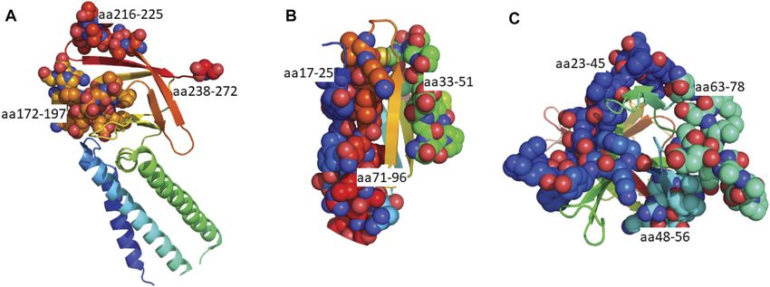
Moody et al.                                                                                            COVID-19 Epitopes and Self-Reactive Immunity

epitope within the protein or within the cell (Angileri et al., 2020a;       proteins which may have antibody cross-reactivity with SARS-
Kanduc, 2020; Kanduc and Shoenfeld, 2020; Lucchese and Flöel,                CoV-2 immunogenic regions.
2020; Lyons-Weiler, 2020; Marino Gammazza et al., 2020).
Moreover, except in the case of Kanduc (2020), who identified
identical hexamers between SARS-CoV-2 and human proteins                     METHODS
associated with various system disorders, these studies only
report small numbers of human proteins with similar (Lyons-                  Research Pipeline to Explore Potential
Weiler, 2020) or short identical sequences (Angileri et al., 2020a;          Immune Cross-Reactivity
Kanduc and Shoenfeld, 2020; Lucchese and Flöel, 2020; Marino                 In order to explore the similarities between potential
Gammazza et al., 2020) to SARS-CoV-2 proteins (e.g. association              immunogenic SARS-CoV-2 regions and human proteins, we
with the immune system, molecular chaperones, brainstem                      applied a research pipeline as outlined in Figure 1. B cell
proteins). Therefore, the limited nature of these studies does               epitopes were first predicted within a selection of SARS-CoV-2
not show the full extent of similarities between SARS-CoV-2 and              proteins. Those selected were then compared to the human
human proteins.                                                              proteome. To explore potential immune cross-reactivity, we
    In the present study we have pioneered a different                       applied novel criteria that not only considered short identical
immunoinformatics approach to overcome some of these                         sequences but also amino acid variations with conserved
limitations and further explore the potential for autoimmune                 structural and charge changes that may not impact antibody
cross-reactivity driven by SARS-CoV-2 molecular mimicry.                     binding. We also considered protein localization, with a focus on
B-cell epitopes within the structural proteins and several Orf               extracellular proteins. The alignments of interest found within
proteins (shown to generate antibody responses in COVID-19                   human proteins were cross-checked as potential epitopes within
positive patients (Hachim et al., 2020)), were first predicted using          their own protein sequence. Details of each step described below.
the IEDB B Cell epitope tool. Epitopes predicted in the structural
proteins were compared to the current VOCs (Alpha, Beta,
Gamma and Delta) and VOIs (Eta, Iota, Kappa and Lambda).                     Sequence Identification and Epitope
Eight of the fifteen epitopes contained at least one mutation in              Mapping
one variant. The identified 9-53aa long sequences, in both                    Based on previous mapping and assay-based studies (Hachim
structural and Orf proteins, were then compared to sequences                 et al., 2020; Grifoni et al., 2020), epitope prediction was carried
in human proteins using the NCBI protein BLAST tool (Altschul                out on proteins previously identified from the SARS-CoV-2
et al., 1990). Assessment of potential for cross-reactivity between          isolate Wuhan-hu-1 (Figure 1; Supplementary Table S1)
SARS-CoV-2 and self-proteins with capacity to perpetuate                     (GenBank: MN908947.3) (Wu et al., 2020). As Orf3b is
autoimmune pathology was based on a combination of                           produced by a premature stop codon (Gordon et al., 2020;
immunologically relevant sequence similarity (not just                       Lam et al., 2020) and the sequence could not be identified on
identity) (Angileri et al., 2020a; Kanduc, 2020; Lucchese and                GenBank, the sequence provided in Lam et al. (2020) was used for
Flöel, 2020; Marino Gammazza et al., 2020) and the localization              epitope mapping.
of the protein itself, with a focus on extracellular targets. 11                Linear B cell epitope predictions were performed using the
human proteins, containing amino acid sequences similar to nine              B cell Epitope prediction tool found within Immune Epitope
predicted SARS-CoV-2 B cell epitopes, were identified based on                Database and Analysis Resource (IEDB, http://tools.iedb.org/
these selection criteria. These findings indicate that antibodies             bcell/ (Dhanda et al., 2019)). Using the Bepipred Linear
induced by SARS-CoV-2 could directly interfere with cell                     Epitope Prediction algorithm (Haste Andersen et al., 2006;
function, including that of immune cells, and could help                     Larsen et al., 2006; Ponomarenko and Bourne, 2007),
explain some of the additional pathologies identified in                      predicted epitopes for structural proteins (Spike, Envelope,
COVID-19 patients (Cheng et al., 2020; Cheung et al., 2020;                  Membrane and Nucleoprotein), were selected based on
Han et al., 2020; Hundt et al., 2020; Nalleballe et al., 2020; Oxley         prediction score (>1) and having length greater than 6aa.
et al., 2020; Poyiadji et al., 2020; Xie et al., 2020; Zheng et al.,         Epitopes for the Orf proteins were predicted using the
2020). Finally, comparing the sequences of both predicted spike              Bepipred Linear Epitope Prediction 2.0 algorithm (Jespersen
epitopes and full length spike protein to various human proteins             et al., 2017) and were selected for having the highest peak
implicated in immune thrombocytopenia purpura (ITP) and                      points (prediction score >0.5) of length greater than 6aa. Each
thrombocytopenia syndrome (TTS), our results indicate that                   algorithm was selected based on the IEDB recommended at the
molecular mimicry is unlikely to be the cause of TTS, or                     time epitope mapping was performed, and prediction score cut
vaccine induced prothrombotic immune thrombocytopenia                        offs to select for those more likely to be real epitopes.
(VIPIT) following vaccination with the COVID-19 adenovirus                      Where possible, protein structures were downloaded from the
vector vaccines. To our knowledge, this is the first study to                 protein data bank rcsb.org (Berman et al., 2000). For SARS-CoV-
compare immune epitopes across the circulating VOCs and                      2 proteins, where regions were uncrystallised, homology models
VOIs; highlight multiple similarities between the selected Orf               were created using the MPI Bioinformatics Toolkit (https://
proteins and human proteins; identify proteins with reported                 toolkit.tuebingen.mpg.de/). Specifically, templates were found
associations to autoimmunity as sharing sequences with SARS-                 using HHpred and models were created using Modeller
CoV-2 epitopes; and to highlight novel extracellular human                   (Zimmermann et al., 2018). Any unstructured regions were

Frontiers in Bioinformatics | www.frontiersin.org                        3                                    August 2021 | Volume 1 | Article 709533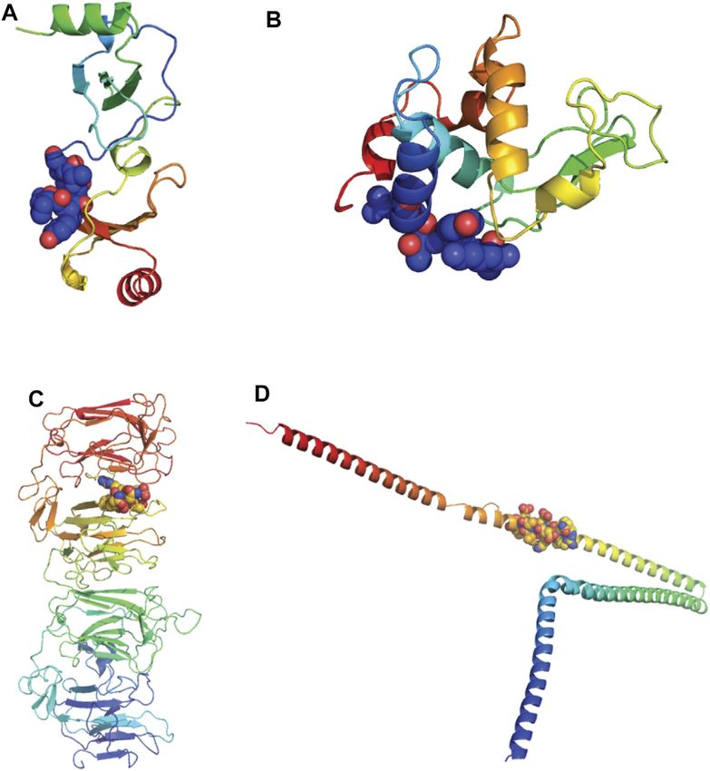
Moody et al. COVID-19 Epitopes and Self-Reactive Immunity
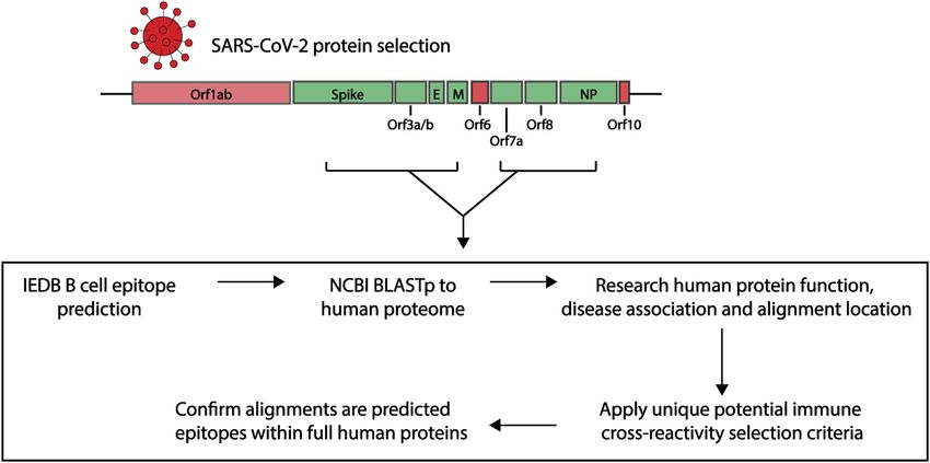
FIGURE 1 | Research pipeline of the project to explore the potential of immune cross-reactivity. Protein sequences for select SARS-CoV-2 proteins were obtained
for epitope predictions (highlighted green. Spike: Surface glycoprotein. E, envelope; M, membrane; NP, nucleoprotein). Using the Immune Epitope Database (IEDB)
epitope prediction tool, B cell epitopes were predicted and those selected were screened against human proteins using the NCBI Blastp tool. Human proteins with
sequence similarities to the SARS-CoV-2 epitopes were investigated for their function, disease association and alignment localization. Potential immune cross-
reactivity between SARS-CoV-2 and human alignments was then explored by applying specific selection criteria. Alignments of interest from the human proteins were
explored as potential epitopes within the human protein sequences.
deleted and figures were created in The PyMOL Molecular BlastN by selecting standard databases and optimizing for highly
Graphics System, Version 2.0 Schrödinger, LLC. All available similar sequences (megablast). Since not all submissions to
human protein models were downloaded from the SWISS- GISAID have also been submitted to NCBI some did not
MODEL Repository (Bienert et al., 2017). exhibit 100% sequence identity. The percentage of sequence
identity for each variant is listed in the table below (column
NCBI blast identity in Supplementary Table S2). For the ones
Sequence Alignment to Other SARS-CoV-2 that exhibited lower than 100% sequence identity, with blastN we
Variants visualized the areas in which the differences were laying. Those
For SARS-CoV-2 variants protein alignment, we used eight specific sequences were then blasted to view where in the viral
different variants and the original Wuhan sequence genome they were appearing.
(Supplementary Table S2). The FASTA sequences were Using JalView (V2.11.1.4) (Waterhouse et al., 2009), we
retrieved from the GISAID database (https://www.gisaid.org/). aligned the amino acid sequences for the surface glycoprotein,
In the EpiCov search section of the GISAID database there is an membrane protein and nucleoprotein areas of all eight variants
available tab that allows for selection of the major circulating plus the original Wuhan sequence, and set the latter as reference
variants. Of these we selected the current VOCs (Alpha, Beta, genome. We then performed alignment using the Multiple
Gamma and Delta) and VOIs (Eta, Iota, Kappa and Lambda). The Sequence Alignment using Fast Fourier Transform (MAFFT)
virus names listed in Supplementary Table S2 are GISAID alignment program with default settings (V7.110) (Katoh and
nomenclature and the specific viruses were selected based on Standley, 2013). MAFFT is a high speed multiple sequence
various conditions: for all variants we selected the conditions in alignment program which utilizes the Fast Fourier Transform
which the sequence was complete, we excluded sequences with to optimize protein alignments based on the amino acid physical
low coverage (>5% unidentified amino acids) and selected the properties.
specific clad of the variant being analyzed. For all variants, each
one was chosen within a specific date based on their historical Sequence BLAST to Compare Epitopes to
appearance. From these we selected the ones that underwent
Illumina sequencing and for which the assembly method was
Human Proteins and Identification of
clearly reported. In order to obtain the specific amino acid Associated Diseases, Expression Location
sequences of the various genomic regions based on the B cell and Function
epitope prediction, we retrieved the FASTA sequence of the entire Using the NCBI protein-protein BLAST (blastp, https://blast.
viral genome from the selected variants and blasted these using ncbi.nlm.nih.gov/ (Altschul et al., 1990)), predicted epitope
Frontiers in Bioinformatics | www.frontiersin.org 4 August 2021 | Volume 1 | Article 709533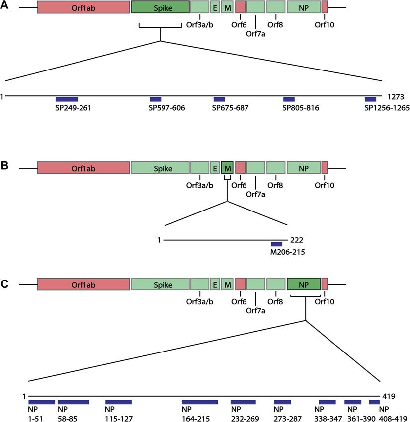
Moody et al.                                                                                           COVID-19 Epitopes and Self-Reactive Immunity

sequences were blasted for the top 100 results, to the non-                four structural proteins of SARS-CoV-2 Wuhan-hu-1 variant:
redundant protein sequences (nr) database, specifically looking             spike (SP), membrane (M), envelope (E) and nucleoprotein (NP),
at Homo sapiens (Taxid ID: 9606). All other algorithm parameters           using the IEDB B cell Epitope Prediction Tool. In B cell epitopes,
remained as default settings, including: automatically adjust              4-6aa often form the minimal binding region (Pieczenik, 2003),
parameters for short input sequences, and expect threshold                and most B cell epitopes have a length between 4 and 25aa (Gupta
0.05. Resulting lists were narrowed by excluding isoform repeats,          et al., 2013; Potocnakova et al., 2016) (but can be as long as 50aa
identical proteins with different nomenclature, uncharacterized/           (Gupta et al., 2013)). As some epitopes are composed of
hypothetical proteins and variable regions of B and T cell                 discontinuous multiple linear segments of 1-6aa (Rubinstein
receptors. Associations of the self-proteins to diseases, their            et al., 2008), we selected the predicted epitopes of 6aa or
expression locations and functions were identified using                    more. Additionally, we applied a cut off of at least score 1,
UniProtKB, GeneCards and PubMed Resources. Any proteins                    selecting those specifically with the higher prediction score
identified through computational analysis or found on an                    and therefore more likely to be real epitopes. Based on these
unreviewed UniProtKB database page were further excluded.                  criteria, we identified 15 epitopes of interest ranging between 10
   For any direct epitope to full protein comparison, “align two or        and 53aa (Figure 2; Supplementary Table S3): five spike epitopes
more sequences” in the NCBI blastp tool was used.                          of which two are C-terminally unstructured (Figure 3A), one
                                                                           unstructured C-terminal membrane epitope, and nine
                                                                           nucleoprotein epitopes of which two are partially structured,
Prediction of Potential Immune                                             and three are C-terminal unstructured motifs (Figure 3B).
Cross-Reactivity Selection Criteria                                        During the course of the present study, four of the five spike
Using the previously narrowed BLAST lists, potential B cell                epitopes (249-261, 597-606, 805-816, 1256-1265), and three of
epitope cross-reactivity was identified based on the following              the nine nucleoprotein epitopes (164-216, 232-269, 361-390) we
criteria:                                                                  predicted, were further shown by others to be regions which
                                                                           overlap with new epitopes identified in laboratory studies
1) Covering at least six consecutive amino acids.                          (Amrun et al., 2020; Poh et al., 2020; Shrock et al., 2020;
2) Where there were amino acid differences (including those                Wang et al., 2020; Yi et al., 2020; Yoshida et al., 2021),
   interrupting a 6aa consecutive sequence), the similarity                providing support to the selected epitope mapping approach.
   between the amino acid structure and charge was                             In addition to the structural proteins, epitopes were also
   compared. Any variances that may impact antibody                        predicted for Orf3a, Orf3b, Orf7a and Orf8 SARS-CoV-2
   function or binding, such as structural and charge changes,             proteins since antibodies against these proteins have been
   were excluded.                                                          identified in COVID-19 patients (Hachim et al., 2020). Similar
3) Expression location or alignment region found in the                    to the structural proteins, epitope sequences were selected to have
   extracellular domain.                                                   a minimal length of 6aa. As the Bepipred v2.0 algorithm
                                                                           (Jespersen et al., 2017) was used to predict these epitopes, the
                                                                           prediction score criteria were modified to have a score greater
Confirmation of B Cell Epitope Within the                                   than 0.55, representing the higher predicted immunogenic
Self-Proteins                                                              regions in these proteins shown to have antibody responses.
Complete protein sequences were obtained from the UniProtKB                Based on these criteria a total of 10 epitopes were identified
database and mapped for potential B cell epitopes using the IEDB           within the Orf proteins (Figure 4; Supplementary Table S3); two
B cell epitope prediction tool. To correspond to the prediction            partially structured, one C-terminal partially structure epitope for
algorithm used for the SARS-CoV-2 proteins, the Bepipred                   Orf3a (Figure 5A), three structured epitopes for Orf7a and Orf8
Linear Epitope Prediction algorithm (Haste Andersen et al.,                (Figures 5B,C, respectively) and a single unstructured
2006; Larsen et al., 2006; Ponomarenko and Bourne, 2007)                   N-terminal epitope within Orf3b. Of these, sequences Orf3a
was used for human proteins where the shared alignments                    172-197, Orf3a 216-225, Orf8 23-45 and Orf8 48-56 have been
were to structural SARS-CoV-2 proteins. Whereas proteins                   validated to be within a region to which COVID-19 patients have
which shared alignment with non-structural Orf proteins were               antibody responses (Shrock et al., 2020; Wang et al., 2020).
mapped using the Bepipred Linear Epitope Prediction 2.0
algorithm (Jespersen et al., 2017). To confirm if any epitopes
had been validated, the UniProtKB ID was used to search the
                                                                           Comparison of Predicted Immunogenic
antigens and epitopes in the IEDB database (Vita et al., 2019).            Regions Among SARS-CoV-2 Variants of
                                                                           Concern and Variants of Interest
                                                                           With the multiple variants of concern and of interest circulating
RESULTS                                                                    worldwide which can escape neutralization and cause more
                                                                           severe disease (Garcia-Beltran et al., 2021), we were interested
Identification and Selection of B Cell                                      in whether these predicted B cell epitopes overlapped with known
Epitopes in SARS-CoV-2 Proteins                                            escape mutations or contain any mutations which may impact
As previous studies have looked at the structural proteins (Grifoni        antibody responses. As the antibody responses to structural
et al., 2020), B cell epitope mapping was initially performed on the       proteins, specifically the spike and nucleoprotein, have been

Frontiers in Bioinformatics | www.frontiersin.org                      5                                     August 2021 | Volume 1 | Article 709533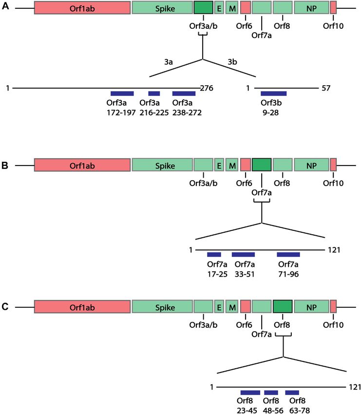
Moody et al.                                                                                                          COVID-19 Epitopes and Self-Reactive Immunity

  FIGURE 2 | Linear schematic of selected B cell epitopes in SARS-CoV-2 Structural proteins. B cell epitopes within the SARS-CoV-2 spike (SP), membrane (M) and
  nucleoprotein (NP) were mapped and selected based as having ≥6aa length and a predicted epitope score ≥1. (A) Five epitopes in the spike protein, beginning at
  positions aa249, 597, 675, 805 and 1256 were selected for downstream analysis. (B) One epitope in the membrane protein at position aa205 was identified. (C) Nine
  epitopes in the nucleoprotein, aa1, 58, 115, 164, 232, 273, 338, 361, 408 were identified. All associated sequences can be found in Supplementary Table S3.

extensively studied and are used for serological assays (Post et al.,                 mutation (Figures 3A, 6A). In this 13aa sequence the Beta variant
2021), potential mutations in these proteins were of interest.                        contains two mutations, Q677H and R682W. At position 681,
Using the GISAID database to obtain sequences for the VOCs:                           both the Delta and Alpha variants contained mutations P681R
Alpha, Beta, Gamma and Delta variants, the structural proteins                        and P681H respectively. In each case, these mutations consist of a
(spike, membrane and nucleoprotein) were blast aligned with                           structural and/or charge mutation which may impact antibody
predicted epitopes to identify any mutations in these regions                         binding. For example, the P681 mutations would be expected to
(Supplementary Figures S1, S2; Supplementary Table S4). Of                            increase flexibility in this region. No mutation was identified
the five predicted spike epitopes only SP675-687 contained any                         within the one membrane epitope predicted (Figure 6B). Among

Frontiers in Bioinformatics | www.frontiersin.org                                 6                                          August 2021 | Volume 1 | Article 709533
Moody et al.                                                                                                                  COVID-19 Epitopes and Self-Reactive Immunity

  FIGURE 3 | Predicted epitopes mapped to protein structure of Surface Glycoprotein and Nucleoprotein. (A) X-ray crystal structure of the Surface Glycoprotein (Cai
  et al., 2020) coloured via spectrum (N to C terminal) with the structured B cell epitopes highlighted via spheres; aa294-261 (cyan), aa597-606 (green), aa675-687 (yellow)
  and aa805-816 (orange). Also labelled are the mutation sites; L249, D253, Q677, P681 and R682. (B) Structure of the nucleoprotein coloured via spectrum (N to C
  terminal). The N-terminal regions are from the X-ray crystal structure (PDB code: 6vyo) followed by an unstructured linear (yellow line) to a C-terminal homology
  model. The structural B cell epitopes are highlighted via spheres; aa58-85 (cyan), aa115-127 (green), aa164-216 (yellow), aa232-269 (orange) and aa273-287 (salmon).
  Also labelled is the location of known mutation sites; P80, P199, R201, S202, R203, T205, G214, N234 and S235.

the nine nucleoprotein epitopes, NP115-127, 273-287, 338-347                               (Figure 6E). Within the nine predicted nucleoprotein epitopes,
and 408-419 did not contain any mutations. NP1-51 contained a                              NP1-51, NP164-216, NP232-269 and NP351-390 contain
mutation in the Alpha variant at D3L. Epitopes NP58-85, 232-                               mutations. The Eta variant contained four mutations in NP1-
269 and 361-390 each only have one mutation: P80R (Gamma),                                 51: Shift at position 1, S2M, D3Y and A12G. There was also a
S235F (Alpha) and D377Y (Delta). Multiple mutations were                                   mutation in NP1-51 in the Lambda variant, P13F. None of these
identified in NP164-216 (Figures 3B, 6C). The Delta variant                                 are shared with VOCs, and only D3Y, given the change in
contained two mutations S202I and R203M. Position 203 was                                  structure and charge may impact antibody binding. As with
additionally mutated in the Alpha and Gamma variants, R203K.                               the VOCs, multiple mutation sites were found within NP164-
These variants were also mutated at position 204, G204R. There                             215 (Figure 6F). Shared with VOCs were R203M (Kappa), R203K
was additionally a mutation in the Beta variant, T205I. Except for                         (Lambda), G204K (Lambda) and T205I (Eta). Additional
mutations R203K and T205I in NP164-216, all mutations                                      mutations were P119L (Iota) and G214C (Lambda). The last
identified result in a structural and/or charge change that may                             two epitopes containing at least one variant with a mutation were
impact antibody binding, resulting in the more severe disease                              NP232-269 and NP361-390, with one and two mutations
seen in these variants.                                                                    respectively. These were M234I (Iota), T366I (Lambda) and
   In addition to looking at the VOCs, we further looked into                              D377Y (Kappa). Of these, D377Y results in a structural and
whether any mutations occurred within the predicted epitopes in                            charge change that may impact antibody binding.
the VOIs: Eta, Iota, Kappa and Lambda (Supplementary Figures
S3, S4; Supplementary Table S4). Within the five predicted spike
epitopes, SP249-261 and SP675-687 contained mutations
                                                                                           Shared Sequence Alignments Between
(Figure 6D). In SP249-261, the Lambda variant was missing                                  SARS-CoV-2 Predicted Epitopes and
aa249-252 and contained the mutation D253N. At this same                                   Human Proteins
residue the Iota variant had the mutation D253G. In each of these                          The similarities between the predicted SARS-CoV-2 B cell
cases a structural and or charge change occur. Both Eta and                                epitopes and the human proteome were identified using the
Kappa variants contained a mutation in SP675-687, Q677H and                                NCBI protein-protein BLAST tool. Each of the 25 epitopes
P681R, respectively. Each of these are shared mutations in VOCs                            were compared, and final lists were narrowed to remove 1) the
Beta and Delta, respectively. As seen in the VOCs, no mutation                             duplicates under alternative nomenclature or sequence ID 2)
was identified in the single predicted membrane epitope                                     different isomer repeats 3) uncharacterised or hypothetical

Frontiers in Bioinformatics | www.frontiersin.org                                      7                                             August 2021 | Volume 1 | Article 709533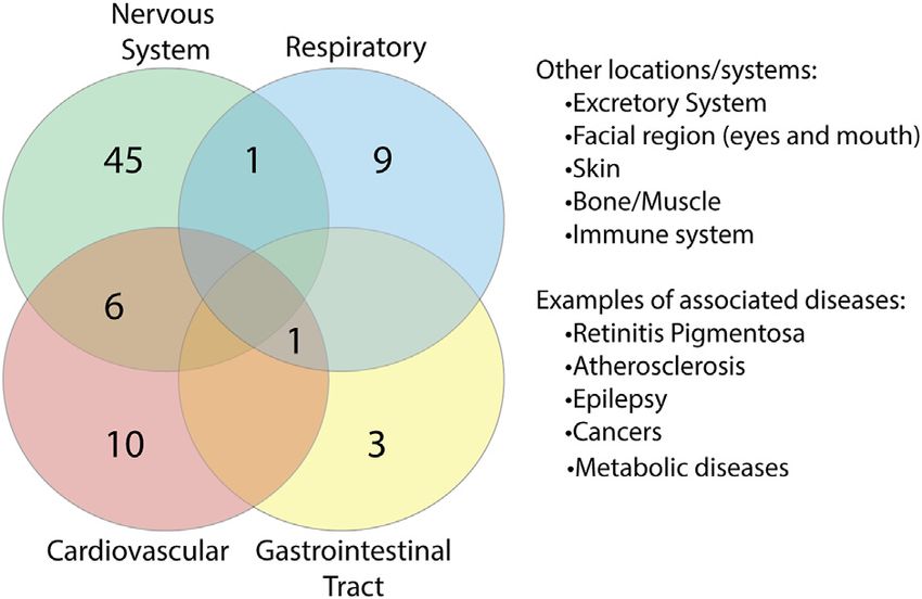
Moody et al.                                                                                                              COVID-19 Epitopes and Self-Reactive Immunity

  FIGURE 4 | Linear schematic of selected B cell epitopes in SARS-CoV-2 Orf proteins. B cell epitopes within the SARS-CoV-2 Orf3a, Orf3b, Orf7a and Orf8 and
  selected based as having ≥6aa length and a predicted epitope score ≥0.55. (A) Three epitopes in Orf3a (aa172-197, 216-225 and 238-272) and one epitope in Orf3b
  (aa9-28) were identified. (B) Three epitopes in Orf7a (aa17-25, 33-51 and 71-96) were identified. (C) Three epitopes in Orf8 (aa23-45, 48-56 and 63-78). All associated
  sequences can be found in Supplementary Table S3.

proteins and 4) variable regions of lymphocyte receptors                                the function, association to diseases (including COVID-19) and the
(Figure 7; Supplementary Table S5). Of the 25 epitopes, two                             specific sequence alignment identified were investigated (Figure 8). In
(NP1-51 and NP164-216) had no significant similarity when                                doing so, it was found that some of the alignments identified were in
blasted to the nr database. Among the 23 other epitopes, a total of                     computationally predicted sequences or unreviewed proteins in the
281 alignments consisting of 256 self-proteins, were identified.                         UniprotKB database. These proteins (Supplementary Table S6), were
                                                                                        removed from those of interest, resulting in a total of 246 alignments,
                                                                                        consisting of 223 self-proteins remaining (Supplementary Table S7).
Self-Proteins Share Disease Associations                                                   While alignments identified within these remaining self-
Which Relate to Symptoms Reported in                                                    proteins did not overlap key functional domains of the human
COVID-19 Patients                                                                       proteins, identified in the UniProtKB database, some of the
For each of the human proteins identified that share similar sequences                   proteins are described to be associated with a variety of
with the SARS-CoV-2 predicted epitopes (Supplementary Table S5),                        diseases. Two of the human proteins identified have been

Frontiers in Bioinformatics | www.frontiersin.org                                   8                                            August 2021 | Volume 1 | Article 709533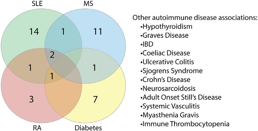
Moody et al.                                                                                                               COVID-19 Epitopes and Self-Reactive Immunity

  FIGURE 5 | Predicted epitopes mapped to the structures of SARS-CoV-2 Orf proteins. (A) Cryo-EM structure of the Orb3a (Kern et al., 2021) coloured via
  spectrum (N to C terminal) with the structured B cell epitopes highlighted via spheres; aa172-197 (orange), aa216-225 (light red), aa238-272 (red). (B) X-ray crystal
  structure of Orb7a (Zhou et al., 2021) with the structured B cell epitopes highlighted via spheres; aa17-25 (blue), 33-51 (green) and aa71-96 (red). (C) X-ray crystal
  structure of Orb7a (Flower et al., 2021) with the structured B cell epitopes highlighted via spheres; aa 23-45 (blue), 1148-56 (cyan) and aa63-78 (light green).

reported in other SARS-CoV-2 related papers (Aishwarya et al.,                           mitochondria/metabolic diseases and the immune system.
2020; Vastrad et al., 2020). Ankryin B, which aligns to SP597-606,                       Among the proteins associated with these, some showed
has been reported to be transcribed in lungs of COVID-19 patients,                       overlap with other regions. This suggests that potential
where it is not usually expressed (Aishwarya et al., 2020), and                          interruptions in some of these proteins could have multi-
ZNF354C, aligning to NP361-390, is a transcription factor which                          organ consequences, which may be associated with COVID-19.
interacts with genes that are differentially expressed in SARS-CoV-
2 infected patients (Vastrad et al., 2020). Additionally, 144 of the
self-proteins are reported to be associated with a range of other
                                                                                         There is an Association Between Human
diseases (Supplementary Table S8). Among the identified diseases,                         Proteins, With Shared SARS-CoV-2
we observed similarities for different forms of the same disease, for                    Sequences, and Autoimmunity
example types of retinitis pigmentosa or types of epilepsy. Some of                      As reports of autoimmunity in COVID-19 continue to emerge
these diseases share similarities with symptoms reported within                          (Bordet et al., 2020; Korem et al., 2020; Unsworth et al., 2020; Lui
COVID-19 patients such as cardiovascular diseases                                        et al., 2021), of key interest was the association between the
(atherosclerosis, cardiomyopathy, hypertension etc.) (Zhou                               identified human proteins and whether they have a role in
et al., 2020b; Madjid et al., 2020); respiratory issues (airway                          autoimmune diseases or are known autoantigens. Of the 223
hyper-responsiveness, inflammation) (Huang et al., 2020);                                 human proteins, 50 were associated with autoimmune diseases, in
neurological diseases (cerebellar ataxia, epilepsy) (Mithani et al.,                     both human and animal model settings (Supplementary Table
2021; Povlow and Auerbach, 2021; Werner et al., 2021); and                               S10). Among these 50 proteins, we found that some overlapped
myopathy (Manzano et al., 2020; Versace et al., 2021). We also                           with multiple autoimmune diseases (Figure 10; Supplementary
found that 42 of these 144 proteins had an association with various                      Table S11). Systemic lupus erythematosus (SLE) was found to
types of cancer.                                                                         have the most protein association, followed by multiple sclerosis
    To identify whether there were any similarities or common                            (combined human and animal model, experimental autoimmune
location associations between the proteins, we grouped each of                           encephalomyelitis (EAE)). SLE shared the most overlap with
the proteins based on the body system/s they were found to be                            other autoimmune diseases and some targets were shared
associated with (Supplementary Table S9). In doing so, we                                across more than two autoimmune diseases. Many of the
identified a range of overlap between proteins and systems                                associations identified were due to gene single nucleotide
(Figure 9). 52 proteins were found to be associated with the                             polymorphisms (SNPs) and altered expression levels. However,
nervous system, six of which overlapped with the cardiovascular                          eight of the proteins are known targets of autoantibodies and
system, which is just under half of the cardiovascular-related                           include key antibodies for assessing or diagnosing the associated
proteins identified. Overlap could additionally be found with the                         diseases such as the myasthenia gravis autoantigen A-kinase
respiratory system and gastrointestinal tract (GIT), systems                             anchor protein 12 (gravin), and histone 3, a nuclear target in
associated with known COVID-19 complications. Additional                                 SLE (Table 1). This suggests that the presence of some of these
locations/systems found to have protein associations included                            autoantibodies in COVID-19 patients without a history of
excretory system, facial region, skin, bone/muscle, thyroid,                             autoimmune disease could be due to immune cross-reactivity.

Frontiers in Bioinformatics | www.frontiersin.org                                    9                                            August 2021 | Volume 1 | Article 709533
Moody et al. COVID-19 Epitopes and Self-Reactive Immunity
FIGURE 6 | Examples of sequence comparison between predicted B cell epitopes and key SARS-CoV-2 variants. Structural protein sequences for a strain from
Wuhan (WIV04/2019), VOCs (Alpha, Beta, Gamma and Delta) and VOIs (Eta, Iota, Kappa and Lambda) were obtained from the GISAID database and blast aligned with
predicted B cell epitopes. Quality refers to the alignment quality based on blosum2 algorithm scores, Consensus indicates the abundance of the amino acids present in a
particular position and Occupancy is the number of aligned positions. (A) SP VOC sequence alignment at position aa675-687. (B) VOC sequence alignment of
membrane aa206-215. (C) NP VOC sequence alignment at position aa164-216. (D) SP VOI sequence alignment at position aa675-687. (E) VOI sequence alignment of
membrane aa206-215. (F) NP VOI sequence alignment at position aa164-216.
Potential Cross-Reactivity of Predicted with any of these proteins, they are more likely to bind and cross-
SARS-CoV-2 B Cell Epitopes to Human react with extracellular proteins on intact cells. Using the
Proteins UniProtKB database, research into the localization of each
protein from Supplementary Table S12 of potential cross-
Short identical alignments (5-6aa) have been reported to be shared
with SARS-CoV-2 and human proteins (Angileri et al., 2020a; reactive human proteins was performed. Those of further
Angileri et al., 2020b; Kanduc, 2020; Kanduc and Shoenfeld, interest were proteins that are reported to be secreted into the
2020; Lucchese and Flöel, 2020; Marino Gammazza et al., 2020). extracellular domain or where the alignment region was reported to
Using the alignments identified from blasting predicted SARS- be extracellular. In doing this, a total of 11 proteins were identified
(Table 2). In these 11 human proteins, the alignment identities to
CoV-2 immune epitopes, the potential for immune cross-
the SARS-CoV-2 epitopes ranged between 62 and 100%.
reactivity was explored. While point mutations in viruses are
Additionally, the SARS-CoV-2 spike and nucleoprotein epitopes
known to create antibody escape variants (Doud et al., 2018),
with aligned human proteins were not found to have a mutation in
this is not always the case, and not all mutations will affect
any variant of concern or variant of interest.
antibody binding (Doud et al., 2018). Some antibodies are Next, we further explored these sequences in the human
unaffected by conserved changes (Rodpothong and Auewarakul, proteins through identifying whether they are potential
2012). Therefore, to look at potential immune cross-reactivity, we epitopes within the self-protein sequence and their structural
applied criteria that not only relied on a minimum 6aa length, but accessibility. Complete human protein sequences were obtained
took into account amino acid variations that had conserved charge from the UniprotKB database and epitope mapping performed
and non-structural changes and therefore is less likely to impact (Table 3). The alignments in seven of the 11 proteins were
antibody binding. Applying these unique criteria, a total of 136 identified as epitopes, namely Bone morphogenetic protein 1,
alignments from both intra- and extra-cellular proteins were Lysozyme-like 1, Lysozyme-like 2, CCL22, COL6A3, tubby like
identified as potential cross-reactive targets (Supplementary protein 3 and alpha-internexin. Additionally, those of COL6A3,
Table S12). Although antibodies have a potential to cross-react tubby like protein 3 and alpha-internexin were found within large
Frontiers in Bioinformatics | www.frontiersin.org 10 August 2021 | Volume 1 | Article 709533Moody et al. COVID-19 Epitopes and Self-Reactive Immunity
FIGURE 7 | Workflow to identify human proteins that share sequence similarities with SARS-CoV-2 immunogenic regions. The predicted SARS-CoV-2 B cell
epitopes were compared to the human proteome using the NCBI protein BLAST tool. Two epitopes, NP1-51 and NP164-216 had no sequence similarities to human
proteins. The top 100 sequence alignments from the remaining 23 epitopes were narrowed by removing duplicates (alternative nomenclature/sequence IDs),
uncharacterized/hypothetical proteins and the variable regions of lymphocyte receptors. This resulted in a final list of 281 sequence alignments that was comprised
of 256 human proteins.
sequences (greater than 50aa). For the proteins Reelin, LST-3 and et al., 2020; Sadoff et al., 2021). To explore whether molecular
lactase-phlorizin hydrolase, some, but not all, of the amino acids mimicry is a potential mechanism causing the syndrome we used
within the alignments were identified as potential epitopes. The the blastp protein alignment tool to align the predicted spike
final protein of interest, Mucin-12 did not yield any results when epitopes to PF4 (UniprotKB: P02776). No similarity was found
using the Bepipred Linear Epitope Prediction 2.0 algorithm, the between any predicted epitope in the spike protein to sequences
algorithm used for the matching to SARS-CoV-2 protein, Orf3a. in the full-length PF4 protein. To further check if there may be
similarity between the spike and PF4 outside the selected epitope
However, using version 1 of this prediction algorithm, the
regions, the complete spike protein sequence was compared to
alignment partially sits within a predicted epitope. Given these
PF4, which resulted in no similarity results. PF4 interacts with a
epitopes were predicted, we searched IEDB to explore whether variant of CXCR3 (CXCR3B) (Lasagni et al., 2003), the receptor
any of these B cell epitopes may have been previously validated, of a number of key chemokines (such as CXCL10, a pro-
however none were. Despite this, most of these alignments do inflammatory cytokine important for chemotaxis and the
appear as potential epitopes within the human proteins, further activation of peripheral immune cells (Lasagni et al., 2003)).
suggesting a likelihood that antibody cross-reactivity may occur We therefore explored whether the SARS-CoV-2 spike
between SARS-CoV-2 and these targets. epitopes were similar to CXCR3B and thus potentially
Of the 11 extracellular proteins, five had homology model involved with interrupting PF4-CXCR3B interactions. Spike
structures available which covered the alignment regions of epitopes were blasted to the CXCR3-B UniprotKB sequence
interest: CCL22, Lysozyme-like 1, Lysozyme-like 2, Reelin and (P49682-2), and no similarities were identified. Finally, as
Alpha Internexin (Table 2). Each of these alignment motifs were deficiency of ADAMTS13, a metalloprotease, has been
mapped to the corresponding structures (Figure 11). In every case, implicated in patients with ITP, spike epitopes were blasted to
the motifs of interest were found towards the protein surface, making the ADAMTS13 UniprotKB sequence (Q76LX8), where no
them accessible to antibody binding and therefore the potential of sequence similarity was found. This suggests that molecular
cross-reactivity. Furthermore, for those proteins which did not have mimicry is unlikely to be the cause of the described cases of TTS.
a structure readily available, all alignment motifs were predicted to
reside in unstructured protein regions. These unstructured regions,
usually unstructured loops or N-/C-terminal tails, are also readily DISCUSSION
available for antibody cross-reactivity.
Using in silico immunoinformatic tools, potential B cell
immunogenic epitopes in the SARS-CoV-2 proteome were
Thrombosis and Thrombocytopenia predicted and further used to compare to global variants as
Syndrome Following COVID-19 Vaccination well as explore the similarity to human proteins. In doing so,
is Not due to Molecular Mimicry we identified eight structural epitopes containing mutations in at
Autoantibodies targeting PF4 have been implicated in the least one strain within these immunogenic regions. When
thrombosis and thrombocytopenia syndrome (TTS) induced in comparing the epitopes to the human proteome, a variety of
rare cases following vaccination with the ChAdOx1 nCoV-19 human proteins were identified to share sequences similar to
(AstraZeneca) or the Ad26.COV2.S (Johnson & Johnson) SARS-CoV-2 proteins. Many of the identified human proteins
COVID-19 vaccines (Greinacher et al., 2021; Muir et al., 2021; were found to be associated with diseases, some of have which
Scully et al., 2021). Both these vaccines consist of adenovirus been reported to be related to COVID-19 symptoms and
vectors encoding the spike protein of SARS-CoV-2 (Folegatti complications. Additionally, we show associations of these
Frontiers in Bioinformatics | www.frontiersin.org 11 August 2021 | Volume 1 | Article 709533Moody et al. COVID-19 Epitopes and Self-Reactive Immunity
FIGURE 8 | Investigation of human proteins and association with diseases. Using the UniProtKB, GeneCards and Pubmed online resources, research into
expression location, protein function and association with diseases was performed on all proteins listed in Supplementary Table S5 (n 256). This resulted in a further
exclusion of proteins (listed in Supplementary Table S6), which were computationally predicted or unreviewed protein sequences, leaving 246 sequence alignments
comprising of 223 proteins (Supplementary Table S7). Two proteins have been reported to be associated with COVID-19, 144 have an association with diseases
(Supplementary Table S8) and 50 with autoimmunity (Supplementary Table S10).
proteins to autoimmune diseases, such as SLE and MS. We
further identified sequence similarities between SARS-CoV-2
immunogenic regions and human proteins which are localized
in the extracellular region. These similarities and potential ease of
access to circulating antibodies suggests the potential damaging
cross-reactivity that can perpetuate a pathological condition.
Finally, we analyzed and found that molecular mimicry may
not be the mechanism for the thrombosis and thrombocytopenia
syndrome occurring following vaccination with the AstraZeneca
and Johnson & Johnson COVID-19 vaccines.
To the best of our knowledge, this is the first study to predict
B cell epitopes and compare to the highlighted VOCs and VOIs
known to escape the immune response and be more infectious.
However, a recent study aligned 10,664 SARS-CoV-2 genomes, to
FIGURE 9 | Overlap of proteins between body systems. Proteins found identify conserved regions and predicted both B and T cell
to be associated with diseases were grouped based on body system location epitopes specifically within these regions (Ghosh et al., 2021).
of the diseases. Key systems with known complications in COVID-19 disease Of their highlighted B cell epitopes, only our single predicted
were found to have overlapping protein associations. membrane epitope crossed over. This may be due to different
epitope prediction algorithms used or single mutations in these
genomes eliminating regions of interest due to not being
conserved. While this method, may help in the design of
epitope-based synthetic vaccines, due to targeting conserved
regions, it does not indicate the immunogenic regions of the
full proteins and how mutations may impact immune responses.
Typically, the mutations reported in the VOCs and VOIs are
those that lie in the spike protein due to its key role in infection
and pathogenesis (Shang et al., 2020). D614G was one of the
earliest mutations in variants that emerged as more infectious
than the initial SARS-CoV-2 variant and became globally
dominant (Korber et al., 2020). Each of the VOCs contain this
mutation along with various others. The Alpha strain contains a
N501Y mutation in the ACE2 receptor binding domain (RBD)
FIGURE 10 | Proteins found to share alignments with SARS-CoV-2 epitopes
have associations with various autoimmune diseases. Proteins associated with
(Garcia-Beltran et al., 2021). Examples of other mutations
autoimmune diseases were grouped based on the specific disease or diseases characteristic to the different strains used in the present study
(human and animal model combined) they are found to be associated with include K417N/T and E484K (Garcia-Beltran et al., 2021), and
Systemic lupus erythematosus (SLE), multiple sclerosis (MS), rheumatoid arthritis P681H, L425R, P681R, E484Q, among others (COVID-19
(RA) and diabetes had the most protein associations. Proteins were also found to be
Weekly Epidemiol, 2021). Of these, one mutation of interest
associated with other autoimmune diseases to a lesser extent.
in the spike protein, P681H/R, is located next to the furin cleavage
Frontiers in Bioinformatics | www.frontiersin.org 12 August 2021 | Volume 1 | Article 709533Moody et al. COVID-19 Epitopes and Self-Reactive Immunity
TABLE 1 | Identified human proteins which are known antigens of autoantibodies in autoimmune diseases.
SARS-CoV-2 Human protein Autoimmune disease References
SP 675-687 Serine/arginine-rich splicing factor 7 Multiple sclerosis Ayoglu et al. (2016)
NP 361-390 RNA polymerase-associated protein RTF1 SLE and systemic vasculitis Avila et al. (2008), Luo et al. (2019)
homolog
Orf3a Golgin subfamily B member 1 RA, SLE and Sjögren’s syndrome Rodríguez et al. (1982), Hong et al. (1992), Nozawa et al. (2004)
172-197
Orf3a Tubby-related protein 3 SLE Luo et al. (2019)
216-225
Orf3a Centromere protein Q SLE and systemic sclerosis Song et al. (2013)
238-272
Orf3b 9-28 A-kinase anchor protein 12 (Gravin) Myasthenia gravis Gordon et al. (1992), Sasaki et al. (2001)
Orf7a 71-96 Alpha-Internexin Type 1 diabetes, Hypothyroidism Rajasalu et al. (2004)
Orf8 48-56 Histone H3.1 SLE Sun et al. (2008), Didier et al. (2018)
TABLE 2 | Extracellular human proteins with the potential for antibody cross-reactivity with SARS-CoV-2 immunogenic regions.
SARS-CoV-2 Human protein
Epitope Sequence Predicted Variantsa Protein Alignment Alignment
Score sequenceb Identities
(%)
SP1256- FDEDDSEPVL >1.5 Alpha, Beta, Gamma, Bone morphogenetic protein 1 EEDDSEP 86
1265 Delta, Eta, Iota, (BMP1)
Kappa, Lambda
NP115- TGPEAGLPYGANK >1.5 Alpha, Beta, Gamma, CC chemokine STCP-1 (CCL22) EAG-PYGAN 89
127 Delta, Eta, Iota,
Kappa, Lambda
NP273- AFGRRGPEQTQGNFG >2.0 Alpha, Beta, Gamma, Collagen alpha 3 (VI) chain FGRRGP 100
287 Delta, Eta, Iota, (COL6A3)
Kappa, Lambda
NP408- QQSMSSADSTQA >1.5 Alpha, Beta, Gamma, Lysozyme like -1 SVSSADSTE 78
419 Delta, Eta, Iota, Lysozyme like -2 SVSSADSTE 78
Kappa, Lambda
Orf3a GDGTTSPISEHDYQIGGYTEKWESGV 0.63 NA Mucin-12 GESTTSPIS 78
172-197
Orf3a STQLSTDTGV 0.6 NA Tubby like protein 3 SSQNSTDTGI 80
216-225
Orf3b SCCFSERFQNHNPQKEMATS 0.61 NA Reelin SERFQN 100
9-28
Orf7a EPCSSGTYEGNSPFHPLAD 0.62 NA Putative solute carrier organic TYDGNSP 86
33-51 anion transporter family member
1B7 (LST3)
Orf7a VKHVYQLRARSVSPKLFIRQEEVQEL 0.6 NA Lactase-phlorizin hydrolase ISP---VRQEEVQ 62
71-96 Alpha Internexin FVRQVHDEEVAEL 62
a
Variants which contain no mutation.
b
Bold letters indicate identical residues between human and SARS-CoV-2 alignments.
site which is important for invading host cells (Papa et al., 2021; not overlapping a validated epitope. Despite this, there may still
Peacock et al., 2021). Specifically, P681R is of interest as it is be antibody responses to this epitope in the population, and
found within the Delta variant. Following the wave of devastation therefore the mutations within the SP675-687, such as P681R,
in India, with its increased transmissibility, the Delta variant has may be impacting antibody binding due to structure and charge
become the dominant variant in multiple countries (Bolze et al., changes that could be impacting a functional role of these
2021; Mishra et al., 2021; Sheikh et al., 2021). An early study antibodies. Among the other predicted spike epitopes only
suggests mutation P681R is associated with the increased viral SP249-261 contained at least one mutation in VOIs, Iota and
pathogenicity (Saito et al., 2021). This mutation site can be Lambda. With no mutations identified in the other three spike
identified within the predicted epitope SP675-687. This epitopes, this suggests that although found overlapping validated
epitope is the only spike predicted epitope that contained at regions, these antibodies may not play a role in viral
least one mutation in multiple VOCs (Alpha, Beta and Delta) and neutralization. We additionally found mutations in at least one
VOIs (Eta and Kappa) but is also the only predicted spike epitope variant in six of the nine predicted nucleoprotein epitopes.
Frontiers in Bioinformatics | www.frontiersin.org 13 August 2021 | Volume 1 | Article 709533Moody et al. COVID-19 Epitopes and Self-Reactive Immunity
TABLE 3 | The identification of the human protein alignment as potential B cell epitope.
Human Alignment location (aa) Predicted epitope Prediction algorithm Structured protein
protein (UniProtKB ID) (start-end) region
(Y/N)
Bone morphogenetic protein 1 (BMP1, P13497) 34-40 Y (31-48) Bepipred Linear Epitope N
Prediction V1.0
C-C motif chemokine 22 (CCL22, O00626) 23-30 Y (25-48) Bepipred Linear Epitope Y
Prediction V1.0
Collagen alpha 3(VI) chain (COL6A3, P12111) 2200-2205 Y (2111-2374) Bepipred Linear Epitope N
Prediction V1.0
Lysozyme-like 1 (Q6UWQ5-2) 16-24 Y (11-34) Bepipred Linear Epitope Y
Prediction V1.0
Lysozyme-like 2 (Q7Z4W2-2) 16-24 Y (11-33) Bepipred Linear Epitope Y
Prediction V1.0
Mucin-12 (Q9UKN1) 936-961, 2019-2044, 3576- NA Bepipred Linear Epitope N
3601, 4659-4684 Prediction V2.0
Tubby Related protein 3 (O75386) 159-168 Y (5-187) Bepipred Linear Epitope N
Prediction V2.0
Reelin (P78509) 1719-1724 N Bepipred Linear Epitope Y
Prediction V2.0
Putative solute carrier organic anion transporter family 394-400 N Bepipred Linear Epitope N
member 1B7 (LST-3, G3V0H7) Prediction V2.0
Lactase-phlorizin hydrolase (P09848) 1863-1869 N Bepipred Linear Epitope N
Prediction V2.0
Alpha-Internexin (Q16352) 226-238 Y (154-241) Bepipred Linear Epitope Y
Prediction V2.0
Y, Protein structure available or alignment is within a predicted epitope in the human protein; N, Protein structure not available or the full alignment could not be found within a predicted
epitope; NA, epitope prediction results were not obtained.
NP164-216 was found to contain multiple mutations among all higher risk of infection and greater severity and risks in COVID-
VOCs and VOIs examined, all of which were around the same 19 (Chen et al., 2020; Huang et al., 2020; Ramlall et al., 2020; Yu
region (position 199-205 and 214). It has recently been reported et al., 2021). Many of the proteins identified to be associated with
that high anti-NP responses may be associated with poorer disease are intracellular and are therefore less likely to be immune
outcomes in COVID-19 (Batra et al., 2021). With more severe targets. However, as the SARS-CoV-2 virus is an intracellular
disease associated with these variants, it raises questions of pathogen, the sequence similarities could alternatively have an
whether the mutations in these epitope regions increase impact on cellular functions which may result in the observed
antibody binding, rather than decrease binding, or whether the pathologies, independently of having the potential to be
different strains have unique epitope regions. recognized by antibodies.
COVID-19 is associated with a series of multi-organ Autoantibody (AAb) targeted proteins found within a range of
complications (Huang et al., 2020; Zaim et al., 2020). Many of autoimmune diseases (SLE, Myasthenia Gravis, T1D etc.) were
the human proteins identified in this study, that share amino acid found to share similar sequences to some of the new predicted
sequence similarities with the SARS-CoV-2 virus, play key roles SARS-CoV-2 epitopes, as well as known SARS-CoV-2 epitopes
in cellular functions, which if interrupted may result in altered (Shrock et al., 2020; Wang et al., 2020). Studies have shown that
cell function and therefore pathology. We found that clusters of within COVID-19 patients, known AAbs associated with
proteins could be grouped based on their relationship to similar autoimmune diseases, including but not limited to, anti-
diseases and overlap to multiple body systems, some of which cardiolipin, anti-SSA/Ro and anti-nuclear antibody (Zhou et al.,
have been implicated in COVID-19 pathology, including 2020a; Vlachoyiannopoulos et al., 2020) are increased, indicating a
respiratory, cardiovascular, gastrointestinal tract and nervous breaking of immune tolerance (the mechanisms which regulate
systems. Some of the broad examples of such diseases include responses and ensure immune cells do not attack self). As
epilepsy, cardiomyopathy and cerebellar ataxia, all of which have identified in the present study, histone H3 shares an identical 6aa
been reported in COVID-19 patients (Siripanthong et al., 2020; sequence with the SARS-CoV-2 Orf8 protein, which additionally sits
Mithani et al., 2021; Povlow and Auerbach, 2021; Werner et al., in a region identified as an epitope in COVID-19 patients (Wang
2021). However, some of the diseases associated with the similar et al., 2020), indicating the possibility of immune cross-reactivity.
proteins may not result in a complication but instead confer a Gravin, the myasthenia gravis autoantigen, was also found to have
higher risk. Alzheimer’s Disease, macular degeneration and sequence similarity with the Orf3b viral protein. While, to our
cardiovascular diseases were all diseases identified with knowledge, anti-gravin AAbs have not been reported in COVID-
proteins that shared sequence similarities to SARS-CoV-2 19 patients, there has been a case report of post-COVID-19 infection
capable of making them the targets of autoantibodies. Pre- onset of myasthenia gravis (Huber et al., 2020). With several viral
existing diagnosis for each of these have been found to predict infections being associated with autoimmune diseases, these data
Frontiers in Bioinformatics | www.frontiersin.org 14 August 2021 | Volume 1 | Article 709533You can also read