OLFACTORY TRANSMUCOSAL SARS-COV-2 INVASION AS A PORT OF CENTRAL NERVOUS SYSTEM ENTRY IN INDIVIDUALS WITH COVID-19
←
→
Page content transcription
If your browser does not render page correctly, please read the page content below

Articles
    https://doi.org/10.1038/s41593-020-00758-5

Olfactory transmucosal SARS-CoV-2 invasion as a
port of central nervous system entry in individuals
with COVID-19
Jenny Meinhardt1,25, Josefine Radke 1,2,3,25, Carsten Dittmayer1,25, Jonas Franz 4,5,6, Carolina Thomas 4,6,
Ronja Mothes1, Michael Laue7, Julia Schneider 8, Sebastian Brünink8, Selina Greuel9, Malte Lehmann10,
Olga Hassan1, Tom Aschman1, Elisa Schumann1,3, Robert Lorenz Chua 11, Christian Conrad11,
Roland Eils 11,12, Werner Stenzel1, Marc Windgassen13, Larissa Rößler13, Hans-Hilmar Goebel1,
Hans R. Gelderblom7, Hubert Martin1, Andreas Nitsche7, Walter J. Schulz-Schaeffer14, Samy Hakroush15,
Martin S. Winkler16, Björn Tampe 17, Franziska Scheibe 18,19, Péter Körtvélyessy18,20, Dirk Reinhold21,
Britta Siegmund 10, Anja A. Kühl22, Sefer Elezkurtaj9, David Horst9, Lars Oesterhelweg13,
Michael Tsokos13, Barbara Ingold-Heppner23, Christine Stadelmann 4, Christian Drosten8,
Victor Max Corman 8, Helena Radbruch1,26 and Frank L. Heppner 1,2,19,24,26 ✉

The newly identified severe acute respiratory syndrome coronavirus 2 (SARS-CoV-2) causes COVID-19, a pandemic respira-
tory disease. Moreover, thromboembolic events throughout the body, including in the CNS, have been described. Given the
neurological symptoms observed in a large majority of individuals with COVID-19, SARS-CoV-2 penetrance of the CNS is likely.
By various means, we demonstrate the presence of SARS-CoV-2 RNA and protein in anatomically distinct regions of the naso-
pharynx and brain. Furthermore, we describe the morphological changes associated with infection such as thromboembolic
ischemic infarction of the CNS and present evidence of SARS-CoV-2 neurotropism. SARS-CoV-2 can enter the nervous system
by crossing the neural–mucosal interface in olfactory mucosa, exploiting the close vicinity of olfactory mucosal, endothelial and
nervous tissue, including delicate olfactory and sensory nerve endings. Subsequently, SARS-CoV-2 appears to follow neuro-
anatomical structures, penetrating defined neuroanatomical areas including the primary respiratory and cardiovascular control
center in the medulla oblongata.

T
       here is increasing evidence that SARS-CoV-2 not only affects            ache, fatigue, nausea and vomiting in more than one-third of indi-
       the respiratory tract but also impacts the CNS, resulting in            viduals with COVID-19 (refs. 1,2). Moreover, acute cerebrovascular
       neurological symptoms such as loss of smell and taste, head-            disease and impaired consciousness have been reported3. While

1
 Department of Neuropathology, Charité–Universitätsmedizin Berlin, corporate member of Freie Universität Berlin, Humboldt-Universität zu Berlin and
Berlin Institute of Health, Berlin, Germany. 2Berlin Institute of Health (BIH), Berlin, Germany. 3German Cancer Consortium (DKTK), Partner Site Berlin,
CCCC (Campus Mitte), Berlin, Germany. 4Institute of Neuropathology, University Medical Center, Göttingen, Germany. 5Campus Institute for Dynamics
of Biological Networks, University of Göttingen, Göttingen, Germany. 6Max Planck Institute for Experimental Medicine, Göttingen, Germany. 7Centre
for Biological Threats and Special Pathogens (ZBS), Robert Koch Institute, Berlin, Germany. 8Institute of Virology, Charité–Universitätsmedizin Berlin,
corporate member of Freie Universität Berlin, Humboldt-Universität zu Berlin, and Berlin Institute of Health and German Centre for Infection Research,
Berlin, Germany. 9Institute of Pathology, Charité–Universitätsmedizin Berlin, corporate member of Freie Universität Berlin, Humboldt-Universität zu
Berlin and Berlin Institute of Health, Berlin, Germany. 10Division of Gastroenterology, Infectiology and Rheumatology, Medical Department, Charité–
Universitätsmedizin Berlin, corporate member of Freie Universität Berlin, Humboldt-Universität zu Berlin and Berlin Institute of Health, Berlin, Germany.
11
  Center for Digital Health, Berlin Institute of Health (BIH) and Charité–Universitätsmedizin Berlin, corporate member of Freie Universität Berlin and
Humboldt-Universität zu Berlin, Berlin, Germany. 12Health Data Science Unit, Faculty of Medicine, University of Heidelberg, Heidelberg, Germany.
13
   Institute of Legal Medicine and Forensic Sciences, Charité–Universitätsmedizin Berlin, corporate member of Freie Universität Berlin, Humboldt-Universität
zu Berlin and Berlin Institute of Health, Berlin, Germany. 14Institute of Neuropathology, University of the Saarland, Homburg, Germany. 15Institute of
Pathology, University Medical Center Göttingen, Göttingen, Germany. 16Department of Anaesthesiology and Intensive Care Medicine, University Medical
Center Göttingen, Göttingen, Germany. 17Department of Nephrology and Rheumatology, University Medical Center Göttingen, Göttingen, Germany.
18
   Department of Neurology, Charité–Universitätsmedizin Berlin, corporate member of Freie Universität Berlin, Humboldt-Universität zu Berlin and Berlin
Institute of Health, Berlin, Germany. 19Cluster of Excellence, NeuroCure, Berlin, Germany. 20German Center for Neurodegenerative Diseases (DZNE),
Magdeburg, Germany. 21Institute of Molecular and Clinical Immunology, Otto-von-Guericke-University Magdeburg, Magdeburg, Germany. 22Charité–
Universitätsmedizin Berlin, corporate member of Freie Universität Berlin, Humboldt-Universität zu Berlin and Berlin Institute of Health, and iPATH.Berlin,
Berlin, Germany. 23Institute of Pathology, DRK Kliniken Berlin, Berlin, Germany. 24German Center for Neurodegenerative Diseases (DZNE) Berlin, Berlin,
Germany. 25These authors contributed equally: Jenny Meinhardt, Josefine Radke, Carsten Dittmayer. 26These authors jointly supervised this work: Helena
Radbruch, Frank L. Heppner. ✉e-mail: frank.heppner@charite.de

168                                                                Nature Neuroscience | VOL 24 | February 2021 | 168–175 | www.nature.com/natureneuroscience
NATuRe NeuROSCIenCe                                                                                                              Articles
recent studies have described the presence of viral RNA in the brain             we found no reliable ACE2 immunoreactivity in the parenchyma
and cerebrospinal fluid (CSF), they have lacked proof of genuine                 of the CNS, namely, in the olfactory bulb and medulla oblongata
SARS-CoV-2 infection4,5. A systematic analysis of autopsy brains                 (Supplementary Fig. 1).
and peripheral tissues aimed at understanding the port of entry
and distribution for SARS-CoV-2 within the CNS has therefore                     Regional mapping of SARS-CoV-2 RNA in olfactory mucosa, its
been missing6.                                                                   nervous projections and distinct CNS regions. Assessment of viral
    Currently, there are seven types of coronavirus (CoV) that nat-              load by means of RT–qPCR in regionally defined tissue samples
urally infect humans7,8, and, of these, at least two endemic strains             including the olfactory mucosa (R1), olfactory bulb (R2), olfactory
have been shown to enter and persist in the CNS. In one autopsy                  tubercle (R3), oral mucosa (uvula; R4), trigeminal ganglion (R5),
study, 48% of the investigated cases carried detectable human CoV                medulla oblongata (R6) and cerebellum (R7) demonstrated the
RNA in the CNS9. Additionally, the neuroinvasive potential of                    highest levels of viral RNA for SARS-CoV-2 within the olfactory
SARS‐CoV and Middle East respiratory syndrome (MERS)-CoV,                        mucosa sampled directly beneath the cribriform plate (n = 20 of
which are evolutionarily closely related to SARS-CoV-2, has previ-               30; Fig. 1a). Lower levels of viral RNA were found in the cornea,
ously been described10–12.                                                       conjunctiva and oral mucosa, highlighting the oral and ophthal-
    SARS-CoV, including SARS-CoV-2, are known to enter human                     mic routes as additional potential sites of SARS-CoV-2 CNS entry
host cells primarily by binding to the cellular receptor angioten-               (Fig. 1b–d). In only a few COVID-19 autopsy cases, the cerebellum
sin‐converting enzyme 2 (ACE2) and by the action of the serine                   (n = 3 of 24) was positive for SARS-CoV-2 by means of RT–qPCR.
protease TMPRSS2 for spike (S) protein priming13. Supporting evi-                The carotid artery wall served as a control tissue for excluding or
dence comes from animal studies demonstrating that SARS‐CoV                      proving systemic (vascular) entry routes to the CNS and was found
is capable of entering the brain upon intranasal infection of mice               to be negative in 12 of the 13 samples analyzed; the one positive
expressing human ACE2 (refs. 12,14). In the lung, bronchial transient            result was derived from the carotid artery of an individual with
secretory cells express ACE2 and TMPRSS2 (ref. 15). Similarly, there             acute COVID-19 disease. Subgenomic RNA (sgRNA) is used as a
is evidence for ACE2 expression in neuronal and glial cells in the               surrogate for active viral replication. We obtained a positive result
human CNS16. In human olfactory mucosa, ACE2 was shown to be                     in 4 of 20 olfactory mucosa samples positive for SARS-CoV-2
expressed by non-neuronal cells under physiological conditions17,                RNA and 1 of 6 uvula samples positive for SARS-CoV-2 RNA, but
while little is known about ACE2 expression in an inflammatory or                in none of the other tissues analyzed in this study (Fig. 1b–d and
septic setting18.                                                                Supplementary Table 2). Disease duration inversely correlated with
    Because knowledge of SARS-CoV-2 neurotropism and potential                   the amount of detectable SARS-CoV-2 RNA in the CNS, with high
mechanisms of CNS entry and viral distribution is key for a better               SARS-CoV-2 RNA levels found in individuals with COVID-19 who
understanding of COVID-19 diagnosis, prognosis and interven-                     had relatively short disease duration, whereas individuals with pro-
tional measures, we assessed olfactory mucosa, its nervous projec-               longed COVID-19 disease typically had low RNA load (correlation
tions and several defined CNS regions in 33 individuals who died in              coefficient r = −0.5, **P = 0.006 from n = 29 individuals; Fig. 1e).
the context of COVID-19.
                                                                                 The olfactory mucosal–nervous milieu as a SARS-CoV-2 CNS
Results                                                                          entry-prone interface. Anatomical proximity of neurons, nerve
We analyzed the cellular mucosal–nervous micromilieu as a first                  fibers and mucosa within the oro- and nasopharynx (Fig. 2a–f)
site of viral infection and replication, followed by thorough regional           and the reported clinical–neurological signs related to alterations
mapping of the consecutive olfactory nervous tracts and defined CNS              in smell and taste perception suggest that SARS-CoV-2 exploits
regions, in autopsy material from 33 individuals with COVID-19                   this neural–mucosal interface as a port of entry into the CNS. The
(n = 22 male and n = 11 female) examined between March and                       olfactory epithelium is organized as a pseudostratified epithelial
August of 2020 (Supplementary Tables 1 and 2). The median age at                 structure mainly composed of olfactory sensory neurons (OSNs),
death was 71.6 years (interquartile range, 67–79 years; range, 30 to             apical sustentacular cells, Bowman’s gland (BG), microvillous cells
98 years), and the time from onset of COVID-19 symptoms to death                 and neural stem cells19. Horizontal basal cells (HBCs) and glo-
ranged from 4 to 79 days, with a median of 31 days. Cases were not               bose basal cells (GBCs) complete the spectrum of immature and
preselected with regard to clinical and/or neurological symptoms,                mature neural/neuronal cells20,21. On the apical side of the olfactory
which, owing to the pandemic situation, in some instances were not               mucosa, the dendrites of OSNs project into the nasal cavity, while
fully documented or not possible to retrieve. Clinically documented              on the basal side the axons of OSNs merge into fila, which pro-
COVID-19-associated neurological alterations included impaired                   trude through the cribriform plate directly into the olfactory bulb
consciousness (n = 5), intraventricular hemorrhage (n = 2), head-                (Fig. 2), thereby also having contact with CSF22. OSNs are bipolar
ache (n = 2) and behavioral changes (n = 2); acute cerebral ischemia             cells, and somatic (including dendritic and axonal) expression of
was reported for 2 individuals, while neuropathological postmor-                 olfactory membrane protein (OMP) indicates their mature state
tem workup revealed acute infarcts in 6 individuals (Supplementary               (Fig. 2g), while expression of class III β-tubulin (TuJ1) corresponds
Table 2). Coexisting conditions included diabetes mellitus (n = 4),              to both immature and mature neuronal cells within the olfactory
hypertension (n = 21), cardiovascular disease (n = 9), hyperlipid-               mucosa20 (Fig. 2h). The neuronal cells coalesce with the other cells
emia (n = 2), chronic kidney disease (n = 2), prior stroke (n = 6) and           to the epithelial layer.
dementia (n = 5) (Supplementary Table 1). Although all 33 individ-
uals required mechanical ventilation and at the time of autopsy were             SARS-CoV-2 tropism within the olfactory mucosa. When assess-
found to have suffered from COVID-19-associated lung disease, 9                  ing the local distribution of SARS-CoV-2 within SARS-CoV-2
did not receive mechanical ventilation according to the will of the              PCR-positive tissue at the cellular level, we found that SARS-CoV S
respective individual. Additional clinical information on comorbid-              protein was most prevalent in the olfactory mucosa. Using immuno-
ities is provided in Supplementary Table 1. Thirty-one individuals               histochemistry, distinct immunoreactivity for SARS-CoV S protein,
were proven to be positive by quantitative PCR with reverse tran-                including a characteristic granular, partly perinuclear pattern, was
scription (RT–qPCR) for SARS-CoV-2 before death (n = 31 of 33),                  found in morphologically distinct cell types indicative of neuronal/
while 2 individuals showed a clinical presentation highly suggestive             neural origin (Fig. 3a). SARS-CoV-2 RNA was detectable in cells
of COVID-19 (n = 2 of 33). Correspondingly, ACE2 was detectable                  of the olfactory epithelium and in olfactory mucus by RNAScope
in olfactory mucosa by means of immunohistochemistry, while                      in situ hybridization (ISH) in formalin-fixed and paraffin-embedded

Nature Neuroscience | VOL 24 | February 2021 | 168–175 | www.nature.com/natureneuroscience                                                         169
Articles NATuRe NeuROSCIenCe
a b c
log10 SARS-CoV-2 RNA copies per
log10 SARS-CoV-2 RNA copies per
8 8
6 6
10,000 cells
10,000 cells
R2 R3
R7
4 4
R1 R5
R6 2 2
R4
0 0
a ry
ry
at lla
le ry
a
a
la
n al
ti v
ne
os to
l b to
rc to
vu
ng u
lio in
lo ed
nc
uc ac
bu lfac
be ac
a
or
ng em
U
C
ob M
ju
m Olf
tu Olf
4)
O
ga Trig
on
6)
(R
1)
2)
3)
C
(R
(R
(R
(R
5)
(R
d e 5
log10 SARS-CoV-2 RNA copies per
Max. CNS log10 SARS-CoV-2
8
RNA copies per 10,000 cells
4 P1 P2 P3 P4 P5
P6 P7 P8 P9 P10
6
10,000 cells
3 P11 P12 P13 P14 P15
P16 P17 P18 P19 P20
4 2
P21 P22 P23 P24 P25
P26 P28 P29 P30 P31
2 1
P32 P33
0 0
(R7) Cerebellum Carotis 0 20 40 60 80 100
Duration of illness (days)
Fig. 1 | Detection of SARS-CoV-2 in deceased individuals with COVID-19 in anatomically distinctly mapped oro- and nasopharyngeal as well as CNS
regions. a, Cartoon depicting the anatomical structures sampled for histomorphological, ultrastructural and molecular analyses including SARS-CoV-2
RNA measurement from fresh (non-formalin-fixed) specimens of deceased individuals with COVID-19. Specimens were taken from the olfactory mucosa
underneath the cribriform plate (anatomical region R1, blue, n = 30), the olfactory bulb (R2, yellow, n = 31), the olfactory tubercle (R3, n = 7), different
branches of the trigeminal nerve (including conjunctiva (n = 16) and cornea (n = 13)), mucosa covering the uvula (R4, n = 22), the respective trigeminal
ganglion (R5, orange, n = 22), the cranial nerve nuclei in the medulla oblongata (R6, dark blue, n = 31), the cerebellum (R7, n = 24) and the carotid artery
wall (n = 13). b–d, Quantitative data for each individual shown on a logarithmic scale normalized on 10,000 cells. e, Correlation of disease duration and
viral RNA load in the CNS (typically measured in the olfactory bulb or medulla oblongata). The length of disease duration correlates inversely with the
amount of detectable SARS-CoV-2 RNA (correlation coefficient r = −0.5, **P = 0.006 from n = 29 individuals). Females are represented by triangles and
males are represented by circles; no data for P27 is shown because no viral testing could be performed on naive or cryopreserved tissue of P27.
(FFPE) samples (Fig. 3b). On the ultrastructural level, we were able The results of the different approaches used to detect SARS-CoV-2
to detect intact CoV particles in an individual with high viral RNA including SARS-CoV S immunostaining, ISH for SARS-CoV-2
load and presence of sgRNA (Fig. 3c–f). Re-embedding of FFPE RNA and ultrastructural analyses to visualize CoV particles at vari-
olfactory mucosa for electron microscopy (EM) allowed selec- ous sites and regions are summarized in a heatmap-like manner
tive assessment of a tissue region with a strong SARS-CoV-2 RNA (Fig. 5), ultimately supporting the hypothesis of a site-specific, local
ISH signal (Fig. 3b). Characteristic CoV substructures within the CNS infection by SARS-CoV-2.
respective cellular compartments were found as expected, including
surface projections (spikes) and partially visible membrane enve- The SARS-CoV-2-mediated neuroinflammatory response. As an
lope as well as a heterogeneous and partly granular electron-dense indirect sign of local infection and ongoing inflammation, we looked
interior due to the presence of ribonucleoprotein (RNP). Only for small cell clusters of early activated macrophages expressing
subtle ultrastructural differences as compared to CoV-infected cell myeloid-related protein 14 (MRP14), which were detected in the
cultures were noted. These were clearly discernible from intrinsic olfactory epithelium (Supplementary Fig. 1). These cells can initiate
cellular structures or artifacts and can be entirely explained by the and regulate an immune cascade that, upon influenza virus infec-
FFPE re-embedding procedure23–28 (Supplementary Fig. 2). tion, has been shown to act as an endogenous damage-associated
To further pinpoint which cells within the olfactory mucosa har- molecular pattern (DAMP), ultimately initiating a virus-associated
bor SARS-CoV-2, we performed colocalization studies using vari- inflammatory response via TLR4–MyD88 signaling29. In the
ous neuronal markers and SARS-CoV S protein, finding perinuclear CNS, we found strong upregulation of human leukocyte antigen
SARS-CoV S protein immunoreactivity in TuJ1+ (Fig. 4a–d), neu- (HLA)-DR on microglia/macrophages, which were often arranged
rofilament 200 (NF200)+ (Fig. 4e–h) and OMP+ (Fig. 4i–l) neural/ in so-called microglial nodules, in 13 of 25 individuals analyzed
neuronal cells in three individuals with COVID-19 where adequate (Supplementary Table 2 and Supplementary Fig. 4b–f). There was
tissue for this type of analysis was available; olfactory mucosa from no evidence of MRP14+ cells nor of infiltrating lymphomono-
two individuals without COVID-19 was used as a negative con- cytic cells within the CNS. A correlate of this HLA-DR-positive,
trol and showed no immunoreactivity for SARS-CoV S protein in presumably myeloid-driven inflammatory response was found
otherwise equally detectable TuJ1+ or OMP+ neural/neuronal cells in the CSF, where levels of inflammatory mediators such as inter-
(Supplementary Fig. 3g,h). leukin (IL)-6, IL-18, CC-chemokine ligand 2 (CCL2) and soluble
170 Nature Neuroscience | VOL 24 | February 2021 | 168–175 | www.nature.com/natureneuroscience
NATuRe NeuROSCIenCe                                                                                                                  Articles
                         a                                 b                                      c

                                                                                                                    *
                                                                             *

                                                            Masson–Goldner                       PAS ∣ S100

                        d                                  e                                      f

                                                   BG

                                OSN          OSN

                                   *
                                                          AE1/AE3 ∣ CD56                          OLIG2

                        g                                                           h

                        OMP                                                        TuJ1

Fig. 2 | Close anatomical proximity of nervous and epithelial tissues in the olfactory mucosa. a–c, Cartoon (a) and histopathological coronal
cross-sections (b,c; individual P9) depicting the paranasal sinus region with the osseous cribriform plate (turquoise asterisk and dotted line in b; pink
asterisk and dotted line in c) and the close anatomical proximity of the olfactory mucosa (green in b, purple in c) and nervous tissue characterized by nerve
fibers immunoreactive for S100 protein (c, brown). d, Cartoon representing the olfactory mucosa, which is composed of pseudostratified ciliated columnar
epithelium (asterisk), basement membrane and lamina propria and also contains mucus-secreting BGs and bipolar OSNs, which coalesce to the epithelial
layer. e,f, Immunohistochemical staining of the olfactory mucosa showing epithelial cells (e, immunoreactivity for the pan-cytokeratin marker AE1/AE3,
red, individual P9), which closely intermingle with staining for OLIG2 specifying late neuronal progenitor cells and newly formed neurons
(f, nuclear staining, brown, individual P27)45. In e, the basement membrane underneath the columnar AE1/AE3-positive epithelium is discontinued due to
CD56-positive (brown) nerve fibers of either olfactory or trigeminal origin (arrow). g, Cell bodies (arrows) and dendrites (arrowheads) of OMP-positive
mature OSNs (brown, control individual C6 without COVID-19) are shown. h, Immunostaining for TuJ1 corresponding to both immature and mature
neural/neuronal cells and their dendrites (brown, control individual C6 without COVID-19). Scale bars: 0.5 cm (b,c), 30 µm (e,g,h) and 50 µm (f).

intercellular adhesion molecule-1 (sICAM-1) were found to be                     RNA. As shown for the CNS, microthromboembolic events were
increased (Supplementary Fig. 4a)30.                                             also detectable in the olfactory mucosa in one individual.

Cerebral microthrombosis and acute CNS infarcts. In line with                    Discussion
recent clinical data demonstrating thromboembolic CNS events in                  Several recent tissue-based studies assessing CNS alterations in
a few individuals with COVID-19 (ref. 31), we found in 18% of the 33             fatal COVID-19 have provided the first hints at histopathological
individuals investigated (n = 6 of 33) a histopathological correlate             changes occurring in COVID-19 such as hypoxia-related pathol-
of microthrombosis and subsequent acute territorial brain infarcts               ogy including CNS infarction due to cerebral thromboembolism
(Fig. 6a–c and Supplementary Table 2). Of note, there was increased              and signs of a CNS-intrinsic myeloid cell response32–38 and/or
immunoreactivity to SARS-CoV S protein (which is thought to also                 have presented data on the presence of viral RNA in the CNS4,39.
recognize other CoV types) in endothelial cells within these acute               To extend existing knowledge and to provide further proof for the
cerebral infarcts (Fig. 6b,c) in comparison to a weaker but similarly            presence and distribution of SARS-CoV-2 in the olfactory mucosa
distributed endothelial staining pattern in some control individu-               and within the CNS, we visualized viral RNA and protein using
als (Supplementary Fig. 5). Because of the limitations in obtain-                ISH and immunohistochemical staining techniques. This allowed
ing accessible and appropriate frozen, unfixed CNS tissue from                   us to dissect the cells harboring the virus and shed light on the
these acute infarcts, we were only able to assess an infarct located             mechanism of SARS-CoV-2 CNS entry at the neural–muco-
within the medulla oblongata in one individual (P3) by means of                  sal interface in olfactory mucosa. We were also able to visualize
RT–qPCR, finding that this sample was positive for SARS-CoV-2                    intact CoV particles at the ultrastructural level. Such data are often

Nature Neuroscience | VOL 24 | February 2021 | 168–175 | www.nature.com/natureneuroscience                                                               171
Articles                                                                                                                NATuRe NeuROSCIenCe

                         a                                                           b

                                                                                          *

                         SARS-CoV S                                                   SARS-CoV ISH

                         c                                                           d

                                           *

                                                                                     e                          f
                                                       *

                                                 *

Fig. 3 | Immunohistochemistry-, in situ hybridization- and electron microscopy-based detection of SARS-CoV within the olfactory mucosa. a, CoV
antigen detected by anti-SARS-CoV S protein antibodies (brown, individual P30) exhibits a cytoplasmic, often perinuclear, signal for CoV-positive
cells resembling epithelial cells and cells harboring dendrite-like projections (arrowhead) with tips (arrows), which morphologically qualify as OSNs.
b, SARS-CoV-2 RNA ISH showing intense signals in the mucus layer and cells (arrows) of the epithelium (asterisk) (brown, individual P15). c–f,
Ultrastructural images of re-embedded FFPE material showing numerous extracellular CoV particles (c, arrows) attached to kinocilia (c, white asterisks)
and intracellular CoV particles (d–f, increasing magnification) in a ciliated cell (individual P15, punch biopsy from the area in b). In e and f, intracellular
CoV particles are located within cellular compartments of different sizes and are similar in their size and substructure. In f, at high magnification,
five particles in this region show a particularly well-recognizable substructure (black arrows) that includes characteristic surface projections (black
arrowhead), a heterogeneous and partly granular electron-dense interior, most likely representing RNP (white arrowheads), and a membrane envelope
(white arrows). Scale bars: 20 µm (a), 50 µm (b), 1 µm (c), 2 µm (d), 500 nm (e) and 200 nm (f).

misinterpreted40, especially when conclusions are solely based on                 possibility that virus-infected (neuronal) cells might die and thus
relatively ill-defined virus-like substructures41. In tissues positive            evade detection.
for SARS-CoV-2 RNA, we found SARS-CoV S protein in the cyto-                          As we were able to detect SARS-CoV-2 RNA in some individu-
plasm of endothelial cells, in contrast to the findings of Solomon                als in CNS regions that have no direct connection to the olfactory
et al.38; the different results are most likely due to methodologi-               mucosa, such as the cerebellum, there may be other mechanisms
cal differences between the staining protocols used. The presence                 or routes of viral entry into the CNS, possibly in addition to or
of SARS-CoV-2 in the CNS was found to result in a local CNS                       in combination with axonal transport. For instance, migration of
response mediated through HLA-DR+ microglia as effectors of                       SARS-CoV-2-carrying leukocytes across the blood–brain barrier
a myeloid-driven inflammatory response. This innate immune                        (BBB) or viral entry along CNS endothelia cannot be excluded.
response has a correlate in the CSF, where the levels of inflamma-                The latter is a valid possibility, at least in addition to a presumably
tory mediators were found to be increased.                                        axonal route, as we found immunoreactivity to SARS-CoV S pro-
    Presence of intact CoV particles together with SARS-CoV-2 RNA                 tein in cerebral and leptomeningeal endothelial cells (Fig. 6b,c and
in the olfactory mucosa, as well as in neuroanatomical areas receiv-              Supplementary Fig. 5).
ing olfactory tract projections (Fig. 1b), may suggest SARS-CoV-2                     Widespread dysregulation of the cardiovascular, pulmonary and
neuroinvasion occurring via axonal transport. However, morpho-                    renal systems has been thought to be a leading cause of disease in
logical detection of single viral particles in axons is (if possible at all)      severe or lethal COVID-19 cases42. In light of previous reports of
very difficult owing to the very low number of viral particles that               infection by SARS-CoV and other CoVs in the nervous system43
are expected, given that the viral reproduction apparatus is thought              and our observations of SARS-CoV-2 in the brainstem, which com-
to be located in the neuronal somata. This difficulty in visualizing              prises the primary respiratory and cardiovascular control center, it
SARS-CoV-2 within the CNS on a cellular level is further aggra-                   is possible that SARS-CoV-2 infection, at least in some instances,
vated by the fact that the olfactory bulb is a relatively small CNS               might aggravate respiratory or cardiac insufficiency—or even cause
region with a limited number of neurons, which is evidenced by the                failure—in a CNS-mediated manner44. The presence of acute infarcts
small amount of viral RNA that was obtained in COVID-19 cases                     in the brainstem (n = 2 of 6 individuals analyzed; Supplementary
harboring SARS-CoV-2 PCR-positive olfactory bulbs. In addition,                   Table 2) might support this notion. Even in the absence of clear
the ability to detect SARS-CoV-2 may also be affected by the dura-                signs of widespread distribution of SARS-CoV-2 in neuronal or glial
tion of COVID-19 infection, as the duration determines the viral                  cells of the CNS parenchyma in the COVID-19 autopsy cases inves-
load at a given time point and location, and we cannot exclude the                tigated here, SARS-CoV-2 in the CNS endothelium might facilitate

172                                                                   Nature Neuroscience | VOL 24 | February 2021 | 168–175 | www.nature.com/natureneuroscience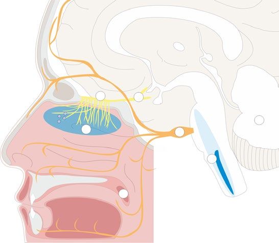
NATuRe NeuROSCIenCe                                                                                                                           Articles
                        a                            b                             c                          d

                        Merge                        TuJ1                          SARS-CoV S                 DAPI

                        e                            f                             g                          h

                        Merge                        NF200                         SARS-CoV S                 DAPI

                        i                            j                             k                          l

                        Merge                        OMP                           SARS-CoV S                 DAPI

Fig. 4 | Colocalization of SARS-CoV spike protein with neural/neuronal cells in distinct olfactory mucosa samples from individuals with COVID-19.
a–l, Representative maximum-intensity projections of confocal (a–d and i–l) or epifluorescence (e–h) microscopy images of olfactory mucosa showing
intracytoplasmic staining for SARS-CoV S protein within TuJ1+ (a–d, individual P27), NF200+ (e–h, individual P27) and OMP+ (i–l, individual P27)
OSNs. Staining for TuJ1, NF200 and OMP (magenta, Alexa Fluor 488) marks cells of neuronal origin, staining for SARS-CoV S protein (yellow,
Alexa Fluor 555) visualizes the presence of SARS-CoV and DAPI staining (petrol) identifies all cell nuclei (n = 3 individuals with COVID-19
(P27, P30 and P32) were analyzed; n = 2 individuals without COVID-19 served as controls; shown are representative images from P27). Scale bars,
(all panels) 10 µm.

                                P3    P5      P6         P9   P10     P11    P15       P20     P23    P29    P30        P31      P32   P33

                      PCR

                             NA      NA                       NA     NA
                       IHC
                                                         NA                                    NA      NA    NA          NA      NA
                             NA      NA                       NA     NA                      NA                                NA      NA
                       ISH
                                                       NA                                      NA      NA     NA         NA      NA      NA
                             NA      NA     NA       NA       NA     NA                NA    NA      NA     NA         NA      NA      NA
                        EM
                                NA     NA      NA        NA     NA     NA    NA         NA      NA     NA    NA          NA       NA    NA

                                                                                                             NA        OM/CNS
                                       OM positive            CNS positive              OM/CNS negaitive               Not available
                                                                                                                  NA

Fig. 5 | Summary of various SARS-CoV detection measures in deceased individuals with COVID-19. Various SARS-CoV-related investigations of the
individuals who tested positive for SARS-CoV-2 by RT–qPCR in the olfactory mucosa (OM), the CNS or both. SARS-CoV-2 RT–qPCR positivity was
combined with results derived from SARS-CoV-specific immunohistochemistry (IHC) and ISH as well as EM in appropriate tissue as available.

Nature Neuroscience | VOL 24 | February 2021 | 168–175 | www.nature.com/natureneuroscience                                                           173
Articles                                                                                                              NATuRe NeuROSCIenCe

                         a

                        H&E
                                                            SARS-CoV S       c                                            SARS-CoV S
                         b

Fig. 6 | Signs of (micro)thromboembolic events and SARS-CoV-2 immunostaining in the CNS of deceased individuals with COVID-19. a, Hematoxylin
and eosin (H&E)-stained FFPE section of the thalamus obtained from a deceased individual with COVID-19 (individual P26). Several small vessels exhibit
fresh thrombi (pink, indicated by arrows) resulting in a large infarct of surrounding CNS tissue characterized by a substantial reduction of detectable
neuronal and glial nuclei, edema and vacuolation. b,c, SARS-CoV S protein observed in the endothelial cells of small CNS vessels. Tissue with no obvious
ischemic damage exhibits only sparse staining intensity in endothelial cells (b, medulla oblongata, n = 3 of 6; red, indicated by arrows, individual P3) when
compared to endothelial cells within acute infarct areas (c, pons, n = 3 of 4; red, indicated by arrows, individual P4; inset depicts a magnified vessel from a
different region of the same specimen exhibiting SARS-CoV S protein deposits within endothelial cells). Scale bars: 30 µm (a), 50 µm (b), 200 µm (c) and
40 µm (inset in c).

vascular damage and allow the virus to spread more widely to other               particles at the ultrastructural level in an individual with an 82-hour
brain regions over time, thus eventually contributing to a more                  postmortem interval (P15) and important pathogenetic insights,
severe or even chronic disease course, depending on various fac-                 thus enabling further, more detailed and mechanistic investigations
tors such as the duration of viral persistence, viral load and immune            while encouraging further autopsy studies including broad sam-
status, among others.                                                            pling to allow multiple complementary analyses and the application
   Taking our findings together, we provide evidence that                        of state-of-the-art methodologies. Such studies will allow identifica-
SARS-CoV-2 neuroinvasion can occur at the neural–mucosal inter-                  tion of the precise cellular and molecular SARS-CoV-2 entry mech-
face by transmucosal entry via regional nervous structures. This                 anism as well as receptors on OSNs, where non-neuronal pathways
may be followed by transport along the olfactory tract of the CNS,               may also have a role17.
thus explaining some of the well-documented neurological symp-
toms in COVID-19, including alterations of smell and taste percep-               Online content
tion. One caveat to note with the COVID-19 cases reported here is                Any methods, additional references, Nature Research report-
the relatively long postmortem interval, an almost insurmountable                ing summaries, source data, extended data, supplementary infor-
obstacle in autopsy studies, especially when performed under the                 mation, acknowledgements, peer review information; details of
emergency-like conditions encountered during a pandemic situa-                   author contributions and competing interests; and statements of
tion. Analysis of these samples is limited by well-known restrictions            data and code availability are available at https://doi.org/10.1038/
resulting from autolysis of cells and tissues, ultimately complicat-             s41593-020-00758-5.
ing the interpretation of morphological and molecular analyses. In
spite of these limitations, we were able to retrieve numerous valu-              Received: 13 June 2020; Accepted: 12 November 2020;
able insights. These included the detection of well-preserved CoV                Published online: 30 November 2020

174                                                                  Nature Neuroscience | VOL 24 | February 2021 | 168–175 | www.nature.com/natureneuroscience
NATuRe NeuROSCIenCe                                                                                                                                Articles
References                                                                           24. Goldsmith, C. S., Miller, S. E., Martines, R. B., Bullock, H. A. & Zaki, S. R.
1. Huang, C. et al. Clinical features of patients infected with 2019 novel               Electron microscopy of SARS-CoV-2: a challenging task. Lancet https://doi.
    coronavirus in Wuhan, China. Lancet 395, 497–506 (2020).                             org/10.1016/S0140-6736(20)31188-0 (2020).
2. Conde Cardona, G., Quintana Pájaro, L. D., Quintero Marzola, I. D., Ramos         25. Goldsmith, C. S. & Miller, S. E. Modern uses of electron microscopy for
    Villegas, Y. & Moscote Salazar, L. R. Neurotropism of SARS-CoV 2:                    detection of viruses. Clin. Microbiol. Rev. 22, 552–563 (2009).
    mechanisms and manifestations. J. Neurol. Sci. 412, 116824 (2020).               26. Goldsmith, C. S. et al. Ultrastructural characterization of SARS coronavirus.
3. Mao, L. et al. Neurologic manifestations of hospitalized patients With                Emerg. Infect. Dis. 10, 320–326 (2004).
    coronavirus disease 2019 in Wuhan, China. JAMA Neurol. https://doi.              27. Ksiazek, T. G. et al. A novel coronavirus associated with severe acute
    org/10.1001/jamaneurol.2020.1127 (2020).                                             respiratory syndrome. N. Engl. J. Med. 348, 1953–1966 (2003).
4. Puelles, V. G. et al. Multiorgan and renal tropism of SARS-CoV-2. N. Engl. J.     28. Blanchard, E. & Roingeard, P. Virus-induced double-membrane vesicles.
    Med. https://doi.org/10.1056/NEJMc2011400 (2020).                                    Cell. Microbiol. 17, 45–50 (2015).
5. Moriguchi, T. et al. A first case of meningitis/encephalitis associated with      29. Tsai, S.-Y. et al. DAMP molecule S100A9 acts as a molecular pattern to
    SARS-coronavirus-2. Int. J. Infect. Dis. 94, 55–58 (2020).                           enhance inflammation during influenza A virus infection: role of DDX21–
6. Otero, J. J. Neuropathologists play a key role in establishing the extent of          TRIF–TLR4–MyD88 pathway. PLoS Pathog. 10, e1003848 (2014).
    COVID-19 in human patients. Free Neuropathology https://doi.org/10.17879/        30. Körtvelyessy, P. et al. Serum and CSF cytokine levels mirror different
    FREENEUROPATHOLOGY-2020-2736 (2020).                                                 neuroimmunological mechanisms in patients with LGI1 and Caspr2
7. Zubair, A. S. et al. Neuropathogenesis and neurologic manifestations of the           encephalitis. Cytokine 135, 155226 (2020).
    coronaviruses in the age of coronavirus disease 2019: a review. JAMA Neurol.     31. Oxley, T. J. et al. Large-vessel stroke as a presenting feature of Covid-19 in the
    https://doi.org/10.1001/jamaneurol.2020.2065 (2020).                                 young. N. Engl. J. Med. 382, e60 (2020).
8. Cyranoski, D. Profile of a killer: the complex biology powering the               32. Deigendesch, N. et al. Correlates of critical illness-related encephalopathy
    coronavirus pandemic. Nature 581, 22–26 (2020).                                      predominate postmortem COVID-19 neuropathology. Acta Neuropathol. 140,
9. Arbour, N., Day, R., Newcombe, J. & Talbot, P. J. Neuroinvasion by human              583–586 (2020).
    respiratory coronaviruses. J. Virol. 74, 8913–8921 (2000).                       33. Schurink, B. et al. Viral presence and immunopathology in patients with
10. Glass, W. G., Subbarao, K., Murphy, B. & Murphy, P. M. Mechanisms of host            lethal COVID-19: a prospective autopsy cohort study. Lancet Microbe
    defense following severe acute respiratory syndrome-coronavirus                      https://doi.org/10.1016/S2666-5247(20)30144-0 (2020).
    (SARS-CoV) pulmonary infection of mice. J. Immunol. 173,                         34. Jensen, M. P. et al. Neuropathological findings in two patients with fatal
    4030–4039 (2004).                                                                    COVID‐19. Neuropathol. Appl. Neurobiol. https://doi.org/10.1111/nan.12662
11. Li, K. et al. Middle East respiratory syndrome coronavirus causes multiple           (2020).
    organ damage and lethal disease in mice transgenic for human dipeptidyl          35. Reichard, R. R. et al. Neuropathology of COVID-19: a spectrum of vascular
    peptidase 4. J. Infect. Dis. 213, 712–722 (2016).                                    and acute disseminated encephalomyelitis (ADEM)-like pathology.
12. Netland, J., Meyerholz, D. K., Moore, S., Cassell, M. & Perlman, S. Severe           Acta Neuropathol. 140, 1–6 (2020).
    acute respiratory syndrome coronavirus infection causes neuronal death in        36. Schaller, T. et al. Postmortem examination of patients with COVID-19. JAMA
    the absence of encephalitis in mice transgenic for human ACE2. J. Virol. 82,         323, 2518–2520 (2020).
    7264–7275 (2008).                                                                37. von Weyhern, C. H., Kaufmann, I., Neff, F. & Kremer, M. Early evidence of
13. Hoffmann, M. et al. SARS-CoV-2 cell entry depends on ACE2 and                        pronounced brain involvement in fatal COVID-19 outcomes. Lancet 395,
    TMPRSS2 and is blocked by a clinically proven protease inhibitor. Cell 181,          e109 (2020).
    271–280 (2020).                                                                  38. Solomon, I. H. et al. Neuropathological features of Covid-19. N. Engl. J. Med.
14. Doobay, M. F. et al. Differential expression of neuronal ACE2 in transgenic          383, 989–992 (2020).
    mice with overexpression of the brain renin–angiotensin system. Am. J.           39. Matschke, J. et al. Neuropathology of patients with COVID-19 in Germany: a
    Physiol. Regul. Integr. Comp. Physiol. 292, R373–R381 (2007).                        post-mortem case series. Lancet Neurol. 19, 919–929 (2020).
15. Lukassen, S. et al. SARS-CoV-2 receptor ACE2 and TMPRSS2 are primarily           40. Dittmayer, C. et al. Why misinterpretation of electron micrographs in
    expressed in bronchial transient secretory cells. EMBO J. 39, e105114 (2020).        SARS-CoV-2-infected tissue goes viral. Lancet https://doi.org/10.1016/
16. Khan, S. & Gomes, J. Neuropathogenesis of SARS-CoV-2 infection. eLife 9,             S0140-6736(20)32079-1 (2020).
    e59136 (2020).                                                                   41. Paniz‐Mondolfi, A. et al. Central nervous system involvement by severe acute
17. Brann, D. H. et al. Non-neuronal expression of SARS-CoV-2 entry genes in             respiratory syndrome coronavirus‐2 (SARS‐CoV‐2). J. Med. Virol. 92,
    the olfactory system suggests mechanisms underlying COVID-19-associated              699–702 (2020).
    anosmia. Sci. Adv. 6, eabc5801 (2020).                                           42. Wiersinga, W. J., Rhodes, A., Cheng, A. C., Peacock, S. J. & Prescott, H. C.
18. Butowt, R. & Bilinska, K. SARS-CoV-2: olfaction, brain infection, and the            Pathophysiology, transmission, diagnosis, and treatment of coronavirus
    urgent need for clinical samples allowing earlier virus detection. ACS Chem.         disease 2019 (COVID-19): a review. JAMA 324, 782–793 (2020).
    Neurosci. 11, 1200–1203 (2020).                                                  43. Desforges, M., Le Coupanec, A., Brison, E., Meessen-Pinard, M. & Talbot, P.
19. Schwob, J. E. Neural regeneration and the peripheral olfactory system.               J. Neuroinvasive and neurotropic human respiratory coronaviruses:
    Anat. Rec. 269, 33–49 (2002).                                                        potential neurovirulent agents in humans. Adv. Exp. Med. Biol. 807,
20. Holbrook, E. H., Wu, E., Curry, W. T., Lin, D. T. & Schwob, J. E.                    75–96 (2014).
    Immunohistochemical characterization of human olfactory tissue: human            44. Baig, A. M., Khaleeq, A., Ali, U. & Syeda, H. Evidence of the COVID-19
    olfactory tissue characterization. Laryngoscope 121, 1687–1701 (2011).               virus targeting the CNS: tissue distribution, host–virus interaction, and
21. Carter, L. A., MacDonald, J. L. & Roskams, A. J. Olfactory horizontal basal          proposed neurotropic mechanisms. ACS Chem. Neurosci. 11, 995–998 (2020).
    cells demonstrate a conserved multipotent progenitor phenotype. J. Neurosci.     45. Wang, Y.-Z. et al. Olig2 regulates terminal differentiation and maturation of
    24, 5670–5683 (2004).                                                                peripheral olfactory sensory neurons. Cell. Mol. Life Sci. https://doi.
22. van Riel, D., Verdijk, R. & Kuiken, T. The olfactory nerve: a shortcut for           org/10.1007/s00018-019-03385-x (2019).
    influenza and other viral diseases into the central nervous system. J. Pathol.
    235, 277–287 (2015).                                                             Publisher’s note Springer Nature remains neutral with regard to jurisdictional claims in
23. Varga, Z. et al. Endothelial cell infection and endotheliitis in COVID-19.       published maps and institutional affiliations.
    Lancet 395, 1417–1418 (2020).                                                    © The Author(s), under exclusive licence to Springer Nature America, Inc. 2020

Nature Neuroscience | VOL 24 | February 2021 | 168–175 | www.nature.com/natureneuroscience                                                                                175
Articles                                                                                                                       NATuRe NeuROSCIenCe

Methods                                                                                immunohistological staining, control tissues harboring or lacking the expected
Study design. Thirty-three deceased individuals with COVID-19 either confirmed         antigens were used. Staining patterns were compared to expected results as
by PCR for SARS-CoV-2 (n = 31 of 33) or with clinical features highly suggestive       specified in the Supplementary Information (Supplementary Fig. 1a–d
of COVID-19 (n = 2 of 33) were included (Supplementary Table 1). Individuals           (ACE2), Supplementary Fig. 5e (SARS-CoV S), Supplementary Fig. 6 (NF200,
were not preselected with regard to their clinical symptoms. Autopsies were            SARS-CoV S) and Supplementary Fig. 3 (S100, OLIG2, HLA-DR, CD56, CD45,
performed at the Department of Neuropathology and the Institute of Pathology,          AE1/AE3, OMP and TuJ1)).
Charité–Universitätsmedizin Berlin (n = 25 of 33), including one referral from             For immunofluorescence, the protocol was adapted as follows: Alexa
the Institute of Pathology, DRK Kliniken Berlin, the Institutes of Pathology and       Fluor 488-conjugated goat anti-rabbit (1:100; Jackson, 111515003) and Alexa
of Neuropathology, University Medical Center Göttingen (n = 6 of 33) and the           Fluor 555-conjugated goat anti-mouse (1:100; Invitrogen) were used as secondary
Institute of Forensic Medicine, Charité–Universitätsmedizin Berlin (n = 1 of 33).      antibodies. Nuclei were counterstained with DAPI (Invitrogen, D3571),
This study was approved by the local ethics committees (Berlin: EA1/144/13,            and sections were subsequently mounted on slides with Dako mounting
EA2/066/20 and EA1/075/19; Göttingen: 42/8/20) as well as by the Charité–BIH           medium (S3023).
COVID-19 research board and was in compliance with the Declaration of Helsinki;
autopsies were performed on the legal basis of §1 of the Autopsy Act of the            SARS-CoV S immunohistochemistry and immunofluorescence.
state Berlin and §25(4) of the German Infection Protection Act. In all deceased        Immunohistochemical staining with mouse monoclonal anti-SARS spike
individuals, a whole-body autopsy was performed, which included a thorough             glycoprotein antibodies (clone 3A2, ab272420, Abcam, 1:100) was performed
histopathological and molecular evaluation comprising virological assessment           using 1-μm-thick FFPE tissue sections. Slides were cooked in sodium citrate
of SARS-CoV-2 RNA and/or SARS-CoV S protein levels in the carotid artery,              (pH 6.0; 95–100 °C) for 20 min, followed by enzymatic antigen retrieval with
cornea, conjunctiva, optic nerve, uvula, olfactory mucosa, olfactory bulb, olfactory   Triton X-100 and hydrogen peroxide for 15 min. Slides were blocked with 10%
tract, trigeminal ganglion, medulla oblongata and cerebellum as indicated in           normal goat serum. Primary antibody (1:100, diluted in ProTaqs Antibody
Supplementary Table 2. To exclude cross-contamination, clean instruments for           Diluent for IHC (Quartett) with 10% normal goat serum) was applied, and samples
the preparation and sampling of each organ and region were always used. All            were incubated overnight. Then, secondary antibody (Biotin-SP-AffiniPure
individuals with COVID-19 with known disease duration and available PCR-tested         Goat Anti-Mouse IgG (H+L), Jackson ImmunoResearch Laboratories), diluted
appropriate CNS tissue were included (n = 29 of 33) to calculate the correlation       1:100 in ProTaqs Antibody Diluent for IHC, was applied, and samples were
coefficient for the correlation between disease duration and CNS SARS-CoV-2            incubated for 2 h. Next, Streptavidin-HRP Reagent (RE7104, Leica Biosystems)
viral load. Therefore, we could not perform randomization as all individuals           and DAB substrate–chromogen (Agilent) were applied according to the
with SARS-CoV-2 RNA were included in the COVID-19 group and controls                   manufacturer’s instructions. Slides were rinsed, counterstained with hematoxylin,
were defined as individuals negative for SARS-CoV-2 by PCR. Where available,           dehydrated and mounted.
clinical records were assessed thoroughly for preexisting medical conditions and           Staining signals were compared to those of non-COVID-19 control
medications and progression of the disease as well as COVID-19-related symptoms        samples with respect to the staining intensity and staining pattern as specified
before death, with a special focus on neurological symptoms including alterations      in the Supplementary Information (Supplementary Fig. 5a–d; SARS-CoV S,
in olfaction and taste.                                                                Supplementary Table 2).

SARS-CoV- and SARS-CoV-2-specific PCR including subgenomic RNA                         Image acquisition and processing. For fluorescence microscopy, an Olympus BX63
assessment. For PCR-based assessment of SARS-CoV-2 RNA, unfixed and, where             (DP80 camera) automated fluorescence microscope was used, if not specified
possible, non-cryopreserved (i.e., native) tissue samples were used. RNA was           otherwise, for confocal images. For confocal microscopy, fluorescence signals were
purified from ∼50 mg of homogenized tissue obtained from all organs by using the       collected with an Olympus FluoView FV1000 confocal microscope using a ×60
MagNAPure 96 system and the MagNAPure 96 DNA and Viral NA Large Volume                 oil-immersion objective. For post‐acquisition image processing, the image analysis
kit (Roche) according to the manufacturer’s instructions.                              software Fiji was used48. For data handling of whole-slide images, an OME-TIFF
     Quantitative real-time PCR for SARS-CoV-2 was performed on RNA extracts           workflow was used49.
with RT–qPCR targeting the SARS-CoV-2 E gene. Quantification of viral RNA was
performed using photometrically quantified in vitro RNA transcripts as described       Electron microscopy. Autopsy tissues were fixed with 2.5% glutaraldehyde
previously46. Total DNA was measured in all extracts by using the Qubit dsDNA          in 0.1 M sodium cacodylate buffer, postfixed with 1% osmium tetroxide in
HS Assay kit (Thermo Fisher Scientific). The RT–qPCR analysis was replicated at        0.05 M sodium cacodylate, dehydrated using a graded acetone series and then
least once for each sample.                                                            infiltrated and embedded in Renlam resin. Block-contrasting with uranyl acetate
     Detection of sgRNA, as a correlate of active virus replication in the tested      and phosphotungstic acid was performed at the dehydration step with 70%
tissue, was performed by using oligonucleotides targeting the leader transcriptional   acetone. 500-nm semithin sections were cut using an ultramicrotome (Ultracut
regulatory sequence and a region within the sgRNA encoding the SARS-CoV-2 E            E, Reichert-Jung) and a Histo Jumbo diamond knife (Diatome), transferred onto
gene, as described previously47.                                                       glass slides, stretched at 120 °C on a hot plate and stained with toluidine blue at
                                                                                       80 °C. 70-nm ultrathin sections were cut using the same ultramicrotome and
Histological and immunohistochemical techniques. FFPE tissue blocks were               an Ultra 35° diamond knife (Diatome), stretched with xylene vapor, collected
taken at the day of autopsy when the postmortem interval was shorter than 24 h         onto pioloform-coated slot grids and then stained with lead citrate. Standard
and fixed for 24 h in 4% paraformaldehyde. Otherwise, brain tissue was fixed           transmission EM was performed using a Zeiss 906 microscope in conjunction
for 14 d in 4% paraformaldehyde before cutting. Routine histological staining          with a 2k CCD camera (TRS). Large-scale digitization was performed using a
(H&E, Masson–Goldner, periodic acid–Schiff (PAS) reaction and toluidine                Zeiss Gemini 300 field-emission scanning electron microscope in conjunction
blue) was performed according to standard procedures. Immunohistochemical              with a STEM detector via Atlas 5 software at a pixel size of 4–6 nm. Regions of
staining was performed either on a Benchmark XT autostainer (Ventana Medical           interest from the large-scale datasets were saved by annotation (‘mapped’) and then
Systems) with standard antigen retrieval methods (CC1 buffer, pH 8.0, Ventana          recorded at very high resolution using a pixel size of 0.5–1 nm.
Medical Systems) or manually using 1-μm- or 4-μm-thick FFPE tissue sections.               Alternatively, ultrastructural analysis was performed from FFPE tissues.
The following primary antibodies were used: polyclonal rabbit anti-S100 (Dako,         For virus detection, we took 3-mm punch biopsy cylinders from
Z0311; 1:3,000), monoclonal mouse anti-AE1/AE3 (Dako, M3515; 1:200),                   paraffin-embedded tissue. The respective regions were selected on the basis of
monoclonal mouse anti-MRP14 (Acris, BM4026B; 1:500, pretreatment protease),            the SARS-CoV ISH or immunohistochemistry signal. After deparaffinization
monoclonal mouse anti-CD56 (Serotec, ERIC-1; 1:200), mouse monoclonal                  in xylene, samples were rehydrated and postfixed in 1% formaldehyde and 2.5%
anti-SARS spike glycoprotein (Abcam, ab272420; 1:100), goat anti-OMP                   glutaraldehyde in 0.05 M HEPES buffer (pH 7.2) for a minimum of 2–4 h at room
(Wako, 019-22291; 1:1,000), rabbit monoclonal anti-βIII tubulin (Abcam,                temperature. Postfixation, block contrasting (tannic acid, uranyl acetate) and
ab215037; 1:2,000), rabbit polyclonal anti-NF200 (Sigma, N4142; 1:100), rabbit         embedding in epon resin were performed according to a standard protocol50.
polyclonal anti-ACE2 (Proteintech, 21115-1-AP; 1:3,000) and rabbit polyclonal          Ultrathin sections were analyzed using a transmission electron microscope
anti-OLIG2 (IBL, 18953; 1:150, pretreatment Tris-EDTA + microwave). Briefly,           operated at 120 kV (Tecnai Spirit, Thermo Fisher), and images were recorded
primary antibodies were applied and developed by using either the iVIEW DAB            using a CCD camera (MegaviewIII, EMSIS).
Detection kit (Ventana Medical Systems) and the ultraView Universal Alkaline
Phosphatase Red Detection kit (Ventana Medical Systems) or manual application          Cytokine array. To analyze cytokine levels in CSF samples from deceased
of biotinylated secondary antibodies (biotinylated donkey anti-sheep-goat (1:200;      individuals with COVID-19 and controls (each n = 4), a human cytokine array
Amersham, RPN 1025), biotinylated donkey anti-rabbit (1:200), biotinylated             (Bio-techne) was used according to the manufacturer’s instructions27. We analyzed
sheep anti-mouse (1:200; Amersham, RPN 1001), rabbit immunoglobulin                    all CSF samples from individuals with COVID-19 accessible at the time of analysis.
(RPN1004), peroxidase-conjugated avidin and diaminobenzidine (DAB; Sigma,              This assay enables the semiquantitative measurement of 36 cytokines and related
D5637) or 3-amino-9-ethylcarbazol (AEC). Sections were counterstained with             proteins (CCL1, CCL2, MIP-1α, CCL5, CD40L, C5/C5a, CXCL1, CXCL10,
hematoxylin, dehydrated in a graded alcohol and xylene series, mounted and             CXCL11, CXCL12, G-CSF, GM-CSF, ICAM-1, IFN-γ, IL-1α, IL-1β, IL-1ra, IL-2,
coverslipped. Immunohistochemistry sections were evaluated by at least two             IL-4, IL-5, IL-6, IL-8, IL-10, IL-12 p70, IL-13, IL-16, IL-17A, IL-17E, IL-18, IL-21,
board-certified neuropathologists with concurrence. To biologically validate all       IL-27, IL-32a, MIF, serpin E1, TNF-α, TREM-1). Twelve of these 36 cytokines

                                                                                                             Nature Neuroscience | www.nature.com/natureneuroscience
NATuRe NeuROSCIenCe                                                                                                                                         Articles
were above the detection limit in our samples. Using the software Kodak D1 3.6           References
(Eastman Kodak), a semiquantitative analysis was performed by determining the            46. Corman, V. M. et al. Detection of 2019 novel coronavirus (2019-nCoV) by
background-corrected sum intensity for each region of interest on the membrane.              real-time RT–PCR. Euro. Surveill. 25, 2000045 (2020).
Separate membranes were normalized to each other using the results for the               47. Wölfel, R. et al. Virological assessment of hospitalized patients with
positive controls. We could not perform a statistical test owing to the limited access       COVID-2019. Nature 581, 465–469 (2020).
to sufficient CSF samples. Data represent single data points and the mean, range         48. Schindelin, J. et al. Fiji: an open-source platform for biological-image analysis.
and 25th and 75th percentiles.                                                               Nat. Methods 9, 676–682 (2012).
                                                                                         49. Besson, S. et al. Bringing open data to whole slide imaging. Digit. Pathol.
In situ hybridization. For detection of mRNA, the RNAScope 2.5 HD Reagent                    2019, 3–10 (2019).
Kit-BROWN (ACD Europe SRL) was used. Briefly, paraffin sections were freshly             50. Laue, M. Electron microscopy of viruses. Methods Cell. Biol. 96, 1–20 (2010).
cut, dried for 1 h at 60 °C and dewaxed before mild unmasking with Target
Retrieval buffer and protease. Pretreated sections were hybridized with specific
probes to Ppib as a positive control and irrelevant probe to dapβ as a negative          Acknowledgements
control (both ACD Europe SRL). Virus-specific probe V-ncoV2019-S (ACD                    We are indebted to F. Egelhofer, R. Koll, P. Matylewski, K. Permien, V. Wolf, S. Meier,
Europe SRL) was used for samples from individuals with COVID-19 and was                  R. Müller, U. Scheidt, K. Guttek, F. Paap, B. Maruschak and K. Schulz for excellent
accompanied by an additional slide with FFPE lung tissue from an individual              technical assistance and advice. We thank the Core Facility for Electron Microscopy of
with COVID-19 (P15) as a further positive control. After hybridization signal            the Charité for support in acquisition of the data. We also thank H. Bohnenberger, S.
amplification, binding of probes was visualized with DAB. Nuclei were stained with       Küffer and the Institute of Pathology of the University Medical Center, Göttingen, for
hematoxylin, and sections were coverslipped with Ecomount.                               their support. The authors are most grateful to the individuals with COVD-19 and their
    Images were acquired using an AxioImager Z1 microscope (Carl Zeiss                   relatives for consenting to autopsy and subsequent research, which were facilitated by
MicroImaging).                                                                           the Biobank of the Department of Neuropathology, Charité–Universitätsmedizin Berlin.
                                                                                         Cartoon images were partially created with Biorender.com. This work was supported
Statistics and reproducibility. All statistical analyses were performed and all          by the Deutsche Forschungsgemeinschaft (DFG, German Research Foundation) under
graphs were created in GraphPad Prism 8 (GraphPad Software). No statistical              Germany’s Excellence Strategy EXC-2049-390688087, as well as SFB TRR 167 and
methods were used to predetermine sample sizes; sample sizes in the current              HE 3130/6-1 to F.L.H., SFB 958/Z02 to J.S., SFB TRR 130 TP17 to H.R., EXC 2067/1-
study are similar to those in previous COVID-19 autopsy reports by others4,32,38.        390729940, SFB TRR 274 and STA 1389/5-1 to C.S. and SFB TRR 241 to B.S. and A.A.K.,
We included all individuals with COVID-19 and material that were available               by the German Center for Neurodegenerative Diseases (DZNE) Berlin, by the European
as specified in the description of study design. The RT–qPCR analysis was                Union (PHAGO, 115976; Innovative Medicines Initiative-2; FP7-PEOPLE-2012-ITN:
replicated at least once for each positive sample. We did not exclude any data           NeuroKine) and by the Ministry for Science and Education of Lower Saxony through the
points from the performed analyses. To compute correlation between disease               program ‘Niedersächsisches Vorab’ to J.F. Furthermore, we thank the Charité Foundation
duration and viral load, Spearman nonparametric correlation was used. A                  (Max Rubner Preis 2016) for financial support to C. Dittmayer.
two-tailed P value was calculated. Values were considered to be significant at
P < 0.05. Statistical details for each analysis (for example, n, P and r values) are     Author contributions
mentioned in each figure legend or in the respective part of the text. Owing to          J.M., J.R., R.M., J.F., C.T., O.H., M.W., L.R., H.M., W.J.S.-S., C.S., S.H., M.S.W., B.T., F.S.,
the nature of the investigation, data collection and analyses could not always           P.K., M.L., A.A.K., T.A., S.G., E.S., D.R., A.N., B.S., R.L.C., C.C., R.E., S.E., D.H., L.O.,
be done in a blinded fashion. Histological staining, immunohistochemistry,               M.T, B.I.-H., H.R. and F.L.H. performed clinical workup and section and/or histological
immunofluorescence, ISH and EM results were analyzed independently                       analyses. C. Dittmayer, H.-H.G., H.R.G., M.L. and W.S. performed ultrastructural
by various neuropathologists in two distinct neuropathological institutions              analyses. J.S., S.B., C. Drosten and V.M.C. carried out viral RT–qPCR analyses. All
(Berlin and Göttingen) for each individual, region and specific staining/method.         authors contributed to the experiments and analyzed data. H.R. and F.L.H. designed and
Histological staining, immunohistochemistry, immunofluorescence and                      supervised the study. J.M., J.R., H.R. and C. Dittmayer prepared the figures. All authors
ISH analyses were replicated at least once. EM was performed in one individual.          wrote, revised and approved the manuscript.
The representative micrographs shown were adjusted in brightness and
contrast to different degrees (depending on the need resulting from the
range of brightness and contrast of the raw images), rotated and cropped in              Competing interests
Adobe Photoshop.                                                                         The authors declare no competing interests.

Reporting Summary. Further information on research design is available in the
Nature Research Reporting Summary linked to this article.
                                                                                         Additional information
                                                                                         Supplementary information is available for this paper at https://doi.org/10.1038/
                                                                                         s41593-020-00758-5.
Data availability                                                                        Correspondence and requests for materials should be addressed to F.L.H.
The datasets generated and/or analyzed during the current study are available from
the corresponding author on reasonable request. The three electron microscopy            Peer review information Nature Neuroscience thanks Roxana Carare and the other,
datasets in Supplementary Fig. 2 are available for open access pan-and-zoom              anonymous, reviewer(s) for their contribution to the peer review of this work.
analysis at http://www.nanotomy.org/.                                                    Reprints and permissions information is available at www.nature.com/reprints.

Nature Neuroscience | www.nature.com/natureneuroscience
You can also read