Insights into the pathophysiology of DFNA10 hearing loss associated with novel EYA4 variants - Nature
←
→
Page content transcription
If your browser does not render page correctly, please read the page content below
www.nature.com/scientificreports

            OPEN           Insights into the pathophysiology
                           of DFNA10 hearing loss associated
                           with novel EYA4 variants
                           Matias Morín 1, Lucía Borreguero1, Kevin T Booth2,3, María Lachgar1, Patrick Huygen4,
                           Manuela Villamar1, Fernando Mayo 1, Luis Carlos Barrio5, Luciana Santos Serrão de Castro1,
                           Carmelo Morales6, Ignacio del Castillo 1, Beatriz Arellano7, Dolores Tellería1, Richard J. H. Smith2,
                           Hela Azaiez2 & M. A. Moreno Pelayo1*
                           The mutational spectrum of many genes and their contribution to the global prevalence of hereditary
                           hearing loss is still widely unknown. In this study, we have performed the mutational screening of
                           EYA4 gene by DHLPC and NGS in a large cohort of 531 unrelated Spanish probands and one Australian
                           family with autosomal dominant non-syndromic hearing loss (ADNSHL). In total, 9 novel EYA4 variants
                           have been identified, 3 in the EYA4 variable region (c.160G > T; p.Glu54*, c.781del; p.Thr261Argfs*34
                           and c.1078C > A; p.Pro360Thr) and 6 in the EYA-HR domain (c.1107G > T; p.Glu369Asp, c.1122G > T;
                           p.Trp374Cys, c.1281G > A; p.Glu427Glu, c.1282-1G > A, c.1601C > G; p.S534* and an heterozygous
                           copy number loss encompassing exons 15 to 17). The contribution of EYA4 mutations to ADNSHL in
                           Spain is, therefore, very limited (~1.5%, 8/531). The pathophysiology of some of these novel variants
                           has been explored. Transient expression of the c-myc-tagged EYA4 mutants in mammalian COS7 cells
                           revealed absence of expression of the p.S534* mutant, consistent with a model of haploinsufficiency
                           reported for all previously described EYA4 truncating mutations. However, normal expression pattern
                           and translocation to the nucleus were observed for the p.Glu369Asp mutant in presence of SIX1.
                           Complementary in silico analysis suggested that c.1107G > T (p.Glu369Asp), c.1281G > A (p.Glu427Glu)
                           and c.1282-1G > A variants alter normal splicing. Minigene assays in NIH3T3 cells further confirmed
                           that all 3 variants caused exon skipping resulting in frameshifts that lead to premature stop codons.
                           Our study reports the first likely pathogenic synonymous variant linked to DFNA10 and provide further
                           evidence for haploinsufficiency as the common underlying disease-causing mechanism for DFNA10-
                           related hearing loss.

                           Sensorineural hearing loss (SNHL) is a common human genetic sensory defect. It is estimated that 1 of every 1000
                           children born in developed countries have hearing loss for which the genetic factors account for approximately
                           two-thirds of the cases1. Among all the hereditary nonsyndromic SNHL, the autosomal recessive forms accounts
                           for about 70–80% of cases and are typically congenital or prelingual. The population that suffers postlingual hear-
                           ing loss, however, is significantly higher affecting 10% of the population by age 60, and 50% by age 80, although
                           the proportion that is due to genetic causes has not been precisely determined2. The autosomal dominant hearing
                           loss (ADNSHL) represents about 10–20% of all cases of hereditary SNHL; is characterized by high genetic and
                           clinical heterogeneity and, in contrast to the recessive conditions, has a delayed onset (postlingual) and escapes
                           the neonatal hearing screening. To date, more than 70 ADNSHL loci have been mapped and in 47 of them the
                           responsible gene has been identified3. The contribution of many of these genes to the pool of ADNSHL is still

                           1
                            Servicio de Genética, Ramón y Cajal Institute of Health Research (IRYCIS) and Biomedical Network Research Centre
                           on Rare Diseases (CIBERER), 28034, Madrid, Spain. 2Molecular Otolaryngology and Renal Research Laboratories,
                           Department of Otolaryngology, Head & Surgery, University of Iowa, Iowa City, Iowa, 52242, USA. 3Harvard Medical
                           School, Department of Neurobiology, Boston, Massachusetts, 02115, USA. 4Department of Otorhinolaryngology,
                           Radboud University Nijmegen Medical Centre, Nijmegen, Netherlands. 5Departamento de Investigación, Ramón
                           y Cajal Institute of Health Research (IRYCIS), Unidad de Neurología Experimental, 28034, Madrid, Spain. 6Servicio
                           de Otorrinolaringología, Hospital Universitario Marqués de Valdecilla, 39008, Santander, Spain. 7Servicio de
                           Otorrinolaringología, Hospital Universitario Puerta de Hierro, Majadahonda, 28922, Madrid, Spain. *email:
                           mmorenop@salud.madrid.org

Scientific Reports |   (2020) 10:6213 | https://doi.org/10.1038/s41598-020-63256-5                                                          1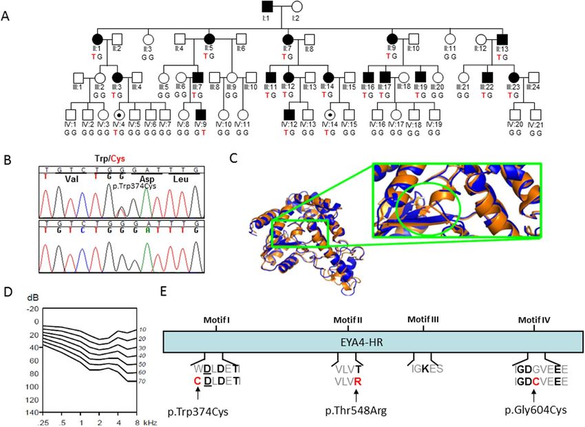
www.nature.com/scientificreports/                                                                www.nature.com/scientificreports

                           unknown as comprehensive mutational screening has not yet been conducted in most populations worldwide.
                           Amongst these genes is the transcriptional coactivator EYA4 (Eyes Absent Homolog 4) belonging to the family
                           of Eya genes, vertebrate orthologs (Eya1–Eya4) of the Drosophila melanogaster eyes absent gene (Eya). EYA4
                           participates in the development of multiple organs, including the eye, pituitary gland, muscle, kidney, inner ear
                           and heart4. Eya proteins contain an N-terminal variable region (VR) with transactivating function that includes
                           the Neuronal helix-loop-helix transcription factor domain (Neuro-bHLH) and a highly conserved C-terminal
                           Eya domain (eyaHR) that mediates interactions with members of the sine oculis family of proteins (Six1-Six6)5.
                           Within each of these domains are several motifs that are essential for all EYA protein enzymatic activity6. More
                           broadly, the EYA proteins are functionally multifaceted and belong to a highly conserved and complex network
                           involving several families of transcriptional proteins that is known collectively as the Pax-Six-Eya-Dach network
                           (PSEDN)7,8. The formation of the PSEDN is essential for proper development of many organs including the lungs,
                           craniofacial skeleton, muscle, eyes, heart and ears9.
                               Since Eya proteins lack a DNA-binding domain they require to interact with the transcription factors Six and
                           Dach to mediate the regulatory effects10. Six binding by Eya proteins is required for nuclear translocation5 where
                           they participate in the regulation of gene expression11,12 through the phosphatase activity of the Eya domain that
                           releases Dach-Six–mediated transcriptional repression of genes13.
                               In humans, mutations in two members of EYA gene family (EYA1 and EYA4) cause syndromic and
                           non-syndromic sensorineural hearing loss, respectively. Human EYA1 (OMIM 601653) mutations cause bran-
                           chiootorenal (BOR) syndrome (OMIM 113650), a disorder that presents with branchial arch defects including
                           lachrymal duct abnormalities, preauricular fistulae, and hearing loss (mixed conductive and sensorineural), with
                           abnormal shaped external ears, cochlear malformations, and renal anomalies14. EYA4 mutations, in contrast,
                           are responsible for the non-syndromic SNHL subtype DFNA10 (OMIM 601316), with no external ear or other
                           craniofacial structures affected. So far, 38 variations in EYA4 have been reported associated with DFNA10-related
                           hearing loss. These include 11 frameshift variants15–24, 6 nonsense variants15,20,25–28, 16 missense variants15–17,29–39
                           and 5 splice-altering variant15,40–43. The age of onset of the hearing loss due to EYA4 mutations shows a broad var-
                           iation ranging from early childhood to adulthood44. Moreover, more complex mutations involving EYA4 (large
                           deletions affecting the gene structure) have been associated only with SNHL15 or with other clinical entities that
                           encompass SNHL, dilated cardiomyopathy, and/or mental retardation7,45–47. Familiar reports of otofaciocervical
                           syndrome (OTFS), without a cardiac phenotype have also been reported in patients carrying a large 3.7MB dele-
                           tion in 6q23.1q23.2 that includes the entire EYA4 gene48.
                               In this study, we have performed a comprehensive mutation screening in a large population of 531 ADSNHL
                           Spanish patients and in an isolated Australian family. We have identified 9 novel genetic variations in EYA4 gene
                           associated with DFNA10 hearing loss: three missense, two nonsense, one frameshift, one splice-site, one silent
                           mutation affecting splicing and one copy number loss encompassing exon 15 to exon 17. We have also investi-
                           gated the physiopathological mechanism of four of these mutations by means of immunocytochemistry, western
                           blotting and minigene assays.

                           Patients and methods
                           Sample collection and clinical evaluation.             Patients and healthy relatives were recruited from the
                           University Hospital Ramón y Cajal (Madrid-Spain) or the University of Iowa, Iowa City, Iowa, USA. This study
                           was designed in compliance with the tenets of the Helsinki Declaration, and patient enrollment was approved
                           by the ethics committee and the human research Institutional Review Boards of Hospital Ramón y Cajal (IRB
                           number: 288-17) and University of Iowa (IRB number: 199701065). All participants provided written informed
                           consent prior to their participation in this study.
                               A large cohort of 531 unrelated Spanish probands with ADNSHL was enrolled without any hearing loss phe-
                           notype preselection. Whole blood samples were obtained from all participants by venipuncture and genomic
                           DNA was extracted by using standard procedures. Clinical history ruled out environmental factors as the cause
                           of the hearing loss in the probands and physical examination did not reveal any evidence of syndromic features.
                           No other clinically significant manifestations including balance or visual problems were reported by any of the
                           affected individuals. Tympanometry indicated proper functioning of the middle ear. Pure tone audiometry with
                           air and bone conduction at frequencies ranging 250-8.000 HZ was performed for all consenting families’ mem-
                           bers according to standard protocols.
                               Family 10880 is a large four-generation Australian family segregating postlingual progressive ADNSHL.
                           Clinical examination of the subjects was completed to exclude any additional and/or syndromic findings.
                           Pure tone audiometry was performed to determine air conduction thresholds at 0.25, 0.5, 1, 2, 3, 4 and 8 kHz.
                           Audiometric data were analyzed to determine the Age Related Typical Audiogram (ARTA) and Annual Threshold
                           Deterioration (ATD) as previously described49. After obtaining written informed consent to participate in this
                           study, saliva samples were obtained from 44 family members, 17 affected and 27 presumed unaffected, and
                           genomic DNA was extracted following the manufacture’s protocol.

                           Targeted genomic sequencing and data analysis. OtoNGSpanel V1 or OtoNGSpanel V2, custom gene
                           panels were developed at the Hospital Ramón y Cajal. OtoNGSpanel V1 was based on HaloPlex technology to
                           capture all exons and 25 bp of intronic flanking regions of 71 genes involved in hereditary hearing loss. The ver-
                           sion 2 of the panel is based on IDT probes capture system and Sophia Genetics’ software and included 117 genes
                           associated with SNHL. Version 1 and V2 were used to screen a cohort of 111 and 108 Spanish independent famil-
                           ial cases with ADSNHL, respectively. Sequencing of captured enriched-libraries was done on the Illumina MiSeq
                           (Illumina, Inc., San Diego, CA). The sequence data were mapped against the human genome sequence (build
                           GRCh37/hg19) and data analysis was performed using the Variant Studio and Sophia Genetics’ softwares, the

Scientific Reports |   (2020) 10:6213 | https://doi.org/10.1038/s41598-020-63256-5                                                               2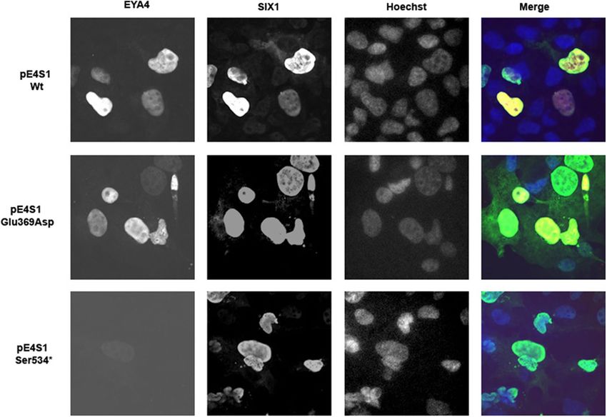
www.nature.com/scientificreports/                                                               www.nature.com/scientificreports

                           latter enabling the copy number variation (CNV) analysis of the targeted exonic sequences. Variant prioritization
                           was carried out using a custom filtering strategy.
                                           ®
                               OtoSCOPE v6, a next-generation sequencing platform, was used to screen 109 genes implicated in NSHL
                           and USH for possible mutations in two affected individuals, II.1 and II.13 of the Australian family 10880, as
                           described50–52. Following sequencing on the Illumina HiSeq. 2000 (Illumina, Inc., San Diego, CA) using
                           100 bp paired-end reads, bioinformatics analysis was performed on a custom Galaxy pipeline running on the
                           high-performance computing cluster at the University of Iowa as described51,52. Both samples underwent CNVs
                           evaluation by assessing the read-depth ratio, as described53.

                           DHPLC analysis and Sanger sequencing. In a Spanish initial cohort of 312 unrelated probands with
                           ADSNHL, the exonic regions of EYA4 gene [NM_004100.5] [MIM: 603550] were amplified using a standard pro-
                                                      ®
                           tocol on a GeneAmp PCR System 9700 (Applied Biosystems, Foster City, CA). PCR amplimers of each exon of
                           the EYA4 gene were screened for mutations by Denaturing High Performance Liquid Chromatography (DHPLC)
                           on a WaveTM DNA fragment analysis system (TransgenomicTM, USA) according to manufacturer’s protocol.
                           The different DHPLC heteroduplex profiles were characterized by sequencing a product of a second PCR ampli-
                           fication as previously described54.
                               Segregation analysis for the Spanish families and for the Australian family 10880 was achieved by PCR amplifi-
                           cation of the respective target exons and flanking introns of EYA4 followed by Sanger Sequencing. In all instances,
                           sequencing was completed with a BigDyeTM v3.1 Terminator Cycle Sequencing kit (Applied Biosystems, Foster
                           City, CA), according to the manufacturer’s instructions. The sequencing procedure was carried out in an ABI
                           3730 s sequencer (Perkin Elmer, Waltham, MA).

                           Copy number variation confirmation by using Real Time PCR. EYA4 DNA copy numbers in family
                           S2532 was determined in an Applied Biosystems 7300 Real-time PCR system using SYBR premix Ex Taq (Takara)
                           according to the manufacturer’s directions. All primers were designed with the Primer Express 2.0 software
                           (Applied Biosystems). The following primers were used to amplify a fragment of EYA4 containing the exon 16:
                           5′-CAACTGATGGCTTCCATGCA-3′ and 5′-TCTTACACCTGTTGGCAAACAAA-3′. As a chromosomal dip-
                           loid reference we have amplified a region of ALB gene, which encodes the human albumin, by using the following
                           primers: 5′-GCTGTCATCTCTTGTGGGCTGT-3′ and 5′-ACTCATGGGAGCTGCTGGTTC-3′. For each assay,
                           we have included one replicate for each of the different DNA concentrations used (5 ng, 2 ng and 0.2 ng) in healthy
                           relatives and patients. The cycling conditions were as follows: 2 min at 50 °C, 10 min at 95 °C, 40 cycles at 95 °C for
                           15 s, 59 °C for 30 s and 72 °C for 30 s. The threshold cycle (Ct) and the amplification efficiency (E) were obtained by
                           using the 7300 system SDS software. Where E = 10(−1/slope). Relative quantification (Q) was determined as follows:
                           Q = E EYA4(ΔCt EYA4)/E ALB (ΔCt ALB), where ΔCt=mean Ct Healthy relatives-mean Ct patients. After performing
                           relative quantification, statistical analysis was carried out by using SPSS software, version 15 (SPSS Inc.). To ana-
                           lyze the variance between the healthy relatives and patients, one-way ANOVA was used. A Pvalue of ≤0.001 was
                           deemed significant.

                           Expression of EYA4 mutants in COS7 cells.               Expression vectors for EYA4 (pE4, encoding the EYA4-HR
                           domain) and SIX1 (pS1) wild type proteins were kindly provided by Prof. RJH Smith55. In order to generate a Wt
                           bicistronic plasmid vector (pE4S1-Wt) to express both, the EYA4 and SIX1 proteins, a HindIII-XbaI fragment
                           containing the c-myc encoded tag in frame with a distal DNA fragment that encodes the EYA4-HR domain
                           (codons 322 to 616) was extracted from the pE4-HR vector and subsequently subcloned into the expression
                           vector pS1. The final construct (pE4S1) was a bicistronic expression vector including the c-myc-tagget EYA4-HR
                           domain driven by the CMV promoter and the HA-tagged SIX1 under the control of the EF-1 promoter (Suppl
                           Fig. 1). The wild type individual (pE4-Wt) and bicistronic (pE4S1-Wt) vectors was then mutagenized using the
                           QuickChange Site-Directed Mutagenesis Kit (Stratagene) to generate the mutated vectors (pE4Glu369Asp, pE4Ser534*,
                           pE4S1Glu369Asp and pE4S1Ser534*) that were subsequently verified by Sanger sequencing.
                               For the assessment of EYA4 expression, we proceed similarly to our previous works56. COS7 cells were cul-
                           tured in DMEM medium supplemented with 10% fetal calf serum (FCS), 100 U/ml of penicillin and 100 mg/ml
                           of streptomycin on 12 mm glass coverslips (Menzel-Glaser, GmbH, Braunschweig, Germany) in flat-bottomed
                           24-well multititer plates (Falcon; BD Biosciences, San Jose, CA, USA) for immunocytochemistry studies.
                           Cells were transiently transfected with 800 ng of the wild-type or with each of the two mutant constructs using
                           Lipofectamine TM 2000 (111668, Invitrogen) according to the manufacturer’s directions. For immunocytochem-
                           istry, cells on coverslips were fixed 48 h after transfection in 4% paraformaldehyde in phosphate buffer (0.1 M
                           NaH2PO4 pH 7.04) for 10 min and then washed three times briefly in PBS.
                               To study the intracellular protein localization of HA-tagged SIX1 and the c-myc tagged EYA4 proteins,
                           cells were permeabilized for 10 min at room temperature with 0.5% Triton X-100 in PBS and blocked with 3%
                           BSA in PBS for 30 min, washed twice with PBS and incubated for 1 h at room temperature with a 1:2000 and
                           1:1000 dilution of the rabbit primary antibody against the HA epitope (Abcam) and the mouse primary anti-
                           body against the Myc epitope (Sigma), respectively. Cells were then washed three times with PBS, and incubated
                           for 1 h at room temperature with a 1:1000 dilution of the secondary antibody, Alexa FluorTM 488 conjugated
                           goat anti-rabbit IgG (Molecular Probes A-11034; Invitrogen) or with the antibody Alexa 594 conjugated goat
                           anti-mouse IgG (Molecular Probes A-11020; Invitrogen). After washing three times in PBS, slides were incubated
                           with Hoechst 1μg/ml (Sigma, Hoechst 33342) for 10 min and washed one time in PBS. Slides were mounted using
                           FluorsaveTMReagent (Calbiochem, USA)

                           Western blot analysis. Transfected COS7 cells were homogenized in lysis buffer (150 mM NaCl,
                           50 mM Tris–Cl, pH 7.4, 5 mM EGTA, 5 mM EDTA, 1% Triton X-100, 25 lg/ml leupeptin, and 1 mM

Scientific Reports |   (2020) 10:6213 | https://doi.org/10.1038/s41598-020-63256-5                                                              3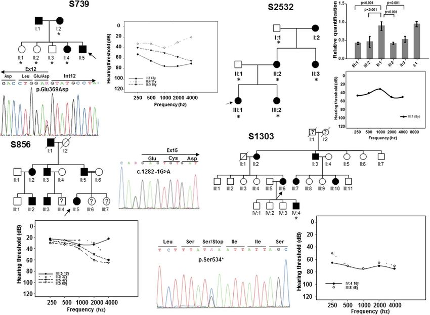
www.nature.com/scientificreports/ www.nature.com/scientificreports
DNA change Potein Change Exon Origin Phenotype Detection Degree Audiogram profile Reference
c.84-2A > G Intron 3 Chinese SNHL N.A N.A N.A Chen et al. 201640
c.152C > T p.Ser51Phe 4 N. American SNHL N.A N.A N.A Sloan-Heggen et al. 201629
c.160G > T p.Glu54* 4 Spanish SNHL 42 y Mild Mid/flat freq. This work
c.222_223del p.Val75Phefs*32 5 Japanese SNHL 61 y Mild to moderate High/Low freq. Shinagawa et al. 202015
Swedish SNHL N.A N.A N.A Neveling et al. 201316
c.464del p.Pro155Glnfs*43 # 8
Dutch SNHL Childhood Moderate Mid or high freq. Van Beelen et al. 201617
c.498del p.Thr167Leufs*31 8 Japanese SNHL 13 Mild Low freq. Shinagawa et al. 202015
c.511G > C p.Gly171Arg 8 Chinese SNHL 2nd decade Moderate to severe Gently sloping Liu et al. 201530
c.517C > T p.Gln173* 8 Japanese SNHL 48 y Moderate Flat Shinagawa et al. 202015
c.579_580insTACC p.Asp194Tyrfs*52 8 Swedish SNHL 4–40 y Mild to profound Variable Frykholm et al., 201518
c.580 + 1G > A Intron 8 Japanese SNHL 45 y Moderate Flat Shinagawa et al. 202015
Moderate to
c.612dup p.Glu205Argfs*40 9 Chinese SNHL 20–40 y High/flat freq. Huang et al. 201519
profound
c.781del p.Thr261Argfs*34 10 Spanish SNHL 26–44 y Mild to moderate Gently downsloping This work
c.804G > C p.Gln268His 10 Slovak SNHL 10–40 y Moderate Gently downsloping Varga et al. 201931
Korean SNHL N.A Moderate Reverse U-shaped Baek et al. 201225
c.863C > A p.Ser288* 11
Korean SNHL N.A Moderate to severe Flat freq. Kim et al. 201526
Miszalski-Jamka et al.
c.866C > T p.Thr289Met 11 N. American SNHL N.A N.A N.A
201732
c.910del p.Ser305Leufs*15 11 Japanese SNHL 30 Severe Flat Shinagawa et al. 202015
c. 978C > G p.Phe326Leu 12 Korean SNHL N.A Moderate Down sloping Choi et al. 201333
c.988C > T p.Gln330* 12 Japanese SNHL 16 y Moderate Flat Shinagawa et al. 202015
Moderate to
c.1026_1027dup p.Thr343Lysfs*62 12 N. American SNHL 1st, 3rd decade Flat/Gently sloping Wayne et al. 200120
profound
2nd-4th
c.1048_1049dup p.Arg352Profs*53 12 N. American SNHL Moderate to severe Mid to high freq. Makishima et al. 200721
decade
c.1078C > A p.Pro360Thr 12 Spanish SNHL 44 y Mild to moderate Gently downsloping This work
c.1107G > T p.Glu369Asp 12 Spanish SNHL 10–11 y Mild to severe Gently downsloping This work
c.1109G > A p.Arg370His 13 Philippines SNHL N.A N.A. N.A Truong et al. 201934
c.1109G > C p.Arg370Pro 13 Japanese SNHL 30 y Mild to moderate Mid freq. Shinagawa et al. 202015
c.1111G > A p.Val371Met 13 Belgium SNHL N.A N.A N.A Sommen et al. 201635
c.1115_1118dup p.Trp374Cysfs*6 13 Hungarian SNHL Postlingual Profound Variable Pfister et al. 200222
c.1122G > T p.Trp374Cys 13 Australian SNHL 10–25 y Mild to severe Gently downsloping This work
c.1154C > T p.Ser385Leu 13 Italian SNHL Postlingual Mild to profound Mid-freq. Cesca et al. 201836
SNHL Moderate to
Korean N.A Mid/High freq. Kim et al. 201526
SNHL + minor profound
c.1177C > T p.Gln393* 13 Japanese 26 y Mid/flat freq. Abe et al. 201827
DCM Moderate
Japanese 26 y Flat Shinagawa et al. 202015
SNHL Moderate
c.1194del p.Met401Trpfs*3 14 Korean SNHL 1st decade Moderate Down sloping Choi et al. 201623
c.1216G > C p.Gly406Arg 14 Japanese SNHL 5y Moderate Flat Shinagawa et al. 202015
Miszalski-Jamka et al.
c.1223G > A p.Arg408His 14 N. America SNHL N.A N.A N.A
201732
Moderate to
c.1281G > A p.Glu427Glu 14 Spanish SNHL 26 y Flat freq. This work
profound
c.1282 -12T > A Intron 14 Australian SNHL 1st-4th decade Mild to profound Mid/flat freq. Hildebrand et al. 200741
c.1282 -1G > A — Intron 14 Spanish SNHL 12 y Mild to moderate Gently downsloping This work
c.1301T > A p.Ile434Lys 15 Chinese SNHL 8-38 y Mild to severe Mid/flat freq. Tan et al. 201437
c.1341 -19T > A Intron 15 Germany SNHL N.A N.A N.A Vona et al. 201442
c.1601C > G p.Ser534* 17 Spanish SNHL 3–16 y Moderate to severe Mid/flat freq. This work
c.1643C > G p.Thr548Arg 18 Chinese SNHL 17–40 y Mild to profound Variable Sun et al. 201538
c.1663G > C p.Ala555Pro 18 Japanese SNHL 25 y Moderate High freq. Shinagawa et al. 202015
c.1739 -1G > A Intron 18 N. America SNHL 50 y N.A N.A Cirino et al. 201743
c.1759C > T p.Arg587* 19 Belgian SNHL 6–40 y Mild to moderate Mid freq. Wayne et al. 200120
c.1790del p.Val597Glyfs*4 19 Japanese SNHL 35 Moderate Flat Iwasa et al. 201624
Swedish Neveling et al. 201316
c.1810G > T p.Gly604Cys # 19 SNHL N.A N.A N.A
Dutch Van Beelen et al. 201617
c.1834A > T p.Lys612* 19 Chinese SNHL 27 y Moderate Gently downsloping Hu et al. 201828
c.1855T > G p.Trp619Gly 20 Chinese SNHL N.A N.A N.A Xiao et al. 201939
Deletion 7689 bp (Ex7 to Ex11) Japanese SNHL 25 y Moderate to severe Low/High freq. Shinagawa et al. 202015
Deletion 9.5 Mb (Ex4 to Ex 20) Japanese SNHL 13 y Severe Low/High freq. Shinagawa et al. 202015
Deletion 2747 bp (Ex15 to Ex17) Spanish SNHL 8y Moderate Flat This work
Continued
Scientific Reports | (2020) 10:6213 | https://doi.org/10.1038/s41598-020-63256-5 4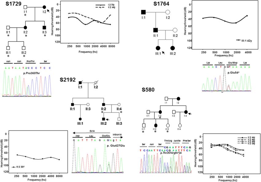
www.nature.com/scientificreports/                                                                             www.nature.com/scientificreports

DNA change                         Potein Change       Exon   Origin     Phenotype     Detection    Degree               Audiogram profile   Reference
                                                                         SNHL
Deletion 9 Mb at 6q23.1-24.1       No protein          -      Polish                   N.A          N.A                  N.A                 Dutrannoy et al. 200945
                                                                         +DCM + MR
Deletion 4,846pb incl. intron 9,
                                                                                                                                             Schönberger et al. 2000;46
exon 10 and partial intron 10      p.Asp194Glyfs*30    9–10   N.A        SNHL + DCM    2nd decade   Moderate to severe   Mid/flat freq.
                                                                                                                                             20057
c.581_804del
~10.4 Mb, incl. promoter and ex.
                                   —                   —      Japanese   MR            20-months-old N.A                 N.A                 Abe et al. 200947
1-2 at 6q22.31-q23.2
Deletion 3.7MB in 6q23.1q23.2      —                   —      Italian    OTFCS + SNHL 12 y (SNHL)   Mild                 N.A                 Gana et al. 201948

                                       Table 1. All known EYA4 (DFNA10) mutations including those identified in this study in boldface. EYA4
                                       mRNA and protein sequences are: NM_004100.5 and NP_004091.3. Nucleotide numbering reflects cDNA
                                       coordinates with +1 corresponding to the A of the ATG initiation codon of the CDS. SNHL sensorineural
                                       hearing loss, DCM autosomal dominant dilated cardiomyopathy, MR mental retardation, OTFCS
                                       otofaciocervical syndrome, NA not available. All variant names were checked using Mutalyzer 2.0.beta-21
                                       software59. #These mutations were found in the same patient.

                                       phenylmethylsulphonylfluoride) and maintained on ice for 30 min. After centrifugation at 10,000 g (4 °C) for
                                       5 min, 100 μg of the supernatant was mixed 1:1 with SDS-PAGE sample buffer (50 mM Tris–Cl, pH 6.8, 50 mM
                                       DTT, 2% SDS, 10% glycerol, 5% βME). The proteins were separated in 10% SDS-PAGE gels and transferred elec-
                                       trophoretically to nitrocellulose membranes. After blocking with 5% skimmed dry milk in 1% TBS (pH 7.6)/0.1%
                                       Tween 20 for 1 h at room temperature, the blots were incubated with the primary rabbit anti-HA antibody
                                       (1:2000) (Abcam) for 1 hour. After washing, the first antibody was detected by a secondary sheep anti-rabbit IgG
                                       antibody (1:1000) conjugated with HorseRadish peroxidase (HRP, Amersham Biosciences) for 1 h at room tem-
                                       perature. Bands were visualized by the enhanced chemiluminiscence (ECL; Amersham Biosciences) and exposed
                                       to X-ray film. The blots were then stripped, blocked and incubated with the primary mouse anti-Myc antibody
                                       (1:1000) (Sigma) for 1 hour. Secondary goat anti-mouse IgG antibody conjugated with HRP 1:1000 (Amersham
                                       Biosciences) was incubated for 1 hour. Visualization was carried out as previously described57.

                                       RNA extraction and cDNA synthesis. Total RNA was extracted from transfected COS7 cells using
                                                   ®
                                       QIAamp RNA kit following the manufacturer’s instructions (QIAGEN). Reverse transcription was carried
                                       out using 1μg of RNA and random hexamers with the Transcriptor First Strand cDNA Synthesis Kit (Roche).
                                       EYA4 and Zeocin cDNAs were amplified using the primers EYA4F 5′-CGAGGAAGAGGCCGGAAA-3′ and
                                       EYA4R 5′-CAGCTTCCTCATCCAGTCCAC-3′ and ZeoF 5′-GGCTGCTCGCCGATCTCG-3′ and ZeoR
                                       5-′GACCGGCTCGGGTTCTCC-3′ respectively and the 390 and 230 pb generated products were separated in a
                                       2% agarose gel.

                                       Construction of splicing minigene and expression.               Exons 12, 14, 15–16 and 17 of the EYA4 human
                                       gene and its flanking introns were amplified by PCR using genomic DNA from controls and patients. PCR frag-
                                       ments were cloned into the pSPL3 exon trapping vector (Gibco BRL) digested with XhoI and PstI within the
                                       multiple cloning site. The pSPL3 vector58 contains an HIV genomic fragment with truncated tat exons 2 and 3
                                       inserted into rabbit β-globin coding sequences. NIH3T3 cells were transfected with 4μg of the DNA plasmid
                                       using lipofectamin transfection reagent (Invitrogene). Cells were harvested 24 h post- transfection.
                                           First strand cDNA was synthesized from 1 μg of total RNA by random-primed reverse transcription with the
                                       Transcriptor First Strand cDNA Synthesis Kit (Roche). To evaluate the transcriptional splicing pattern from the
                                       transfected minigenes, we used the forward primer SA (5′TCTGAGTCACCTGGACAACC-3′) and the reverse
                                       SD primer (5′-ATCTCAGTGGTATTTGTGAGC-3′), both of which annealed to the pSPL3 vector sequence. All
                                       spliced products were verified by Sanger sequencing.

                                       Results
                                       Identification of nine novel variants in EYA4 gene associated with DFNA10.                           In this study, we
                                       have enrolled, without phenotype-guided preselection, a cohort of 531 unrelated probands from Spanish families
                                       with non-syndromic SNHL in which the mode of inheritance was compatible with an autosomal dominant pat-
                                       tern. A first set of 312 patients were subjected to mutation screening by DHPLC enabling the detection of three
                                       novel variants in EYA4 (Table 1). The first variant was identified in subject II:5 of family S739. It is a c.1107G > T
                                       transversion in exon 12 (Fig. 1) that replaces a glutamic acid at position 369 by aspartate (p.Glu369Asp). The
                                       second variant, c.1282-1G > A, was identified in subject III:5 of family S856 (Fig. 1) and affects the canonical 3′
                                       acceptor splice-site of intron 14. The third nucleotide variation was the c.1601C > G transversion in exon 17 of
                                       EYA4 in subject III:6 of family S1303 (Fig. 1) that produces a termination codon predicted to generate a truncated
                                       protein of 534 amino acids (p.Ser534*). Two extra subsets of 111 and 108 Spanish patients with ADSNHL were
                                       screened using OtoNGSpanel versions 1 and 2, respectively. Five extra novel pathogenic variants were identified
                                       using this methodology. A heterozygous deletion of 2747 bp in the index case (III:1) of family S2532 represent-
                                       ing a copy variant loss encompassing exon 15 to exon 17 (Fig. 1). A stop mutation (p.Glu54*) in exon 4 in the
                                       index case (III:1) of family S1764 (Fig. 2); a frameshift deletion, c.781del in exon 10 detected in the index case
                                       (II:4) of family S580 predicted to create an aberrant protein tail of 34 amino acids and a premature stop codon
                                       (p.Thr261Argfs*34) (Fig. 2); a missense variant, c.1078C > A (p.Pro360Thr) in the index case (I:2) of family

Scientific Reports |               (2020) 10:6213 | https://doi.org/10.1038/s41598-020-63256-5                                                                            5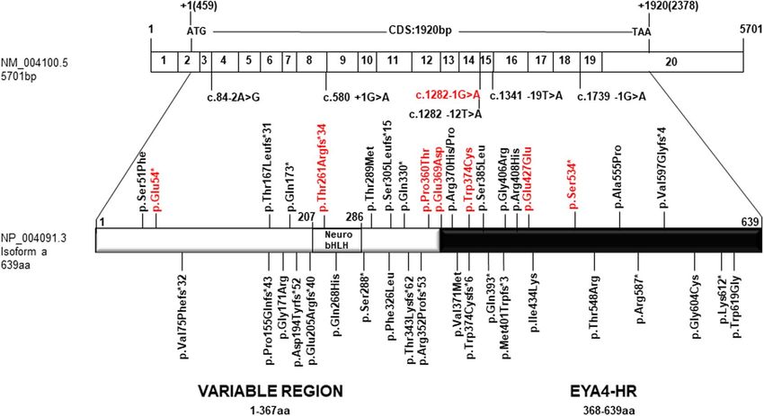
www.nature.com/scientificreports/                                                             www.nature.com/scientificreports

                           Figure 1. Pedigrees of the Spanish families S739, S856, S1303 and S2532. Black and white symbols indicate the
                           affected and the unaffected subjects, respectively. The index cases are pointed by black arrows. The subjects of
                           whom DNA samples were available for segregation analysis were marked by asterisks. The relative quantification
                           of the 2747 bp deletion (CNV) by Real-Time PCR in the subjects of family S2532 is displayed. Only affected
                           members showed a significant reduction (~50%) in the number of copies of EYA4. The audiograms showing the
                           air conduction values obtained from several different patients of each family and the electropherograms of the
                           mutations are also displayed. Each graph point represents the average hearing loss for the right and left ears.

                           S1729 (Fig. 2), and a silent synonymous mutation, c.1281G > A (p.Glu427Glu) at the last nucleotide of exon 14 in
                           subject III:2 of family S2192 (Fig. 2).
                               Finally, a novel missense variant, c.1122G > T (p.Trp374Cys) was identified in the Australian family (10880)
                           using the gene panel OtoSCOPE (Fig. 3). Segregation with the hearing loss for the different mutations was con-
                           firmed in all the extended families of whom DNA samples were available. The segregation of the CNV (dele-
                           tion of exon 15 to 17) in members of family S2532 was assessed by relative quantification of the exon 16 by
                           Real-time PCR (Fig. 1). The classification for each SNV was done taking into account the American College
                           of Medical Genetics and Genomics (ACMG) guidelines and the prediction algorithms used by Varsome60,61,
                           several predictors of pathogenicity, the Human Splice Finder (HSF) predictor62, the allele frequencies in the
                           Genome Aggregation Database (GnomAD)63, the international genome sample resource (IGSR, https://www.
                           internationalgenome.org/data), the Exome Aggregation Consortium-ExAC (http://exac.broadinstitute.org/), the
                           Collaborative Spanish Variant Server-CSVS database (http://csvs.babelomics.org/) and the Deafness Variation
                           Database (DVD, http://deafnessvariationdatabase.org/). This analysis indicated that six out of eight single nucle-
                           otide variants (SNVs) identified in this study were classified as “Pathogenic/Likely pathogenic”, whereas two
                           remained as “variants of uncertain significance” (VUS) (Table 2).
                               All mutations identified so far in EYA4 gene, including those reported in the present work, are summarized in
                           Table 1 and Fig. 4. The hearing loss observed in our patients with EYA4 mutations is characteristic of the DFNA10
                           phenotype. Affected subject show a bilateral and progressive hearing loss of postlingual onset (ranging from the
                           first to fourth decade) with a flat/gently downsloping audiometric profile (Figs. 1, 2). The ARTA of the Australian
                           family (10880) provided evidence that the hearing loss progresses at a rate that ranges between 0.5 dB/year at
                           0.25 kHz and 1.3 dB/year at 8 kHz (Fig. 3D).

                           Effect of p.Glu369Asp and p.Ser534* mutations on EYA4 localization. To further explore the
                           mechanism of pathogenesis underlying p.Glu369Asp and p.Ser534* mutations, we investigated their effect on

Scientific Reports |   (2020) 10:6213 | https://doi.org/10.1038/s41598-020-63256-5                                                          6
www.nature.com/scientificreports/ www.nature.com/scientificreports
Figure 2. Pedigrees of the Spanish families S580, S1729, S1764, S2192 showing the audiograms and
electropherograms as in Fig. 1.
interaction with SIX1 and ability of the SIX1-EYA4 protein complex to translocate to the cell nucleus. To do
this we investigated the expression pattern and cellular localization of EYA4 mutants in transiently transfected
COS7 cells in the absence or presence of SIX1. When SIX1 protein is not present, the c-myc-tagged EYA4-HR-Wt
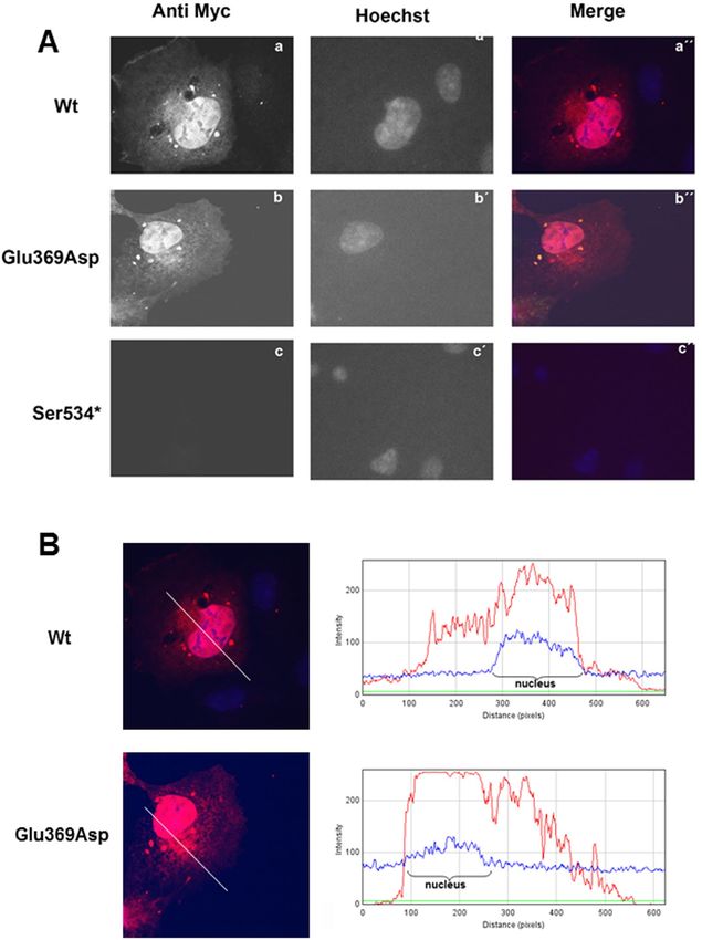
and the EYA4-HR-Glu369Asp proteins are detected in both, cytoplasm and nucleus, in the majority of COS7
cells forty-eight hours after transfection (Fig. 5A,B). However, no signal was observed for the mutant p.Ser534*
(Fig. 5A). When COS7 cells were transfected with the bicistronic plasmid containing SIX1 and EYA4-HR
(pS1E4), the protein complex was detected mostly in the nucleus for both Wt and p.Glu369Asp mutant, but
not for p.Ser534* mutant (Fig. 6; Supp Fig. 2A,B). Taken together, these observations suggest that p.Glu369Asp
mutation does not significantly alter the interaction with SIX1 and consequently does not impair the nuclear
translocation of the EYA4-SIX1 complex.
To confirm the results of the immunocytochemistry assays, we performed a western-blot analysis of COS7
cells transfected with the pE4S1Wt, pE4S1Glu369Asp and pE4S1Ser534* vectors. Lysates were separated on a
10% gel followed by blotting and immunostaining with the anti-myc and anti-HA antibodies. As shown in Fig. 7A
a clear band of 37 kDa corresponding to the EYA4-HR domain was detected for the Wt and p.Glu369Asp mutant
using anti-myc antibodies directed against the c-myc tag cloned in frame in the Nt portion of the EYA4-HR
domain (Suppl Fig. 1). However, by using the same anti-myc antibodies the 25 kDa band expected for the puta-
tive truncated protein p.Ser534*, was not detected in the corresponding lane, whilst a clear band of 33 kDa was
observed when anti-HA was used to detect the HA-tagged SIX1 protein (Suppl Fig. 1) indicating an efficient
transfection with the bicistronic plasmid. Therefore, the p.Ser534* mutant protein could not be detected in trans-
fected cells neither by immunocytochemistry nor by Western blotting analysis.
The lack of detection of p.Ser534* mutant could be explained by a degradation at the mRNA or protein levels.
To test this hypothesis, we obtained total RNA from COS7 cell transfected with the Eya constructs (pE4-Wt,
pE4Glu369Asp and pE4Ser534*) from three independent experiments and RT-PCR assays were then carried out.
Expected PCR-amplimers of 390 pb were obtained in the Wt and both mutants by using specific primers to
amplify an EYAHR fragment. No statistical significant differences in mRNA expression levels were found between
the wild type and mutant transcripts (Fig. 7B) once they were normalized with the expression level obtained from
the Zeocin mRNA, the antibiotic selection marker of the vector.
In Silico splicing analysis of EYA4 mutations. To further investigate the pathogenic mechanism of
p.Glu369Asp missense mutation we performed computational analysis of the genomic region surrounding
c.1107G > T which corresponds to the last nucleotide of exon 12, a position important for normal splicing. We
Scientific Reports | (2020) 10:6213 | https://doi.org/10.1038/s41598-020-63256-5 7www.nature.com/scientificreports/ www.nature.com/scientificreports
Figure 3. (A) Pedigree of the family 10880 showing the novel EYA4 (c.1122G > T) variant segregating with the
hearing loss phenotype. The genotype of all the subjects analysed is indicated in capital letters (Wt = GG, Hz =
GT). Black dots represent normal hearing individuals carrying the mutation but under the age of onset in this
family. (B) Representative chromatograms from wild-type and mutant sequences. (C) Molecular modelling of
the p.Trp374Cys in EYA4. Mutated site is highlighted by a green circle and locally zoomed. Mutant (orange) is
superimposed on wild-type (blue) for comparison. The p.Trp374Cys alters local conformation and disrupts beta
sheet backbone folding resulting in decrease structural stability. (D) Age Related Typical Audiogram (ARTA)
analysis. The hearing loss progresses at a rate that ranges between 0.5 dB/year at 0.25Khz and 1.3 dB/year at
8 kHz. E) EYA4-HR domain showing the four Tyr-phosphatase motifs that mediate the protein enzymatic
activity. The p.Trp374Cys mutation identified in this work, and two previously identified missense mutations
(p.Thr548Arg and p.Gly604Cys) are all affecting conserved residues at motif I, II and IV, respectively.
used several different web-based tools: Esefinder, MaxenScan, BDGP Splice Site Prediction by Neural Netwok and
NetGene264–68. Esefinder and Netgene2 predict a complete loss of the splicing donor site for the mutant transcript.
MaxEntScan evaluates 5′ splice site strengths, being the typical MaxEnt 5′ score in wild type sequences of around
11.81. In our analysis MaxEntScan yielded a score of 9.12 for the Wt 5′splice site of intron 12 in EYA4 and 3.89 for
the c.1107G > T mutant. These data suggest that c.1107G > T alters the 5′ splice site leading to skipping of exon 12
and generation of a truncated protein of 331 amino acids. Similar analysis was performed for the c.1282-1G > A
mutation identified in family S856. ESEfinder and MaxEntscan softwares indicated that c.1282-1G > A would
abrogate the 3′ splicing acceptor site of intron 14 leading to skipping of exon 15. The aberrant splicing of exon 15
would produce a putative truncated protein of 433 amino acids or, as an alternative, a peptide of 455 amino acids
if the cryptic splice acceptor site within the exon 16 was used, as previously described4. The same analysis was
performed for the nonsense mutation (c.1601C > G, p.Ser534*) and for the other missense mutations identified
in this study (p.Pro360Thr; p.Trp374Cys) but no alteration of splicing was predicted. Finally, we analyzed the
silent mutation c.1281G > A (p.Glu427Glu) as it affects the last nucleotide of exon 14 and the in silico analysis
carried out by ESEfinder and MaxEntscan software predicted this mutation could be compromising the splicing
donor site of intron 14 (Table 2).
In vivo minigene splicing assays of EYA4 mutations. To assess the predicted effects of EYA4 mutations
on splicing, we carried out minigene assays in NIH3T3 cells (Fig. 8A). Results of the minigene indicated that the
wild type exon 12 was correctly incorporated in more than 85% of the transcripts (400 bp band), while the mutant
c.1107G > T (p.Glu369Asp) failed to be retained in the mature transcript and was eliminated during the splicing
Scientific Reports | (2020) 10:6213 | https://doi.org/10.1038/s41598-020-63256-5 8www.nature.com/scientificreports/ www.nature.com/scientificreports
Number of scores CSVS
supporting Allele GnomAD
DNA change Protein change ACMG pathogenicity HSF DVD Freq. Allele Freq.
Activation of an exonic cryptic donor
Pathogenic site. Potential alteration of splicing.
c.160G > T p.Glu54* 6/9 N.A 0 0
(PVS1, PM2, PP1, PP3) Alteration of an exonic ESE site.
Potential alteration of splicing.
Pathogenic Alteration of an exonic ESE site.
c.781del p.Thr261Argfs*34 N.A N.A 0 0
(PVS1, PM2, PP1, PP3) Potential alteration of splicing.
VUS 1.06e-5 (3 in
c.1078C > A p.Pro360Thr 16/19 No impact on splicing VUS 0
(PM2, PP1, PP3, BP1) 281904)
Likely pathogenic Alteration of Wt donor site. Alteration
c.1107G > T p.Glu369Asp 11/20 N.A 0 0
(PS3, PM2, PP1, PP3, BP1) of an exonic ESE site
VUS Activation of an exonic cryptic donor
c.1122G > T p.Trp374Cys 19/19 N.A 0 0
(PM2, PP1, PP3, BP1) site.
Likely pathogenic Alteration of Wt donor site, most
c.1281G > A p.Glu427Glu 1/1 N.A 0 0
(PS3, PM1, PM2, PP1, BP4) probably affecting splicing
Alteration of the Wt acceptor site, most
Pathogenic probably affecting splicing. Activation
c.1282-1G > A — 6/6 N.A 0 0
(PVS1, PS3, PM2, PP1, PP3) of an intronic cryptic acceptor site.
Potential alteration of splicing.
Pathogenic
c.1601C > G p.Ser534* 7/9 Creation of an exonic ESS site N.A 0 0
(PVS1, PS3, PM1, PM2, PP1, PP3)
Table 2. Classification of Single Nucleotide Variants (SNVs) identified in this work. ACMG criteria: PVS1
(Pathogenic Very Strong): null variant (nonsense, frameshift, canonical ±1 or 2 splice sites, initiation codon,
single or multiexon deletion) in a gene where LOF is a known mechanism of disease. PS3 (Pathogenic Strong
3): well-established in vitro or in vivo functional studies supportive of a damaging effect on the gene or gene
product. PM1 (Pathogenic Moderate 1): located in a mutational hot spot and/or critical and well-established
functional domain (e.g., active site of an enzyme) without benign variation. PM2 (Pathogenic Moderate 2):
absent from controls (or at extremely low frequency if recessive) in Exome Sequencing Project, 1000 Genomes
Project, or Exome Aggregation Consortium. PP1 (Pathogenic Supporting 1): cosegregation with disease
in multiple affected family members in a gene definitively known to cause the disease. PP3 (Pathogenic
Supporting 3): multiple lines of computational evidence support a deleterious effect on the gene or gene
product (conservation, evolutionary, splicing impact, etc.). BP1 (Benign Supporting 1): missense variant in a
gene for which primarily truncating variants are known to cause disease. BP4 (Benign Supporting 2): multiple
lines of computational evidence suggest no impact on gene or gene product (conservation, evolutionary,
splicing impact, etc.). All the databases were searched on the 22nd of March 2020.
process (263 bp band). These results confirm the in silico analysis indicating that this mutation compromises the
acceptor splicing site of exon 12. This would result in a frameshift and a premature termination codon generating
a truncated EYA4 protein (Fig. 8B, Supp Fig. 3A).
The wild type minigene including exons 15–16 generated two splicing products, a 483 bp band that matched
with the expected size for the normal splicing of exon 15 and 16 and a 415 bp band corresponding to normal
exon 15 and a shorter exon 16 that lacks the first 68 nucleotides (Fig. 8B, Supp Fig. 3B). The shorter exon 16 was
previously identified in human lymphoblastoid and lens epithelial cell lines4. The c.1282-1G > A mutant mini-
gene showed a PCR product of 354 nucleotides that lacks exon 15 and includes the shorter exon 16 (Fig. 8B, Supp
Fig. 3B).
Results of the minigene assays for the Wt and c.1601C > G (p.Ser534*) mutant in exon 17, revealed a single
378 bp PCR product corresponding to the mature transcript in which the exon 17 is retained. These data indi-
cate that this mutation has no effects on splicing as the in silico analysis had previously indicated (Fig. 8B, Supp
Fig. 3C).
Finally, the minigene construct containing both, the Wt and c.1281G > A (p.Glu427Glu) mutant in exon 14,
revealed that wild type exon 14 is incorporated in more than 90% of the transcripts (352 bp band). However, the
intensity of this band was dramatically reduced in the mutant. Additionally, extra bands were also observed, indi-
cating an impairment of the splicing process (Fig. 8C).
Discussion
Transcription factors and co-factors are large group proteins that are fundamental for proper gene regulation.
These factors often exhibit a combination of spatial and temporal expression and are critical for cellular events
including development, function and lifelong maintenance. In the inner ear, disruption of transcription factors
belonging to the POU, SIX, LMX and EYA families have all been shown to be essential for proper hearing in
humans. Here we report 9 novel mutations that disrupt the expression or function of the EYA4 gene, segregating
in 8 Spanish families and one Australian family. In doing so we report the first synonymous variant, classified
as likely pathogenic following the ACMG criteria and the minigene assay, linked to DFNA10 and provide fur-
ther evidence for haploinsufficiency as the common underlying disease-causing mechanism for DFNA10-related
hearing loss.
In previous mutational screenings of ADNSHL families in the Spanish population, only 15 TECTA
(DFNA8/12), two ACTG1 (DFNA20/26) and two miR-96 (DFNA50) mutations were identified54,56,69. In this
Scientific Reports | (2020) 10:6213 | https://doi.org/10.1038/s41598-020-63256-5 9www.nature.com/scientificreports/ www.nature.com/scientificreports
Figure 4. Scheme depicting the mRNA structure and coding sequence (CDS) of the NM_004100.5 EYA4
transcript and the NP_004091.3 protein isoform showing the variable region and the EYA4 homologous region
(EYA4-HR domain). The novel genetics variants (in red) and those previously identified in EYA4 are shown.
study, we have identified 8 novel variants in EYA4 gene after mutational screening of 531 Spanish familial cases.
Therefore, the global prevalence of DFNA10 in the Spanish hearing impaired is about 1.5% (8/531), suggesting
that it is rare form of ADNSHL in Spain. Moreover, we have identified a novel missense mutation in EYA4 in an
Australian family enrolled in this study. The exact prevalence of EYA4/DFNA10-related hearing loss is not known
as it was only detected in Caucasian and East Asian population to date. However, studies from Japan, Korea and
Slovakia estimate the prevalence of EYA4-related hearing loss to be 0.9%15, 7.4% (1/14)33 and 5.56% (1/17)31. The
substantial increased prevalence in the last two populations is most likely due to the reduced number of probands
screened and the phenotypic preselection of patients.
Prior to this study, 47 families with ADSNHL at the DFNA10 locus have been identified. Despite the different
mutations (nonsense, missense, splice-site, frameshift or large genomic deletions) so far reported in the DFNA10
families and the different affected protein domains in EYA4, no dramatic auditory phenotypic differences have
been observed. To the best of our knowledge, all families carrying mutations in EYA4 exhibit a similar phenotype
consisting of a postlingual progressive hearing impairment affecting high frequencies at greater rates. ARTA
analysis in DFNA10 families for which audiograms were available (primary or published data) demonstrates a
fairly similar picture (Suppl Fig. 4). Relatively discordant phenotypes showing poorer thresholds, i.e. of> 45 dB
at ages of> 20 years at all frequencies, were seen in a few patients who carried two mutations, p.Pro155Glnfs*43
and p.Gly604Cys17.
The ATD indicated more progression at high frequencies in most families. Progression varied from ~0.5 -
~0.8 dB/year at 0.25–1 kHz to ~1.5 - ~1.8 dB/year at 4–8 kHz. Exceptions were for LMG 265 with the p.Arg-
352Profs*53 mutation21 showing progression with an ATD of ~0.8 dB/year at all frequencies, and the Australian
family harboring a splice-site mutation c.1282-12 T > A, which is predicted to result in exon 15 skipping41, that
showed more progression at the lower frequencies (ATD ~1.5 dB/year at 0.25–1 kHz) than at the higher frequen-
cies (ATD ~1.0 dB at 4–8 kHz).
Mutations in EYA4 have also been linked to HL and dilated cardiomyopathy (DCM) in two families7,45,46.
The genotype-phenotype correlation for DFNA10 versus HL with DCM is not well understood. Initially, it was
proposed that mutations resulting in the loss of the EYA-domain would result in HL with DCM. However, this
was later challenged as several families have been reported with truncating mutations in the variable region with
no DCM phenotype (Table 1). In this work we reports the most N-terminal nonsense mutation identified to date
in the variable region (c.160G > T; p.Glu54*) that is only associated with SNHL in a Spanish family. Interestingly,
one mutation (p.Gln393*), has been linked to both DFNA1015,26 and HL with a mild-DCM27. The differences
in the cardiovascular phenotype could be due to environmental factors, variable penetrance or a coincidental
segregation of different genetic etiology for mild-DCM in the family described by Abe et al. (2018). In our study,
all reported families have non-syndromic DFNA10-related hearing loss regardless of mutation type and location.
It is noteworthy that families with available clinical data and large deletions encompassing either the whole
gene or part of the gene, have other clinical phenotypes which include, dilated cardiomyopathy (DCM) and/or
isolated mental retardation and OTFC syndrome7,45,47,48. It is unclear whether these additional phenotypes are
a result of contiguous gene deletions or more complex, mechanisms are at play. For example, the c.579_580in-
sTACC, p.Asp194Tyrfs*52; mutation associated with SNHL18 is indeed very similar to the truncated isoform
(c.581_804del; p.Asp194Glyfs*30) resulting from the 4,846pb DNA deletion spanning the intron 9-exon
10-partial intron 10 described in the family with SNHL and dilated cardiomyopathy7,46. Moreover, we have
Scientific Reports | (2020) 10:6213 | https://doi.org/10.1038/s41598-020-63256-5 10www.nature.com/scientificreports/ www.nature.com/scientificreports
Figure 5. (A) Expression pattern of the EYA4-HR Wt (a), Glu369Asp (b) and Ser534* (c) in COS7 cells.
EYA4-HR protein shows a cytoplasmic and nuclear distribution for the Wt and Glu369Asp mutant but no signal
was observed for the mutant Ser534*. Hoechst staining was used to identify the nucleus. (B) IMAGE J graphical
section showing the fluorescence intensities plotted against the distances (pixels). EYA4-HR staining (in red) for
the Wt and Glu369Asp mutant is detected in both the nucleus and the cytoplasm.
identified the first gross heterozygous deletion reported in an Spanish family spanning exon 15 to 17 that is only
associated with SNHL, and a recent work15 also reports two more CNVs (affecting exon 7 to 11 and exon 4 to
20, respectively) linked to SNHL. Thus, one possibility is that the reported cardiomyopathy in other families was
caused by other genetic factors present in the large deleted region or created by the event of deletion.
Prior to this study, 16 out of 38 variations identified in EYA4 are missense mutations (Table 1). The vari-
ant p.Trp374Cys identified in this work causes a substitution from a large, bulky and hydrophobic tryptophan
to a small thiol-containing cystine at amino acid 374. We predict this change might alter protein folding or
Scientific Reports | (2020) 10:6213 | https://doi.org/10.1038/s41598-020-63256-5 11www.nature.com/scientificreports/ www.nature.com/scientificreports
Figure 6. Nuclear translocation of the EYA4-HR-SIX1 Wt and mutant protein complex in COS 7 cells. In
presence of SIX1, EYA4-HR Wt and Glu369Asp were detected mostly in the nucleus, consequently, the capacity
of nuclear translocation of the complex EYA4-SIX1 seems not to be impaired by the p.Glu369Asp mutation. No
signal was observed for p.Ser534* mutant.
conformation, possibly by allowing the thiol of the substituted cystine to create new disulfide bonds and disrupt-
ing proper protein folding. Improper protein folding or formation may interfere with its other protein binding
partners and an inability to properly form the essential PSED network7,20,44. An alternative hypothesis would be a
detrimental effect on the activation and enzymatic activity of EYA4 by modifying metal cofactor binding ability
of magnesium ions (Mg2+). There are three predicted metal binding sites (amino acids 375, 377 and 603) and two
active sites (amino acids 375 and 377) in the EYA domain (Fig. 3C,E). Interestingly, all three of these residues are
Aspartic Acid and are all integral parts of catalytic motifs fundamental to EYA4’s Tyr phosphatase activity. Unlike
traditional Tyr phosphatases which require Cystine residues at the heart of their catalytic core, EYA proteins
depend on Aspartic Acid residues and require divalent ion, such as Mg2+ for activation6. The first motif (AA
sequence [WDLDETI]) starts at residue 374 and extends 6 residues to 380 (Fig. 3E). The Aspartic Acid at position
375 acts as the catalytic center and is essential for EYA4 activation. It is possible that this variant does not cause
a dysregulation of the PSEDN, but rather alters EYA4’s enzymic activity, either physically (changing the protein
conformation of the binding pocket) or chemically (altering binding affinity and efficiency of its neighboring
Aspartic Acids through the cystine thiol-group known to have a high affinity for other metals)70 or a combination
of both. This possible activation impairment could adversely affect the enzymic roles of EYA4 in the inner ear.
In any case, more functional studies are required to fully assess the consequences of this variant. Interestingly,
a variant has already been reported at this amino acid position, (p.Trp374Cysfs*6) in a Hungarian family with
progressive ADNSHL22. The reported family exhibits a similar phenotype as family 10880. Surprisingly, mutation
type and location do not seem to influence the hearing phenotype as seen in other deafness-causing genes54.
The variant c.1078C > A; p.Pro360Thr was not present in the disease or hearing-loss genetic variant databases
consulted and it presented an extremely low allelic frequency in the GnomAD. It causes the substitution from
the non-polar, aliphatic residue proline to a polar non-charged residue threonine at position 360. We predict this
change might alter protein folding or conformation, as the distinctive cyclic structure of proline’s side chain gives
proline an exceptional conformational rigidity compared to other amino acids. This proline is fully conserved
through evolution and the computational analysis (16 out of 19, including Polyphen-2, Sift and others) indicated
the p.Pro360Thr is a damaging change. However, in the absence of functional analysis it is classified as a VUS
following the ACMG criteria.
The p.Glu369Asp mutation identified in this work initially categorized as a missense mutation does not affect a
key position for EYA4 activation or enzymatic activity. Furthermore, it behaved as the wild type isoform in the in
vivo experiments we have carried out as it did not impact the translocation of the mutant SIX1-EYA4Glu369Asp
complex to the nucleus. Both in silico predictions and minigene assays, confirmed that the c.1107G > T (pGlu-
369Asp) affects the donor splice-site of exon 12. The human 5′ss consensus sequence is MAG|GURAGU (M is
A or C; R is purine), spans positions −3 to +6 relative to the exon-intron junction71. Pathogenic splicing alter-
ations caused by point mutations in both 5′ and 3′ splice sites are an important mechanism through which gene
Scientific Reports | (2020) 10:6213 | https://doi.org/10.1038/s41598-020-63256-5 12www.nature.com/scientificreports/ www.nature.com/scientificreports
Figure 7. (A) Western blot analysis of EYA4 Wt and mutants’ production. A clear band of 37 kDa
corresponding to the EYA4-HR domain detected by the anti-myc antibodies directed against the c-myc tag
cloned in frame in the Nt portion of the EYA4-HR domain (Suppl Fig. 1) is observed in extracts from COS7
cells transfected with pE4S1 Wt and Glu369Asp bicistronic plasmid and in the control pEYA4. No signal was
detected in the mutant Ser534* when we use the same anti-Myc antibodies (at the top), whereas a robust band
of 33kDA corresponding to SIX1 was seen when we use anti-HA antibodies (at the bottom) to detect the HA-
tagged SIX1 protein (Suppl Fig. 1). The full-length blots revealed at different time exposures with anti-Myc and
anti-HA antibodies are displayed in Suppl Fig. 5. (B) (top) RT-PCR assay performed on total RNA extracted
from COS7 cells transfected with Eya constructs (wild-type and mutants) showed a band corresponding to
the amplification of the tagged EYA4-HR mRNA. This band was not detected in the control lanes (-RT) in
which the mRNA was nod added; (B) (bottom) cDNA amplification of Zeocin (the plasmid antibiotic resistant
cassette) using specific primer was used for normalization purposes. Quantification of the relative amounts
of EYA4 cDNA amplifications once they were normalized with Zeocin levels did not show any statistically
significant differences after Wt and mutants comparisons. t Student p values are shown.
mutations cause human diseases72,73, including deafness50–52, and it has been estimated that around 15% of all
disease-causing point mutations result in defective splicing. Moreover the ‘G’ nucleotide at the last base of exon
present in almost 80% of the human 5′ss74. Interestingly, another EYA4 variation, c.804G > C affecting the last
nucleotide of exon 10, was also initially classified as a missense mutation (p.Gln268His). Subsequently, in vitro
experiments have demonstrated that it leads to exon 10 skipping resulting in a frameshift that introduces a prema-
ture stop codon31. These results suggest that for some predicted EYA4 missense mutations, the hidden mechanism
of pathogenesis is rather haploinsufficiency as most mutations identified so far are frameshifts and nonsense pre-
dicted to trigger nonsense-mediated mRNA decay (NMD)75. Based on this hypothesis, the nonsense c.1601C > G
mutation could provoke a complete degradation of the mutant messenger by NMD. Our RT-PCR experiments
have shown that there were no differences in the expression levels of mutant and Wt mRNAs, however, we could
not detect the p.Ser534* mutant protein by western blot. Moreover, the minigene assay has shown that this muta-
tion does not result in an alternative splicing. Although this assay is not able to detect NMD because the generated
minigene only contained one exon (exon 17); we propose that protein degradation, alone or in combination with
NMD, as the possible underlying mechanism of pathogenesis of this mutation. In a previous study, Zhang and
co-workers55 showed the same effect for the p.Arg587* EYA4 mutation, further supporting the notion of hap-
loinsufficiency as the pathogenic mechanism. Finally, we have identified the c.1282-1G > A mutation in family
S856. This mutation destroys the acceptor splice-site of intron 14 leading to skipping of exon 15 as shown by the
minigene assay and is predicted to generate a truncated protein. Hitherto, this is the second DFNA10-causing
mutation affecting the acceptor splice-site of intron 14 in EYA4. The first mutation, c.1282-12T > A has been
previously detected in an Australian family41.
It is of key interest the results obtained for the silent mutation c.1281G > A (p.Glu427Glu) as it is classified as
“Likely pathogenic” according to the ACMG criteria by altering the donor site of intron 14, most probably affect-
ing the splicing (Table 2); a point confirmed by the minigene assay (Fig. 8). Albeit additional experiments are
further required to fully understand the mechanism of pathogenesis of this variant, to the best of our knowledge
this can represent the first silent mutation to be linked to DFNA10-related hearing loss. Therefore, our findings
Scientific Reports | (2020) 10:6213 | https://doi.org/10.1038/s41598-020-63256-5 13You can also read