What Role Might Non-Mating Receptors Play in Schizophyllum commune?
←
→
Page content transcription
If your browser does not render page correctly, please read the page content below
Journal of
              Fungi
Article
What Role Might Non-Mating Receptors Play in
Schizophyllum commune?
Sophia Wirth † , Daniela Freihorst † , Katrin Krause * and Erika Kothe

                                          Friedrich Schiller University Jena, Institute of Microbiology, Microbial Communication,
                                          25 07743 Neugasse Jena, Germany; sophia.wirth@uni-jena.de (S.W.); danielafreihorst@aol.com (D.F.);
                                          erika.kothe@uni-jena.de (E.K.)
                                          * Correspondence: katrin.krause@uni-jena.de; Tel.: +49-(0)3641-949-399
                                          † These authors contributed equally to the work.

                                          Abstract: The B mating-type locus of the tetrapolar basidiomycete Schizophyllum commune encodes
                                          pheromones and pheromone receptors in multiple allelic specificities. This work adds substantial
                                          new evidence into the organization of the B mating-type loci of distantly related S. commune strains
                                          showing a high level of synteny in gene order and neighboring genes. Four pheromone receptor-like
                                          genes were found in the genome of S. commune with brl1, brl2 and brl3 located at the B mating-type
                                          locus, whereas brl4 is located separately. Expression analysis of brl genes in different developmental
                                          stages indicates a function in filamentous growth and mating. Based on the extensive sequence
                                          analysis and functional characterization of brl-overexpression mutants, a function of Brl1 in mating
                                          is proposed, while Brl3, Brl4 and Brl2 (to a lower extent) have a role in vegetative growth, possible
                                          determination of growth direction. The brl3 and brl4 overexpression mutants had a dikaryon-
                                          like, irregular and feathery phenotype, and they avoided the formation of same-clone colonies on
                                solid medium, which points towards enhanced detection of self-signals. These data are supported
                                   by localization of Brl fusion proteins in tips, at septa and in not-yet-fused clamps of a dikaryon,
Citation: Wirth, S.; Freihorst, D.;       confirming their importance for growth and development in S. commune.
Krause, K.; Kothe, E. What Role
Might Non-Mating Receptors Play in        Keywords: Schizophyllum commune; pheromone receptor-like genes; B mating-type locus; mating;
Schizophyllum commune? J. Fungi 2021,     self-recognition
7, 399. https://doi.org/10.3390/
jof7050399

Academic Editor: Ulrich Kück              1. Introduction
                                                Schizophyllum commune is a well-known wood-decaying basidiomycete [1]. Unlike
Received: 31 March 2021
Accepted: 18 May 2021
                                          many other basidiomycetes, S. commune can be cultured in the laboratory on artificial media
Published: 20 May 2021
                                          and can be genetically modified, allowing the intensive studying of mating and mushroom
                                          development at the molecular level [2,3].
Publisher’s Note: MDPI stays neutral
                                                The tetrapolar mating system of S. commune is not required for the initial fusion
with regard to jurisdictional claims in
                                          between mating partners but is involved in the stable formation of a dikaryotic mycelium
published maps and institutional affil-   involving nuclear migration and clamp cell fusion [4]. With two unlinked multigenic
iations.                                  mating-type loci, the tetrapolar mating system of S. commune is conserved in basidiomycetes
                                          including Coprinopsis cinerea [5], Laccaria bicolor [6], Flammulina velutipes [7] and Ustilago
                                          maydis [8]. The A locus encodes a set of divergently transcribed homeodomain transcription
                                          factors [9], while the B locus comprises pheromone receptors surrounded by several
Copyright: © 2021 by the authors.
                                          pheromone precursor genes [3,10]. Generally, two allelic versions of pheromone receptors
Licensee MDPI, Basel, Switzerland.
                                          occur in each S. commune strain, which thereby define the specificity of the locus by
This article is an open access article
                                          recognizing either Bα or Bβ pheromones.
distributed under the terms and                 The publicly available S. commune genome sequences allow a comparison of different
conditions of the Creative Commons        S. commune strains from different geographic locations and with unknown mating-type
Attribution (CC BY) license (https://     loci organization and specificities. All annotated basidiomycete pheromone receptors are
creativecommons.org/licenses/by/          orthologs of Ste3 of Saccharomyces cerevisiae, which is also true for the Bα and Bβ mating
4.0/).                                    receptors of S. commune (Bar and Bbr, respectively) [11]. Generally, these proteins are

J. Fungi 2021, 7, 399. https://doi.org/10.3390/jof7050399                                                        https://www.mdpi.com/journal/jof
J. Fungi 2021, 7, 399                                                                                            2 of 18

                        characterized by the presence of seven transmembrane domains and their association with
                        intracellular G-proteins (GPCR) triggering signaling pathways to coordinate B-dependent
                        development. They are further characterized by a short N-terminal extracellular domain
                        and a long cytoplasmic C-terminal tail. Binding of a pheromone from a non-self mating
                        partner stimulates the associated heterotrimeric G-proteins to dissociate into the Gα and
                        Gβγ subunits, which trigger the subsequent G-protein signaling pathways. The specifici-
                        ties, and hence sequences, of the strains differ to allow for different pheromones to be
                        recognized [12,13]. A strain carrying a deletion of the B receptor and pheromone genes
                        that shows no B-regulated development in matings with any wild-type strain is available.
                        Monokaryotic mutant strains with constitutively active B mating-type genes exist as well,
                        which show an aberrant phenotype typically found in semi-compatible mating [10].
                              Among the 75 encoded proteins with predicted transmembrane domains [14], six
                        fungal pheromone receptors are found in a given strain. Since mating-type specificities
                        are different, allelic versions of these six pheromone receptors are to be considered. In any
                        genome, only the Bar and Bbr mating receptors have been functionally characterized. Of
                        these, specifically the pheromone receptors Bbr2 [10] and Bar2 [11], as well as Bbr1 [15],
                        have received the most attention. These true pheromone-recognizing receptors induce
                        pheromone/receptor-dependent development resulting in fertile dikaryons. The remaining
                        four B-receptor-like genes per genome (brl1 through brl4) are orthologs of S. cerevisiae Ste3
                        a-factor receptor, with three (brl1–3) surrounding the bar and bbr gene loci within an
                        81 kb region, while brl4 was identified on a separate scaffold [11]. The brl genes are not
                        surrounded by pheromone genes, indicating that they are not directly associated with
                        mating. In addition, they are highly similar in sequence, which indicates that they do not
                        differ regarding recognition of ligands. This finding is in line with the identification of
                        pheromone receptor-like genes in the genome of many basidiomycetes [16–18], including
                        F. velutipes [7], Postia placenta [19] and Phanerochaete chrysosporium [20], and the mycorrhizal
                        species L. bicolor [6]. To unravel the function of the B receptor-like genes, expression
                        analysis in L. bicolor showed association with different life stages [6]. For the receptor-like
                        Crp2 protein from C. neoformans, a role in cell fusion and sporulation has been postulated,
                        since it activates the same G-protein-coupled signaling pathway as do the true mating-type
                        receptors [21].
                              Here, it could be established that overexpression of the pheromone receptor-like genes
                        brl3 and brl4 increase self-sensing of S. commune and induce an asymmetrical colony growth,
                        while the function of brl1 could be attributed to an early mating response.

                        2. Materials and Methods
                        2.1. Culture Conditions and Strains
                              S. commune strains (Table S1) were routinely grown on minimal medium (MM [22])
                        with the addition of 4 mM tryptophan for trp- strains and 0.1 mM uracil for ura- strains at
                        28 ◦ C. The sister strains S. commune 4–39 and W22 are derived from a lineage of 40 back-
                        crosses and are co-isogenic, differing only in mating-type to allow for out-crossing to
                        S. commune 12–43. To assess growth and biomass formation on other carbon sources,
                        glucose was replaced by 4% xylose or 3.4% sucrose, and colonies were grown on agar
                        medium covered with a cellophane foil for easy biomass recovery using five biological
                        replicates. Mycelium was dried overnight and weighed. Statistical analysis was done with
                        a paired, two-tailed t-test. A modified sandwich system was used to grow compatible
                        monokaryons for mating interactions allowing mycelial harvesting 6, 12, 24 and 48 h after
                        mating and in the 8-day-old, established dikaryon [23].
                              For growth in liquid culture, a mycelial homogenate was prepared using a laboratory
                        blender (neoLab, Heidelberg, Germany), and cultures were grown at 250 rpm in 500 mL
                        Erlenmeyer flasks containing 200 mL MM.
J. Fungi 2021, 7, 399                                                                                        3 of 18

                        2.2. Gene Overexpression and Protein Labeling
                              Overexpression vectors for brl1 (ID: 2638177), brl2 (ID: 2704867), brl3 (ID: 2638261)
                        and brl4 (ID: 2691538) were constructed using yeast recombinational cloning with the
                        binary shuttle vector pRS415 carrying the leu2 gene for yeast selection and the bla gene
                        for E. coli selection [24]. For overexpression, brl genes and a 758 bp long fragment of the
                        tef1 promoter (elongation factor 1α; ID: 84142) were amplified from genomic DNA of S.
                        commune H4–8 by PCR using PhusionTaq (Thermo Fisher Scientific, Waltham, MA, USA)
                        and specific primer pairs (Table S2). PCR products were recombined in the vector pRS415
                        (linearized with HindIII and BamHI) by using overhangs of at least 30 bp. The plasmids were
                        isolated from the transformed yeast cells [25] and checked by PCR (M13 standard primers),
                        and the correct fusion product was verified by sequencing (GATC Biotech, Konstanz,
                        Germany). Electrocompetent E. coli were transformed using 1 µL of DNA, followed by
                        colony PCR verification and plasmid isolation (GeneJET Plasmid-Miniprep-Kit, Thermo
                        Fisher Scientific, Waltham, USA). Restriction with BamHI, SalI and HindIII was used
                        to confirm the expected products, pRSbrl1OE, pRSbrl2OE, pRSbrl3OE and pRSbrl4OE.
                        Subsequently, plasmid DNA was transformed into protoplasts of the tryptophan and uracil
                        auxotrophic strain S. commune T33 [26], and successful transformation was verified by
                        sequencing (GATC Biotech, Konstanz, Germany). An empty vector control (evc) was used
                        for control.
                              For protein labeling, brl1 and brl2 were amplified from genomic DNA of strain H4–8
                        including ca. 500 bp of the native promoter. The promoter regions of the genes were
                        analyzed by MEME server before primer design. The reverse primer for each receptor gene
                        amplification included the sequences for a myc tag and His tag. The PCR amplicons (for
                        primers, see Table S2) were generated by proofreading polymerase Q5 Hot Start (NEB) and
                        cloned into pJET1.2/blunt (Thermo Fisher Scientific, Waltham, MA, USA). Plasmids were
                        sequenced to verify the correct sequence and tag presence at the 3‘ end. The S. commune
                        selection marker gene ble was introduced into XbaI sites of pJET1.2/blunt vector, resulting
                        in the vectors pbrl1myc and pbrl2His. Subsequently, constructs were introduced into
                        S. commune T33 and transformants selected for 4 days at 30 ◦ C on MM plates containing
                        4 mM tryptophan, 10 mM uracil and 15 µg mL−1 phleomycin. Successful transformation
                        was verified by sequencing (GATC Biotech, Konstanz, Germany).

                        2.3. In Silico Analyses
                             The genome sequence of S. commune strains H4–8, TatD and LoeD was screened for the
                        presence of pheromone receptor genes by blast searches, and gene models for pheromone
                        receptor genes were manually annotated. The promoter sequences of the brl genes and
                        bar3 and bbr2 (approximately 1000 bp upstream of ATG) were investigated by Multiple Em
                        for Motif Elicitation (MEME) [27] and Motif Alignment & Search Tool (MAST) [28]. The
                        predicted motifs were analyzed in detail using Tomtom Motif Comparison Tool v4.10.1 [28].
                             Secondary protein structures of Brl1, Brl2, Brl3 and Brl4 encoded proteins were pre-
                        dicted using the online server I-TASSER and GPCR-I-TASSER [29–32]. Proteins were
                        submitted without putative signal peptides which were cut from sequence after prediction
                        by SignalP server [33].
                             Transmembrane domains were predicted using Split4.0 [34], Kyte and Doolittle
                        algorithm [35], the MPEx software (http://blanco.biomol.uci.edu/mpex: accessed on
                        28 March 2021) based upon Kyte and Doolittle (1982) and others [36] and the TMHMM
                        server tool [37].
                             The software ChromoMapper [38] was used to display the bar and bbr loci compared
                        to the three available genome sequences. Conceptually translated proteins were used for
                        Blast searches to display similarities. The DNA and protein sequences were analyzed with
                        MAFFT alignments using the G-INS-i strategy and BLOSUM80 scoring matrix especially
                        for proteins [39,40], and similarity was calculated using BioEdit.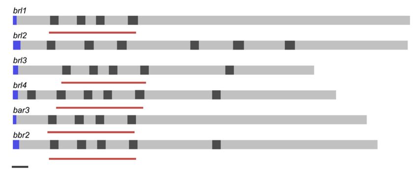
J. Fungi 2021, 7, 399                                                                                             4 of 18

                        2.4. Quantitative PCR
                             Total RNA was isolated from S. commune using the RNeasy Plant Mini Kit (Qiagen,
                        Hilden, Germany) according to the manufacturer’s instructions, and RNA concentration
                        was measured spectrophotometrically (NanoVue Photometer, GE Healthcare, Chicago, IL
                        USA). For reverse transcription, the QuantiTect Reverse Transcription Kit (Qiagen, Hilden,
                        Germany) was used with Maxima SYBR Green 2x Master Mix (Thermo Fisher Scientific,
                        Waltham, USA) in a total volume of 12.5 µL. Each reaction mix contained gene-specific
                        primer pairs (0.5 µL each of 10 pmol/µL stocks; see Table S2), 2 µL cDNA, 3.25 µL A. dest.
                        and 6.25 µL SYBR Green 2x Master Mix. For each primer pair, the amplification efficiency
                        was calculated using a cDNA dilution. Measurements took place in 48-well white PCR
                        plates (Multiplate Low-Profile Unskirted PCR Plates, BioRad, Hercules, USA) sealed with
                        optical foil (Microseal ‘C’ Optical Seals, BioRad, Hercules, CA, USA). The reaction was
                        performed in a MiniOpticon cycler (BioRad, Hercules, CA, USA) with initial denaturation
                        at 95 ◦ C for 10 min and 35 cycles (95 ◦ C for 15 s, 55–60 ◦ C for 10–23 s, 72 ◦ C for 30 s)
                        followed by melting curve analysis. Three biological and three technical replicates and two
                        controls, one without reverse transcriptase and one without template DNA, were used in
                        every run. The genes act1, tef1 and ubi were used as reference genes. The ct values of target
                        genes were normalized with respect to the reference genes and calculated for relative and
                        normalized fold change by the equation 2-∆∆Ct [41,42].

                        2.5. Whole-Genome Microarrays
                              The microarrays were carried out as previously described [11] using 50-mer oligos for
                        all 13,181 predicted genes of S. commune H4–8. Whole RNA was labelled with biotin, and
                        1 µg of total RNA was used for each array. The preprocessing of the data was done using
                        LIMMA packages of the Bioconductor software [43]. For background correction, the inten-
                        sities of blank probes were used (single T nucleotides). The resulting median background
                        intensity was then subtracted from the actual spot intensity values. Any negative value was
                        converted into a low positive value, and signal intensities were log2 transformed. Repli-
                        cates were averaged, and the data were processed by quantile normalization. Statistical and
                        quality tests were done. A p value in Student’s t-test under 0.05 was set to show significant
                        differential expression values. Microarray data were deposited in NCBI’s Gene Expression
                        Omnibus webpage and are accessible through Platform GPL11376 and GEO Series acces-
                        sion number GSE26401 (http://www.ncbi.nlm.nih.gov/geo/query/acc.cgi?accGSE26401;
                        accessed on 28 March 2021).

                        2.6. Phylogenetic Analysis
                              Fungal pheromone receptor protein and DNA sequences were selected using the
                        NCBI database, Unite or the respective genome databases (Joint Genome Institute, Walnut
                        Creek, USA). Alignments were made with the server MAFFT version 7 [39,40], using the
                        F-INS-i or G-INS-i strategies and BLOSUM80 as well as the unalign-level-setting 0.0 or
                        0.8. The software MrBayes 3.2.2. [44] and Markov chain Monte Carlo method (Dayhoff
                        modelling) were used for protein tree calculation with the following settings: prset aamod-
                        elpr = fixed(dayhoff); prset ratepr = variable; mcmcp nruns = 2 ngen = 3,000,000 printfreq
                        = 1000 samplefreq = 100 nchains = 4; mcmc. The mixed trees were calculated using these
                        settings: prset applyto = (2) ratepr = variable; lset applyto = (2) nst = 6 rates = gamma; prset
                        applyto = (1) aamodelpr = fixed(dayhoff); prset applyto = (all) ratepr = variable; unlink
                        statefreq = (all) revmat = (all) shape = (all) pinvar = (all); mcmcp nruns = 2 ngen = 1,000,000
                        printfreq = 1000 samplefreq = 100 nchains = 4; mcmc; sumt burnin = 2000.
                              The Cyberinfrastructure for Phylogenic Research (CIPRES) server was used for com-
                        puting power (http://www.phylo.org/index.php; accessed last on 28 March 2021). The
                        software FigTree v1.4.2 and Inkscape 0.91 were chosen to edit the resulting trees [45], while
                        Tracer software was used to verify the results [46].
J. Fungi 2021, 7, 399 5 of 18
2.7. Immunofluorescence Staining
Immunofluorescence staining of epitope-tagged proteins was performed according
to [11]. In brief, methanol (HPLC grade) was added and incubated for 10 min at −20 ◦ C.
Mycelium was fixed with 3.7% formaldehyde in PME (50 mM PIPES pH 6.7, 25 mM EGTA
pH 8.0, 5 mM MgSO4 ) for 90 min at room temperature. Three times washing with PME
followed, the last time for 10 min. Then, 30 mg lysing enzyme of T. harzianum (Sigma-
Aldrich, Taufkirchen, Germany) was added to egg white in PME, mixed vigorously, and
incubated for 15 min at 37 ◦ C to solve and activate the enzyme. The lysing solution was
added to the mycelium and incubated for 20 to 30 min at room temperature. Three times
washing with PBS (137 mM NaCl, 2.7 mM KCl, 8 mM Na2 HPO4 , 1.8 mM KH2 PO4 ), the
last time for 5 min, and then a permeabilization with PBS + 0.3% Triton X-100 for 10 min
followed. The wash steps were done again with PBS. Unspecific binding was blocked by
1 to 3% BSA in PBS (5 min at 37 ◦ C). The first antibody was 1:200 diluted in PBS + 1 to
3% BSA and incubated on the mycelium overnight at 4 ◦ C. Wash steps were carried out
with PBS (last time 10 min), and the second antibody, which was FITC- or TRITC-coupled,
was added (1:100 diluted in PBS + 1 to 3% BSA) for 60 min at 37 ◦ C in the dark. Again,
wash steps were carried out with PBS, and the coverslips were embedded upside down
into freshly made embedding medium (0.1 M Tris/HCl pH 8.0, 50% glycerol, 1 mg/mL
phenylene diamine, 0.1 mg/mL DAPI). An incubation of at least 24 h at 4 ◦ C followed,
before microscopy ensued.
2.8. Microscopy
Hyphae of S. commune were routinely grown on coverslips which were laid on top
of solid medium for five to ten days at 30 ◦ C. For early dikaryons, two coverslips of the
mating-type partners were sandwiched and stained after 24 h (incubation on medium).
Morphology of hyphae was studied with the Axioplan 2 microscope (Carl Zeiss, Jena,
Germany). Images were taken with the digital camera system Insight Firewire 4 (Diagnostic
Instruments, Sterling Heights, MI, USA) and the software Spot Version 4.6 (Diagnostic
Instruments). For detailed imaging of hyphae, the laser scanning microscope LSM780
(Carl Zeiss, Jena, Germany) was used with GaAsP-detector and software ZEN black. All
immunofluorescence specimens were grown on special coverslips (high performance,
D = 0.17 mm ± 0.005 mm, Carl Zeiss, Jena, Germany).
3. Results
3.1. In Silico Analyses of brl Genes from Different Genotypes
In any strain analyzed so far, six genes belonging to the fungal pheromone receptor
sub-family of seven transmembrane domain receptors are found. These six genes (bar3, bbr2,
brl1, brl2, brl3, brl4) showed the basidiomycete typical intron lengths with 50 bp. Regarding
intron positions, brl1 and bar3 do not show introns in the 30 part of the transcript. The 50
region of brl1, brl3, brl4, bar3 and bbr2 is characterized by three stringed exon length regions
of 110/113 bp, 67 bp and 138 bp (Figure 1). The genes coding for the four Brls in S. commune
H4–8, brl1 (ID 2638177), brl2 (ID 2704867), brl3 (ID 2638261) and brl4 (ID 2691538), are true
orthologs of S. cerevisiae Ste3 [11], with three genes, brl1, brl2 and brl3, located at the B
mating-type locus, whereas brl4 is localized separately.
From the conceptually translated protein sequences, seven transmembrane helices
typical for GPRCs could be confirmed, and the Brls contain a long third cytoplasmic loop
(more than 50% of the protein in case of Brl2) and carboxy terminus, like the Bar and
Bbr pheromone receptors. They contain protein–protein interaction sites and polynu-
cleotide/DNA binding sites, as well as putative domains, responsible for receptor recy-
cling/degradation (for motif assignment, Figure S1).J. Fungi 2021,
J. Fungi 7, x7,FOR
2021, 399 PEER REVIEW 6 of 18 6 of 19
Figure 1. Gene structure of ste3-like genes of S. commune strain H4–8. Exons (grey), introns (dark
grey), signal peptide (blue) and scaling according to JGI annotation. Black bar (bottom left) indi-
cates 100 bp. Red bars indicate similar exon-length regions.
From the conceptually translated protein sequences, seven transmembrane helice
typical for GPRCs could be confirmed, and the Brls contain a long third cytoplasmic loo
(more than 50% of the protein in case of Brl2) and carboxy terminus, like the Bar and Bb
pheromone receptors. They contain protein–protein interaction sites and polynucleo
tide/DNA binding sites, as well as putative domains, responsible for receptor recy
Figure 1. Gene structure
cling/degradation of ste3-like
(for genes of S. commune
Figurestrain H4–8. Exons (grey), introns (dark
Figure 1. Gene structure of motif assignment,
ste3-like genes of S. commune S1).
strain H4–8. Exons (grey), introns (dark
grey), signal peptide (blue) and scaling according to JGI annotation. Black bar (bottom left) indicates
A comparison
grey), signal peptide (blue)of the
and genomic loci fromto
scaling according three available S.Black
JGI annotation. commune genome
bar (bottom sequence
left) indi-
100 bp. Red bars indicate similar exon-length regions.
cates 100 bp. Red bars indicate similar exon-length regions.
showed high synteny (Figure 2). The B locus spans 32 kb, with a distance of 7 kb betwee
the two mating-specific
A comparison sub-loci
of the genomic Bαfrom
loci andthree
Bß. The brl1 genes
available are located
S. commune genomeupstream
sequencesof the B
From
pheromone
showed the conceptually
receptor(Figure
high synteny gene, 2).translated
bar,The
flanked
B locusprotein
byspans
a zincsequences,
32finger seven
transcription
kb, with transmembrane
a distance offactor (MYND,
7 kb between helices
FYVE
typical
the twoA
type). for GPRCs could
mating-specific
protein kinase be confirmed,
sub-loci
and two and
Bα hypothetical the
and Bß. The brl1 Brls contain
genes are
proteins, both a long
located third cytoplasmic
upstream
located upstream of the loop
of brl1, ar
(more
Bα than
pheromone 50% of the
receptor protein
gene, in
bar, case
flankedof Brl2)
by a and
zinc carboxy
finger terminus,
transcription
highly conserved between the two strains S. commune H4–8 and S. commune LoeD. N like
factor the Bar
(MYND, and Bbr
FYVE-type).
pheromone Agenes
pheromonereceptors.proteinare kinase
seenand
They two hypothetical
contain
surrounding brl1. proteins,
protein–protein
The genes bothbrl2located
interaction upstream
andsites
brl3, and
which brl1,
ofpolynucleo-
are locate
are highly
tide/DNA conserved
binding between
sites, as the
welltwo strains
as S.
putative commune
domains,H4–8 and S. commune
responsible forLoeD. No recy-
receptor
downstream of the Bα pheromone receptor bar, are flanked by two hypothetical genes
pheromone genes are
cling/degradation (forseen
motifsurrounding
assignment, brl1. S1). brl2 and
The genes
Figure brl3, which are located
which show a high degree of similarity. All Ste3-like pheromone
downstream of the Bα pheromone receptor bar, are flanked by two hypothetical genes,
receptors and phero
A
moneshowcomparison
genes retainedof the genomic loci from three available S. commune genome sequences
which a high degreetheir position,
of similarity. Allwhile more
Ste3-like distal genes
pheromone receptorswereandmore variable,
pheromone wit
showed
genes high
relatedsynteny
to the (Figure 2).
cytoskeleton The B locus spans
interspersed. 32
The kb,
gene with
genes retained their position, while more distal genes were more variable, with genes brl4a distance
is not of
linked 7 kb between
but again i
the two
flanked
related mating-specific
towith genes similar
the cytoskeleton sub-loci Bα and
in all strains,
interspersed. Bß.
ThewithThe brl1
geneabrl4Con-6genes are
domain
is not located
linked protein upstream
but againclose of the Bα
to brl4 (Figur
is flanked
pheromone
with receptor
3). genes similar gene,
in all bar, with
strains, flanked by adomain
a Con-6 zinc finger transcription
protein close to brl4factor
(Figure(MYND,
3). FYVE-
type). A protein kinase and two hypothetical proteins, both located upstream of brl1, are
highly conserved between the two strains S. commune H4–8 and S. commune LoeD. No
pheromone genes are seen surrounding brl1. The genes brl2 and brl3, which are located
downstream of the Bα pheromone receptor bar, are flanked by two hypothetical genes,
which show a high degree of similarity. All Ste3-like pheromone receptors and phero-
mone genes retained their position, while more distal genes were more variable, with
genes related to the cytoskeleton interspersed. The gene brl4 is not linked but again is
flanked with genes similar in all strains, with a Con-6 domain protein close to brl4 (Figure
3).
Figure2.2.Chromosomal
Figure Chromosomal organization of B of
organization mating-type
B mating-type S. commune
loci ofloci strain H4–8
of S. commune (scaffold
strain 10),
H4–8 (scaffold 10)
TatD (scaffold 230, 617, 960) and LoeD (scaffold 212, 284 and 343). The rectangle under the genes
TatD (scaffold 230, 617, 960) and LoeD (scaffold 212, 284 and 343). The rectangle under the genes
marks the highly syntenic regions (protein identity values are displayed in grey gradient 40–100%).
Figure 2. Chromosomal organization of B mating-type loci of S. commune strain H4–8 (scaffold 10),21, 7, x FOR PEER REVIEW 7 of 19
J. Fungi 2021, 7, 399 7 of 18
marks the highly syntenic regions (protein identity values are displayed in grey gradient 40–
100%).
Figure 3. Chromosomal
Figureorganization of theorganization
3. Chromosomal brl4 surrounding
of theinbrl4
S. commune strain
surrounding in H4–8 (scaffold
S. commune strain H4–8 (scaffold 8),
8), TatD (scaffold 232)
TatDand LoeD (scaffold
(scaffold 60). The
232) and LoeD rectangle
(scaffold 60).under the genes
The rectangle marks
under thethe highly
genes marks the highly syntenic
syntenic regions (protein
regionsidentity
(proteinvalues arevalues
identity displayed in grey gradient
are displayed in grey 40–100%).
gradient 40–100%).
Alignments of pheromone
Alignments receptor genes and
of pheromone conceptually
receptor genes translated proteinstranslated
and conceptually of S. proteins of
commune H4–8 revealed
S. commune thatH4–8
Bar3 revealed
and Bbr2that areBar3
moreand closely
Bbr2 related
are more to closely
each other
relatedthanto each other than
either is related to any Brl
either proteinto(Table
is related any BrlS3). Interestingly,
protein (Table S3). Brl Interestingly,
sequences areBrl more closely are more closely
sequences
related to Bar3 and Bbr2 to
related than
Bar3either
and is related
Bbr2 thanto another
either Brl protein,
is related indicating
to another either indicating
Brl protein, that either that
a divergence event separatingevent
a divergence Brl proteins fromBrl
separating Bar3 and Bbr2
proteins fromoccurred
Bar3 andbefore the two before the two
Bbr2 occurred
mating
mating types evolved or types evolved
that Bar3 or that
and Bbr2 haveBar3 and Bbr2 more
undergone have recent
undergone
gene more recent gene conversion.
conversion.
The alignments alsoTheshowed
alignments alsosimilarity
higher showed higher
for the similarity
N-terminal forprotein
the N-terminal protein parts, suggesting
parts, suggesting
a strong
a strong conservation conservation
of the N-terminaloftransmembrane
the N-terminal transmembrane
domains and extra- domains and extra- or intracellular
or intracellu-
lar loops (Figure S2). A highly similar part of the C-terminal part of the second the
loops (Figure S2). A highly similar part of the C-terminal part of andsecond
the and the third
extracellular loops extending into the adjacent
third extracellular loops extending into the adjacent transmembrane domains was found transmembrane domains was found and
and highlighted in highlighted
the 3D models in theand
3D inmodels and in the
the sequence sequence(Figure
alignment alignmentS3). (Figure S3).
The phylogeneticThe phylogenetic
clustering among clustering among the
the pheromone pheromone
receptors showedreceptors
brl gene prod-brl gene products
showed
ucts well separatedwell separated
from from pheromone-recognizing
pheromone-recognizing mating-typemating-type receptorsin
receptors (clustering (clustering
Bα in Bα and
Bβ receptors). The Brl1 group is most related
and Bβ receptors). The Brl1 group is most related to the known mating-type receptors to the known mating-type receptors Bbr2
of strain 4–39 and H4–8, while Brl2 clusters together
Bbr2 of strain 4–39 and H4–8, while Brl2 clusters together with Bβ receptor sequences of with Bβ receptor sequences of TatD,
4–40 The
TatD, 4–40 and LoeD. and Brl3
LoeD.andThe Brl3
Brl4 and Brl4
proteins areproteins
contained areincontained
a clade with in aBrl1
clade with Brl1 as well, but
as well,
but grouped separately. The Brl proteins of TatD and LoeD are always more closely re-closely related to
grouped separately. The Brl proteins of TatD and LoeD are always more
lated to each othereachthanother than
to the to the respective
respective H4–8 protein
H4–8 protein (compare (compare
Figure 4).Figure
With4).thisWithin- this information,
is now possible to define four general clusters for the four brl
formation, it is now possible to define four general clusters for the four brl genes, set apart set apart from
it genes,
the two pheromone-recognizing
from the two pheromone-recognizing receptors inreceptors
S. commune. in S. commune.
Thus, Thus,different
potentially potentially different but
but overlapping roles for each of the Brl gene products can be envisioned. The analysis of The analysis of
overlapping roles for each of the Brl gene products can be envisioned.
expression and therefore,
expression and overexpression, overexpression,
was used therefore, was used
to specifically to specifically
address address the regulation
the regulation
and role
and role in cells of S. commune. in cells of S. commune.x FOR PEER REVIEW
J. Fungi 2021, 7, 399 88of 19
of 18
Figure
Figure 4.4. Phylogram
Phylogram of of Ste3-like receptor proteins
Ste3-like receptor of S.
proteins of S. commune strains TatD,
commune strains TatD, LoeD
LoeD and others. S.
and others. S. cerevisiae
cerevisiae Ste3
Ste3 protein
protein
served
served asas outgroup
outgroup (accession
(accessionnumber
numberP06783).
P06783).Protein
ProteinandandDNA
DNAalignments
alignmentswere were used
used inin combination
combination forfor calculation
calculation of
of
thethe tree
tree with
with Markov
Markov chain
chain Monte
Monte Carlo
Carlo method
method (Dayhoff
(Dayhoff model).
model). Node Node labels
labels indicate
indicate posterior
posterior probabilities.
probabilities. “Bar”“Bar”
and
and
“Bbr”“Bbr”
referrefer to true
to true mating
mating receptors
receptors (marked
(marked with
with grey);
grey); “Brl”
“Brl” referstotoBBreceptor-like
refers receptor-likeproteins.
proteins. Used
Used sequences
sequences and
accession numbers (NCBI or JGI databases) of S. commune are: Bar1, Bar1, strain
strain 4–40
4–40 (X77949);
(X77949); Bar1,
Bar1, strain
strain 1–69
1–69 (X94996);
(X94996); Bar,
Bar,
strain LoeD
strain LoeD (ID(ID 284719);
284719); Bar, strain TatD
Bar, strain TatD (ID
(ID 373755);
373755); Bar3,
Bar3, strain
strain H4–8
H4–8 (X3027970);
(X3027970); Bar2,
Bar2, strain
strain 12–43
12–43 (X91164);
(X91164); Brl1,
Brl1, strain
strain
LoeD (artificial);
LoeD (artificial); Brl1,
Brl1, strain
strain TatD
TatD(ID(ID208691);
208691);Brl1,
Brl1,strain
strainH4–8
H4–8(ID(ID112464);
112464);Bbr2,
Bbr2,strain
strain4–39
4–39(AF148501);
(AF148501);Bbr2,
Bbr2,strain
strainH4–8
H4–
8 (EFI93340); Brl3, strain LoeD (ID 238914); Brl3, strain H4–8 (ID 258344); Brl3, strain TatD (ID 422121); Brl4, strain LoeD
(EFI93340); Brl3, strain LoeD (ID 238914); Brl3, strain H4–8 (ID 258344); Brl3, strain TatD (ID 422121); Brl4, strain LoeD
(ID 289019); Brl4, strain TatD (ID 373780); Brl4, strain H4–8 (ID 111749); Bbr, strain LoeD (ID 161825); Bar8, strain V142–5
(ID 289019); Brl4, strain TatD (ID 373780); Brl4, strain H4–8 (ID 111749); Bbr, strain LoeD (ID 161825); Bar8, strain V142–5
(AAR99618); Bbr1, strain 4–40 (AAB41858); Bbr, strain TatD (ID 373756); Brl2, strain TatD (ID 215071); Brl2, strain LoeD
(AAR99618); Bbr1,strain
(ID 168872); Brl2, strainH4–8
4–40 (ID
(AAB41858);
112482). Bbr, strain TatD (ID 373756); Brl2, strain TatD (ID 215071); Brl2, strain LoeD (ID
168872); Brl2, strain H4–8 (ID 112482).
3.2. Transcriptome Expression Analysis of Brls
3.2. Transcriptome Expression Analysis of Brls
To evaluate signals for transcriptional control, the brl promotor sequences were ana-
lyzedTo evaluate
using signals
1000 bp long for transcriptional
regions control,
before the start the brl
codons. promotormotifs
Regulatory sequences
in thewere an-
first 500
alyzed using 1000 bp long regions before the start codons. Regulatory
to 600 bp of each promotor included a CTTCTTCCTCCCTTCTGCCTT motif. This is motifs in the firstJ. Fungi 2021, 7, 399 9 of 18
500 to 600 bp of each promotor included a CTTCTTCCTCCCTTCTGCCTT motif. This is
present in all promotor regions, including the pheromone-recognizing bbr2 and bar2. This
motif shows similarities to the yeast Tec1 transcription factor recognition site [47]. A second
motif (consensus GACGCAaa) very similar to yeast’s Fhl1-binding motif [48] was found in
the promoter regions of bar3, bbr2, brl1, brl2 and brl4 (Figure S4). To evaluate potentially
similar regulation, a transcriptome analysis was performed.
Expression profiles of brl genes in wild-type 4–39 and 12–43 monokaryon, W22 × 12–43
dikaryon and several signal transduction mutants (thn mutant, ras1G12V and SccdcG12V )
were determined by whole-genome microarrays (Figure S5). In order to compare the two
monokaryon genetic backgrounds in a dikaryon, a co-isogenic line was used. From this,
strain 4–39 was used for a back-cross to create a compatible mating type for crossings,
strain W22. The high sequence similarity of brl genes in different strains facilitates their
investigation within all stages on the array (Figure S5). The brl3 gene showed differences
in regulation between the two compared monokaryons (S. commune 12–43 and S. commune
4–39), suggesting a strain-specific function. Since S. commune 12–43 is auxotrophic for uracil,
a cultivation-dependent regulation seems possible as well. For the dikaryon, a > 2-fold
decrease in brl1 and brl2 was observed, while brl3 was >2-fold upregulated in the dikaryon.
This might indicate a specific function of Brl3 in the dikaryon.
Involvement of downstream signaling cascades into the analysis of differential expres-
sion will allow for the inclusion of those into function prediction. The constitutively active
Ras signaling in a mutant ras1G12V strain (12–43 vs. ras1G12V ) showed dow-regulation of
brl3 by > 2-fold, which is in accordance with a function in mating-type signaling, as Ras
activation is a result of pheromone binding to the cognate pheromone receptor. The gene
brl2 was > 2-fold downregulated in a cdc42 mutant background (4–39 vs. SccdcG12V ), and
brl4 was > 2-fold downregulated in thn mutant (12–43 vs. thn). Since both Cdc42 and regu-
lation through the regulator of G-protein signaling (RGS) are connected to the cytoskeleton
and phenotypes in growth patterns, these data suggest involvement in dikaryon-specific
growth types.
3.3. Overexpression of brl2, brl3 and brl4 Induces Asymmetrical Growth
The genes brl1, brl2, brl3 and brl4 were overexpressed in S. commune T33 using the
translation elongation factor tef1 promotor. Monokaryotic colonies of the empty vector
control (evc) and brl overexpressing strains (brl1OE, brl2OE, brl3OE and brl4OE) were
analyzed for their phenotypes. S. commune brl1OE showed a straight growing phenotype
with reduced aerial mycelia formation, while the remaining overexpressing strains grew
irregularly, with bundles of hyphae or hyphal knots being formed locally (Figure 5). In
addition, mycelial sectors growing faster than others were formed, resembling more the
colony shape of a dikaryon (compare Figure S6). This phenotype was stronger on minimal
than on complex medium or medium supplemented with wood chips.
Since overexpressing transformants in S. commune can show slightly different pheno-
types depending on integration site, several transformants were screened for each brl gene.
Only S. commune brl3OE yielded different phenotypes, with 30% of the colonies growing in
a feathery manner with less aerial mycelia, while 70% grew more irregularly with more
biomass. Quantitative PCR using RNA from representative transformants showed that
in the feathery transformant (brl3–1OE), brl3 was expressed at a lower level compared to
transformants forming more aerial mycelia (brl3–2OE, Figure S7); brl3–1OE was selected
for further experiments.
In all different overexpression transformants, a reduced growth rate without loss of
colony biomass was observed (Figure S8). Thus, biomass formation and radial growth were
assessed on sucrose and xylose in addition to the normally used glucose. Less biomass
was observed for the brl overexpressing strains (Brl1OE, 0.8-fold only on xylose; Brl2OE,
0.7-fold on either sucrose or xylose; Brl3OE as well as Brl4OE, 0.6- and 0.5-fold reduction,
respectively). The reduced biomass formation was accompanied by a reduced radial
growth rate of Brl1OE, Brl3OE and Brl4OE on sucrose and xylose, which was not observedbrl4 was > 2-fold downregulated in thn mutant (12–43 vs. thn). Since both Cdc42 and reg-
ulation through the regulator of G-protein signaling (RGS) are connected to the cytoskel-
eton and phenotypes in growth patterns, these data suggest involvement in dikaryon-
specific growth types.
J. Fungi 2021, 7, 399 10 of 18
3.3. Overexpression of brl2, brl3 and brl4 Induces Asymmetrical Growth
The genes brl1, brl2, brl3 and brl4 were overexpressed in S. commune T33 using the
translation elongation factor tef1 promotor. Monokaryotic colonies of the empty vector
for BrlOE2.
control (evc)Microscopical analysis revealed
and brl overexpressing strainsthat overexpression
(brl1OE, of brl1 and
brl2OE, brl3OE andbrl4 resulted
brl4OE) in
were
vacuole-rich,
analyzed widephenotypes.
for their hyphae (suppl. Figure S9).
S. commune brl1OE showed a straight growing phenotype
with Dikaryotic-like growth of
reduced aerial mycelia Brl2OE, Brl3OE
formation, and
while the Brl4OE strains
remaining could enhance
overexpressing mating
strains grew
or fruiting body formation; thus, they were mated with compatible partners.
irregularly, with bundles of hyphae or hyphal knots being formed locally (Figure 5). In The mating
behavior was
addition, not different
mycelial sectors from the control
growing and there
faster than was
others neither
were a lack
formed, of time normore
resembling a higher
the
speed in
colony mating
shape of a (Figure
dikaryonS10). Clamps
(compare and fruiting
Figure S6). Thisbodies developed
phenotype indistinguishably
was stronger on minimal
from on
than wild type. medium or medium supplemented with wood chips.
complex
Figure 5. Morphology of of empty
empty vector
vector control
controlstrain
strain(A) andbrl
(A)and brloverexpressing
overexpressingstrains
strains(B–F).
(B–F).Strain
StrainBrl1
Brl1OE
OE(B)
(B)showed
showed a
a straight
straight growth
growth butbut produced
produced lessless dense
dense aerial
aerial hyphae,
hyphae, while
while strain
strain brl2OE
brl2OE (C) grows
(C) grows moremore irregularly,
irregularly, forming
forming dense dense
aerial
aerial hyphae.
hyphae. The mycelia
The mycelia of brl3of brl3 overexpressing
overexpressing strain strain Brl3–1OE
Brl3–1OE (D) grows
(D) grows in a feathery
in a feathery manner manner with sectors
with sectors growing growing
faster
than the rest of the mycelium, while Brl3–2OE (E) grows in a feathery manner but with dense aerial hyphae. Strain brl4OE
(F) grows more irregularly when compared to the empty vector control strain forming dense aerial hyphae. Self-recognition
of brl overexpressing strains (G–J). (G) Distance towards same clone mycelia was measured after 14 days from 5 biological
replicates. (H) represents the empty vector control strain, while (I) shows the non-self-avoiding phenotype of the Brl1OE
strain and the self-avoiding phenotype of Brl3OE (J) as an example. The Student’s t-test was used to determine the P value
between control and transformants; (*) p < 0.005.
3.4. Brl2, Brl3 and Brl4 are Involved in Self-Recognition
When the brl overexpressing strains were cultivated with independent inocula grow-
ing towards each other on one plate, a self-avoidance phenotype became visible (Figure 5).
This is in contrast to the wild type that will show intermingling mycelia without a gap or
with only a minor gap between same-clone mycelia of up to 0.42 cm ± 0.21 cm. In the case
of the brl3 and brl4 overexpressing mutants, the space between the mycelia was larger, with
0.77 ± 0.26 cm (p > 0.001) for Brl3OE and 0.75 ± 0.46 cm (p > 0.001) for Brl4OE. (Figure 5).
In contrast, overexpression of brl1 did not enhance self-recognition. Confrontation of
Brl1OE, Brl2OE, Brl3OE and Brl4OE with each other did not result in any growth reduction
(Figure S11).
3.5. Brl1 and brl4 Expression is Induced During Mating
The shared sequence identity between Bar3 and Bbr2 with pheromone receptor-like
proteins suggested that Brls could have a function in pheromone sensing and signaling.
Consistently, overexpression of brl1 induces a flat growth with a reduced aerial mycelia
formation also known for mutants in RGS signaling [49]. Thus, we investigated the pattern
of brl1 expression over a time period of 48 h during mating between 12–43 and 4–39
(Figure 6). Quantitative real-time expression analysis showed that brl1 is downregulated
6 h after mating followed by a gradual increase up to 3-fold after 24 h (Figure 6). In contrast,
overexpression of brl2, brl3 and brl4 led to an irregular colony growth with knot-like
structures, which is typical for dikaryons. Thus, their expression was determined in the
early dikaryon (24 h after mating) and in the established dikaryon (8 days after mating)
corresponding to the growth conditions used for the microarray-based transcriptomeJ. Fungi 2021, 7, 399 11 of 18
J. Fungi 2021, 7, x FOR PEER REVIEW 11 of 19
analysis. Expression analysis showed that brl2 and brl3 were ≥2-fold repressed in the
dikaryon,
establishedsuggesting a role of brl2
dikaryon, suggesting andof
a role brl3 inand
brl2 vegetative growth. Ingrowth.
brl3 in vegetative comparison, brl4 was
In comparison,
≥2-fold
brl4 wasupregulated in the early
≥2-fold upregulated dikaryon,
in the indicating
early dikaryon, a temporal
indicating regulation
a temporal of brl4of
regulation inbrl4
re-
sponse to mating
in response (see (see
to mating Figure 6). All
Figure 6). four brl genes
All four areare
brl genes expressed in strain
expressed V153–21
in strain V153–21[50], a
[50],
UV-radiated
a UV-radiatedmutant,
mutant,which
whichisisunable
unabletotomate
matedue
duetotoaadeletion
deletionofofthe
theBBlocus
locus(Figure
(Figure S12)
S12)
suggesting a mating-independent function or an involvement beyond mating.
Figure 6. Expression of brl1, brl2, brl3 and brl4 in S. commune during mating (qRT-PCR). The expression of brl1 was monitored
Figure 6. Expression of brl1, brl2, brl3 and brl4 in S. commune during mating (qRT-PCR). The expression of brl1 was moni-
over a time period of 48 h, while brl2, brl3 and brl4 were analyzed 24 h and 8 days after mating, corresponding to an early
tored over a time period of 48 h, while brl2, brl3 and brl4 were analyzed 24 h and 8 days after mating, corresponding to an
dikaryon
early and established
dikaryon dikaryon,
and established respectively.
dikaryon, The data
respectively. Thewere
datanormalized to the to
were normalized expression in the monokaryon
the expression 4–39 (set
in the monokaryon 4–
to 1) and relatively quantified to three reference genes. (*) Significant differences ≥ 2-fold regulation.
39 (set to 1) and relatively quantified to three reference genes. (*) Significant differences ≥ 2-fold regulation.
A Role
3.6. A Role in
in Clamp
Clamp Fusion
Fusion
To visualize protein localization, Brl1 and Brl2 were were tagged
tagged with
with C-terminal
C-terminal codon-
codon-
optimized epitope tags, creating Brl1::myc and Brl2::his strains. For For localization
localization studies,
studies,
brl genes
the brl genes were
were under
under the
the control
control of
of their
their native
native promoter,
promoter, ensuring natural expression
levels, which resulted
resulted in
in transformants
transformants morphologically
morphologically indistinguishable from wild-type
strains. Brl1::myc
strains. Brl1::myc was
wasnot
notdetectable
detectableininmonokaryons,
monokaryons, and
andonly
onlya weak
a weakfluorescence could
fluorescence be
could
observed in Brl1::myc dikaryons (Figure 7A,B) using a wild-type strain as a mate.
be observed in Brl1::myc dikaryons (Figure 7A,B) using a wild-type strain as a mate. How- However,
in matings
ever, with awith
in matings prolonged clamp clamp
a prolonged cell fusion using the
cell fusion S. commune
using Bar2 receptor
the S. commune mutant
Bar2 receptor
G11 as aG11
mutant mate,asBrl1 localization
a mate, to pseudoclamps
Brl1 localization could be obtained
to pseudoclamps could be(see Figure 7C–F).
obtained (see Figure
7C–F).The protein Brl2::his was detectable in monokaryons in vesicles and at the hyphal tips
(Figure 7G–J, Figure S13). Brl2 was recruited to the membrane of hyphal tips when many
hyphae were in close proximity to each other. To confirm the results, the transformant was
crossed with S. commune G11, allowing a double staining of Brl2::his and Bar2::egfp. For
both receptors, a membrane localization in clamps and pseudoclamps could be shown
(Figure 7M–P and Figure S13). Moreover, an accumulation of both receptors in vesicles
could be seen. Taken together, the specific subcellular localization indicates an involvement
of Brl1 and Brl2 in clamp cell fusion. For Brl2, an additional role in hyphal communication
is indicated.J. Fungi 2021, 7, x FOR PEER REVIEW 12 of 19
J. Fungi 2021, 7, 399 12 of 18
Figure 7. Localization of Brl1::myc in dikaryotic hyphae and Brl2::his in monokaryotic and dikaryotic hyphae. Localization
of Myc-tagged Brl1 in dikaryon with wild-type 12–43 (A,B) and in Brl1::myc dikaryon with G11 (C–F) as shown by
Figure 7. Localization of Brl1::myc in dikaryotic hyphae and Brl2::his in monokaryotic and dikaryotic hyphae. Localization
immunofluorescence
of Myc-tagged Brl1 instaining
dikaryon with
withanwild-type
anti-myc 12–43
antibody 24and
(A,B) h after mating. Staining
in Brl1::myc dikaryonwith
withDAPI (E) and
G11 (C–F) asmerging
shown byofimmu-
DAPI
and FITC signalstaining
nofluorescence (F). Localization of Brl2::his
with an anti-myc in unmated
antibody 24 h hyphae (G–J).Staining
after mating. Localization
with of His-tagged
DAPI (E) and Brl2 in dikaryon
merging of DAPIwith
and
wild-type
FITC signal12–43
(F). (K–L) and in Brl1::his
Localization dikaryon
of Brl2::his with G11
in unmated (M–P)
hyphae as shown
(G–J). by immunofluorescence
Localization of His-tagged Brl2staining with an
in dikaryon anti-his
with wild-
type 12–43
antibody 24(K–L)
h afterand in Brl1::his
mating. Stainingdikaryon with(O)
with DAPI G11and
(M–P) as shown
merging by immunofluorescence
of DAPI stainingare
and FITC signal (P). Clamps with an anti-his
marked with
antibody Bar
asterisks. 24 hrepresents
after mating. Staining with DAPI (O) and merging of DAPI and FITC signal (P). Clamps are marked with
10 µm.
asterisks. Bar represents 10 µm.
4. Discussion
The protein
4.1. What Brl2::hisComparing
Is the Difference: was detectable in monokaryons Pheromone
the Ligand-Recognizing in vesiclesReceptors
and at theandhyphal
Brls
tips (Figure 7G–J, Figure S13). Brl2 was recruited to the membrane of hyphal tips when
The occurrence of four Brl genes in the first Schizophyllum genome had prompted
many hyphae were in close proximity to each other. To confirm the results, the trans-
the question of whether these are merely remnants of gene duplication events in this
formant was crossed with S. commune G11, allowing a double staining of Brl2::his and
fungus known for its excessive number of mating specificities in nature [51,52]. Here, we
Bar2::egfp. For both receptors, a membrane localization in clamps and pseudoclamps
compared three genomic sequences of S. commune and identified syntenic regions carrying
could be shown (Figure 7M–P and Figure S13). Moreover, an accumulation of both recep-
pheromone-recognizing Bα and Bß and three brl receptor genes, as well as one copy for brl4
tors in vesicles could be seen. Taken together, the specific subcellular localization indicates
placed outside the mating-type locus. The organization of the B mating-type locus, with
an involvement of Brl1 and Brl2 in clamp cell fusion. For Brl2, an additional role in hyphal
fungal pheromone receptor genes, pheromone genes and neighboring pheromone receptor-
communication is indicated.
like genes is conserved in many basidiomycetes [53]. Pheromone receptor-like genes have
been found adjacent to the B mating-type loci in the genomes of other basidiomycetes as
4. Discussion
well, including L. bicolor, F. velutipes, P. placenta and P. chrysosporium [6,7,20]. An organiza-
4.1. What
tion Is the
similar Difference:
to S. communeComparing
was found theinLigand-Recognizing Pheromone
S. lacrymans [54] and Receptors
C. cinerea and Brls
[55], where two
pheromone receptor-like
The occurrence genes
of four are located
Brl genes in thenext
firstto the B locus. genome
Schizophyllum Since all had brl genes are
fourprompted the
expressed
question ofand share similarity
whether and synteny,
these are merely remnantstheyofmust
genehave a yet-unknown
duplication events infunction.
this fungus
The brl genes show a high sequence identity in closely related S. commune strains
and a higher sequence divergence is shown in two more distantly related strains (TatDJ. Fungi 2021, 7, 399 13 of 18
and LoeD). Still, they cluster in accordance with their placing within the extended mating-
type locus in distinct groups, which is specifically well visible when including other
fungal mating-type receptors and putative pheromone receptor-like proteins (for detailed
phylogram, see [56]). Brl1 was closely related to C. cinerea Rcb2B6 (pheromone-recognizing
receptor; [5]) and F. velutipes Ste3.2 (pheromone-recognizing receptor; [7]), while Brl3
clusters with the four pheromone receptor-like proteins of F. velutipes [7]. Brl2 was found in
close association with Ste3.3 of V. volvacea and other pheromone-recognizing receptors from
L. edodes, P. placenta and T. vaccinum [56]. The location of brl4 outside of the mating-type
locus may be indicative of a non-mating-specific function associated with the extensively
high sequence conservation between the three S. commune strains. This specific assignment
of related groups of Brls might indicate different functions.
4.2. Is There an Involvement of One or More Brls in Mating?
The close relation of Brls with pheromone-recognizing receptors of basidiomycetes
suggests a function in mating. In response to mating and in an established dikaryon,
qRT-PCR confirmed that the genes are not pseudogenes or silent products of receptor gene
duplications. Overexpression of brl genes did not influence mating with compatible strains,
and Brls are expressed in a mating-deficient strain carrying a large deletion covering the
pheromone-recognizing receptors Bar and Bbr [10,50]. This evidence implies that the role
of Brl proteins is not directly linked to mate recognition.
A conceivable function for Brls would be in heterodimerization with Bar and/or
Bbr pheromone receptors. Mammalian GPCRs were found to undergo such oligomeriza-
tion [57], thereby increasing the number of ligand binding sites. The new ligand binding
pockets in such dimers or oligomers would fit nicely to the differential recognition of
over 20 pheromones per mating-type receptor necessary for the discrimination of nine
allelic specificities found for Bα and another nine for Bß recognition of non-self versus
self pheromones [58]. The options for different protein–protein interactions of receptors
can be outlined from interaction domains and dimerization signaling sequences present
in all receptor sequences. For this function, direct pheromone recognition of the Brls on
their own might not be possible, explaining earlier findings. The receptor proteins Brl1 and
Brl2 were localized in the hyphae of S. commune transformants carrying the fusion proteins
Brl1::myc-tag and Brl2::his-tag. Brl1 was found to localize in unfused clamps in a ∆gap1
background, known for aberrant clamp formation [59], which might therefore elongate
the time frame when Brl1 is expressed. At the same time, this was the place where the
pheromone-recognizing Bar2 receptor was localized [11]. The successful detection of Brl1
in dikaryotic hyphae is correlated with the proposed function in mating and a potential
contribution to self versus non-self recognition.
4.3. Functions Downstream of Direct Pheromone Interaction
The receptor protein Brl2::his-tag was also detected in clamps, pseudoclamps, in
hyphae (most likely in vesicles used for receptor recycling and transport), at tips of the
hyphae, at tips just after branching and near septa. The difference in spatial distribution
between Brl1 and Brl2 points towards different functions of the two Brl proteins. The
high abundance of Brl2::his-tag at tips, after branching and also where many hyphae were
clumped together possibly indicates its importance during growth, determination of the
growth direction and orientation of hyphae towards each other. Double staining of the
fusion proteins Bar2::HA and Brl2::his-tag revealed the localization of both in unfused
clamps of the dikaryon. Hence, Brl1 and potentially also Brl2 are connected to mating and
sexual development with clamp fusion.
Consistently, promotor analysis put the brl genes in the context of mating and fil-
amentous growth, identifying a prominent motif similar to the yeast Tec1 transcription
factor binding site. Tec1 is involved in the transcription of filamentation genes and forms
a complex with Ste12, which is a transcription factor regulating mating-dependent gene
expression [47]. The pheromone-recognizing receptor gene bar2 [11], brl1 and brl4 showedJ. Fungi 2021, 7, 399 14 of 18
a similar pattern, with upregulation shortly after mating in the early dikaryon. Thus,
different roles for the single B-like receptors may be expected, and those functions likely are
related to processes induced by mating interactions. Thus, we propose for Brl2 a function
after mating and in clamp fusion.
A different regulation of brl2 and brl3 was seen in RasGap1 and Cdc42 mutant back-
ground. This rather places them into signaling pathways influencing cytoskeleton forma-
tion via Cdc42, cAMP and MAPK cascades, mainly influencing fruiting body formation
and pheromone response [60]. The transcription factor Fst4 seems to influence the expres-
sion of brl2, which was significantly induced in the ∆fst4∆fst4 dikaryon. In contrast, the
pheromone-recognizing bar3 and bbr2 genes were stably expressed [61]. A motif similar
to the Fhl1-binding motif of yeast was found in bbr2 and with a lower similarity in bar2,
brl1, brl2 and brl4. The transcription factor Fhl1 is known to bind Rap1, and the complex
then regulates the expression of several genes and is involved in cell wall maintenance [48],
while the localization of Brl2 near the hyphal tip indicated a role in hyphal growth.
4.4. Self-Recognition through Brls
Both brl2 and brl3 did not change their expression in the early dikaryon but instead
were repressed in the established dikaryon.
This is in line with the differential expression pattern of brl genes in the lifecycle.
The genes coding for dynamitin and Con-6 are located as direct neighbors to brl4, and an
induction of con-6 was confirmed in mating interactions, mostly in W22 × 12–43 but also in
the signal transduction mutants of S. commune [11,60]. The upregulation of con-6 indicates
an involvement in dikaryon and mushroom formation, similar to N. crassa Con proteins
functioning in asexual spore development [62].
We propose a function of Brl3 and Brl4 in hyphal self-sensing and -avoidance based
on the phenotypes observed in overexpression analyses. The receptors, however, so far are
orphans. Potential ligands could be self-pheromones that after recognition do not lead to
hyphal attraction but rather induce hyphae to grow away from the pheromone source. This
would create the observed self-avoidance phenotype and be in line with the asymmetrical,
dikaryon-like growth of brl3 overexpressing mutants and the downregulation of brl3 in the
established dikaryon. Thus, self-pheromones would act as auto-inhibitors, which had been
postulated earlier to explain feathery hyphal growth in dikaryons [63].
The function of an inhibitor secreted by the mycelium would slowly diffuse, producing
an inhibitory front. Hyphae ahead of this front are not inhibited, causing the leading growth
and finally asymmetry of the colony. Aside from self-pheromones acting as auto-inhibitors,
other ligands might be involved. The volatile compounds 1-octen-3-ol and 3-octanone,
which control conidia germination of A. nidulans [64], are known to inhibit the growth
of other fungi [65]. In addition, small secreted proteins common to fungi are suggested
to play a role in development and fruiting [66]. A study identified 10 putative small
secreted proteins in the genome of S. commune, which might function as auto-inhibitory
substances [61]. Our results indicate that Brl3 and Brl4 are involved in sensing this auto-
inhibitor, creating the typical asymmetrical colony.
As to intracellular signaling involved in response to an auto-inhibitor, the transcription
factors hom2 and bri1 have been reported to be involved in mycelial growth, and dikaryons
homozygous for ∆hom2 and ∆bri1 form symmetrical colonies under conditions that would
usually show asymmetrical growth, again postulated to be exerted by regulation through a
postulated auto-inhibitor. Inactivation of these transcription factors abolished early stages
of fruiting body formation [61].
In contrast to the symmetrically growing monokaryons, dikaryons of S. commune form
asymmetrical colonies, which are able to produce fruiting bodies only in response to light.
In the dark, dikaryons grow symmetrically and mushroom formation is abolished. It could
be shown that the blue light receptor genes wc-1 and wc-2 are involved this light-stimulated
development [2]. Hence, yet another layer of regulation is imposed on self-avoidance.You can also read