The Cell Cycle Checkpoint System MAST(L)- ENSA/ARPP19-PP2A is Targeted by cAMP/PKA and cGMP/PKG in Anucleate Human Platelets - MDPI
←
→
Page content transcription
If your browser does not render page correctly, please read the page content below
cells
Article
The Cell Cycle Checkpoint System MAST(L)-
ENSA/ARPP19-PP2A is Targeted by cAMP/PKA and
cGMP/PKG in Anucleate Human Platelets
Elena J. Kumm 1 , Oliver Pagel 2 , Stepan Gambaryan 1,3 , Ulrich Walter 1 , René P. Zahedi 2,4 ,
Albert Smolenski 5 and Kerstin Jurk 1, *
 1    Center for Thrombosis and Hemostasis (CTH), University Medical Center of the Johannes
      Gutenberg-University Mainz, 55131 Mainz, Germany; elena.kumm@unimedizin-mainz.de (E.J.K.);
      s.gambaryan@klin-biochem.uni-wuerzburg.de (S.G.); ulrich.walter@uni-mainz.de (U.W.)
 2    Leibniz-Institut für Analytische Wissenschaften—ISAS—e.V., 44227 Dortmund, Germany;
      oliver.pagel@isas.de (O.P.); Rene.Zahedi@ladydavis.ca (R.P.Z.)
 3    Sechenov Institute of Evolutionary Physiology and Biochemistry, Russian Academy of Sciences,
      St. Petersburg 194223, Russia
 4    Proteomics Centre, Lady Davis Institute, Jewish General Hospital, Montréal, QC H3T1E2, Canada
 5    UCD Conway Institute, UCD School of Medicine and Medical Science, University College Dublin,
      D04 V1W8 Dublin, Ireland; albert.smolenski@ucd.ie
 *    Correspondence: kerstin.jurk@unimedizin-mainz.de; Tel.: +49-6131-17-8278

 Received: 23 January 2020; Accepted: 14 February 2020; Published: 18 February 2020

 Abstract: The cell cycle is controlled by microtubule-associated serine/threonine kinase-like (MASTL),
 which phosphorylates the cAMP-regulated phosphoproteins 19 (ARPP19) at S62 and 19e/α-endosulfine
 (ENSA) at S67and converts them into protein phosphatase 2A (PP2A) inhibitors. Based on initial
 proteomic data, we hypothesized that the MASTL-ENSA/ARPP19-PP2A pathway, unknown until
 now in platelets, is regulated and functional in these anucleate cells. We detected ENSA, ARPP19
 and various PP2A subunits (including seven different PP2A B-subunits) in proteomic studies of
 human platelets. ENSA-S109/ARPP19–S104 were efficiently phosphorylated in platelets treated
 with cAMP- (iloprost) and cGMP-elevating (NO donors/riociguat) agents. ENSA-S67/ARPP19-S62
 phosphorylations increased following PP2A inhibition by okadaic acid (OA) in intact and lysed
 platelets indicating the presence of MASTL or a related protein kinase in human platelets. These data
 were validated with recombinant ENSA/ARPP19 and phospho-mutants using recombinant MASTL,
 protein kinase A and G. Both ARPP19 phosphorylation sites S62/S104 were dephosphorylated by
 platelet PP2A, but only S62-phosphorylated ARPP19 acted as PP2A inhibitor. Low-dose OA treatment
 of platelets caused PP2A inhibition, diminished thrombin-stimulated platelet aggregation and
 increased phosphorylation of distinct sites of VASP, Akt, p38 and ERK1/2 MAP kinases. In summary,
 our data establish the entire MASTL(like)–ENSA/ARPP19–PP2A pathway in human platelets and
 important interactions with the PKA, MAPK and PI3K/Akt systems.

 Keywords: platelets; serine/threonine protein phosphatases; cyclic AMP; cyclic GMP; ENSA; ARPP19;
 MAP kinase

1. Introduction
     Platelets are small, anucleate blood cells, which are essential in physiological and pathological
haemostasis but also have important roles in inflammation, atherosclerosis and cancer [1–5]. In the
process of haemostasis, platelets are regulated by multiple endogenous activating and inhibitory
factors that normally prevent spontaneous platelet adhesion to the vessel wall and subsequent platelet

Cells 2020, 9, 472; doi:10.3390/cells9020472                                      www.mdpi.com/journal/cells
Cells 2020, 9, 472                                                                                  2 of 28

aggregation, thrombus formation and occlusion of blood vessels. Upon vascular injury, platelets adhere
to the injured endothelium and to subendothelial matrix proteins such as collagen to form localized
thrombi and prevent blood loss. Either inherited or acquired dysfunctions of human platelets may
cause serious, even lethal bleeding or thrombotic complications [3,6]. In addition to their essential
regulation of haemostasis and coagulation, activated platelets release and secrete, primarily from
their α- and δ-granules, more than 300 biomolecules and proteins, which affect other platelets, blood,
vascular and tissue cells [7,8]. These secreted platelet factors regulate multiple physiological and
pathophysiological processes such as microvascular integrity, wound healing, inflammation, tumour
stability and metastasis [7,8]. Among the important factors released are thromboxane A2 (TXA2) and
adenosine 5’-diphosphate (ADP), which enhance the initial platelet response and recruit additional
platelets to the growing thrombus. Targeting and blocking of enhanced platelet activation by TXA2
synthesis inhibitors (aspirin) and/or ADP-receptor antagonists (thienopyridines) have been well
established as the most effective intervention to prevent and attenuate complications of various acute
and chronic cardiovascular diseases [9].
     In vivo, platelets are activated by subendothelial collagen predominantly via their cell membrane
GPVI/FcRγ-chain receptor complex. Other platelet agonists such as von Willebrand factor (vWF) and
podoplanin bind to and stimulate specific platelet membrane-receptor complexes such as GPIb/V/IX
and the C-type lectin receptor CLEC-2, respectively. Soluble agonists such as thrombin, ADP and
TXA2 bind to and activate G-protein coupled receptors (GPCRs). The resulting platelet activation is a
multistep process, characterized by cytoskeletal rearrangements, integrin activation, granule secretion,
TXA2 synthesis/release, and exposure of anionic phospholipids, leading to platelet shape change,
adhesion, aggregation and platelet-dependent coagulation [5,10,11]. Platelet activation responses are
mediated by tyrosine protein kinases [11], Ca2+ /calmodulin-dependent protein kinases protein kinase C
(PKC) [12–14] and phospholipases. On the other hand, endothelial cell-derived prostacyclin and nitric
oxide (NO) represent the two major endogenous platelet inhibitors, which increase the level of platelet
cAMP and cGMP, respectively [15–17]. Elevated cAMP and cGMP levels regulate specific effector
systems in platelets such as certain phosphodiesterases (PDEs) and cAMP- and cGMP-dependent
protein kinases A (PKA) and G (PKG), which phosphorylate multiple substrates involved in platelet
inhibition [18,19]. Overall, responses of both platelet agonists and inhibitors are mediated by an
extensive membrane receptor-activated intracellular signalling network that includes intracellular
serine/threonine protein kinases/phosphatases and tyrosine protein kinases/phosphatases [10,11,20,21].
     Many serine/threonine protein kinases, tyrosine protein kinases and tyrosine protein phosphatases
have been extensively studied also in platelets [11,14,22], but much less is known about platelet
serine/threonine protein phosphatases and in particular PP2A. This is perhaps due to the substantial
heterogeneity and complexity of PP2A. In addition, for a long time serine/threonine protein phosphatases
were considered unregulated enzymes, which ‘simply’ limit protein kinase action. However, PP2A
is now known to be a heterotrimeric enzyme with more than 90 different forms in humans, playing
important roles in cell growth and signalling [23–26]. PP2A is expressed in most cells from yeast to man
and exists in the cell predominantly as a heterotrimeric holoenzyme composed of a catalytic subunit (C),
a scaffold subunit (A) and a targeting/regulatory subunit (B) [25,27]. In humans, A- and C-subunits of
PP2A each have two possible variants (α, β) whereas the B-subunits are encoded by 15 different genes,
which yields at least 23 isoforms due to alternative promoters and alternative splicing. Based on this,
human tissues/cells are expected to contain up to 92 different trimeric PP2A holoenzymes and additional
four dimeric (AC) forms [25]. Overall, the B-type subunits are true ‘regulatory’ subunits, which determine
substrate specificity of the associated PP2A C-subunit and modulate PP2A catalytic activity. They are
often expressed in a specific way and determine the intracellular location of the PP2A holoenzymes.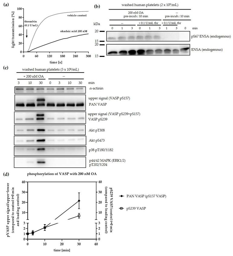
Cells 2020, 9, 472                                                                                 3 of 28

     The cAMP-regulated phosphoproteins (ARPP) with ENSA (also known as ARPP19e or α-
endosulfine), ARPP19 and its isoform ARPP16 were discovered and studied extensively as PKA
substrates in cells other than platelets, with limited functional information [28]. However, considerable
progress was made in 2010 when two independent groups reported for Xenopus oocytes that both ENSA
and ARPP19 inhibit PP2A (only B55δ-subunit) and thereby control mitosis, when phosphorylated by a
special kinase called the Greatwall kinase (Gwl) [29,30]. Whereas PKA phosphorylated ENSA/ARPP19
at their C-terminal site S109/S104 with unknown effect, ENSA/ARPP19 phosphorylation at S67/S62 by
Gwl was required for the potent inhibition of PP2A [31]. ENSA and ARPP19 are highly conserved
[especially the central region with the Gwl phosphorylation site] in a broad spectrum of systems such as
plants, Xenopus, Drosophila, and a wide range of eukaryotes including yeast and humans [32]. Since the
original demonstration in 2010, multiple studies established that the Gwl/MASTL-ENSA/ARPP19–PP2A
pathway is an important check-point for controlling mitosis and its M- and S-phases from yeast to
man [33]. However, ENSA/ARPP19 and their targets were not investigated in anucleate platelets until
now. In our own proteomic/phosphoproteomic studies with human platelets we discovered both ENSA,
ARPP19 [34,35] and in this study other PP2A components. Although possible platelet functions of PP2A
were investigated in early studies using global PP2A inhibitors [36–39], the PP2A composition in human
platelets and the regulation and role of ENSA/ARPP19 have not been addressed so far. In the light of
the possible, but unknown major regulatory role of the PP2A system as opponent of serine/threonine
protein kinases in human platelets, we developed the hypothesis that a MASTL-ENSA/ARPP19-PP2A
pathway is present and regulated in human platelets, with important interactions with the cAMP/PKA
system. Therefore, we investigated the spectrum of platelet protein kinases that phosphorylate/regulate
ARPP19/ENSA, and searched for a Gwl/MASTL-like protein kinase activity, which phosphorylates
ARPP19/ENSA and affects serine/threonine protein phosphatases of the PP2A family. Finally, we asked
whether the ENSA/ARPP19/PP2A pathway regulates the phosphorylation state of important signaling
proteins in platelets.

2. Materials and Methods

2.1. Materials
      Recombinant GST-ARPP19 and active MASTL kinase were from SignalChem (Richmond, BC V6V
2J2, Canada); DNA for ENSA production, C-subunit of PKA (bovine) and PKGIβ (human) were
kindly provided by Prof. E. Butt, University of Würzburg, Würzburg, Germany; 8-Bromo-cGMP
sodium salt and fostriecin sodium salt were from Cayman chemical, USA; okadaic acid ammonium
salt (OA) was from Enzo Life Sciences, Lörrach, Germany; tautomycetin was from Tocris/Bio-Techne
GmbH, Wiesbaden, Germany bovine serum-albumin fraction V (BSA) was from Capricorn Scientific
GmbH, Ebsdorfergrund, Germany; isopropyl β-D-1-thiogalactopyranoside (IPTG) and ZebaTM spin
desalting columns (0.5 mL, 7K MWCO) were from Thermo Fisher Scientific, Waltham, MA USA;
phostagTM AAL-107 was from Wako Chemicals, Neuss, Germany; ClarityTM Western ECL substrate,
HRP-conjugated anti-rabbit antibody were from BioRad Laboratories, Hercules, CA, USA; primary
antibodies (ENSA pS67/ARPP19 pS62; ENSA general; AKT T308/S473; VASP pS239; p38 MAP kinase
pT180/pY182 (12F8); p44/42 MAPK (ERK1/2) pT202/Y204 and α-actinin) were from Cell Signaling
Technology® , Danvers, MA USA; anti-ARPP19 antibody (rabbit polyclonal serum), anti-PP2A B56δ
and anti-PP2A B56δ pS573 antibodies (both rabbit) were kindly provided by Prof. A. Nairn, Yale
University, New Haven, CT USA; anti-ENSA pS109 antibody (rabbit) was kindly provided by Prof.
Satoru Mochida (Kumamoto University, Kurokami, Chuo Ward, Japan); PAN VASP antibody and
antibody against ENSA pS109/ARPP19 pS104 were custom prepared by ImmunoGlobe® , Himmelstadt,
Germany; HisTrap columns (1 mL) and PD-10 desalting columns were from GE Healthcare, Chicago,
IL USA; E. coli BL21 (DE3) were from New England Biolabs (NEB), Frankfurt am Main, Germany;
pET28a vector was from Novagen/Merck KGaA, Darmstadt, Germany; human embryonic kidney cells
293 (HEK293 cells) were kindly provided by the clinic for obstetrics and women’s health (University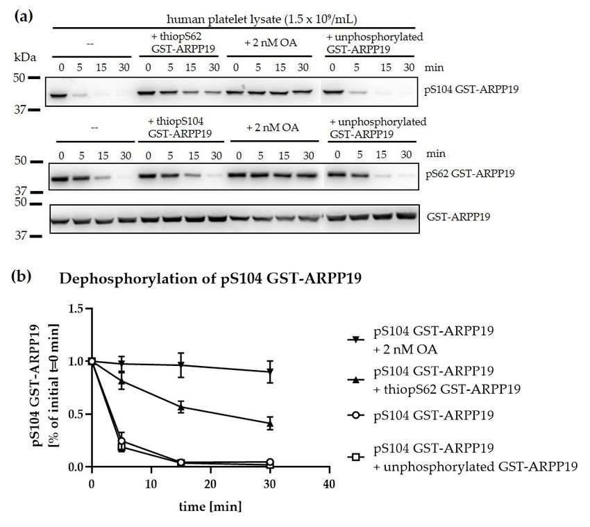
Cells 2020, 8, x 4 of 29
Cells 2020, 9, 472 4 of 28
embryonic kidney cells 293 (HEK293 cells) were kindly provided by the clinic for obstetrics and
women’s health (University Medical Center of the Johannes Gutenberg-University Mainz, Mainz,
Medical Center of the Johannes Gutenberg-University Mainz, Mainz, Germany); HEK293 growth
Germany); HEK293 growth medium (Dulbecco’s Modified Eagle Medium/DMEM) was from Life
medium (Dulbecco’s Modified Eagle Medium/DMEM) was from Life Technologies Inc./Thermo Fisher
Technologies Inc./Thermo Fisher Scientific; pCMV-3Tag-1A vector for FLAG-ENSA was from
Scientific; pCMV-3Tag-1A vector for FLAG-ENSA was from Agilent Technologies, Santa Clara, CA
Agilent Technologies, Santa Clara, CA USA; PolyJetTM Transfection reagent was from SignaGen®
USA; PolyJetTM Transfection reagent was from SignaGen® Laboratories, Rockville, MD USA; Ser/Thr
Laboratories, Rockville, MD USA; Ser/Thr phosphatase activity assay for quantification of PP2A
phosphatase activity assay for quantification of PP2A activity was from Promega Corporation, Madison,
activity was from TM
Promega Corporation, Madison, WI USA; cOmplete 0
TM protease inhibitor mini,
WI USA; cOmplete protease inhibitor mini, thioATP (adenosine 5 -[3-γ-thio]triphosphate) lithium
thioATP (adenosine 5′-[3-γ-thio]triphosphate) lithium salt, α-thrombin/factor IIa (from human
salt, α-thrombin/factor IIa (from human plasma) were from Roche Diagnostics International AG,
plasma) were from Roche Diagnostics International AG, Rotkreuz, Switzerland; ATP (adenosine 5′-
Rotkreuz, Switzerland; ATP (adenosine 50 -triphosphate) and forskolin were from Sigma-Aldrich
triphosphate) and forskolin were from Sigma-Aldrich GmbH/Merck KGaA.
GmbH/Merck KGaA.
2.2. Canonical
2.2. Canonical Sequence
Sequence of
of ENSA
ENSA and and ARPP19
ARPP19
In the
In the following
following(Figure
(Figure1),1),the
thecanonical
canonical sequences
sequences of the
of the ENSA
ENSA andand ARPP19
ARPP19 proteins
proteins usedused for
for this
this study
study (without
(without tags)
tags) are are shown
shown in comparison.
in comparison. Stars
Stars show show of
identity identity of amino
amino acids. acids. alignment
Sequence Sequence
alignment was performed with Clustal
was performed with Clustal Omega (Version 1.2.1).Omega (Version 1.2.1).
Figure 1. Amino
Figure 1. Amino Acid
Acid sequence
sequence alignment
alignment of
of ENSA
ENSA isoform
isoform 11 and
and ARPP19.
ARPP19. Clustal
Clustal Omega
Omega (Version
(Version
1.2.1) was used for sequence alignment. S62/67 is marked in red, S104/109 in yellow. Stars demonstrate
1.2.1) was used for sequence alignment. S62/67 is marked in red, S104/109 in yellow. Stars demonstrate
identical
identical amino
amino acids. Empty space
acids. Empty space shows
shows that
that there
there is
is no
no amino
amino acid
acid sequence
sequence similarity
similarity between
between
ARPP19 and ENSA.
ARPP19 and ENSA.
2.3. Recombinant Wildtype and Mutant ENSA Protein Expression and Purification
2.3. Recombinant Wildtype and Mutant ENSA Protein Expression and Purification
E. coli BL21 were transfected with pET28 vectors including the DNA for wildtype or mutant
E. coli BL21 were
(S67A/S109A/S109D) transfected
HisENSA. with
Protein pET28was
expression vectors including
induced the DNA
with isopropyl for wildtype or mutant
β-d-1-thiogalactopyranoside
(S67A/S109A/S109D)
(IPTG) HisENSA.
(0.1 mM) and proteins Protein
were isolated 20 h expression was
after induction. induced
Therefore, with
E. coli were isopropyl β-d-1-
pelleted at 4225× g
thiogalactopyranoside
◦ (IPTG) (0.1 mM) and proteins were isolated 20 h after induction.
for 10 min at 4 C. The pellets were resuspended in ice-cold lysis buffer (50 mM NaH2 PO4 , 300 mM
Therefore, E.
coli were pelleted at 4225× g for 10 min at 4 °C. The pellets were resuspended in ice-cold lysis buffer
NaCl, 1 mM MgCl2 , 15 mM imidazole, pH 8.0, cOmpleteTM protease inhibitor cocktail) and four times
(50 mM NaH2PO4, 300 mM NaCl, 1 mM MgCl2, 15 mM imidazole, pH 8.0, cOmpleteTM protease
sonicated with 30 s of resting on ice in between. The lysate was centrifuged for 45 min at 16,900× g and
inhibitor cocktail) and four times sonicated with 30 s of resting on ice in between. The lysate was
4 ◦ C. The recombinant wildtype and mutant HisENSA proteins were purified using metal ion affinity
centrifuged for 45 min at 16,900× g and 4 °C. The recombinant wildtype and mutant HisENSA
chromatography (1 mL HisTrap HP (Ni2+ -ions) columns and an ÄKTA prime, GE Healthcare). The proteins
proteins were purified using metal ion affinity chromatography (1 mL HisTrap HP (Ni2+-ions)
were eluted with elution buffer (20 mM Na3 PO4 , 500 mM NaCl, 500 mM imidazole, pH 7.4), afterwards,
columns and an ÄKTA prime, GE Healthcare). The proteins were eluted with elution buffer (20 mM
the buffer was exchanged to 1× TBS buffer (1.37 M NaCl, 0.2 M tris, pH 7.4) with PD-10 desalting columns
Na3PO4, 500 mM NaCl, 500 mM imidazole, pH 7.4), afterwards, the buffer was exchanged to 1× TBS
(GE Healthcare).
buffer (1.37 M NaCl, 0.2 M tris, pH 7.4) with PD-10 desalting columns (GE Healthcare).
2.4. Culture, Treatment, and Sample Generation of HEK293 Cells
2.4. Culture, Treatment, and Sample Generation of HEK293 Cells
HEK293 cells were grown in DMEM at 37 ◦ C and 5% (v/v) CO2 on 6-well plates until they reached
HEK293 cells
80% confluence. were
They grown
were in DMEM
transfected at 37
with °C and 5% (v/v)vector
pCMV-3Tag-1A CO2 on 6-well plates
including until theysequence
FLAG-ENSA reached
80% confluence.
using PolyJet They
TM were transfected
transfection reagent (1with
µg pCMV-3Tag-1A
of vector/well). vector including
One day FLAG-ENSAcells
after transfection, sequence
were
using PolyJet TM transfection reagent (1 µg of vector/well). One day after transfection, cells were
incubated in the presence or absence of 10 µM forskolin or vehicle control (0.1% (v/v) DMSO) or iloprost
incubated
(1 µM) for 15 in min
the or
presence or absence
in the presence of 10 µM
or absence forskolin(0.1
of thrombin or vehicle
U/mL) forcontrol
10 min (0.1%
at 37(v/v) DMSO)
◦ C and or
5% (v/v)
iloprost
CO (1 µM) for 15 min or in the presence or absence of thrombin (0.1 U/mL) for 10 min at 37 °C
2 . As control, non-transfected cells were used. Cells were lysed with 2× lysis buffer (0.1 M tris,
and 5% (v/v)
0.3 M NaCl, 10 COmM2. As control,
MgCl non-transfected cells were used. Cells were lysed with 2× lysis buffer
2 , 2% (v/v) triton, pH 7.5) and western blot samples were generated in 3×
(0.1 M tris,
Laemmli 0.3 heated
buffer, M NaCl, for10
10 mM
min atMgCl , 2% (v/v) triton, pH 7.5) and western blot samples were
95 ◦2C.
generated in 3× Laemmli buffer, heated for 10 min at 95 °C.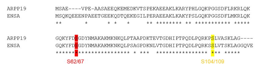
Cells 2020, 9, 472                                                                                5 of 28

2.5. Preparation of Washed Human Platelets
     Washed human platelets were isolated from citrate-anticoagulated (3.2% (v/v) tri-sodium-citrate)
whole blood, taken from healthy volunteers not on platelet-affecting drugs for at least 10 days before
blood collection. All subjects gave their informed consent for inclusion before they participated in the
study. The study was conducted in accordance with the Declaration of Helsinki, and the protocol was
approved by the local Ethics Committee of the University Medical Center Mainz (Study No. 837.302.12;
25.07.12; FF109/2015). Platelet-rich plasma (PRP) was generated by centrifugation of citrated blood,
supplemented with 2 mM EGTA, at 200× g for 10 min at room temperature (RT). PRP was diluted 1:1
with CGS buffer (120 mM NaCl, 12.9 mM trisodium citrate dihydrate, 30 mM d-glucose, pH 6.5) and
centrifuged at 69× g for 10 min at RT, to pellet the leukocytes. The supernatant was centrifuged at
400× g for 10 min at RT. The platelet pellet was resuspended in 3 mL CGS buffer and centrifuged again
at 400× g for 10 min at RT. Finally, platelets were resuspended in HEPES buffer (150 mM NaCl, 5 mM
KCl, 1 mM MgCl2 , 10 mM d-glucose, 10 mM HEPES, pH 7.4). The isolated platelets were adjusted to
5 × 10⁸ or 1 × 10⁹ or 2 × 10⁹ platelets/mL and kept at 37 ◦ C for 15 min.

2.6. Generation of Platelet Lysates from Washed Human Platelets
    Washed human platelets were incubated with 1 mM Ca2+ in the presence or absence of 2 µM
okadaic acid (OA) for 15 min at 37 ◦ C. Platelets were centrifuged at 16,900× g for 1 min at 4 ◦ C.
The pellet was resuspended in 50% (v/v) of the original volume of lysis buffer (50 mM tris, 150 mM
NaCl, 1 mM EDTA, 0.1 mM EGTA, 0.25% (v/v) NP-40, pH 7.4; cOmpleteTM protease inhibitor cocktail)
and vortexed.

2.7. Western Blot Analysis
     Western blot samples were prepared with 3 x Laemmli buffer (200 mM tris/HCl, 15% (v/v) glycerol,
6% (w/v) SDS, 0.06% (w/v) bromphenol blue; 1:10 β-mercaptoethanol). The samples were heated at
95 ◦ C for 5 min at 350 rpm. Proteins were separated by gel electrophoresis with 8%, 10% or 14%
SDS-PAGE gels and transferred to polyvinylidene difluoride (PVDF) membranes. After transfer,
membranes were blocked with 5% BSA in TBST (20 mM tris, 140 mM NaCl, 0.1% (v/v) Tween®-20,
pH 7.4) for 1 h at RT and then incubated overnight with the respective antibodies in 5% BSA TBST
at 4 ◦ C. Membranes were washed three times with 1× TBST and incubated for 1 to 2 h at RT with
the secondary-HRP-conjugated antibodies in 5% BSA-TBST. After three times of washing with TBST,
membranes were developed by ECL detection.

2.8. Western blot Analysis Using Zn2+ -PhostagTM -Gel Electrophoresis
     Western blot samples were prepared with 3× Laemmli buffer for phostag (200 mM tris, 15% (v/v)
glycerol, 6% (w/v) SDS, 2% (w/v) bromphenol blue, 1:10 β-mercaptoethanol) in the absence of EDTA.
The samples were heated at 95 ◦ C for 5 min. Proteins were separated according to their phosphorylation
ratio using 6% (v/v) acrylamide phostag gels. The gels did not contain SDS, but contained phostagTM
compound (35 µM, depending on the protein of interest) and ZnCl2 (69 µM) in the separating gel.
Phostag running buffer was used for gel electrophoresis (0.1 M tris, 0.1 M MOPS, 0.1% (w/v) SDS, 5 mM
sodium bisulfite, pH 7.8). Prior to protein membrane transfer, gels were washed twice for 10 min with
transfer buffer containing 1 mM EDTA, to remove the Zn2+ -ions. A third washing step was performed
with 1× transfer buffer without EDTA before the proteins were transferred to polyvinylidene difluoride
(PVDF) membranes using a transfer buffer for phostag (25 mM tris, 192 mM glycine, 10% (v/v) methanol,
5% (w/v) SDS, pH 8.4).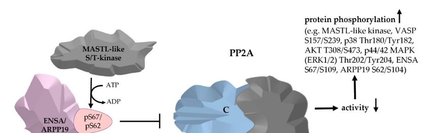
Cells 2020, 9, 472                                                                                   6 of 28

2.9. Phosphorylation of GST-ARPP19 or HisENSA with Recombinant Kinases for Western Blot Analysis
     Recombinant GST-ARPP19 (76 nM) or HisENSA (WT or S109 mutants, 76 nM) was incubated
with active recombinant MASTL kinase (4 µM) or with PKA C-subunit (27 µM) or PKGIβ (16 µM
plus 5 µM of 8-bromo-cGMP sodium salt), 1 mM ATP or thio-ATP in kinase dilution buffer (5 mM
MOPS, 5 mM MgCl2 , 1 mM EGTA, 0.4 mM EDTA, 0.05 mM DTT, 100 ng/µL BSA, pH 7.2). For the
consecutively phosphorylation experiments, the second kinase was added 20 min after the first kinase
and western blot samples were taken before (0 min) and after 5, 10, 20, 25, 30 and 40 min of incubation
at 30 ◦ C. For western blot analysis of single phosphorylation sites, samples were taken before (0 min)
and after 0.5, 2 and 10 min (for HisENSA) or 5, 10 and 20 min (GST-ARPP19) of incubation at 30 ◦ C.

2.10. Phosphorylation of GST-ARPP19 or HisENSA with Recombinant Kinases for Dephosphorylation
Experiments in Platelet Lysates and for PP2A Phosphatase Activity Assay
     Recombinant GST-ARPP19 or HisENSA were phosphorylated as described in 2.9. For western
blot analysis, samples were taken before (0 min) and after 20 min of incubation. The phosphorylation
reaction was stopped with 6 mM EDTA. For the PP2A activity assay, samples were desalted and buffer
was exchanged with ZebaTM spin desalting columns (7K MWCO, 0.5 mL) to 5× PP2A reaction buffer
(250 mM imidazole, 1 mM EGTA, 0.1% (v/v) 2-mercaptoethanol, 0.5 mg/mL BSA, pH 7.2).

2.11. Phosphorylation of Recombinant HisENSA and GST-ARPP19 in Platelet Lysates
    Kinase phosphorylation buffer (10×: 100 mM HEPES, 50 mM MgCl2 , 10 mM DTT, 0.2 mM EDTA,
pH 7.2) and 1 mM ATP was added 1:10 to platelet lysates. As controls, lysates were incubated in the
absence or presence of OA (2 µM or 2 nM). The reaction was started with addition of recombinant
HisENSA (250 nM) or GST-ARPP19 (87 nM). Western blot samples were taken directly after protein
addition (0 min), after 3, 10 and 30 min or after 10, 20 and 40 min of protein incubation at 30 ◦ C, in 3×
Laemmli buffer and heated for 5 min at 95 ◦ C.

2.12. Dephosphorylation of GST-ARPP19 in Platelet Lysates
      Isolated human platelets were pelleted and lysed in lysis buffer (50 mM tris, 150 mM NaCl, 1 mM
EDTA, 0.1 mM EGTA, 0.25% (v/v) NP-40, 30 ng/µL BSA, pH 7.4) with 50% (v/v) volume of HEPES buffer
to a final concentration of 1.5 × 10⁹ platelets/mL. GST-tagged ARPP19 (62.3 nM final concentration)
phosphorylated by PKA at S104 or MASTL at S62 was added. Western blot samples were taken directly
after protein addition (0 min) and after 5, 15 and 30 min of incubation at 37 ◦ C, respectively. OA (2 nM),
thio-phosphorylated GST-ARPP19 (62.3 nM final concentration; thiopS62 GST-ARPP19 for pS104
GST-ARPP19 dephosphorylation; thiopS104 GST-ARPP19 for pS62 GST-ARPP19 dephosphorylation) or
non-phosphorylated GST-ARPP19 (62.3 nM) served as controls. All experiments were performed in the
presence of 5× PP2A reaction buffer (250 mM imidazole, 1 mM EGTA, 0.1% (v/v) 2-mercaptotethanol,
0.5 mg/mL BSA, pH 7.2).

2.13. Ser/Thr-Protein Phosphatase Inhibition by OA in Intact Human Platelets
     Washed human platelets were incubated in the presence of 1 mM Ca2+ with OA (50 nM, 200 nM,
2 µM or 10 µM) or vehicle control (0.1% (v/v) or 0.25% (v/v) EtOH) at 37 ◦ C for up to 40 min. Western
blot samples were taken directly after OA/vehicle addition (0 min) and after 10, 20, 30 and 40 min of
OA/vehicle addition into 3× Laemmli buffer and heated for 5 min at 95 ◦ C.

2.14. PKA/PKG Effects in Intact Human Platelets
     Washed human platelets (2 × 10⁹ platelets/mL) were incubated with 5 nM iloprost (PKA) or 10 µM
riociguat (PKG) or kept resting (–) at 37 ◦ C. Western blot samples were taken directly after (0 s) and 15,
30, 120 and 300 s after iloprost addition or 120, 300 and 600 s after riociguat addition into 3× Laemmli
buffer and heated for 5 min at 95 ◦ C.
Cells 2020, 9, 472                                                                               7 of 28

2.15. Colorimetric Ser/Thr/PP2A Phosphatase Activity Assay
      To assess PP2A activity in platelet lysates, a colorimetric molybdate-based Ser/Thr phosphatase
assay was used according to the manufacturer’s protocol (Promega Serine/Threonine Phosphatase
Assay System V2460). Platelet lysate was generated as described in 2.6. and free phosphate was
removed using the spin columns provided by the assay system. Platelet count was adjusted to 1 ×
108 platelets/mL with lysis buffer (50 mM tris, 150 mM NaCl, 1 mM EDTA, 0.1 mM EGTA, 0.25%
(v/v) NP-40, pH 7.4; cOmpleteTM protease inhibitor cocktail). 35 µL platelet lysate were added per
well to start the reaction and incubated with phosphopeptide in PP2A reaction buffer for 60 min
at 37 ◦ C. 0 min control samples were stopped immediately after lysate addition with a mixture of
molybdate dye/additive (50 µL/well). After 60 min samples were also stopped, the reaction was
incubated additional 15 min at RT in the dark. For inhibitory experiments, OA, fostriecin, tautomycetin
or recombinant proteins were added to the platelet lysate. As recombinant proteins were in 5× PP2A
reaction buffer (250 mM imidazole, 1 mM EGTA, 0.1% (v/v) 2-mercaptoethanol, 0.5 mg/mL BSA, pH 7.2),
the amount of additional 5× PP2A reaction buffer was adjusted, according to the amount of added
recombinant protein. P2A activity was measured by absorbance of 630 nm using a 96-well plate reader
(Dynex Opsys MRTM , Tarporley, CW6 9BL UK).

2.16. Light Transmission Aggregometry
     Washed human platelets were adjusted to 2 × 10⁸ /mL with HEPES-buffer including 1 mM CaCl2
and pre-incubated with OA (50 nM; 200 nM or 1 µM) or vehicle control (0.01% or 0.25% (v/v) EtOH) for
10 min at 37 ◦ C. Alternatively, washed human platelets were pre-incubated with tautomycetin (20 µM
or 30 µM) or vehicle control (1.5% (v/v) DMSO) for 10 or 30 min at 37 ◦ C. Aggregation was started by
addition of α-thrombin (0.05 U/mL or 0.1 U/mL final concentration) and monitored for 5 min at 37 ◦ C
under stirring (1000 s−1 ) using a photometric aggregometer (Apact 4S Plus; DiaSys, Flacht, Germany).
Platelet aggregation was expressed as % of aggregation, which represents % of light transmission.

2.17. (Phospho-)Proteomic Sample Measurement
     Washed human platelets were incubated with buffer (control), sodium S-nitrosocysteine (SNC,
5 µM for 2 min), sodium nitroprusside (SNP, 5 µM for 2 min), DEA-NO (5 µM for 2 min) or riociguat
(10 µM for 5 min) at 37 ◦ C. After incubation, samples were stopped by the addition of 4× SDS/lysis
buffer (50 mM tris, 150 mM NaCl, 4% (w/v) SDS, pH 7.5), shock frozen in liquid nitrogen and analysed
by quantitative phosphoproteomics as described [40]. The fold stimulation of phosphorylation of
various phosphosites compared to control is shown as average ratio.

2.18. Statistical Analysis
     Experiments were performed at least three times with at least three different healthy donors when
platelet samples were involved. Data are presented as means ± standard deviation (SD). Statistical
analysis was performed using GraphPad Prism 8 for Windows (GraphPad Software, San Diego, CA,
USA). One-way ANOVA multiple comparisons test was used for comparison of more than two groups.
p < 0.05 was considered as significant.

3. Results and Discussion

3.1. The PP2A Inhibitors ENSA and ARPP19 are Present in Human Platelets and Phosphorylated by Both PKA
and PKG
     In our earlier proteomic studies, we detected significant expression of both ENSA and ARPP19 in
the small anucleate human platelets [34]. Subsequently, in our analysis of the iloprost/cAMP-stimulated
phosphoproteome of human platelets, more than 130 cAMP/PKA regulated phosphoproteins were
detected including ENSA (phosphorylated at S109) and ARPP19 (at S104) [35]. Platelets are strongly
inhibited by both cAMP- and cGMP-elevating agents mediated by the corresponding cAMP- and
Cells 2020, 9, 472 8 of 28
cGMP-dependent protein kinases (PKA/PKG), which have overlapping specificity [18,19]. Therefore,
we investigated the effect of the cGMP/PKG–pathway on ENSA/ARPP19 phosphorylation using various
NO-donors [S-nitrosocysteine (SNC), sodium-nitroprusside (SNP), diethylamine NONOate (DEA-NO)]
and the soluble guanylyl cyclase (sGC)-stimulator riociguat in comparison to the iloprost (cAMP)
pathway, using vasodilator-stimulated phosphoprotein (VASP) as established PKA/PKG substrate
and marker. Both ENSA (at S109) and ARPP19 (at S104) were strongly phosphorylated (several-fold
stimulation over basal level, similar to VASP S239) in response to different cGMP-elevating agents
as well as in response to the cAMP-elevating iloprost (Table 1). We also noted that both cAMP- and
cGMP-elevating conditions reduced the basal phosphorylation of a second ENSA phospho-site, S67,
(Table 1).
Table 1. cAMP-elevating (iloprost) and cGMP-elevating (riociguat, various NO donors) platelet inhibitors
regulate VASP and ENSA phosphorylation.
Av. Av. Av. Av.
Av. Ratio Copy
Gene Uniprot P-Site Ratio Ratio Ratio Ratio
Riociguat Number/Platelet
Iloprost DEA-NO SNC SNP
VASP P50552 S239 7.17 12.19 9.22 7.67 6.93 44,600
ENSA O43768 S109 17.97 14.04 16.03 13.88 12.46 7800
ENSA O43768 S67 0.53 0.82 0.62 0.72 0.82 7800
Washed human platelets were incubated without (control) or with the following test substances : iloprost (5 nM,
2 min); diethylamine NONOate (DEA-NO 5 µM, 2 min.); riociguat (10 µM, 5 min); sodium S-nitrosocysteine (SNC,
5 µM, 2 min) or sodium nitroprusside (SNP, 5 µM, 2 min). The reaction was stopped with 4× lysis buffer, samples
were shock frozen in liquid nitrogen and analysed by quantitative phosphoproteomics as described [40]. The change
of various phosphosites in response to the test substance compared to control (increase or decrease) is shown as
average ratio (av. ratio). The copy number indicates the number of molecules of a given protein per platelet obtained
in earlier studies [34].
Previously, we demonstrated that under these conditions riociguat had no detectable effect on cAMP/PKA
in human platelets [41]. Therefore, we performed a second, comprehensive phosphoproteomic analysis of
iloprost- and riociguat–treated human platelets with three biological replicates. These phosphoproteomic data
demonstrated that both ENSA (at S109) and ARPP19 (at S104) are phosphorylated in response to cAMP/PKA
as well as to cGMP/PKG stimulation in intact human platelets, similar to VASP S239 (Table 2).
Table 2. Iloprost- and riociguat- stimulated phosphorylation of selected phosphosites of ENSA, ARPP19,
PP2A-B56δ (PPP2R5D), and VASP in human platelets.
Copy P-site (1st) Iloprost (cA) Riociguat (cG)
Gene Uniprot Protein Name
Number/Platelet (#of Averaged
Av. p-Value Av. p-Value
Peptides)
Ratio Fraction Ratio Fraction
ENSA O43768 ENSA/ARPP19e 7800 S109 (3) 4.81 100% 3.75 100%
ARPP19 P56211 ARPP19 2500 S104 (1) 3.53 100% 2.18 100%
PP2A B-subunit B’-δ
PPP2R5D Q14738 1300 S573 (1) 4.52 100% 2.13 100%
(B56δ)
VASP P50552 VASP 44600 S157* (1) 2.03 100% 1.67 100%
VASP P50552 VASP 44600 S239 (4) 3.92 100% 4.52 100%
Washed human platelets from three healthy donors (three biological replicates) were incubated with buffer (control),
iloprost (5 nM, 2 min at 37 ◦ C) or riociguat (10 µM, 5 min at 37 ◦ C). After incubation, samples were stopped by
the addition of 4× lysis buffer, shock frozen in liquid nitrogen and analysed by quantitative phosphoproteomics
as described [40]. The fold increase of phosphorylation of various phosphosites compared to control is shown as
average ratio of all quantified peptides bearing
P the corresponding site. As a measure of reliability, the ‘p-value
peptides p < 0.05/ site-bearing peptides) and # of averaged peptides is presented.
P
fraction’ (for each site:
The change of various phosphosites in response to the test substance compared to control (increase or decrease) is
shown as average ratio (av. ratio). The p-value fraction of all sites shown here is 100%, which represents an excellent
reliability of these phosphosite measurements. * All trypsin digests except VASP S157 (subtilisin digest).Cells 2020, 9, 472 9 of 28
Phosphorylation of the PKA-preferred site of VASP, S157, was also detected but required a
special protease treatment (subtilisin) for detection by phosphoproteomics. It is of special interest
for this study that phosphorylation of the serine/threonine protein phosphatase 2A (PP2A) at its
regulatory B56δ subunit (S573) was also detected for the iloprost/cAMP pathway and as well but
less phosphorylated for the riociguat/cGMP pathway. Others previously showed that the PKA
phosphorylation of PP2A B56δ at S573 activates this special PP2A heterotrimer, leading to a decrease
in protein phosphorylation [42]. We also confirmed this important B56δ S573 phosphorylation in our
experiments with iloprost-/riociguat-treated human platelets, using the specific phospho-antibody as
described [42], see Figure S1.
Our phosphoproteomic data show that both ENSA and ARPP19 are phosphorylation targets
in response to the platelet inhibitory pathways cAMP and cGMP. It is indeed surprising to find the
important phosphoproteins and cell cycle regulators ENSA and ARPP19 in non-dividing human
platelets. We therefore confirmed our proteomic data via western blot analysis of washed human
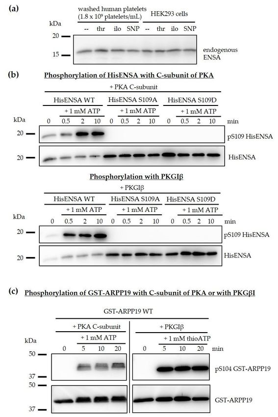
platelets and HEK293 cells using a well-established anti-ENSA antibody. Both human platelets and
human HEK293 cells contained one ENSA species, (apparent 17 kDa in SDS-PAGE), independent of the
experimental conditions (Figure 2a). We also cloned, expressed and purified human ENSA of the known
canonical sequence to study the direct PKA-/PKG-specific phosphorylation of human recombinant
HisENSA (including S109A and S109D phosphosite mutants) and, in comparison, recombinant
GST-ARPP19 using pure PKA (C-subunit) and PKG (Figure 2b,c).
HisENSA and GST-ARPP19 were strongly phosphorylated by both PKA and PKG, which was
abolished by ENSA S109A or S109D mutations. Other data showed that the PKA- and PKG-induced
phosphorylation of ARPP19 S104 resulted in a complete shift of GST-ARPP19 in phostag-gels, indicating
stoichiometric phosphorylation of ARPP19 at S104.
All data obtained with intact human platelets and with recombinant proteins proved that ENSA
and ARPP19 are not only PKA but also excellent PKG targets. In fact, the extent of S109 ENSA
phosphorylation in response to various cGMP-elevating platelet inhibitors (Tables 1 and 2) compared to
VASP 239 places especially ENSA among the best PKG targets studied. In contrast, the iloprost/cAMP
pathway was more effective than the riociguat/cGMP pathway with respect to the phosphorylation of
PP2A-B56δ (PPP2R5D) at S573, an established PKA substrate [42]. It is of special interest that both
cGMP- and cAMP-elevating platelet inhibitors increased the phosphorylation of S109 ENSA while
they decreased S67 ENSA phosphorylation (Table 1). Therefore, the properties of S67 ENSA and S62
ARPP19 phosphorylation in platelets were addressed next.
3.2. Phosphorylation of ENSA S67/ARPP19 S62 by a MASTL-Related Protein Kinase in Human Platelets
As introduced earlier, ENSA and ARPP19, when phosphorylated at S67/S62, strongly inhibit
certain holoenzymes of PP2A. Considering this important functional role of S67 ENSA/S62 ARPP19
phosphorylation in other cell systems such as Xenopus oocytes and our preliminary detection of these
phosphosites in human platelets by phosphoproteomics (Table 1), we investigated the possible S67
ENSA/S62 ARPP19 phosphorylation in human platelets and their lysates. With the Cell Signaling®
anti-pS67 ENSA/anti-pS62 ARPP19 phospho-antibody, which recognizes the conserved pS67 ENSA
site as well as pS62 ARPP19 (sequence 100% similar), positive signals were initially detectable only in
HEK293 cell samples (Figure S2, used as positive control). However, it has been reported for other
systems (Xenopus oocytes, HeLa cells) that Gwl/MASTL-phosphorylated ENSA/ARPP19 are rapidly
dephosphorylated by PP2A, which could be completely prevented by the PP2A inhibitor okadaic
acid (OA) [43,44]. In our experiments pre-incubation of intact platelets with OA at low (200 nM) and
high (10 µM) concentrations induced a similar time–dependent phosphorylation of endogenous ENSA
S67 (Figure 3a). This ENSA pS67 band may include some ARPP19 pS62 since the phospho-antibody
recognizes both ENSA pS67 and ARPP19 pS62, and platelets contain about 3-fold more ENSA than
ARPP19 (Table 2).regulatory B56δ subunit (S573) was also detected for the iloprost/cAMP pathway and as well but less
phosphorylated for the riociguat/cGMP pathway. Others previously showed that the PKA
phosphorylation of PP2A B56δ at S573 activates this special PP2A heterotrimer, leading to a decrease
in protein phosphorylation [42]. We also confirmed this important B56δ S573 phosphorylation in our
experiments with iloprost-/riociguat-treated human platelets, using the specific phospho-antibody as
Cells 2020, 9, 472 10 of 28
described [42], see Figure S1.
Human ENSA
Figure 2. Human ENSA andand ARPP19
ARPP19 areare substrates
substrates of
of both
both PKA
PKA andand PKG.
PKG. (a)
(a) The molecular weight
of endogenous
of endogenous ENSA
ENSA inin human
human platelets
platelets and
and HEK293
HEK293 cells
cells is
is similar
similar and
and is
is not
not affected
affected by
by different
different
treatments (thrombin (thr), iloprost (ilo) or sodium nitroprusside (SNP)). (b) Recombinant HisENSA
wildtype and phosphosite mutants S109A and S109D were phosphorylated in the presence of the
C-subunit of PKA or of PKGIβ (with 5 µM cGMP for PKG) and 1 mM ATP. Samples were taken
in Laemmli buffer before (0 min) and after 0.5, 2 and 10 min of ATP addition and analysed by
immunoblotting. HisENSA S109A and S109D mutants were not phosphorylated confirming S109 is the
phosphorylated amino acid. (c) Recombinant GST-ARPP19 was phosphorylated in the presence of
PKA C-subunit or PKGIβ (and 5 µM cGMP) and 1 mM ATP. Samples were taken in Laemmli buffer
before (0 min) and after 5, 10 and 20 min of ATP addition. The western blots showing HisENSA and
GST-ARPP19 phosphorylation are representative for n = 3 independent experiments.part). This experiment additionally validated the phosphorylated site and the phospho-antibody
studied. A consistent phosphorylation of endogenous ENSA at S67 already in the beginning of the
lysate incubation can also be noted, probably due to the intracellular OA pre-incubation effects before
lysis. OA added to lysates of untreated platelets also induced the phosphorylation of S67 HisENSA,
and asCells
well of 9,S62
2020, 472GST-ARPP19 (Figure 3c). 11 of 28
Figure 3. Effect of okadaic acid (OA) treatment on the phosphorylation of endogenous ENSA (S67)
Figurein3. intact
Effect human
of okadaic acid (OA)
platelets and treatment on the phosphorylation
on the phosphorylation of added of endogenous
HisENSA ENSA (S67) in (S62)
(S67)/GST-ARPP19
intact in
human
platelet lysates. (a) Isolated human platelets (2 × 10 platelets/mL) were incubated(S62)
platelets and on the phosphorylation of added 9HisENSA (S67)/GST-ARPP19 within200 nM
platelet lysates. (a) Isolated human platelets (2 × 10 9 platelets/mL) were incubated with 200 nM or 10
or 10 µM of OA or vehicle (0.1% (v/v) ethanol) for 0, 10, 20 and 40 min. Both OA concentrations
µM of induced
OA or vehicle (0.1% (v/v) ethanol)
a S67 phosphorylation of for 0, 10, 20 and
endogenous 40 min.
ENSA, Both
with OA concentrations
noticeable induced
effects already after a10 min.
S67 phosphorylation
(b) S67 HisENSA of phosphorylation
endogenous ENSA, waswith
onlynoticeable
detectable effects
when OAalready
was after
added 10tomin. (b)platelets
intact S67 or
HisENSA phosphorylation was only detectable when OA was added to intact platelets or
platelet lysates. Intact platelets were pre-incubated without or with 2 µM OA and lysed afterwards. platelet
lysates.HisENSA
Intact platelets
and 1 were pre-incubated
mM ATP were added without
to theor with 2lysate.
platelet µM OASamples
and lysed afterwards.
were taken in HisENSA
Laemmli buffer
and 1 before
mM ATP were added to the platelet lysate. Samples were taken in Laemmli
(0 min) and after 3, 10 and 30 min of 1 mM ATP addition. In contrast to endogenous buffer before (0 and
ENSA
added HisENSA(wt), the added S67A HisENSA mutant was not phosphorylated, serving as important
negative control. (c) HisENSA or GST-ARPP19 as well as 1 mM ATP were added to platelet lysate
without (only with HisENSA) or with 2 µM OA in the lysate. Samples were taken before (0 min)
and after 10, 20, 40 and 60 min of 1mM ATP addition. The western blots are representative for n = 3
independent experiments.Cells 2020, 9, 472 12 of 28
We then performed phosphorylation experiments with lysates from human platelets, pre-incubated
with 2 µM of OA or vehicle control. Only lysates from OA pre-treated platelets (but not controls)
supported a time–dependent S67-phosphorylation of added recombinant HisENSA wildtype (Figure 3b,
upper part) whereas a S67A HisENSA mutant was negative (Figure 3b, lower part). This experiment
additionally validated the phosphorylated site and the phospho-antibody studied. A consistent
phosphorylation of endogenous ENSA at S67 already in the beginning of the lysate incubation can
also be noted, probably due to the intracellular OA pre-incubation effects before lysis. OA added to
lysates of untreated platelets also induced the phosphorylation of S67 HisENSA, and as well of S62
GST-ARPP19 (Figure 3c).
A control without OA showed no S67 HisENSA signal after 40 min of incubation. These results
clearly indicate the presence of a protein kinase in human platelets that is able to phosphorylate ENSA
and ARPP19 at S67 or S62, respectively. The protein kinase seems to be active in intact platelets
as well as in platelet lysates but its activity can only be detected, when an ENSA/ARPP19-specific
serine/threonine protein phosphatase (most likely PP2A) is inhibited.
These experiments with intact human platelets and their lysates establish for the first
time a significant activity of a protein kinase, which phosphorylates endogenous and/or added
HisENSA/GST-ARPP19 at the sites S67/S62 known in other systems to be responsible for PP2A
inhibition. The absent S67 ENSA phosphorylation under basal conditions but clear detection when
PP2A is inhibited (Figure 3) suggests that the protein kinase responsible for S67 ENSA phosphorylation
in the platelets is inhibited by PP2A-mediated dephosphorylation. In addition/or alternatively a very
fast dephosphorylation of pS67 ENSA under basal and other conditions in human platelets is mediated
by PP2A and inhibited by OA.
In mammalian cells, the only two protein kinases with the capacity to phosphorylate S67 ENSA/S62
ARPP19 (at the identical phosphosite motif KGQKYFDSGDYNMAK) described so far are MASTL
(microtubule-associated serine/threonine kinase-like, the human orthologue of the Greatwall kinase,
Gwl) [45,46] and the related MAST3 kinase [47,48]. However, in our proteomic studies of human
platelets [34] and related studies of murine platelets [49] neither MASTL nor MAST3 were detected.
Also, a comprehensive analysis of the human and platelet transcriptome did not detect significant
expression levels of MASTL or MAST3, in contrast to significant expression levels of ENSA and
ARPP19 [50]. At present, there are two possible explanations for our data on MASTL kinase protein
(negative) and MASTL activity (positive) in human platelets. Despite the negative protein expression
data so far, there are perhaps low, not yet detectable MASTL/MAST3 levels in platelets, which mediate
the observed S67 ENSA/S62 ARPP19 phosphorylation. Alternatively, platelets may contain other
MASTL-related protein kinases, which catalyse ENSA/ARPP19 phosphorylation, since human platelets
express, at the protein level, about 150 different protein kinases from all classes [34,51]. Clearly, it will
be important to elucidate the identity of the S67 ENSA/S62 ARPP19 protein kinase and its regulation
within platelets (see also our limitation paragraph, end of the discussion).
It is of considerable interest that the discovery of MASTL as human Gwl orthologue [45] also
defined a MASTL E167D mutation in humans, which is associated with thrombocytopenia [45,46,52].
Very recently, a megakaryocyte specific MASTL mutation (E167D knock-in) and a complete MASTL
KO in mice was reported with a reduced platelet count and decreased half-life of platelets in MASTL
KO and mutant knock-in mice. In addition, increased annexinV-levels, probably associated with
platelet apoptosis or platelet activation, increased bleeding times and defective clot retraction were
observed [53]. Overall, this MASTL mutation (E167D) was considered a gain of MASTL kinase function,
leading to a stronger inhibition of PP2A. However, the study did not address the presence of MASTL
protein or activity in platelets.Cells 2020, 8, x 13 of 29
Cells 2020,As
9, 472 13 of 28
already shown, PKA and PKG strongly phosphorylated HisENSA S109 and GST-ARPP19
S104. The ENSA S109 phosphorylation was abolished with ENSA S109A/D mutants but not with the
S67A ENSA mutant (Figure S3). Importantly, PKA and PKG phosphorylation of GST-ARPP19 caused
3.3. Phosphorylation of HisENSA and GST-ARPP19 by Human MASTL (S67/S62) and by PKA C-Subunit/
a complete time-dependent mobility shift of GST-ARPP19 in phostag-gels (shown for PKA C-subunit
PKGIβ (S109/S104) or Combinations
in Figure 4c; data for PKG similar shown in Figure S4b) demonstrating stoichiometric (complete)
An important goal
phosphorylation of this
of the projectphosphosite
PKA/PKG was to investigate
underthe possible
these inhibitory
conditions, effect of ENSA
as phostag-gels and ARPP19
are separating
on the
serine/threonine protein
proteins by their phosphatases
number in humansites
of phosphorylated platelets.
and notAsby
prerequisite, the phosphorylation
their molecular weight. of the
recombinant HisENSA and GST-ARPP19 proteins was studied in more detail (Figure 4).
Figure 4. Site-specific phosphorylation of HisENSA or GST-ARPP19 by MASTL and PKA in combination.
Figure 4. Site-specific phosphorylation of HisENSA or GST-ARPP19 by MASTL and PKA in
(a) HisENSA was phosphorylated consecutively in the presence of MASTL, C-subunit of PKA and 1
combination. (a) HisENSA was phosphorylated consecutively in the presence of MASTL, C-subunit
mM ATP. One of the kinases was added first, the second kinase after 20 min. Samples were taken after 5,
of PKA and 1 mM ATP. One of the kinases was added first, the second kinase after 20 min. Samples
10, were
20, 25taken
(5 min after
after addition
5, 10, of min
20, 25 (5 second kinase),
after 30of
addition and 40 min
second of incubation
kinase), 30 and 40and
minstopped by Laemmli
of incubation and
buffer. The western blots are representative for n = 3 independent experiments. (b) GST-ARPP
stopped by Laemmli buffer. The western blots are representative for n = 3 independent experiments. was
phosphorylated consecutively in the presence of MASTL and C-subunit of PKA
(b) GST-ARPP was phosphorylated consecutively in the presence of MASTL and C-subunit of PKAand 1 mM ATP. One
of the
andkinases wasOne
1 mM ATP. added first,
of the the second
kinases kinase
was added after
first, the20 min. kinase
second The reaction
after 20was
min.stopped afterwas
The reaction 5, 10,
20,stopped
25 (5 min after
after addition
5, 10, 20, 25 (5ofmin
second
afterkinase),
addition30ofand
second40 min by Laemmli
kinase), 30 and 40buffer.
min by(c) Corresponding
Laemmli buffer.
phostag-gel of the same samples as shown in (b). The 6% acrylamide-phostag-gel (with 35 µM phostag)
separated the proteins by their number of phosphorylated sites. The western blots are representative
for n = 3 independent experiments.Cells 2020, 9, 472 14 of 28
As already shown, PKA and PKG strongly phosphorylated HisENSA S109 and GST-ARPP19 S104.
The ENSA S109 phosphorylation was abolished with ENSA S109A/D mutants but not with the S67A
ENSA mutant (Figure S3). Importantly, PKA and PKG phosphorylation of GST-ARPP19 caused a
complete time-dependent mobility shift of GST-ARPP19 in phostag-gels (shown for PKA C-subunit
in Figure 4c; data for PKG similar shown in Figure S4b) demonstrating stoichiometric (complete)
phosphorylation of the PKA/PKG phosphosite under these conditions, as phostag-gels are separating
the proteins by their number of phosphorylated sites and not by their molecular weight.
This phostag-gel analysis worked very well with GST ARPP19 but did not work with HisENSA,
maybe due to interference of the His-Tag with Zn2+ -ions of the phostag-gel. Recombinant human
MASTL kinase phosphorylated HisENSA and GST-ARPP19 strongly and time-dependently at a site
recognized by the pS67 ENSA/pS62 ARPP19 antibody. With GST-ARPP19, MASTL phosphorylation
caused a time-dependent complete mobility shift of GST-ARPP19 in phostag-gels, demonstrating
stoichiometric phosphorylation of the MASTL-site S62. Next, consecutive phosphorylation of HisENSA
or GST-ARPP19 by kinase combinations (MASTL+PKA, Figure 4b,c; MASTL+PKG, Figure S4a) were
investigated. The two important phosphorylation sites (MASTL: S67/S62; PKA/PKG: S109/S104)
were effectively phosphorylated by the responsible kinases without interference by the other kinase.
Specifically, MASTL had no major effect on PKA/PKG-mediated ENSA/ARPP19 phosphorylation, and
PKA/PKG had no major effect on the MASTL-mediated ENSA/ARPP19 phosphorylation. This was
clearly demonstrated by phostag-analysis of ARPP19 phosphorylation and its two independent shifts
by S62 and S104 phosphorylation (Figure 4b,c, Figure S4b). At present, minor interactions between both
kinases cannot be excluded since detailed kinetics analyses were not performed. Also, we cannot rule
out interferences of the protein tags used in our experiments with ENSA/ARPP19 proteins. However,
we have no evidence for this, and such tagged ENSA/ARPP19 proteins have been successfully used by
many investigations in this field. Finally, the effects may be different in intact cells and may be cell
type-/system-specific, as discussed below.
Mochida reported in 2014 the different phosphorylation sites of ENSA in Xenopus oocytes and
their influence on PP2A inhibition [31]. Whereas human ENSA (and ARPP19) apparently only have
two major phosphorylation sites (S67/S62 for MASTL, S109/S104 for PKA), Xenopus ENSA and ARPP19
have a third site, T28 for ENSA and S28 for ARPP19. It was concluded that the phosphorylation of
all three sites influences PP2A inhibition. However, pS67 ENSA was shown to be the most potent
PP2A inhibitor interacting with both B55- and C-subunits and blocking the catalytic centre of PP2A.
Mochida calculated the IC50 values for pENSA and PP2A B55 inhibition for his experiments using
Xenopus oocytes and obtained an IC50 of 0.47 nM for pS67 ENSA, an IC50 of 0.52 nM for pS67+pS109
ENSA and much higher values for pS109 ENSA alone [31]. PKA phosphorylation of Xenopus ENSA
S109 had little effect in these systems.
Also in Xenopus oocytes, PKA did not affect Gwl-induced ARPP19 phosphorylation nor the ability
of Gwl-phosphorylated ARPP19 to inhibit PP2A B55δ. The authors concluded that the effect of S67
phosphorylation was dominant over the negative-function of S109-phosphorylation [54], in agreement
with earlier studies by others [31].
Andrade et al. showed in 2017 that brain ARPP16 phosphorylation sites (the shorter isoform of
ARPP19) were reciprocally phosphorylated when forskolin (activator of adenylate cyclase and thus
of PKA) was added to striatal slices [47,48]. Forskolin induced a reduced phosphorylation of the
MASTL/MAST site S46 (similar to S62 ARPP19/S67 ENSA) and stimulated S88 phosphorylation via
PKA (similar to PKA sites S104 ARPP19/S109 ENSA). In that paper, several mechanisms were discussed
to explain how S46 ARPP16 phosphorylation could be blocked resulting in reduced PP2A inhibition:
• PKA phosphorylation of ARPP16 could interfere with the extent of PP2A inhibition by pS46 ARPP16
• PKA could phosphorylate and inhibit the ARPP16 S46 kinase MAST3
• Forskolin-stimulated PKA could activate PP2A by phosphorylation of PP2A B56δ [42] resulting in
reduced S46 ARPP16 phosphorylationCells 2020, 9, 472 15 of 28
Some of these effects may perhaps be specific for brain, as was recently reviewed [55]. However,
in our phosphoproteomic analysis, we clearly detected PP2A B56δ S573 phosphorylation in human
platelets (Table 2) in response to iloprost (cAMP system) and riociguat (cGMP system), which we were
able to confirm using phospho-specific antibodies (Figure S4).
In our studies, PKA-phosphorylation of ENSA/ARPP19 did not affect their properties as MASTL
substrates or in other in-vitro functions tested so far, but further work is required here. In intact
platelets (Table 1) and transfected HEK293 cells (Figure S2) ENSA S109 was strongly phosphorylated
(transfected ENSA protein stronger than endogenous) when PKA was activated and, at the same
time, down-regulation of S67 ENSA phosphorylation was observed (stronger signal with transfected
ENSA, weaker with endogenous ENSA) (Figure S2). Although our data with human platelets and the
published results with murine striatum [47,48] are very similar (PKA reduced ENSA S67 or ARPP16
S62 phosphorylation, respectively), it will be necessary to identify the ENSA S67/ARPP19 S62 protein
kinase in platelets for further mechanistic studies (see also the paragraph limitation).
3.4. Serine/Threonine Protein Phosphatases in Human Platelets
The availability of recombinant ENSA and ARPP19 in various phospho-variants allowed the
analysis of their effects on platelet serine/threonine protein phosphatases. Although the general
importance of serine/threonine protein phosphatase including PP2A as signalling opponent of serine/
threonine protein kinases has been recognized [25,26,56], the diversity and distinct regulatory
mechanisms of serine/threonine protein phosphatases were only recently discovered, also supported
by the complete description of the human protein kinome [57] and protein phosphatome [58]. With
this background and our quantitative human platelet proteome data [34] it was possible to compile an
overview of most serine/threonine protein phosphatases present in human platelets which include
three catalytic subunits of PP1 and two catalytic subunits (α,β) of PP2A (Table S1). Table S1 also shows
the intracellular concentrations of the catalytic subunits of serine/threonine protein phosphatases
present in human platelets. It is remarkable that the major serine/threonine protein phosphatases such
as PPM1, PP1, PP2A are present in human platelets at µM concentrations. This certainly is important
for their catalytic capacity since most potential substrates have a similar intracellular concentration.
Whereas serine/threonine phosphatases were considered for a long time as unregulated
components which ‘simply’ remove phosphates from serine/threonine phosphoproteins, it is now clear
that serine/threonine protein phosphatases, and in particular PP1 and PP2A, are multimeric enzymes
which are tightly controlled by regulatory subunits and additional activators and inhibitors [26].
Considering the enormous heterogeneity of PP2A composition and function, it was important
to establish quantitative information on the expression level of PP2A subunits and ENSA/ARPP19
in human platelets, which is based on our proteomic databases (Table S2). We also estimated the
intraplatelet concentration of these proteins, which is important for ENSA/ARPP19-PP2A interactions
(see below). It is of interest that the PP2A A subunit concentration (1.67 µM) is higher than that of
the two C subunits (1.07 µM) and similar to the concentration of all B subunits (1.73 µM) (Table S2).
This indicates that there is no free PP2A C subunit in platelets, which agrees with observations in other
cells and systems [26].
Human platelets contain the two catalytic subunits (α,β) of PP2A, only one of the two scaffolding
A (α), two B55 subunits (α,δ), five B56 subunits (α,β,γ,δ,ε) and the PP2A activator, which is known to
activate PP2A [59]. Interestingly, the distribution of PP2A isoforms in human platelets is similar to
murine platelets, although some quantitative differences exist. For the scaffolding subunit A, only
the α-isoform was found in human platelets, similar to other human cells [60]. The proteomic data
suggest that 14 different PP2A trimeric holoenzymes and two different dimeric forms may exist in
human platelets, perhaps even more due to splice variants of the regulatory subunits. The content of
serine/threonine protein phosphatases in platelets and their subunit composition (especially of PP2A)
is important to evaluate the following phosphatase assays.You can also read