Rationale for the Use of Radiation-Activated Mesenchymal Stromal/Stem Cells in Acute Respiratory Distress Syndrome - MDPI
←
→
Page content transcription
If your browser does not render page correctly, please read the page content below
cells
Review
Rationale for the Use of Radiation-Activated
Mesenchymal Stromal/Stem Cells in Acute
Respiratory Distress Syndrome
Isabel Tovar 1,2 , Rosa Guerrero 1,2 , Jesús J. López-Peñalver 3 , José Expósito 1,2,4
and José Mariano Ruiz de Almodóvar 5, *
 1    Departamento de Oncología Médica y Radioterapia, Servicio Andaluz de Salud (SAS), Avenida de las
      Fuerzas Armadas 2, 18014 Granada, Spain; mariai.tovar.sspa@juntadeandalucia.es (I.T.);
      rosa.guerrero.sspa@juntadeandalucia.es (R.G.); jose.exposito.sspa@juntadeandalucia.es (J.E.)
 2    Instituto de Investigación Biosanitaria, Ibis Granada, Hospital Universitario Virgen de las Nieves,
      Avenida de las Fuerzas Armadas 2, 18014 Granada, Spain
 3    Unidad de Radiología Experimental, Centro de Investigación Biomédica, Universidad de Granada,
      PTS Granada, 18016 Granada, Spain; jjpenalver@ugr.es
 4    Departamento de Radiología y Medicina Física, Facultad de Medicina, Universidad de Granada,
      PTS Granada, 18016 Granada, Spain
 5    Centro de Investigación Biomédica, Universidad de Granad, PTS Granada, 18016 Granada, Spain
 *    Correspondence: jmrdar@ugr.es

 Received: 31 July 2020; Accepted: 31 August 2020; Published: 2 September 2020

 Abstract: We have previously shown that the combination of radiotherapy with human
 umbilical-cord-derived mesenchymal stromal/stem cells (MSCs) cell therapy significantly reduces the
 size of the xenotumors in mice, both in the directly irradiated tumor and in the distant nonirradiated
 tumor or its metastasis. We have also shown that exosomes secreted from MSCs preirradiated with
 2 Gy are quantitatively, functionally and qualitatively different from the exosomes secreted from
 nonirradiated mesenchymal cells, and also that proteins, exosomes and microvesicles secreted by
 MSCs suffer a significant change when the cells are activated or nonactivated, with the amount of
 protein present in the exosomes of the preirradiated cells being 1.5 times greater compared to those
 from nonirradiated cells. This finding correlates with a dramatic increase in the antitumor activity of
 the radiotherapy when is combined with MSCs or with preirradiated mesenchymal stromal/stem
 cells (MSCs*). After the proteomic analysis of the load of the exosomes released from both irradiated
 and nonirradiated cells, we conclude that annexin A1 is the most important and significant difference
 between the exosomes released by the cells in either status. Knowing the role of annexin A1 in the
 control of hypoxia and inflammation that is characteristic of acute respiratory-distress syndrome
 (ARDS), we designed a hypothetical therapeutic strategy, based on the transplantation of mesenchymal
 stromal/stem cells stimulated with radiation, to alleviate the symptoms of patients who, due to
 pneumonia caused by SARS-CoV-2, require to be admitted to an intensive care unit for patients
 with life-threatening conditions. With this hypothesis, we seek to improve the patients’ respiratory
 capacity and increase the expectations of their cure.

 Keywords: experimental radiotherapy; radiobiology; mesenchymal stem cells; cell therapy; exosome;
 annexin A1; acute respiratory-distress syndrome; COVID-19

1. Introduction
     The investigation into mesenchymal stromal/stem cells (MSCs) has been of outstanding interest
in the recent years [1]. Stromal cells are heterogeneous and contain several populations, including

Cells 2020, 9, 2015; doi:10.3390/cells9092015                                       www.mdpi.com/journal/cells
Cells 2020, 9, 2015                                                                                    2 of 14

stem cells with different multipotential properties, committed progenitors and differentiated cells [2].
In all our experiments, we have used MSCs obtained from the human umbilical cord perivascular area
of Wharton’s jelly [3]. We have described these cells and assessed their phenotype [3], self-renewal
potential, contractibility, differentiation [3–5], clonogenicity, radiosensitivity [6], secretion [5] and
antitumoral activity both in basal conditions and after stimulation with X-rays [7,8].
      We have recently shown that the combination of human umbilical-cord-derived MSCs cell therapy
plus radiotherapy significantly reduces the size of established tumors in mice, both in the directly
irradiated tumor and in the distant nonirradiated tumor [7] or in its metastasis [8]. These results
support the hypothesis that human mesenchymal stromal/stem cells are radiosensitizers for local
tumor radiotherapy, and simultaneously, they represent an effective tool for amplifying the systemic
effects of radiotherapy. These out-of-target radiotherapy effects [9–11], promoted by MSCs are, in our
view, of major interest [9,12].
      We have also proved [7,8], that the preirradiation of MSCs triggers an important cellular change
that transforms the MSCs into a source of molecules with very interesting pharmacologic proprieties.
Amongst these actively secreted molecules, we have identified TRAIL and Dkk3 with very well-known
antitumor activities, and annexin A1, whose activities we have previously reviewed [12] and now
update here to include new data that demonstrate its anti-inflammatory and antiviral activity and its
role in the regulation of hypoxia.
      This secretion activity suggests a mechanistic explanation of how activated cells may positively
spread their effect far from the place where they are applied. On this basis, we believe that exosomes,
heavily loaded with annexin A1, will be liberated in the lungs after cell therapy with irradiated-MSCs
cells, and this action would ameliorate symptoms in patients with sepsis in the lungs and in any other
organs affected by septic shock.
      A significant number of scientific reports are available demonstrating that gap junction, paracrine
pathways and exocrine effects can transmit radiation-induced biological effects far from the place where
the radiation is applied. These effects are frequently referred to as radiation-induced out-of-target effects.
Multiple molecular signaling mechanisms [13] involving oxidative stress [14,15], kinases, inflammatory
molecules [16,17], exosomes [8], microvesicles are postulated to contribute to bystander short- and
long-range effects [12]. The anticancer immune response may also be activated by ionizing radiation,
and a combination of different treatment strategies is promising in this field [11,18]. The activation
of the immune system by the irradiated tumor to trigger the beneficial abscopal effect is decisively
improving radiotherapy applications and their outcomes [19–22].

2. Mesenchymal Stromal/Stem Cells and Radiotherapy
       It is generally acknowledged that MSCs can be found ubiquitously in many tissues and are not
limited to those of mesodermal origin, such as bone marrow, adipose, muscle and bone.
       Previous reports suggested a protective role for MSCs when combined with radiotherapy
(RT) [23,24]. In effect, due to their properties, MSCs may be recognized as a therapeutic tool for
treating radiation-induced tissue damage [25–27]. Several reports have shown that MSCs skillfully
home onto neoplastics tissues [28,29] and together with tissue recovery functions MSCs prepare the
microenvironment by controlling inflammatory processes to reduce the inflammation grade [30,31],
where they might have the greatest therapeutic impact in vivo [32]. However, the amount of
mesenchymal stromal/stem cells that are up-taken into injured tissues may not be sufficient to
explain their strong overall protective effect.
       The bioactivation of MSCs may be obtained indifferent ways [33] and the molecules secreted by
the activated MSCs (MSCs*) might have an impact on several immune-cell lineages, establishing an
advantageous sphere far away from its original location. We have proposed that exosomes liberated
from radiation-activated MSCs* perform important intratumoral and systemic actions [8,12].
       We are aware that cellular therapy with MSCs can be problematic in cancer therapy [12]. Therefore,
it is important to emphasize that following irradiation MSCs become senescent and the senescence
Cells 2020, 9, 2015 3 of 14
is associated with production of a senescent-associated secretory phenotype (SASP). The SASP has
antitumor activity since it may induce senescence of neighboring cells by paracrine action [34].
Nevertheless, SASP can modify its composition and become much richer in proinflammatory factors
and it has become evident that tumor-associated MSCs have a positive effect on tumor growth
and the spread of metastasis [35] through the acquisition of a chemo- and radiotherapy resistance
mechanisms [36], and it has been suggested that tumor cells can misuse SASP for their own growth [37].
On the other hand, it has been communicated that, in an inflammatory condition, the exosomes
contained in the cancer cell secretome might have a role in the change of the normal MSCs cell
phenotype toward a malignant one [36], which could be an impediment to MSCs therapeutic use.
Nevertheless, whether this innate tropism of MSCs toward the tumors and metastatic foci is
related with cancer promotion or suppression remains controversial [35,37–40], and further studies on
the interactions between cancerous cells and stromal components of tumor microenvironments have
been proposed, which is imperative to allow the progress of more suitable treatments for cancer.
It is generally accepted that MSCs-based therapies are of major importance in regenerative
medicine and, perhaps, in the future a solution for many other medical problems. However, the success
of MSCs therapy relies on the efficiency of its administration and the biodistribution, engraftment,
differentiation and secreting paracrine factors at the target sites [41]. Until now, there has been no
universal delivery route for mesenchymal stromal/stem cells (MSCs) for different diseases [42]. In fact,
efficient homing and migration toward lesion sites play an important role and the local transplantation
of MSCs in spatial proximity to the lesion, as well as the systemic administration routes are being
carefully explored [43]. There is growing evidence that mesenchymal stromal/stem cells based
immunosuppression was mainly attributed to the effects of MSCs-derived extracellular vesicles [44]
although it seems clear that transplanted MSCs can indeed leave the blood flow and transmigrate
through the endothelial barrier, and reach the lesion site [45]. So, both mechanisms must be accepted
until the underlying processes are better understood.
We know that in an uninjured mouse, exogenous intravenously injected MSCs rapidly accumulate
within the lungs and are cleared from this site to other organs, such as the liver, within days [46].
As far as up-take in the lungs is concerned, the MSCs are able to release a wide variety of soluble
mediators, including anti-inflammatory cytokines [47], antimicrobial and angiogenic macromolecules,
and exosomes and microvesicles that are secreted to extravascular spaces [48]. All of the above leads
us to believe that the amount of MSCs cells that engraft onto injured tissues may not be sufficient to
account for their robust overall protective effects.
On the other hand, the induction of the mechanisms of epithelial and endothelial wound healing
and the angiogenesis promotion has been attributed to macromolecules included in the exosomes
released by the MSCs cells, which act as tools for defending the intestines from the damage produced
by necrotizing enterocolitis experimentally induced in animal models [29]. This has been highly
promising [23,49], and MSCs may be a well-thought-out therapeutic tool to treat radiation-induced
tissue damage [30]. It is essential to highlight that the group of Chapel et al. has started a phase 2
clinical trial (ClinicalTrials.gov Identifier: NCT02814864) for the handling of severe collateral healthy
tissue damage after radiation therapy in patients with prostate cancer, and this clinical trial is sustained
by numerous reports focused on the use of MSCs for improving the damage severity on normal tissues
after radiation treatment [46,50,51]. However, the damage severity and the mechanisms involved
in the control of side effects after radiotherapy [52], as well as the role of MSCs in healthy-tissue
radio-protection, are quite unknown.
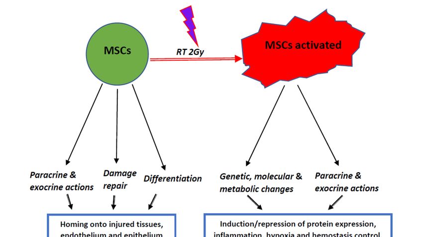
We have included in Figure 1 a graphic summary of the widespread actions done by MSCs
and MSCs*.Cells 2020, 9, 2015 4 of 14
Cells 2020, 9, x FOR PEER REVIEW 4 of 14
possible therapeutic
Figure 1. Graphic and schematic summary of cell actions, tissue response and possible therapeutic
application of mesenchymal stromal/stem
stromal/stem cells
cells (MSCs)
(MSCs) and
and activated
activated MSCs.
MSCs.
3. Radiation-Activated Mesenchymal
3. Radiation-Activated Mesenchymal Stromal/Stem
Stromal/Stem Cells
Cells
When
When we westudied
studied thetheexosome
exosome cargo before
cargo and after
before andthe activation
after of MSCsof
the activation with RT, we
MSCs discovered
with RT, we
significant disparities in the results of the proteomic assessment of both samples.
discovered significant disparities in the results of the proteomic assessment of both samples. We described that there
We
are qualitative, quantitative and functional differences amongst the proteins
described that there are qualitative, quantitative and functional differences amongst the proteins contained in the exosomes
obtained
containedfrom
in thebasal MSCs obtained
exosomes and activated
from MSCs*
basal MSCs [8]. For
andmore information
activated MSCs* [8].in [8]
Forsee Supplementary
more information
Materials, additional file 1.
in [8] see Supplementary Materials, additional file 1.
These
These findings
findingsdemonstrate
demonstratethe profound
the profound metabolic
metabolic change
changethatthat
these activated
these cell exosomes
activated have
cell exosomes
undergone and the consequences after activation with radiation. Amongst
have undergone and the consequences after activation with radiation. Amongst the proteins the proteins representatives
in exosomes released
representatives from MSCs*,
in exosomes we highlight
released from MSCs*,the key wecomponents
highlight the of cell–cell or cell–matrix
key components adhesion
of cell–cell or
and includeadhesion
cell–matrix annexin and and integrins [8]. Between
include annexin them, the
and integrins presence
[8]. Betweenofthem,annexinthe A1 (ANXA1)
presence is very
of annexin
noteworthy
A1 (ANXA1) because
is veryitnoteworthy
is always presentbecausein the
it isexosomes released
always present infrom MSCs* and
the exosomes constantly
released from absent
MSCs* in
MSCs. We verified
and constantly these
absent in results
MSCs. using quantitative
We verified mRNA–PCR
these results to measure the
using quantitative mRNA of this
mRNA–PCR protein
to measure
in MSCs and MSCs* and confirmed that mRNA is spectacularly induced
the mRNA of this protein in MSCs and MSCs* and confirmed that mRNA is spectacularly induced in MSCs after irradiation [8].
After measuring quantitatively the mRNAs of the proteins of TRAIL, Dkk3
in MSCs after irradiation [8]. After measuring quantitatively the mRNAs of the proteins of TRAIL, and ANXA1 in umbilical
cord
Dkk3stromal
and ANXA1stem-cells, before cord
in umbilical and after
stromalcellstem-cells,
stimulation withand
before 2 Gy low-energy
after transferwith
cell stimulation ionizing
2 Gy
radiation, our previously published results [8] show a clear increase in
low-energy transfer ionizing radiation, our previously published results [8] show a clear increase their intracellular levels,
in
compared with the levels found in basal situations (see these results in [8] supplementary
their intracellular levels, compared with the levels found in basal situations (see these results in [8] material,
Figure S2) and notice
supplementary material,that the levels
Figure S2) andof mRNA
notice thatof TRAIL
the levelsand ofDkk3
mRNA at of
48TRAIL
are strongly
and Dkk3increased in
at 48 are
treated cells compared to the basal levels (p < 0.001), whereas the levels
strongly increased in treated cells compared to the basal levels (p < 0.001), whereas the levels of of mRNA of ANXA1 are
strongly
mRNA ofincreased
ANXA1 are at 24 h, and dramatically
strongly increased at 24 at h,
48and
h ofdramatically
cell treatment, with
at 48 the
h of statistical
cell treatment, differences
with theCells 2020, 9, 2015 5 of 14
found 24 and 48 h being very significant (p < 0.0001), which supports the massive presence of ANXA1
in the exosomes released by the radiation-stimulated MSCs.
4. Annexin A1 in the Inflammation and Hypoxia Processes Control
We stated that the existence of ANXA 1 in the exosomes separated from the culture medium of
activated MSCs* and the absence of this protein in the medium withdrawn from the nonirradiated
MSCs is a relevant outcome in our previous studies [8].
In relation with this protein, we would like to emphasize that after more than 30 years of research,
annexins have been clearly recognized as key elements to control immune responses. The prototype
component of this family, ANXA1, has been highly recognized as an anti-inflammatory factor involving
cell mobility and the response of several components of the innate immune system [53]. However,
it has now been recognized that ANXA1 also has important implications in maintaining homeostasis,
fetal development, aging processes and in the evolution of several diseases such as cancer [54,55].
Inflammation is a tightly regulated mechanism, initiated following tissue damage or infection.
If unrestrained or unsolved, the inflammation may lead to further tissue damage and give rise to
persistent inflammatory diseases and autoimmunity with eventual loss of organ function. It is now
evident that the outcome of inflammation is an active process that occurs during an intense inflammatory
incident [56]. After MSCs activation, the released ANXA1 might diminish the gathering of neutrophils
in the tissue injured in several ways. Additionally, ANXA1 promotes neutrophil apoptosis and acts on
macrophages to stimulate the phagocytosis and the removal of dead neutrophils [56,57], and leads to the
rapid reconstruction of tissue homeostasis. Inflammation resolve is controlled by several endogenous
factors involving macromolecules and proteins, such as ANXA1, and their presence is relevant in many
diseases [58]. The study of ANXA1 in relationship with the innate immune system has focused mainly
on the anti-inflammatory and proresolving actions through its binding to the formyl-peptide receptor 2
(FPR2)/ALX receptor. There is much evidence that ANXA1, and its mimetic peptides [58], may have an
important role in alleviating complications associated with ischemia–reperfusion injury [59]. Moreover,
the presence of chronic inflammation in tumors is common and facilitates tumor growth, metastatic
dissemination and treatment resistance [60]. Physical abnormality of tumor vasculature, including its
chaotic structure, enlarged interstitial pressure, increased stiffness and hypoxia, are physical barriers in
tumor treatment [61] are inspiring new anticancer strategies aimed at targeting the tumoral tissue to
normalize these physical irregularities [61,62].
ANXA1 is an endogenous inhibitor of NF-κB that can be induced in cancer cells and experimental
tumors by potent anti-inflammatory glucocorticoids and modified nonsteroidal anti-inflammatory
drugs [49]. In this context, ANXA1 has long been classified as an anti-inflammatory protein due to its
actions on leukocyte-mediated immune responses. However, it is now well known that ANXA1 has
extensive effects further from the immune system, with consequences in maintaining the homeostatic
atmosphere within the whole body due to its capacity to influence cellular signaling, hormonal secretion
and diseases [63]. Upon an injury, epithelial wound shutting is a excellently adjusted process that
re-establishes homeostasis, but in chronic diseases it is related with nonhealing vascular lesions; in this
processes ANXA1 is involved as a preresolving mediator [64].
Moreover, new studies indicating an intracellular function of ANXA1 have now been published.
In effect, using AnxA1 knockout mice, it has been noted that ANSA1 is essential for IL-1β release both
in vivo as in vitro [65]. Furthermore, we know that ANXA1 colocalize and exactly connect with NLRP3,
suggesting the activity of ANXA1 in inflammasome initiation is independent of its anti-inflammatory
role via FPR2 [65]. These mechanisms, which could be of major importance in the resolution of lung
inflammation and in septic shock through cytokine storm control, deserve more research.
5. Annexin A1 in the Treatment of Inflammation
The significance of annexin A1 (ANXA1), a 37 kDa monomeric protein, to stress response is that
its synthesis and release are controlled by glucocorticoids (GCs). After release, it has been shown thatCells 2020, 9, 2015 6 of 14
ANXA1 could strongly downregulate polymorphonuclear leukocyte migration into inflammatory sites
and accelerate their apoptosis, upregulating the monocyte migration into the inflammatory sites [66].
Recently, the role of ANXA1 in the treatment of acute radiation-induced lung damage has been
studied and the causes of its action examined [67]. Neuroinflammation initiated by damage-associated
molecular patterns has been implicated in adverse neurological outcomes following lethal hemorrhagic
shock and polytrauma [68]. Results obtained by Ma Q. et al. [68] show that attractive proresolving
pharmacological approaches, such as annexin-A1 biomimetic peptides, can efficiently attenuate
neuroinflammation and reveal a novel complex role for ANXA1 as a therapeutic and a prophylactic
drug due to its ability to strengthen endogenous proresolving, anti-thrombo-inflammatory mechanisms
in cerebral ischemia–reperfusion injury. Finally, it has been announced that recombinant human ANXA1
may represent a novel candidate for the treatment of diabetes type 2 and/or its complications [69,70].
6. Annexin A1 and Lung Diseases
Endogenous glucocorticoids are proresolving intermediaries, a model of which is the endogenous
glucocorticoid-regulated protein annexin A1. Because silicosis is an occupational lung disease
typified by persistent inflammation and fibrosis, models regarding this illness have been studied
to test the therapeutic properties of the ANXA1 on experimental silicosis [66]. The authors have
demonstrated that the therapeutic administration of N-terminal peptide of ANXA1 (Ac2-26) in
ischemia–reperfusion-provoked lung injury might substantially attenuate the lung edema and
proinflammatory cytokine production, thus reducing oxidative stress, apoptosis, neutrophil infiltration
and lung tissue injury, perhaps via the activation of the N-formyl peptide receptor [66].
A similar result was published in an experimental study made with animals affected by
bleomycin-induced lung fibrosis that were treated with an ANXA1 peptido-mimetic, administrated
prophylactically (from day 0 to 21) or therapeutically (from day 14 onward), which improved signs
of both inflammation and fibrosis [71]. Together these data show a pathophysiological relevance for
ANXA1 in lung inflammation and in fibrosis, and may open up a new approach for the pharmacological
handling of pneumonia and lung fibrosis. Currently, the resolution of inflammation, once considered to
be a passive process, has recently been revealed to be an active and precisely controlled process. In the
resolution stage of acute inflammation, new mediators, including lipoxins and resolvins, which are
members of the specific proresolving mediators of inflammation, are released [72].
Acute lung injury and the more severe forms of acute respiratory distress syndrome, ALI/ARDS,
are relatively common syndromes in seriously ill patients and are related with a high rate of morbidity
and mortality. Recently, new evidence has shown that the resolution of inflammation might be an active
and highly regulated process. Specific proresolving mediators (SPMs), have been proved to produce
strong immune-resolving effects, such as cell proliferation, migration and the clearance of apoptotic
cells and microorganisms. Therefore, the effective and timely control of inflammation could be the key
step to maintain effective host defense and the restoration of homeostasis. Therefore, this reveals a
new mechanism for pulmonary edema fluid reabsorption in which SPMs, amongst them annexin A1,
might offer new chances to design “reabsorption-targeted” treatments with high levels of precision in
controlling acute lung injury [73]. It is also widely acknowledged that to survive, edema fluid should
be removed for patients with ALI/ARDS [74].
Moreover, lung endotoxemia is characterized by neutrophil accumulation, enlarged vascular
permeability and parenchymal damage. In relation with toxic problems, it has been proposed
that the molecular reactions stimulated by ANXA1 peptidomimetic Ac2-26 lead to the control of
leukocyte activation/migration and both cytokine production and lung injury that are generated
by lipopolysaccharides [75]. It was also published that ANXA1 may accelerate the resolution
of inflammation in acute radiation-induced lung damage through the inhibition of IL-6 and
myeloperoxidase inflammatory cytokines, demonstrating that ANXA1 may have a therapeutic role as
treatment target for acute-radiation lung damage [76].Cells 2020, 9, 2015 7 of 14
Moreover, it is well known that pattern recognition receptors (PRRs) are key elements in the
innate immune response. FPR2/ALXR, a receptor modulated for specialized proresolving mediators of
inflammation, amongst them annexin A1, has been shown to be one of the receptors implicated in
inflammation process control. This has encouraged the research community to search for and develop
new anti-inflammatory/proresolution small molecules to control inflammation through the activation
of FPR2/ALXR [44].
We believe that the protective function of the ANXA1-FPR2 signaling axis recently described in
viral infections it is very important [60]. The formyl peptide receptor (FPR) 2 is a pattern recognition
receptor that, in addition to proinflammatory, pathogen-derived compounds, also recognizes the
anti-inflammatory endogenous ligand annexin A1 (ANXA1), and it has been shown that ANXA1,
via FPR2, controls inflammation and bacterial dissemination during pneumococcal pneumonia by
promoting host defenses, suggesting ANXA1-based peptides as a novel therapeutic strategy to control
pneumococcal pneumonia [77].
In this context, it has been described that mice with the influenza A virus (IAV) infection in the
murine model treated with ANXA1 displayed significantly attenuated pathology upon a subsequent
IAV infection with significantly improved survival, impaired viral replication in the respiratory tract
and less severe lung damage.
7. COVID-19: The Magnitude of the Problem
Most countries in the world are suffering a significant spread of SARS-CoV-2, causing pandemic
effects. The clinical presentation of the SARS-CoV-2 infection varies from asymptomatic or with
light symptoms to clinical situations characterized by respiratory insufficiency requiring mechanical
ventilation and intensive care, to multiorgan dysfunction syndrome with signs and symptoms such as
sepsis, septic shock and multisystem failure. It also is true, unfortunately, that all the countries in the
world do not have the capacity to solve this problem due to the lack of therapeutic measures that could
have the appropriate impact. The problem is massive. Therefore, there is a great need to contemplate
new methods to improve patients’ biological resistance to SARS-CoV-2 by using mesenchymal
stromal/stem cells [78]. We know that SARS-CoV-2 invade cells through the ACE2 receptor widely
expressed in human cells, including the alveolar epithelium and the capillary endothelium. The MSCs
are ACE2 negative. So, the transplanted cells are unable to participate in the spread of the infection.
For the healthcare services, the two key imperative necessities in the SARS-CoV-2 infection are to
hinder and reduce infection rates, and to decrease the death rate of those infected. The accumulating
epidemiological analyses, connected with country-based mitigation strategies, and with estimations
that about 80% COVID-19 patients have mild or asymptomatic disease, 14% severe disease, and 6%
are critically ill, support a permanent need for the treatment of SARS-CoV-2 infection and COVID-19
pneumonia in the long term.
According to preliminary estimates of severity that were based on a recent analysis of data from
EU/EEA countries and the UK available in the European Surveillance System TESSy and online country
reports (for countries whose data were incomplete or missing in TESSy) and summarized by the
European Centre for Disease Prevention and Control (ECDC), we know that amongst all the cases of
patients affected, hospitalization has occurred in 32% of cases reported from 26 countries, and cases
with severe illness (requiring ICU and/or respiratory support) have accounted for 2.4% cases reported
from 16 countries. Moreover, amongst hospitalized cases, severe illness was reported in 9.2% of
hospitalized cases in 19 countries and death occurred in the 11% of the hospitalized cases in 21 countries.
The age-specific hospitalization rates amongst all cases showed elevated risk amongst those aged
60 years and over. Finally, a strong estimate for the COVID-19 case death rate is still lacking and
theoretically biased by partial outcome data and differences in testing policies and procedures.
The number of people affected worldwide is progressive and continuously growing,
and SARS-CoV-2 has infected more than 24.5 million people and killed more than 830,000 people inCells 2020, 9, 2015 8 of 14
different countries, areas or territories with cases (ECDC on 28 August 2020). The worldwide lethality
(average) is ≈3.38% with a range of 0.1% to 14.0% depending on the country.
The magnitude of the problem is enormous and terrifying.
8. Clinical Trials of MSCs Transplantation in Patients with COVID-19 Pneumonia
MSC products are quickly arising as promising treatment candidates for the COVID-19 pandemic.
It is well known that septic shock is associated with a considerable viral load in terms of both
mortality and morbidity for survivors of this illness. Preclinical sepsis studies advise that mesenchymal
stromal/stem cells (MSCs) may moderate inflammation, improve pathogen clearance and tissue repair
and reduce death. Because MSCs have not been assessed in humans with septic shock, a clinical
trial that examines safety and tolerability of MSCs is mandatory before proceeding to a randomized
controlled trial to study patient outcomes. This has been performed by L.A. McIntyre et al. [79] and
their results show that the infusion of freshly cultured allogenic bone-marrow-derived MSCs, up to a
dose of 3 million cells/kg, into patients with septic shock seems safe and, consequently, the results of
the phase I dose escalation and safety trial provide researchers with the rationale and argument to now
conduct larger trials to study the efficacy of MSCs in a clinical trial in patients with septic shock [80];
the clinical trial is registered with the www.clinicaltrials.gov (NCT02421484) reference.
Preclinical and early clinical data suggest that human umbilical cord stromal MCSs, because of their
anti-inflammatory and immunomodulatory actions, are able to heal tissues affected and thus improve
recovery rates [81]. Additionally, this treatment also seems to be antimicrobial. Two recent studies
from China [78,82] have examined whether MSCs could be useful for treating SARS-CoV-2/COVID
pneumonia, based on known immune modulatory and reparative abilities of stem cells. Both studies
show an outstanding reversal of symptoms, even in severe to critical circumstances. These clinical
studies not only recognize a novel therapeutic approach, but also the reality of natural processes able
to reduce acute inflammatory pneumonia.
Following the intravenous transplantation of MSCs, a noteworthy population of cells accumulates
in the lung, which together with their immunomodulatory effect, could protect alveolar epithelial cells,
recover the pulmonary microenvironment, avoid pulmonary fibrosis and cure lung dysfunction. It has
been suggested that MSCs have cured or significantly improved the functional outcomes of seven
patients without any detected side effects. The pulmonary function and symptoms of these seven
patients were significantly improved in two days after MSCs transplantation. Furthermore, the gene
expression profile revealed MSCs were ACE2- and TMPRSS2, which showed that the MSCs were free
from the SARS-CoV-2 infection. Thus, the intravenous cellular transplantation was safe and efficient
for handling in patients with COVID pneumonia, particularly for the patients in a seriously severe
condition [78].
Given the uncertainties in this area, Golchin et al. [83] have reviewed published clinical trials and
hypotheses to offer useful information to researchers and those involved in stem-cell therapy. In their
study, they considered a new approach to enhance patients’ immunological responses to COVID-19
pneumonia using MSCs and debating the aspects of this proposed treatment. However, currently,
there are no approved MSC-based approaches for the prevention and/or treatment of COVID-19
patients; nevertheless, clinical trials are ongoing.
The immunomodulatory and anti-inflammatory properties of MSCs in the treatment of respiratory
diseases have been confirmed by 17 completed clinical studies, and also more than 70 trials have been
registered in this regard (https://clinicaltrials.gov).
Many of the critically ill COVID-19 patients are in a hypercoagulable or procoagulant situation and
with a high probability for disseminated intravascular coagulation, thromboembolism and thrombotic
multiorgan catastrophe, another cause of the high death rate. Therefore, it is mandatory to only use
well-characterized and safe MSCs in the most urgent and experimental treatments [84]. Moreover,
in order to alleviate patients with SARS-CoV-2 infection, the obvious risk of adverse thrombotic
reactions after the transplant of high doses of poorly typified cell product, an obligatory a set ofCells 2020, 9, 2015 9 of 14
significant procedures for combining innate immune hemocompatibility examination into the usual
patients’ characterization and clinical procedures, before applying MSCs cell therapies has been
proposed [84].
Of course, cost effectiveness and the speed of medicinal formulation and transport are topics to be
considered for MSCs-based therapy for COVID-19, but without a doubt, whatever the cost the life of a
human being is priceless. Nevertheless, the clinical use of MSCs therapy to treat COVID-19 seems
promising. Therefore, bearing in mind that MSCs therapy could become an important contribution to
terminate the high COVID-19 death rates and prevent long-term functional side effects in those who
survive disease, it is essential that the funding agencies invest more into the development of MSCs
suitable for safe clinical applications [71].
However, it is very important to underline that scientists are tirelessly trying to obtain a
vaccine for SARS-CoV-2 infection and COVID-19 pneumonia, as well as therapeutics to treat this
disease [83], and that now a vaccine to protect against SARS-CoV-2 infection has been assessed for
safety, tolerability and immunogenicity of a recombinant adenovirus type-5 (Ad5) vectored vaccine
expressing the spike glycoprotein of a grave acute respiratory syndrome coronavirus 2 (SARS-CoV-2)
variety [85]. These recently published results show that the vaccine is safe and immunogenic at
28 days postvaccination. Humoral responses against SARS-CoV-2 hit the highest point at day 28
postvaccination in healthy adults, and quick specific T-cell responses were observed from day 14
postvaccination. These findings imply that the Ad5 vectored SARS-CoV-2/COVID vaccine deserves
more research [85] and an ongoing phase 2 trial in China (NCT04341389) will offer more data on the
safety and immunogenicity of the Ad5 vectored SARS-CoV-2/COVID-19 vaccine. The progress in this
field is extremely fast, and an excellent update on the subject can be found in [86].
9. Conclusions and Perspectives
The present global health crisis involving the appearance and rapid spread of a new coronavirus
has encouraged the worldwide scientific community to consider how it can help to combat this
mounting viral pandemic.
Amongst all the different mesenchymal stromal/stem cells that might be used, umbilical cord stem
cells seem to be the most desirable for a series of reasons that have been very well explained by S. Atluri
et al. [81]. Considering together both the previous reports and our own knowledge, and research on the
exceptional abilities of proliferation [5,7], secretion [4] and differentiation [17,71] of the umbilical cord
mesenchymal stromal/stem cells that we have investigated [7,8], we have also decided to recommend
umbilical cord mesenchymal stromal/stem cells as a vehicle for annexin A1 for septic shock treatment.
The activation of these MSCs with a 2 Gy low-LET radiation dose produces an important increase
in the cell-released exosomes and these nanovesicles, which can reach all the tissues and organs affected,
contain a very specific load of proteins, including annexin A1 [8,12], whose activity in situations of
infection, inflammation and hypoxia has been intensively discussed in the previous sections of this
paper. This protein together with the endothelium-repair functions characteristic of MSCs must play a
major role in the treatment of the septic shock and pneumonia related with SARS-CoV-2 infection.
Moreover, it is generally accepted that the efficacy of transplanted MSCs actually seems to be
independent of the physical proximity of the transplanted cells to damaged tissue. Supposedly a
vectorized signaling system, we now believe that the exosomes released from radiation-activated-MSCs
cells can reach other organs different from the lungs, where they will be up-taken after intravenous
injection and thus extend the anti-inflammatory and antimicrobiological effects of the treatment,
to cover systemic problems such as the treatment of patients with septic shock in general and for
COVID-19 at this particular time.
This hypothesis provides a rationale for the therapeutic efficacy of MSCs and their secreted
exosomes in patients with clinical conditions characterized by respiratory failure necessitating
mechanical ventilation and medical assistance in the intensive care unit, for multiorgan insufficiency
and systemic manifestations such as sepsis, septic shock and multiple organ dysfunction cases.Cells 2020, 9, 2015 10 of 14
Lastly, a scheme for our hypothetical cellular therapy in patients with acute respiratory distress
syndrome would be an intravenous infusion of 6 million/kg of patient-weight divided into two
parts: (a) 3 million nonirradiated-MSCs/kg of patient-weight, to take advantage of the protective,
regenerative and repair MSCs-effects at the lung–vasculature and (b) 3 million preirradiated-MSCs*/kg
of patient-weight, to achieve, as soon as possible within the patients, the loaded-exosomes with ANXA1
that clinical-grade umbilical cord MSCs* are able to produce after radiation stimulation and thus, take
advantage of the extensive range of anti-thrombo-inflammatory, antiviral and immunomodulatory
actions associated with this protein.
Finally, we want to clarify that this paper only presents a hypothesis and that the possibility of
treating patients is still far off because we lack the necessary experimental data, which would prove
the applicability, efficiency and security necessary to further the hypothesis in its transition from the
laboratory bench to the patient’s bed. Therefore, more work is necessary to promote this idea and
use activated MSCs* as a therapy for patients with COVID-19, but that is our challenge and we are
optimistic of a positive outcome.
Author Contributions: I.T., R.G., J.J.L.-P., J.E. and J.M.R.d.A. contributed to the study conception and design.
The first draft of the manuscript was written by J.M.R.d.A. and all authors commented on previous versions of the
manuscript. All authors have read and agreed to the published version of the manuscript.
Funding: This work was supported by Ministerio de Economía y Competividad, MINECO: SAF2012-40011-C02-02
and SAF2015-70520-R, RTICC RD12/0036/0026 to J.M.R.d.A.
Acknowledgments: The authors thank A.L. Tate for revising their English text.
Conflicts of Interest: The authors declare no conflict of interest.
References
1. Keating, A. Mesenchymal stromal cells: New directions. Cell Stem Cell 2012, 10, 709–716. [CrossRef]
2. Squillaro, T.; Peluso, G.; Galderisi, U. Clinical Trials With Mesenchymal Stem Cells: An Update. Cell Transplant.
2016, 25, 829–848. [CrossRef]
3. Farias, V.A.; Linares-Fernandez, J.L.; Penalver, J.L.; Paya Colmenero, J.A.; Ferron, G.O.; Duran, E.L.;
Fernandez, R.M.; Olivares, E.G.; O’Valle, F.; Puertas, A.; et al. Human umbilical cord stromal stem cell
express CD10 and exert contractile properties. Placenta 2011, 32, 86–95. [CrossRef]
4. Peñalver, J.L.; Linares-Fernández, J.L.; de Araujo Farías, V.; López-Ramón, M.V.; Tassi, M.; Oliver, F.J.;
Moreno-Castilla, C.; deAlmodóvar, J.M.R. Activated carbon cloth as support for mesenchymal stem cell
growth and differentiation to osteocyte. Carbon 2009, 47, 3574–3577. [CrossRef]
5. de Araújo Farias, V.; López-Peñalver, J.J.; Sirés-Campos, J.; López-Ramón, M.V.; Moreno-Castilla, C.;
Oliver, F.J.; de Almodóvar, J.M.R. Growth and spontaneous differentiation of umbilical-cord stromal stem
cells on activated carbon cloth. J. Mater. Chem. B 2013, 1, 3359–3368. [CrossRef]
6. Gomez-Millan, J.; Katz, I.S.; Farias Vde, A.; Linares-Fernandez, J.L.; Lopez-Penalver, J.; Ortiz-Ferron, G.;
Ruiz-Ruiz, C.; Oliver, F.J.; Ruiz de Almodovar, J.M. The importance of bystander effects in radiation therapy
in melanoma skin-cancer cells and umbilical-cord stromal stem cells. Radiother. Oncol. 2012, 102, 450–458.
[CrossRef]
7. De Araujo Farias, V.; O’Valle, F.; Lerma, B.A.; Ruiz de Almodovar, C.; Lopez-Penalver, J.J.; Nieto, A.;
Santos, A.; Fernandez, B.I.; Guerra-Librero, A.; Ruiz-Ruiz, M.C.; et al. Human mesenchymal stem cells
enhance the systemic effects of radiotherapy. Oncotarget 2015, 6, 31164–31180. [CrossRef]
8. de Araujo Farias, V.; O’Valle, F.; Serrano-Saenz, S.; Anderson, P.; Andres, E.; Lopez-Penalver, J.; Tovar, I.;
Nieto, A.; Santos, A.; Martin, F.; et al. Exosomes derived from mesenchymal stem cells enhance
radiotherapy-induced cell death in tumor and metastatic tumor foci. Mol. Cancer 2018, 17, 122. [CrossRef]
[PubMed]
9. Lara, P.C.; Lopez-Penalver, J.J.; Farias Vde, A.; Ruiz-Ruiz, M.C.; Oliver, F.J.; Ruiz de Almodovar, J.M. Direct
and bystander radiation effects: A biophysical model and clinical perspectives. Cancer Lett. 2015, 356, 5–16.
[CrossRef]
10. Mothersill, C.E.; Moriarty, M.J.; Seymour, C.B. Radiotherapy and the potential exploitation of bystander
effects. Int. J. Radiat. Oncol. Biol. Phys. 2004, 58, 575–579. [CrossRef]Cells 2020, 9, 2015 11 of 14
11. Formenti, S.C.; Demaria, S. Systemic effects of local radiotherapy. Lancet Oncol. 2009, 10, 718–726. [CrossRef]
12. Farias, V.A.; Tovar, I.; Del Moral, R.; O’Valle, F.; Exposito, J.; Oliver, F.J.; Ruiz de Almodovar, J.M. Enhancing
the Bystander and Abscopal Effects to Improve Radiotherapy Outcomes. Front. Oncol. 2019, 9, 1381.
[CrossRef] [PubMed]
13. Decrock, E.; Hoorelbeke, D.; Ramadan, R.; Delvaeye, T.; De Bock, M.; Wang, N.; Krysko, D.V.; Baatout, S.;
Bultynck, G.; Aerts, A.; et al. Calcium, oxidative stress and connexin channels, a harmonious orchestra
directing the response to radiotherapy treatment? BBA Mol. Cell. Res. 2017, 1864, 1099–1120. [CrossRef]
[PubMed]
14. Azzam, E.I.; de Toledo, S.M.; Little, J.B. Oxidative metabolism, gap junctions and the ionizing
radiation-induced bystander effect. Oncogene 2003, 22, 7050–7057. [CrossRef] [PubMed]
15. Kim, S.M.; Oh, J.H.; Park, S.A.; Ryu, C.H.; Lim, J.Y.; Kim, D.S.; Chang, J.W.; Oh, W.; Jeun, S.S. Irradiation
enhances the tumor tropism and therapeutic potential of tumor necrosis factor-related apoptosis-inducing
ligand-secreting human umbilical cord blood-derived mesenchymal stem cells in glioma therapy. Stem cells
2010, 28, 2217–2228. [CrossRef]
16. Mothersill, C.; Seymour, C.B. Radiation-induced bystander effects–implications for cancer. Nat. Rev. Cancer
2004, 4, 158–164. [CrossRef]
17. Prise, K.M.; Schettino, G.; Folkard, M.; Held, K.D. New insights on cell death from radiation exposure. Lancet
Oncol. 2005, 6, 520–528. [CrossRef]
18. Formenti, S.C.; Demaria, S.; Barcellos-Hoff, M.H.; McBride, W.H. Subverting misconceptions about radiation
therapy. Nat. Immunol. 2016, 17, 345. [CrossRef]
19. Golden, E.B.; Apetoh, L. Radiotherapy and immunogenic cell death. Semin. Radiat. Oncol. 2015, 25, 11–17.
[CrossRef]
20. Golden, E.B.; Formenti, S.C. Radiation therapy and immunotherapy: Growing pains. Int. J. Radiat. Oncol.
Biol. Phys. 2015, 91, 252–254. [CrossRef]
21. Petroni, G.; Formenti, S.C.; Chen-Kiang, S.; Galluzzi, L. Immunomodulation by anticancer cell cycle inhibitors.
Nat. Rev. Immunol. 2020. [CrossRef]
22. Demaria, S.; Formenti, S.C. The abscopal effect 67 years later: From a side story to center stage. Br. J. Radiol.
2020, 93. [CrossRef] [PubMed]
23. Chang, P.Y.; Qu, Y.Q.; Wang, J.; Dong, L.H. The potential of mesenchymal stem cells in the management of
radiation enteropathy. Cell Death Dis. 2015, 6, e1840. [CrossRef]
24. Maziarz, R.T.; Devos, T.; Bachier, C.R.; Goldstein, S.C.; Leis, J.F.; Devine, S.M.; Meyers, G.; Gajewski, J.L.;
Maertens, J.; Deans, R.J.; et al. Single and multiple dose MultiStem (multipotent adult progenitor cell) therapy
prophylaxis of acute graft-versus-host disease in myeloablative allogeneic hematopoietic cell transplantation:
A phase 1 trial. Biol. Blood Marrow Transplant. 2015, 21, 720–728. [CrossRef]
25. Klein, D.; Schmetter, A.; Imsak, R.; Wirsdorfer, F.; Unger, K.; Jastrow, H.; Stuschke, M.; Jendrossek, V. Therapy
with Multipotent Mesenchymal Stromal Cells Protects Lungs from Radiation-Induced Injury and Reduces
the Risk of Lung Metastasis. Antioxid. Redox Signal 2016, 24, 53–69. [CrossRef]
26. Bernardo, M.E.; Cometa, A.M.; Locatelli, F. Mesenchymal stromal cells: A novel and effective strategy
for facilitating engraftment and accelerating hematopoietic recovery after transplantation? Bone Marrow
Transplant. 2012, 47, 323–329. [CrossRef]
27. Nicolay, N.H.; Liang, Y.; Lopez Perez, R.; Bostel, T.; Trinh, T.; Sisombath, S.; Weber, K.J.; Ho, A.D.; Debus, J.;
Saffrich, R.; et al. Mesenchymal stem cells are resistant to carbon ion radiotherapy. Oncotarget 2015, 6,
2076–2087. [CrossRef]
28. Loebinger, M.R.; Sage, E.K.; Davies, D.; Janes, S.M. TRAIL-expressing mesenchymal stem cells kill the
putative cancer stem cell population. Br. J. Cancer 2010, 103, 1692–1697. [CrossRef]
29. Loebinger, M.R.; Janes, S.M. Stem cells as vectors for antitumour therapy. Thorax 2010, 65, 362–369. [CrossRef]
30. Nicolay, N.H.; Lopez Perez, R.; Debus, J.; Huber, P.E. Mesenchymal stem cells—A new hope for
radiotherapy-induced tissue damage? Cancer Lett. 2015, 366, 133–140. [CrossRef]
31. Skripcak, T.; Belka, C.; Bosch, W.; Brink, C.; Brunner, T.; Budach, V.; Buttner, D.; Debus, J.; Dekker, A.;
Grau, C.; et al. Creating a data exchange strategy for radiotherapy research: Towards federated databases
and anonymised public datasets. Radiother. Oncol. 2014, 113, 303–309. [CrossRef] [PubMed]
32. Murphy, M.B.; Moncivais, K.; Caplan, A.I. Mesenchymal stem cells: Environmentally responsive therapeutics
for regenerative medicine. Exp. Mol. Med. 2013, 45, e54. [CrossRef] [PubMed]Cells 2020, 9, 2015 12 of 14
33. Lee, R.H.; Yoon, N.; Reneau, J.C.; Prockop, D.J. Preactivation of human MSCs with TNF-alpha enhances
tumor-suppressive activity. Cell Stem Cell 2012, 11, 825–835. [CrossRef]
34. Ozcan, S.; Alessio, N.; Acar, M.B.; Mert, E.; Omerli, F.; Peluso, G.; Galderisi, U. Unbiased analysis of
senescence associated secretory phenotype (SASP) to identify common components following different
genotoxic stresses. Aging 2016, 8, 1316–1329. [CrossRef]
35. O’Malley, G.; Heijltjes, M.; Houston, A.M.; Rani, S.; Ritter, T.; Egan, L.J.; Ryan, A.E. Mesenchymal stromal cells
(MSCs) and colorectal cancer: A troublesome twosome for the anti-tumour immune response? Oncotarget
2016, 7, 60752–60774. [CrossRef]
36. Hass, R.; von der Ohe, J.; Ungefroren, H. Potential Role of MSC/Cancer Cell Fusion and EMT for Breast
Cancer Stem Cell Formation. Cancers 2019, 11, 1432. [CrossRef]
37. Shi, Y.; Du, L.; Lin, L.; Wang, Y. Tumour-associated mesenchymal stem/stromal cells: Emerging therapeutic
targets. Nat. Rev. Drug Discov. 2017, 16, 35–52. [CrossRef]
38. Chowdhury, R.; Webber, J.P.; Gurney, M.; Mason, M.D.; Tabi, Z.; Clayton, A. Cancer exosomes trigger
mesenchymal stem cell differentiation into pro-angiogenic and pro-invasive myofibroblasts. Oncotarget 2015,
6, 715–731. [CrossRef]
39. Wu, S.; Ju, G.Q.; Du, T.; Zhu, Y.J.; Liu, G.H. Microvesicles derived from human umbilical cord Wharton’s jelly
mesenchymal stem cells attenuate bladder tumor cell growth in vitro and in vivo. PLoS ONE 2013, 8, e61366.
[CrossRef]
40. Vieira de Castro, J.; Gomes, E.D.; Granja, S.; Anjo, S.I.; Baltazar, F.; Manadas, B.; Salgado, A.J.; Costa, B.M.
Impact of mesenchymal stem cells’ secretome on glioblastoma pathophysiology. J. Transl. Med. 2017, 15.
[CrossRef]
41. Yun, W.S.; Aryal, S.; Ahn, Y.J.; Seo, Y.J.; Key, J. Engineered iron oxide nanoparticles to improve regenerative
effects of mesenchymal stem cells. Biomed. Eng. Lett. 2020, 10, 259–273. [CrossRef]
42. Liu, Z.; Mikrani, R.; Zubair, H.M.; Taleb, A.; Naveed, M.; Baig, M.; Zhang, Q.; Li, C.; Habib, M.; Cui, X.; et al.
Systemic and local delivery of mesenchymal stem cells for heart renovation: Challenges and innovations.
Eur. J. Pharmacol. 2020, 876. [CrossRef]
43. Nitzsche, F.; Muller, C.; Lukomska, B.; Jolkkonen, J.; Deten, A.; Boltze, J. Concise Review: MSC Adhesion
Cascade-Insights into Homing and Transendothelial Migration. Stem Cells 2017, 35, 1446–1460. [CrossRef]
44. Harrell, C.R.; Jovicic, N.; Djonov, V.; Arsenijevic, N.; Volarevic, V. Mesenchymal Stem Cell-Derived Exosomes
and Other Extracellular Vesicles as New Remedies in the Therapy of Inflammatory Diseases. Cells 2019,
8, 1605. [CrossRef]
45. Carreras-Planella, L.; Monguio-Tortajada, M.; Borras, F.E.; Franquesa, M. Immunomodulatory Effect of MSC
on B Cells Is Independent of Secreted Extracellular Vesicles. Front. Immunol. 2019, 10. [CrossRef]
46. Moussa, L.; Pattappa, G.; Doix, B.; Benselama, S.L.; Demarquay, C.; Benderitter, M.; Semont, A.; Tamarat, R.;
Guicheux, J.; Weiss, P.; et al. A biomaterial-assisted mesenchymal stromal cell therapy alleviates colonic
radiation-induced damage. Biomaterials 2017, 115, 40–52. [CrossRef]
47. Lee, R.H.; Pulin, A.A.; Seo, M.J.; Kota, D.J.; Ylostalo, J.; Larson, B.L.; Semprun-Prieto, L.; Delafontaine, P.;
Prockop, D.J. Intravenous hMSCs improve myocardial infarction in mice because cells embolized in lung are
activated to secrete the anti-inflammatory protein TSG-6. Cell Stem Cell 2009, 5, 54–63. [CrossRef]
48. Hu, S.; Park, J.; Liu, A.; Lee, J.; Zhang, X.; Hao, Q.; Lee, J.W. Mesenchymal Stem Cell Microvesicles Restore
Protein Permeability Across Primary Cultures of Injured Human Lung Microvascular Endothelial Cells.
Stem Cells Transl. Med. 2018, 7, 615–624. [CrossRef]
49. Rager, T.M.; Olson, J.K.; Zhou, Y.; Wang, Y.; Besner, G.E. Exosomes secreted from bone marrow-derived
mesenchymal stem cells protect the intestines from experimental necrotizing enterocolitis. J. Pediatr. Surg.
2016, 51, 942–947. [CrossRef]
50. Bessout, R.; Demarquay, C.; Moussa, L.; Rene, A.; Doix, B.; Benderitter, M.; Semont, A.; Mathieu, N.
TH17 predominant T-cell responses in radiation-induced bowel disease are modulated by treatment with
adipose-derived mesenchymal stromal cells. J. Pathol. 2015, 237, 435–446. [CrossRef]
51. Van de Putte, D.; Demarquay, C.; Van Daele, E.; Moussa, L.; Vanhove, C.; Benderitter, M.; Ceelen, W.; Pattyn, P.;
Mathieu, N. Adipose-Derived Mesenchymal Stromal Cells Improve the Healing of Colonic Anastomoses
Following High Dose of Irradiation Through Anti-Inflammatory and Angiogenic Processes. Cell Transplant.
2017, 26, 1919–1930. [CrossRef] [PubMed]Cells 2020, 9, 2015 13 of 14
52. Lopez, E.; Guerrero, R.; Nunez, M.I.; del Moral, R.; Villalobos, M.; Martinez-Galan, J.; Valenzuela, M.T.;
Munoz-Gamez, J.A.; Oliver, F.J.; Martin-Oliva, D.; et al. Early and late skin reactions to radiotherapy for
breast cancer and their correlation with radiation-induced DNA damage in lymphocytes. Breast. Cancer Res.
2005, 7, R690–R698. [CrossRef] [PubMed]
53. Weyd, H. More than just innate affairs—On the role of annexins in adaptive immunity. J. Biol. Chem. 2016,
397, 1017–1029. [CrossRef] [PubMed]
54. Guo, C.; Liu, S.; Sun, M.Z. Potential role of Anxa1 in cancer. Future Oncol. 2013, 9, 1773–1793. [CrossRef]
[PubMed]
55. Boudhraa, Z.; Bouchon, B.; Viallard, C.; D’Incan, M.; Degoul, F. Annexin A1 localization and its relevance to
cancer. Clin. Sci. 2016, 130, 205–220. [CrossRef]
56. Alessandri, A.L.; Sousa, L.P.; Lucas, C.D.; Rossi, A.G.; Pinho, V.; Teixeira, M.M. Resolution of inflammation:
Mechanisms and opportunity for drug development. Pharmacol. Ther. 2013, 139, 189–212. [CrossRef]
57. Soehnlein, O.; Lindbom, L. Phagocyte partnership during the onset and resolution of inflammation.
Nat. Rev. Immunol. 2010, 10, 427–439. [CrossRef]
58. Fredman, G.; Tabas, I. Boosting Inflammation Resolution in Atherosclerosis: The Next Frontier for Therapy.
Am. J. Pathol. 2017, 187, 1211–1221. [CrossRef]
59. Ansari, J.; Kaur, G.; Gavins, F.N.E. Therapeutic Potential of Annexin A1 in Ischemia Reperfusion Injury.
Int. J. Mol. Sci. 2018, 19, 1211. [CrossRef]
60. Shalapour, S.; Karin, M. Immunity, inflammation, and cancer: An eternal fight between good and evil.
J. Clin. Investig. 2015, 125, 3347–3355. [CrossRef]
61. Hanahan, D.; Weinberg, R.A. Hallmarks of Cancer: The Next Generation. Cell 2011, 144, 646–674. [CrossRef]
[PubMed]
62. Shen, Y.; Wang, X.; Lu, J.; Salfenmoser, M.; Wirsik, N.M.; Schleussner, N.; Imle, A.; Freire Valls, A.;
Radhakrishnan, P.; Liang, J.; et al. Reduction of Liver Metastasis Stiffness Improves Response to Bevacizumab
in Metastatic Colorectal Cancer. Cancer Cell 2020, 37, 800–817. [CrossRef] [PubMed]
63. Sheikh, M.H.; Solito, E. Annexin A1: Uncovering the Many Talents of an Old Protein. Int. J. Mol. Sci. 2018,
19, 1045. [CrossRef] [PubMed]
64. Leoni, G.; Nusrat, A. Annexin A1: Shifting the balance towards resolution and repair. J. Biol. Chem. 2016,
397, 971–979. [CrossRef] [PubMed]
65. Galvao, I.; de Carvalho, R.V.H.; Vago, J.P.; Silva, A.L.N.; Carvalho, T.G.; Antunes, M.M.; Ribeiro, F.M.;
Menezes, G.B.; Zamboni, D.S.; Sousa, L.P.; et al. The role of annexin A1 in the modulation of the NLRP3
inflammasome. Immunology 2020, 160, 78–89. [CrossRef] [PubMed]
66. Trentin, P.G.; Ferreira, T.P.; Arantes, A.C.; Ciambarella, B.T.; Cordeiro, R.S.; Flower, R.J.; Perretti, M.;
Martins, M.A.; Silva, P.M. Annexin A1 mimetic peptide controls the inflammatory and fibrotic effects of silica
particles in mice. Br. J. Pharmacol. 2015, 172, 3058–3071. [CrossRef]
67. Sun, Z.; Shi, K.; Yang, S.; Liu, J.; Zhou, Q.; Wang, G.; Song, J.; Li, Z.; Zhang, Z.; Yuan, W. Effect of exosomal
miRNA on cancer biology and clinical applications. Mol. Cancer 2018, 17. [CrossRef]
68. Ma, Q.; Zhang, Z.; Shim, J.K.; Venkatraman, T.N.; Lascola, C.D.; Quinones, Q.J.; Mathew, J.P.; Terrando, N.;
Podgoreanu, M.V. Annexin A1 Bioactive Peptide Promotes Resolution of Neuroinflammation in a Rat Model
of Exsanguinating Cardiac Arrest Treated by Emergency Preservation and Resuscitation. Front. Neurosci.
2019, 13. [CrossRef]
69. Purvis, G.S.D.; Solito, E.; Thiemermann, C. Annexin-A1: Therapeutic Potential in Microvascular Disease.
Front. Immunol. 2019, 10. [CrossRef]
70. Purvis, G.S.D.; Collino, M.; Loiola, R.A.; Baragetti, A.; Chiazza, F.; Brovelli, M.; Sheikh, M.H.; Collotta, D.;
Cento, A.; Mastrocola, R.; et al. Identification of AnnexinA1 as an Endogenous Regulator of RhoA, and
Its Role in the Pathophysiology and Experimental Therapy of Type-2 Diabetes. Front. Immunol. 2019, 10.
[CrossRef]
71. Damazo, A.S.; Sampaio, A.L.; Nakata, C.M.; Flower, R.J.; Perretti, M.; Oliani, S.M. Endogenous annexin A1
counter-regulates bleomycin-induced lung fibrosis. BMC Immunol. 2011, 12. [CrossRef] [PubMed]
72. Corminboeuf, O.; Leroy, X. FPR2/ALXR agonists and the resolution of inflammation. J. Med. Chem. 2015, 58,
537–559. [CrossRef] [PubMed]
73. Wang, Q.; Yan, S.F.; Hao, Y.; Jin, S.W. Specialized Pro-resolving Mediators Regulate Alveolar Fluid Clearance
during Acute Respiratory Distress Syndrome. Chin. Med. J. 2018, 131, 982–989. [CrossRef] [PubMed]You can also read