Constitutive and Regulated Shedding of Soluble FGF Receptors Releases Biologically Active Inhibitors of FGF-2 - MDPI
←
→
Page content transcription
If your browser does not render page correctly, please read the page content below

International Journal of
               Molecular Sciences

Article
Constitutive and Regulated Shedding of Soluble FGF Receptors
Releases Biologically Active Inhibitors of FGF-2
Anne Hanneken *, Maluz Mercado and Pamela Maher

                                           Department of Molecular Medicine, The Scripps Research Institute, 10550 North Torrey Pines Road,
                                           La Jolla, CA 92037, USA; maluzm@hotmail.com (M.M.); pmaher@salk.edu (P.M.)
                                           * Correspondence: ahanneke@scripps.edu; Tel.: +1-858-784-7713

                                           Abstract: The identification of soluble fibroblast growth factor (FGF) receptors in blood and the
                                           extracellular matrix has led to the prediction that these proteins modulate the diverse biological
                                           activities of the FGF family of ligands in vivo. A recent structural characterization of the soluble
                                           FGF receptors revealed that they are primarily generated by proteolytic cleavage of the FGFR-1
                                           ectodomain. Efforts to examine their biological properties are now focused on understanding the
                                           functional consequences of FGFR-1 ectodomain shedding and how the shedding event is regulated.
                                           We have purified an FGFR-1 ectodomain that is constitutively cleaved from the full-length FGFR-
                                           1(IIIc) receptor and released into conditioned media. This shed receptor binds FGF-2; inhibits FGF-2-
                                           induced cellular proliferation; and competes with high affinity, cell surface FGF receptors for ligand
                                           binding. FGFR-1 ectodomain shedding downregulates the number of high affinity receptors from
                                           the cell surface. The shedding mechanism is regulated by ligand binding and by activators of PKC,
                                           and the two signaling pathways appear to be independent of each other. Deletions and substitutions
                                 at the proposed cleavage site of FGFR-1 do not prevent ectodomain shedding. Broad spectrum

                                           inhibitors of matrix metalloproteases decrease FGFR-1 ectodomain shedding, suggesting that the
Citation: Hanneken, A.; Mercado,
                                           enzyme responsible for constitutive, ligand-activated, and protein kinase C-activated shedding is a
M.; Maher, P. Constitutive and
                                           matrix metalloprotease. In summary, shedding of the FGFR-1 ectodomain is a highly regulated event,
Regulated Shedding of Soluble FGF
                                           sharing many features with a common system that governs the release of diverse membrane proteins
Receptors Releases Biologically
                                           from the cell surface. Most importantly, the FGFR ectodomains are biologically active after shedding
Active Inhibitors of FGF-2. Int. J. Mol.
Sci. 2021, 22, 2712. https://doi.org/
                                           and are capable of functioning as inhibitors of FGF-2.
10.3390/ijms22052712
                                           Keywords: ectodomain shedding; FGF; soluble receptors; downregulation
Academic Editor: Thorsten Maretzky

Received: 5 January 2021
Accepted: 2 March 2021                     1. Introduction
Published: 8 March 2021                          Mounting evidence indicates that ectodomain shedding of cell surface proteins is an
                                           essential element of normal cellular behavior at multiple stages of growth and differentia-
Publisher’s Note: MDPI stays neutral       tion (for reviews, see [1–5]). Among its many roles, ectodomain shedding releases mature
with regard to jurisdictional claims in
                                           growth factors and coreceptors that regulate cellular proliferation and differentiation [6,7],
published maps and institutional affil-
                                           induces conformational changes that lead to receptor activation [8], and alters the display of
iations.
                                           cell adhesion molecules which promote or inhibit cell–cell adhesion and migration [9–11].
                                           Interference with the normal process of ectodomain shedding in Drosophila and mam-
                                           mals leads to a wide variety of developmental and functional defects, emphasizing the
                                           importance of shedding throughout the phylogenetic tree [6,8,10,12].
Copyright: © 2021 by the authors.                The shedding of cell surface receptors is a rapid method for regulating the biological
Licensee MDPI, Basel, Switzerland.         activities of growth factors. Shedding leads to receptor downregulation, decreasing the
This article is an open access article
                                           number of full-length receptors on the cell surface that can respond to ligand binding.
distributed under the terms and
                                           At the same time, the shed receptor ectodomains retain an affinity for their ligands, which
conditions of the Creative Commons
                                           allows them to function as competitive inhibitors in the pericellular environment. Together,
Attribution (CC BY) license (https://
                                           these complementary processes inhibit cytokine activity in physiologically significant ways.
creativecommons.org/licenses/by/
                                           The biological relevance of this mechanism is shown by the finding that germline mutations
4.0/).

Int. J. Mol. Sci. 2021, 22, 2712. https://doi.org/10.3390/ijms22052712                                          https://www.mdpi.com/journal/ijms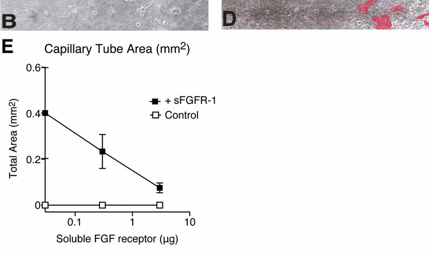
Int. J. Mol. Sci. 2021, 22, 2712                                                                                            2 of 20

                                   in the 55 kDa tumor necrosis factor (TNF) receptor cause a variety of inherited autoimmune
                                   inflammatory syndromes that are thought to be the result of both reduced levels of the
                                   circulating, soluble TNF receptors and impaired TNF receptor clearance [13]. Furthermore,
                                   medical treatments with recombinant soluble receptors can block the biological activity
                                   of cytokines in vivo, as shown by the reduction of retinal edema in patients treated with
                                   a soluble chimeric vascular endothelial cell growth factor (VEGF) receptor (aflibercept,
                                   EyleaTM ) [14] and the reduction of arthritic joint damage in patients treated with a dimeric,
                                   soluble TNF receptor immunoadhesin (Etanercept, EnbrelTM ) [14–16].
                                         Studies with mutant Chinese hamster ovary (CHO) cells have shown that the mech-
                                   anisms regulating the shedding of diverse cell surface proteins have many features in
                                   common [17]. Both constitutive and activated cleavage pathways have been identified,
                                   with the latter being activated by cytokines, growth factors, activators of protein kinase C
                                   (PKC), and calcium ionophores [18–20]. A variety of enzymes, which are members of the
                                   metzincin family of zinc-dependent proteinases, are responsible for ectodomain cleavage.
                                   These include the MMPs (matrix metalloproteases) and the ADAM (a disintegrin and
                                   metalloprotease) family members, ADAM-17 (tumor necrosis factor converting enzyme;
                                   TACE), ADAM-12, ADAM-9, and ADAM-10, among others [8,21–24]. The constitutive and
                                   activated cleavage pathways are independent of a specific consensus sequence and are
                                   regulated via a variety of different protein kinase pathways [19,25].
                                         We have proposed that ectodomain shedding of the high affinity fibroblast growth
                                   factor (FGF) receptors (FGFR-1-4) is an essential mechanism for controlling the diverse
                                   biological activities of the FGF family of growth factors during growth and develop-
                                   ment [26–28]. The FGFs are a family of highly conserved and tightly regulated pleiotropic
                                   growth factors which function in diverse biological processes to induce cellular differen-
                                   tiation and proliferation, promote mesodermal induction and patterning, regulate limb
                                   outgrowth and multi-organ development, and enhance neovascularization and wound
                                   repair [29–34]. The prototype member of the FGF family is FGF-2, which is widely dis-
                                   tributed in cells and the extracellular matrix of both proliferating and nonproliferating
                                   cells [35]. The ubiquitous distribution of the FGFs, especially FGF-2, surrounding cells that
                                   respond to FGF-2 in vitro, but are quiescent in vivo, emphasizes that multiple levels of
                                   regulation exist, which control the biological activity of the FGFs. Mechanisms that have
                                   been proposed to explain this phenomenon include the enzymatic release of FGF-2 from
                                   receptors in the extracellular matrix [36], post-translational modifications of the FGFs [37],
                                   alternative signaling pathways [38], specific temporal and spatial FGF receptor expression
                                   patterns [39], and the shedding of both low and high affinity FGF receptors [7,25,27,39,40].
                                         The FGF receptors consist of two classes of cell surface moieties—low affinity heparan
                                   sulfate proteoglycans and a gene family of four high affinity tyrosine kinase receptors
                                   (FGFR-1-4)—which encode multiple isoforms that are generated by the alternative splicing
                                   of mRNA transcripts [40–42]. The major class of FGF binding, transmembrane heparan
                                   sulfate proteoglycans, known as the syndecans, are constitutively shed from cell mem-
                                   branes and are capable of modulating the biological activity of FGF-2 through interactions
                                   with the high affinity FGF receptors [43]. The syndecans are shed in response to thrombin
                                   and growth factors, such as epidermal growth factor (EGF), and are present in wound
                                   fluids [44,45]. The biological activity of the syndecans is regulated by heparinase-like
                                   proteases, which convert the proteoglycans from inhibitors to activators of FGF-2 [7].
                                         We have identified a class of high affinity, soluble FGF receptors in blood and other
                                   biological fluids, and in the extracellular matrix of vascular endothelial cells [27,28,35,46].
                                   This group of soluble receptors consists of at least five isoforms of the high affinity FGF
                                   receptor gene family, including a two and three Ig-like loop ectodomain of FGFR-1(IIIb) and
                                   FGFR-1(IIIc), and a unique two Ig-domain secreted isoform called FGFR-1(IIIa) (Figure 1).
                                   Analysis of the carboxyl-terminus indicates that the two and three Ig-like loop ectodomains
                                   of FGFR-1 are generated by proteolytic cleavage of the full-length cell-surface FGF receptors,
                                   eight amino acids proximal to the transmembrane domain [26,47].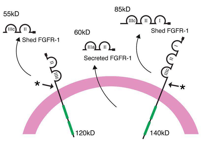
reported. However, recombinant isoforms of the extracellular domains of FGFR-1 and
FGFR2 inhibit FGF-2-induced proliferation [48] and block FGFR2 signaling [34], support-
ing the hypothesis that soluble FGF receptors are modulators of FGFs in vivo. In order to
study the properties of the soluble receptors further, we turned to a cell-based system to
obtain substantial quantities of the proteolytically cleaved ectodomain that is derived
Int. J. Mol. Sci. 2021, 22, 2712 from the extracellular domain of the full-length cell surface receptor and the secreted re-3 of 20
ceptor that is produced by the expression of an alternatively spliced mRNA transcript.
Schematicillustration
Figure1.1.Schematic
Figure illustrationofofthe
the full-length
full-length transmembrane
transmembrane FGFR-1
FGFR-1 receptors
receptors showing
showing the the two
two and three Ig-like extracellular domains (IIIc) that are proteolytically cleaved from
and three Ig-like extracellular domains (IIIc) that are proteolytically cleaved from the cell the cell sur-
surface.
face. A two-loop ectodomain of FGFR-1 is expressed from an alternatively spliced
A two-loop ectodomain of FGFR-1 is expressed from an alternatively spliced transcript of mRNAtranscript of
mRNA and secreted into the extracellular milieu without proteolytic processing.
and secreted into the extracellular milieu without proteolytic processing.
In this
The paper, we
biological focus onofthree
activities important
the native, questions:
circulating (i) IsFGF
soluble the FGFR-1
receptors ectodomain
have not been
biologically active after shedding? (ii) Is the shedding event regulated in vitro? (iii) Does
reported. However, recombinant isoforms of the extracellular domains of FGFR-1 and
the shedding process share similarities with the common system responsible for the shed-
FGFR2 inhibit FGF-2-induced proliferation [48] and block FGFR2 signaling [34], supporting
ding of other transmembrane proteins from the cell surface? Here, we show that the pro-
the hypothesis that soluble FGF receptors are modulators of FGFs in vivo. In order to study
teolytically cleaved three Ig-like loop form of the FGFR-1 ectodomain inhibits the activity
the properties of the soluble receptors further, we turned to a cell-based system to obtain
of FGF-2 by competing with high affinity FGF receptors for ligand binding and blocks
substantial quantities of the proteolytically cleaved ectodomain that is derived from the
FGF-2-induced cell proliferation and in-vitro angiogenesis. Shedding of the FGFR-1 ecto-
extracellular domain of the full-length cell surface receptor and the secreted receptor that
domain is regulated by both ligand binding and activators of PKC and the two signaling
is produced by the expression of an alternatively spliced mRNA transcript.
pathways appear to be independent of each other. Full-length FGFR-1 receptors decrease
In this paper, we focus on three important questions: (i) Is the FGFR-1 ectodomain bi-
as the shedding of the FGFR-1 ectodomain increases, consistent with receptor downregu-
ologically active after
lation. Constitutive, shedding? (ii) Isand
ligand-activated, thePKC-activated
shedding event regulated
shedding in vitro?
appears to be(iii)regu-
Does the
shedding process share similarities with the common system responsible
lated by a metalloprotease family member. FGF receptor mutants, with deletions and sub- for the shedding
of other transmembrane
stitutions intended to disruptproteins from the cell stability
the conformational surface?atHere, we show
the putative that the
cleavage proteolyt-
site, are
still shed in proportion to the expression levels of the high affinity receptor. Therefore, the of
ically cleaved three Ig-like loop form of the FGFR-1 ectodomain inhibits the activity
FGF-2 by
release of acompeting
biologicallywith high
active affinity
FGFR-1 FGF receptors
ectodomain fora ligand
is both binding
constitutive andand blocks
tightly FGF-2-
regu-
induced
lated cellsharing
event, proliferation and in-vitro
many features with angiogenesis.
the system thatShedding
regulates ofthethe FGFR-1
release ectodomain
of other di-
is regulated
verse membraneby both ligand
proteins binding
from and
the cell activators of PKC and the two signaling pathways
surface.
appear to be independent of each other. Full-length FGFR-1 receptors decrease as the
shedding of the FGFR-1 ectodomain increases, consistent with receptor downregulation.
Constitutive, ligand-activated, and PKC-activated shedding appears to be regulated by a
metalloprotease family member. FGF receptor mutants, with deletions and substitutions
intended to disrupt the conformational stability at the putative cleavage site, are still shed
in proportion to the expression levels of the high affinity receptor. Therefore, the release
of a biologically active FGFR-1 ectodomain is both a constitutive and tightly regulated
event, sharing many features with the system that regulates the release of other diverse
membrane proteins from the cell surface.
2. Results
2.1. Identification of a Shed FGFR-1 Ectodomain in the Conditioned Media of Transfected
COS 7 Cells
To explore the possibility that the FGFR-1 ectodomain is shed constitutively from
cells that express full-length FGFR-1, we utilized a transient transfection assay, which hasproteins [17]. We analyzed the serum-free conditioned media from COS 7 cells that were
transfected with the full-length, three Ig-like domain isoform of FGFR-1 (see Figure 2).
Using a specific antibody (Mab6) raised against the extracellular domain of FGFR-1, we
identified a shed FGFR-1 ectodomain in the conditioned media of the FGFR-1-transfected
Int. J. Mol. Sci. 2021, 22, 2712 COS 7 cells, but not in the media of untransfected or vector transfected COS 7 cells. The
4 of 20
FGFR-1 ectodomain was shed into the conditioned media constitutively, increasing in a
time-dependent manner (Figure 2, left). The molecular weight of the shed receptor was
between 70 and 85 kDa, which is identical to the size of the native, three Ig-like domain
previously
form of solublebeen FGFR-1
used to that
evaluate the release
has been purifiedof from
a variety
bloodof [28].
ectodomains from cell surface
proteins [17]. We analyzed the serum-free conditioned
Growth factors and cytokines have been shown to induce media from COS 7 cells
ectodomain that were
shedding by
transfected with the full-length, three Ig-like domain isoform of
the activation of specific signaling cascades [19]. To examine whether FGF-2 couldFGFR-1 (see Figure 2). Us-
en-
ing a specific
hance sheddingantibody
of the(Mab6)
FGFR-1raised against in
ectodomain theanextracellular domainby
autocrine manner of FGFR-1,
binding we identi-
to the cell
fied a shed FGFR-1 ectodomain in the conditioned media of the FGFR-1-transfected
surface FGF receptors and activating downstream signaling pathways, we added exoge- COS 7
cells,
nous but
FGF-2not(0.5
in the media
ng/mL) tooftheuntransfected
serum-free mediaor vector
andtransfected
repeated the COS
time7 cells. The
course. We FGFR-1
found
ectodomain was shed into the conditioned media constitutively,
that shedding of the FGFR-1 ectodomain was increased by the addition of FGF-2, increasing in a time-
most
dependent
notably after manner (Figure
four hours 2, left).2,The
(Figure molecular
right). Image weight
analysisofofthetheshed receptortime
four-hour waspoints
between re-
70 and 85
vealed thatkDa,
the which
increaseis in
identical
shedding to the sizepresence
in the of the native,
of FGF-2 threewasIg-like domain form
approximately three-of
soluble FGFR-1 that has been purified from blood [28].
fold over the level of constitutive shedding.
Figure 2. Constitutive and
Figure 2. and ligand-induced
ligand-inducedFGFR-1
FGFR-1ectodomain
ectodomainshedding.
shedding.COSCOS7 7cells
cellstransfected
transfected
with
with full-length
full-lengthFGFR-1
FGFR-1cDNA
cDNAwere
wereplaced
placedininserum-free
serum-freemedia
mediaforfor
upup
toto
four hours,
four in in
hours, thethe
presence
pres-
ence or absence of recombinant, human fibroblast growth factor (FGF)-2. After collecting the
or absence of recombinant, human fibroblast growth factor (FGF)-2. After collecting the conditioned con-
media, the FGFR-1 ectodomain was precipitated with WGA-Sepharose, eluted with sample buffer,
and analyzed by SDS-PAGE and immunoblotting with an antibody to the extracellular domain
of FGFR-1 (Mab6). The left panel shows the time course of the constitutive release of an 85 kDa
FGFR-1 ectodomain and the right lane shows the time course of the activated release of the FGFR-1
ectodomain in the presence of FGF-2. The negative control consists of the 4 h time point of media
from untransfected cells. The densities of the bands were measured with NIH Image software and
the percent change in intensity of the bands was calculated using the control lane as a reference for
comparison. Standard deviations were calculated for each individual band by the imaging software.
Growth factors and cytokines have been shown to induce ectodomain shedding by
the activation of specific signaling cascades [19]. To examine whether FGF-2 could enhance
shedding of the FGFR-1 ectodomain in an autocrine manner by binding to the cell surface
FGF receptors and activating downstream signaling pathways, we added exogenous FGF-2
(0.5 ng/mL) to the serum-free media and repeated the time course. We found that shedding
of the FGFR-1 ectodomain was increased by the addition of FGF-2, most notably after
four hours (Figure 2, right). Image analysis of the four-hour time points revealed that thereference for comparison. Standard deviations were calculated for each individual band by the
imaging software.
2.2. Ligand-Induced Shedding Leads to a Decrease in Full-Length FGFR-1 Receptors
Int. J. Mol. Sci. 2021, 22, 2712 We explored the dose dependency of ligand-induced shedding in COS 7 and5 of CHO
20
cells to determine whether this event was cell-type specific or representative of a more
general phenomenon. Using transiently transfected COS 7 cells and the stably transfected
CHO cell line FGFR-1/pcDNA3, we observed an increase in the shedding of the FGFR-1
increase in shedding
ectodomain in bothin thetypes
cell presence
after of FGF-2 waswith
stimulation approximately
FGF-2 (Figurethree-fold over theLigand-
3A,B, upper). level
ofinduced
constitutive shedding.
ectodomain shedding was dose-dependent, reaching a maximum at slightly dif-
ferent concentrations of FGF-2 in the two cell types.
2.2. Ligand-Induced Shedding Leads to a Decrease in Full-Length FGFR-1 Receptors
Since the shedding of cell surface proteins has been proposed to be a mechanism of
We
receptorexplored the dose dependency
downregulation of ligand-induced
[3], we examined shedding
the cell lysates in COS 7whether
to determine and CHO thecells
full-
tolength
determine whetherFGF
cell surface thisreceptors
event wasdecreased
cell-type specific or representative
as the shedding of a more
of the FGFR-1 general
ectodomain
phenomenon.
increased. We Using
foundtransiently transfected
that FGF-induced COS 7 cells
shedding and
of the the stably
FGFR-1 transfected
ectodomain ledCHO cell
to a dose-
line FGFR-1/pcDNA3, we observed an increase in the shedding of the FGFR-1 ectodomain
dependent decrease in the 140 kDa full-length cell surface FGF receptor in both cell lysates
in both cell types after stimulation with FGF-2 (Figure 3A,B, upper). Ligand-induced
(Figure 3A,B, lower). These results support the concept that shedding of the FGFR-1 ecto-
ectodomain shedding was dose-dependent, reaching a maximum at slightly different
domain could play a role in the downregulation of cell surface FGF receptors after ligand
concentrations of FGF-2 in the two cell types.
binding.
Figure3.3.Ligand-induced
Figure Ligand-inducedectodomain
ectodomainshedding
shedding downregulates
downregulates full-length
full-length receptors
receptors in in cell
cell ex-
extracts.
tracts. (A) COS 7 cells, transiently transfected with full-length FGFR-1(IIIc) cDNA, and (B) CHO
(A) COS 7 cells, transiently transfected with full-length FGFR-1(IIIc) cDNA, and (B) CHO cells, stably
transfected with full-length FGFR-1(IIIc) cDNA, were placed in serum-free media in the presence
of different concentrations of FGF-2. Conditioned media and cell extracts were collected after two
hours. The shed FGFR-1 ectodomain was precipitated with WGA-Sepharose. Full-length FGFR-1
in the cell lysates was precipitated with an antibody to the C-terminus of FGFR-1 and/or extracted
with sample buffer. The samples were analyzed by SDS-PAGE and immunoblotting with Mab6.
The upper and lower panels show the release of the FGFR-1 ectodomain from COS 7 and CHO cells,
respectively, and the corresponding decrease in the level of the full-length receptor. The densities of
the bands were measured with NIH Image software and the percent change in intensity of the bands
was calculated using the control lane (0 ng/mL) as a reference for comparison.
Since the shedding of cell surface proteins has been proposed to be a mechanism
of receptor downregulation [3], we examined the cell lysates to determine whether the
full-length cell surface FGF receptors decreased as the shedding of the FGFR-1 ectodomain
increased. We found that FGF-induced shedding of the FGFR-1 ectodomain led to acells, stably transfected with full-length FGFR-1(IIIc) cDNA, were placed in serum-free media in
the presence of different concentrations of FGF-2. Conditioned media and cell extracts were col-
lected after two hours. The shed FGFR-1 ectodomain was precipitated with WGA-Sepharose. Full-
Int. J. Mol. Sci. 2021, 22, 2712 length FGFR-1 in the cell lysates was precipitated with an antibody to the C-terminus of FGFR-1 6 of 20
and/or extracted with sample buffer. The samples were analyzed by SDS-PAGE and immunoblot-
ting with Mab6. The upper and lower panels show the release of the FGFR-1 ectodomain from
COS 7 and CHO cells, respectively, and the corresponding decrease in the level of the full-length
receptor. The densities
dose-dependent of the bands
decrease in thewere
140 measured with NIH
kDa full-length cellImage software
surface and the percent
FGF receptor in both cell
change in intensity of the bands was calculated using the control lane (0 ng/mL)
lysates (Figure 3A,B, lower). These results support the concept that shedding as a reference for
of the FGFR-1
comparison.
ectodomain could play a role in the downregulation of cell surface FGF receptors after
ligand binding.
2.3. Purification of the Soluble FGFR-1 Ectodomains
2.3.With
Purification of the Soluble
the evidence FGFR-1
that FGFR-1 Ectodomains
ectodomain shedding is a regulated event, we focused
on theWith
question of whether the soluble FGFR-1
the evidence that FGFR-1 ectodomain ectodomains
shedding are
is a biologically active.we
regulated event, Wefo-
passed
cused FGFR-1 CHO cell
on the question conditioned
of whether media over
the soluble an ectodomains
FGFR-1 FGF-2/heparin-Sepharose affinity
are biologically active.
column to determine
We passed FGFR-1 CHO whether the soluble media
cell conditioned FGFR-1 ectodomains
over could bind FGF-2.affinity
an FGF-2/heparin-Sepharose Both
the shed and
column secreted FGFR-1
to determine whetherectodomains
the solublebound
FGFR-1 avidly to the FGF-2/heparin-Sepharose
ectodomains could bind FGF-2. Both
column.
the shed Coomassie blueFGFR-1
and secreted staining of the eluants
ectodomains revealed
bound avidlymolecular weights of 70–85 kDa
to the FGF-2/heparin-Sepharose
and 55–60 Coomassie
column. kD for the shed
blue FGFR-1
staining and theeluants
of the secreted FGFR ectodomains,
revealed respectively
molecular weights of 70–85 kDa
(Figure 4A,B).kD
and 55–60 Theforpurified
the shedFGFR-1
FGFR-1 ectodomains had theFGFR
and the secreted same electrophoretic character-
ectodomains, respectively
istics as the
(Figure native,
4A,B). The85 kDa and
purified 55 kDectodomains
FGFR-1 soluble FGFhad receptors isolated
the same and purified
electrophoretic from
character-
istics[26,28].
blood as the native, 85 kDa and 55 kD soluble FGF receptors isolated and purified from
blood [26,28].
Figure 4. Purification of the soluble FGFR-1 ectodomains. (A) Coomassie staining of the shed FGFR-1
Figure 4. Purification
ectodomain purifiedoffrom
the soluble FGFR-1
CHO cell ectodomains.
conditioned (A)(B)
media. Coomassie staining
Coomassie of the
staining shedsecreted
of the
FGFR-1 ectodomain purified from CHO cell conditioned media.
FGFR-1 ectodomain purified from CHO cell conditioned media. (B) Coomassie staining of the
secreted FGFR-1 ectodomain purified from CHO cell conditioned media.
2.4. The Shed FGFR-1 Ectodomain Functions as a Biologically Active Inhibitor of FGF-2
2.4. TheWe
Shed FGFR-1
tested Ectodomain
the inhibition of Functions as a Biologically
FGF-2 activity ActiveofInhibitor
with two forms soluble of FGF-2 receptors:
FGFR-1
We tested the inhibition
A proteolytically shed formof ofFGF-2
soluble activity
FGFR-1 with
andtwo forms ofsoluble
a secreted solubleFGFR,
FGFR-1 recep-
expressed
tors:
fromAan proteolytically shed form
alternatively spliced of soluble
transcript FGFR-1
that lacks and a secreteddomain
a transmembrane soluble[26].
FGFR, ex-
We used
a classic
pressed fromFGF-2-induced aortic
an alternatively endothelial
spliced transcriptcell proliferation
that assay to testdomain
lacks a transmembrane the effects[26].of
Wesoluble
used FGF receptors
a classic on FGF-2-induced
FGF-2-induced proliferation,
aortic endothelial celland an 125 I-FGF-2
proliferation assay receptor-binding
to test the ef-
assay to address the mechanism of this activity. We found that
fects of soluble FGF receptors on FGF-2-induced proliferation, and an I-FGF-2 both the
125 shed and secreted
receptor-
FGFR-1 ectodomains inhibited FGF-2-induced aortic endothelial cell
binding assay to address the mechanism of this activity. We found that both the shed and proliferation in a
dose-dependent
secreted manner (Figure
FGFR-1 ectodomains 5A,B). FGF-2-induced aortic endothelial cell prolifera-
inhibited
tion inTo explore the mechanism
a dose-dependent behind 5A,B).
manner (Figure this inhibition, we switched to Swiss 3T3 cells
dueTo toexplore
the higher number of behind
the mechanism cell surface FGF receptors
this inhibition, in this cell
we switched line. 3T3
to Swiss We cells
measureddue
125 I-FGF-2 binding to high affinity, cell surface receptors in both the presence and absence
to the higher number of cell surface FGF receptors in this cell line. We measured I-FGF- 125
2 of the shed
binding FGFR-1
to high ectodomain.
affinity, We found
cell surface thatinthe
receptors shed
both theFGFR-1
presence ectodomain
and absence effectively
of the
reduced 125 I-FGF-2 binding to high affinity FGF receptors (Figure 5C, left), supporting
shed FGFR-1 ectodomain. We found that the shed FGFR-1 ectodomain effectively reduced
the interpretation that the mechanism of inhibition is due to competition between soluble
and cell surface FGF receptors for FGF-2 binding. To further support this interpretation,
we examined the effect of the secreted FGFR-1 ectodomain on cellular proliferation in Swiss
3T3 cells, using a 3 H-thymidine incorporation assay. Similar to the effects of soluble FGFR-1
receptors in the ABAE cell proliferation assays, the secreted FGFR-1 receptor inhibited
FGF-2-induced 3T3 cell proliferation to a similar degree as the inhibition of 125 I-FGF-2
binding (Figure 5D, right). These results support the interpretation that the shed andtation that the mechanism of inhibition is due to competition between soluble and cell
surface FGF receptors for FGF-2 binding. To further support this interpretation, we exam-
ined the effect of the secreted FGFR-1 ectodomain on cellular proliferation in Swiss 3T3
cells, using a 3H-thymidine incorporation assay. Similar to the effects of soluble FGFR-1
Int. J. Mol. Sci. 2021, 22, 2712 7 of 20
receptors in the ABAE cell proliferation assays, the secreted FGFR-1 receptor inhibited
FGF-2-induced 3T3 cell proliferation to a similar degree as the inhibition of 125I-FGF-2
binding (Figure 5D, right). These results support the interpretation that the shed and se-
secreted
creted FGFR-1
FGFR-1 ectodomainsinhibit
ectodomains inhibitthe
the biological
biological activity
activityofofFGF-2
FGF-2ininmultiple
multiplecellcell
types
types
andcompete
and competewith
with the
the high affinity,
affinity,cell
cellsurface
surfaceFGF
FGFreceptors
receptorsforfor
binding to FGF-2.
binding to FGF-2.
Figure
Figure5. The soluble
5. The FGFR-1
soluble receptors
FGFR-1 inhibit
receptors the biological
inhibit activity
the biological of FGF-2.
activity (A,B) (A,B)
of FGF-2. AdultAdult
aorticaortic
endothelial cell prolifer-
endothelial cell
ation is inhibited
proliferation is by solubleby
inhibited FGF receptors.
soluble Cells wereCells
FGF receptors. plated in 24-well
were chambers
plated in in complete
24-well chambers in medium
completewith 0.125with
medium ng/mL
FGF in the
0.125 absence
ng/mL FGF or presence
in the absenceoforincreasing concentrations
presence of of the shedof(A)
increasing concentrations theor secreted
shed (A) or(B) FGFR-1
secreted (B) ectodomain on days 1
FGFR-1 ectodomain
and 3. After five days in culture, the cells were trypsinized and counted with a Coulter Counter. (C) Soluble
on days 1 and 3. After five days in culture, the cells were trypsinized and counted with a Coulter Counter. (C) Soluble FGF receptors
inhibit FGF-2 binding
FGF receptors inhibittoFGF-2
cell surface
bindingFGF receptors
to cell surface in
FGF 125I-FGF-2
thereceptors in the 125 I-FGF-2
ligand bindingligand
assay.binding
Swiss 3T3 cells
assay. were
Swiss grown
3T3 cells to
were grown to confluence ◦ 125
confluence in 24-well plates.inThe
24-well
cellsplates. The cellsfor
were labeled were
2 hlabeled
at 4 °Cfor 2 h125
with atI-FGF-2
4 C with I-FGF-2
(2 ng/mL) in(2the
ng/mL) in the
absence absence of
or presence
or presence of increasing concentrations of the shed FGFR-1 ectodomain. High affinity FGF-2 binding
increasing concentrations of the shed FGFR-1 ectodomain. High affinity FGF-2 binding was determined after removingwas determined
after removing FGF-2 bound to low affinity sites with a high salt wash. Results are presented as the decrease in 125 I-FGF-2
FGF-2 bound to low affinity sites with a high salt wash. Results are presented as the decrease in 125I-FGF-2
3
binding to 3T3
binding to 3T3 cell surface FGFR-1 receptors in the presence of the shed ectodomain. (D) Swiss 3T3 cell H-thymidine
incorporation assays. Swiss 3T3 fibroblasts were grown to confluence in 96-well dishes and switched to low serum media
for 2 days in DME (Dulbecco’s Modified Eagle’s Media) with 0.5% calf serum. The rate of DNA synthesis was measured 24 h
after the addition of FGF-2 and/or secreted FGFR-1 and 5 h after incubation with 0.2 mCi/well of [methyl-3 H]thymidine
(6.7 Ci/mole, ICN). The cultures were then processed for scintillation counting, as previously described [49].cell surface FGFR-1 receptors in the presence of the shed ectodomain. (D) Swiss 3T3 cell 3H-thymidine incorporation as-
says. Swiss 3T3 fibroblasts were grown to confluence in 96-well dishes and switched to low serum media for 2 days in
DME
Int. J. Mol. Sci.(Dulbecco’s
2021, 22, 2712Modified Eagle’s Media) with 0.5% calf serum. The rate of DNA synthesis was measured 24 h after the
8 of 20
addition of FGF-2 and/or secreted FGFR-1 and 5 h after incubation with 0.2 mCi/well of [methyl-3H]thymidine (6.7
Ci/mole, ICN). The cultures were then processed for scintillation counting, as previously described [49].
2.5.
2.5.The
TheSecreted
SecretedFGFR-1
FGFR-1Receptor
ReceptorInhibits
InhibitsCapillary
CapillaryTube
TubeFormation
FormationininCollagen
CollagenGels
Gels
Using
Using a well-known model of in vitro angiogenesis, we tested the ability of
a well-known model of in vitro angiogenesis, we tested the ability of the
the se-
se-
creted
creted FGF receptor to block FGF-2-induced capillary tube growth in collagen gels [50].
FGF receptor to block FGF-2-induced capillary tube growth in collagen gels [50].
Within
Withinseven
sevendays
daysofofgrowth,
growth,the
theABAE
ABAEcells
cellscultured
culturedininthe
thepresence
presenceofofFGF-2
FGF-2invaded
invaded
the
the collagen gel and formed three-dimensional capillary tubes with branching patterns.
collagen gel and formed three-dimensional capillary tubes with branching patterns.
Cells cultured in the absence of FGF-2 showed no evidence of capillary tube formation.
Cells cultured in the absence of FGF-2 showed no evidence of capillary tube formation.
Cells cultured with FGF-2 and increasing concentrations of the secreted FGFR-1 ectodomain
Cells cultured with FGF-2 and increasing concentrations of the secreted FGFR-1 ectodo-
showed progressively less capillary tube formation (Figure 6A–E), demonstrating that the
main showed progressively less capillary tube formation (Figure 6A–E), demonstrating
soluble FGF receptors inhibit FGF-2-induced angiogenesis in the extracellular matrix in a
that the soluble FGF receptors inhibit FGF-2-induced angiogenesis in the extracellular ma-
dose-dependent manner.
trix in a dose-dependent manner.
.
Figure 6. Inhibition of in-vitro angiogenesis and capillary tube formation by soluble FGFR-1 receptors.
Figure 6. Inhibition of in-vitro angiogenesis and capillary tube formation by soluble FGFR-1 recep-
Phase-contrast images of ABAE cells forming three-dimensional capillary structures (arrows) in
tors. Phase-contrast images of ABAE cells forming three-dimensional capillary structures (arrows)
collagen gelsgels
in collagen grown with
grown FGF-2
with (500
FGF-2 pg)pg)
(500 in the absence
in the (A)(A)
absence or presence (B)(B)
or presence of the secreted
of the FGFR-1
secreted
receptor (300 ng). Bar = 200 um. Panels (C,D) show the corresponding capillary tube structures
highlighted in red. (E) The area of capillary tube formation was quantified using ImageJ.
2.6. Inhibition of FGFR-1 Ectodomain Shedding by Metalloprotease Inhibitors
A diverse group of membrane proteins are shed by a mechanism which is sensi-
tive to metalloprotease inhibitors [17]. To examine whether the constitutive and ligand-FGFR-1 receptor (300 ng). Bar = 200 um. Panels (C,D) show the corresponding capillary tube struc-
tures highlighted in red. (E) The area of capillary tube formation was quantified using ImageJ.
Int. J. Mol. Sci. 2021, 22, 2712 9 of 20
2.6. Inhibition of FGFR-1 Ectodomain Shedding by Metalloprotease Inhibitors
A diverse group of membrane proteins are shed by a mechanism which is sensitive
to metalloprotease inhibitors [17]. To examine whether the constitutive and ligand-acti-
activated
vated FGFR-1
FGFR-1 ectodomain
ectodomain shedding
shedding is dependent
is also also dependent onsystem,
on this this system, we tested
we tested the
the effect
of metalloprotease inhibitors on the release of the FGFR-1 ectodomain in COS 7 cells. Cells7
effect of metalloprotease inhibitors on the release of the FGFR-1 ectodomain in COS
cells. Cells
treated withtreated with the metalloprotease
the metalloprotease inhibitors—marimastat
inhibitors—marimastat and CGS27023A—
and CGS27023A—showed a
showed a dose-dependent decrease in constitutive FGFR-1 ectodomain shedding
dose-dependent decrease in constitutive FGFR-1 ectodomain shedding (Figure 7). (Figure 7).
FGF-2-
FGF-2-activated
activated shedding
shedding wasinhibited.
was also also inhibited.
There There was
was no no evidence
evidence of cytotoxicity
of cytotoxicity (data(data
not
not shown).
shown).
Figure
Figure 7. Inhibition
Inhibition of
of constitutive
constitutive FGFR-1
FGFR-1 ectodomain
ectodomain shedding
shedding by
by MMP
MMP inhibitors.
inhibitors. COS
COS 77 cells transiently
transiently transfected
transfected
with FGFR-1(IIIc) were
with FGFR-1(IIIc) wereplaced
placedininserum-free
serum-freemedia
media inin
thethe presence
presence of different
of different concentrations
concentrations of marimastat
of (A) (A) marimastat andCGS
and (B) (B)
CGS 27023A, as indicated. The samples were run in duplicate. The conditioned media was collected after an
27023A, as indicated. The samples were run in duplicate. The conditioned media was collected after an overnight incubation overnight
incubation and the shed FGFR-1 ectodomain was precipitated with WGA-Sepharose. The samples were analyzed by SDS-
and the shed FGFR-1 ectodomain was precipitated with WGA-Sepharose. The samples were analyzed by SDS-PAGE and
PAGE and immunoblotting with Mab6. The densities of the bands were measured with NIH Image software and the
immunoblotting with Mab6. The densities of the bands were measured with NIH Image software and the percent change in
percent change in intensity of the bands was calculated using the control lane (0 uM) as a reference for comparison. Stand-
intensity
ard of thewere
deviations bandscalculated
was calculated using
for each the control
individual bandlane
by(0 uM)
the as a reference
imaging for comparison. Standard deviations were
software.
calculated for each individual band by the imaging software.
2.7. FGFR-1 Ectodomain Shedding Is Not Inhibited by Mutations Surrounding the Cleavage Site
2.7. FGFR-1 Ectodomain Shedding Is Not Inhibited by Mutations Surrounding the Cleavage Site
The putative cleavage site of the FGFR-1 ectodomain is located between Val-Met,
eight The putative
amino acids cleavage
from thesite of the FGFR-1 ectodomain
transmembrane is located
domain [26,47]. between Val-Met,
We constructed eight
an FGFR-1
amino acids from the transmembrane domain [26,47]. We constructed
mutant containing a deletion at the putative cleavage site and analyzed the degree of an FGFR-1 mutant
containing
FGFR-1 a deletion
ectodomain at the putative
shedding following cleavage site and analyzed
the transfection of COS 7 thecellsdegree
with theof FGFR-1
mutant
ectodomain shedding following the transfection of COS 7 cells with
FGFR-1 construct. We found that a seven amino acid deletion surrounding the putativethe mutant FGFR-1
construct.site
cleavage Wedidfound that a seven
not prevent aminoofacid
shedding the deletion surrounding(Figure
FGFR-1 ectodomain the putative cleavage
8). Indeed, the
site did not prevent shedding of the FGFR-1 ectodomain (Figure 8). Indeed,
relative degree of ectodomain shedding was not significantly different from the wild type the relative
degree ofadjusting
receptor, ectodomain for shedding was not
the 50% lower levelsignificantly different
of expression from the wild
of the full-length type receptor,
receptor mutant
adjusting for the 50% lower level of expression of the full-length
(see the histogram in Figure 8). We constructed and examined two additional receptor mutant (seemu-
the
histogram in Figure 8). We constructed and examined two additional mutants:FGFR-1/P2,
tants:FGFR-1/P2, which contains a 10 amino acid deletion with a proline-glycine substitu-
which contains a 10 amino acid deletion with a proline-glycine substitution and FGFR-1/L4,
tion and FGFR-1/L4, which contains a 14 amino acid deletion in the region of the putative
which contains a 14 amino acid deletion in the region of the putative cleavage site. Both
cleavage site. Both of these mutants were also cleaved. Although the extent of ectodomain
of these mutants were also cleaved. Although the extent of ectodomain shedding was
shedding was lower than with the wild type receptor, this difference was eliminated when
lower than with the wild type receptor, this difference was eliminated when the level of
shed receptor was compared to the overall level of expression of the full-length, mutant
receptors. These findings indicate that FGFR-1 ectodomain shedding is not inhibited by
mutations containing deletions and substitutions at the putative cleavage site, which is a
feature that is relatively common for other cell surface proteins.Int. J. Mol. Sci. 2021, 22, x FOR PEER REVIEW 10 of 20
the level of shed receptor was compared to the overall level of expression of the full-
Int. J. Mol. Sci. 2021, 22, 2712 length, mutant receptors. These findings indicate that FGFR-1 ectodomain shedding10isofnot
20
inhibited by mutations containing deletions and substitutions at the putative cleavage site,
which is a feature that is relatively common for other cell surface proteins.
Figure8.8. The
Figure The effect
effect of FGFR-1 mutations
of FGFR-1 mutations ononectodomain
ectodomainshedding.
shedding.COSCOS7 7cells
cells were
were transfected
transfected
with
withvector
vector alone (pcDNA3)
(pcDNA3) or orFGFR-1
FGFR-1constructs,
constructs,asas indicated,
indicated, including
including FGFR-1
FGFR-1 (wild-type),
(wild-type),
FGFR-1/B1, FGFR-1/P2,
FGFR-1/B1, FGFR-1/P2, andandFGFR-1/L4.
FGFR-1/L4.The conditioned
The conditioned media
mediaand
andcell extracts
cell were
extracts collected
were collected
afteran
after anovernight
overnightincubation.
incubation.The
Theshed
shed FGFR-1
FGFR-1 ectodomains
ectodomains were
were precipitated
precipitated with
with WGA-Se-
WGA-Sepharose
pharose
and and analyzed
analyzed by SDS-PAGE
by SDS-PAGE and immunoblotting
and immunoblotting with Mab6.
with Mab6. The densities
The densities of theofbands
the bands
were
were evaluated using NIH Image
evaluated using NIH Image software. software.
2.8.Constitutive
2.8. ConstitutiveFGFR-1
FGFR-1Ectodomain
EctodomainShedding
SheddingIsIsActivated
Activatedby
byTPA
TPA
Several studies
Several studies have
have demonstrated
demonstrated that that the
the constitutive
constitutive shedding
shedding of of diverse
diverse trans-
trans-
membrane
membraneproteins
proteinsisisactivated
activatedby byprotein
proteinkinase
kinaseCCsignaling
signalingpathways
pathways[17]. [17].To
Todetermine
determine
whether
whetheractivators
activatorsofofprotein
proteinkinase
kinaseCCenhance
enhanceshedding
sheddingof ofthe
theFGFR-1
FGFR-1ectodomain,
ectodomain,we we
examined
examinedshedding
sheddingof ofthe
theFGFR-1
FGFR-1ectodomain
ectodomainin in the
the presence
presence of of the
the PKC activator—TPA.
activator—TPA.
FGFR-1-transfected
FGFR-1-transfectedCOS COS 7 cells were
7 cells treated
were withwith
treated TPA inTPAthe in
presence or absence
the presence of G0 6983,
or absence of
which is a specific inhibitor of all PKC isoforms except PKCµ [51].
G06983, which is a specific inhibitor of all PKC isoforms except PKCμ [51]. We found We found that TPA
that
activated FGFR-1
TPA activated ectodomain
FGFR-1 shedding
ectodomain at concentrations
shedding rangingranging
at concentrations from 10 from
to 10010ng/mL
to 100
and thatand
ng/mL thisthat
enhancement was abolished
this enhancement in the presence
was abolished of G0 6983.
in the presence of G(Figure 9A). These
06983. (Figure 9A).
results indicateindicate
These results that shedding is dependent
that shedding on cellular
is dependent signaling
on cellular mechanisms
signaling mechanisms and the
and
activation of PKC pathways.
the activation of PKC pathways.
To
Todetermine
determine whether
whether bothbothconstitutive
constitutive shedding
shedding and and protein
proteinkinase
kinaseC-induced
C-induced
shedding were sensitive to metalloprotease inhibitors, we examined
shedding were sensitive to metalloprotease inhibitors, we examined the effect the effect of marimastat
of mari-
and CGS27023A
mastat on constitutive
and CGS27023A and TPA-activated
on constitutive and TPA-activatedcleavage. We found
cleavage. Wethat
foundmarimastat
that mari-
and CGS27023A
mastat abolishedabolished
and CGS27023A constitutive and TPA-activated
constitutive shedding (Figure
and TPA-activated shedding9B), (Figure
suggesting9B),
that both constitutive, ligand-induced and PKC-activated shedding
suggesting that both constitutive, ligand-induced and PKC-activated shedding pathways pathways involve a
member of the metalloprotease family of enzymes.
involve a member of the metalloprotease family of enzymes.Int. J. Mol. Sci. 2021, 22, 2712 11 of 20
Mol. Sci. 2021, 22, x FOR PEER REVIEW 11 of 20
Figure 9. 12-O-tetradecanoyl
Figure 9. 12-O-tetradecanoyl phorbol
phorbol 13-acetate 13-acetate
(TPA) enhances (TPA) enhances
FGFR-1 FGFR-1
ectodomain ectodomain
shedding. (A) shedding.
COS 7 cells transiently
(A) COS 7 cells transiently transfected with FGFR-1(IIIc) were placed in serum-free
transfected with FGFR-1(IIIc) were placed in serum-free media in the presence of different concentrations media in theof TPA plus
presence of different concentrations of TPA plus or minus G06983. The conditioned media was
or minus G0 6983. The conditioned media was collected after a 36 h incubation and the shed FGFR-1 ectodomain was
collected after a 36 h incubation and the shed FGFR-1 ectodomain was precipitated with WGA-
precipitated with WGA-Sepharose. The samples were analyzed by SDS-PAGE and immunoblotting with Mab6. The densities
Sepharose. The samples were analyzed by SDS-PAGE and immunoblotting with Mab6. The densi-
of the bands were measured
ties of the bands with NIH
were Image software
measured with NIH andImage
the percent change
software and in
theintensity
percent of the bands
change was calculated
in intensity of using
the control lane
the(0 ng/mL)
bands wasas a reference
calculated for the
using comparison.
control laneStandard
(0 ng/mL)deviations were calculated
as a reference for each
for comparison. individual band
Standard
by the imaging software.were
deviations (B) COS 7 cells for
calculated transiently transfected
each individual band with FGFR-1(IIIc)
by the were placed
imaging software. (B) in
COS serum-free media in the
7 cells tran-
presence or absence
siently of 100 ng/mL
transfected TPA
with and the metalloprotease
FGFR-1(IIIc) were placed in inhibitors marimastat
serum-free media in (25
themM) and CGS
presence 27034A (50 mM).
or absence
of 100
The conditioned ng/mL
media wasTPA and the
collected metalloprotease
after a 36 h incubationinhibitors
and the marimastat (25 mM) was
FGFR-1 ectodomain and analyzed
CGS 27034A (50
as described above.
mM). The conditioned media was collected after a 36 h incubation and the FGFR-1 ectodomain
was analyzed as 2.9.described above. Shedding Is Blocked by a Specific Inhibitor of the FGF Receptor Tyrosine
FGF-2-Activated
Kinase, But Not by a PKC Inhibitor
2.9. FGF-2-Activated Shedding Is Blocked by a Specific Inhibitor of the FGF Receptor Tyrosine
We also examined whether stimulation of the FGF receptor tyrosine kinase signaling
Kinase, But Not by a PKC Inhibitor
pathway was necessary for ligand-activated FGFR-1 ectodomain shedding. We analyzed
We also the
examined whether
levels of stimulation
the shed of the FGF in
FGFR-1 ectodomain receptor tyrosine kinase
the conditioned mediasignaling
of FGFR-1 CHO cells
pathway wastreated
necessary for ligand-activated FGFR-1 ectodomain shedding.
with FGF-2 in the presence or absence of a specific inhibitor We analyzed
of the FGF receptor
the levels of the shed kinase—SU5402
tyrosine FGFR-1 ectodomain [52].inWe
thefound
conditioned media
that FGF-2 of FGFR-1
induced CHO cells shedding of
the ectodomain
treated with FGFR-1
FGF-2 inand
thethat
presence
SU5402orreduced
absenceFGF-2-activated
of a specific inhibitor
FGFR-1 of the FGF receptor
ectodomain shedding to the level
tyrosine kinase—SU5402
observed with [52]. We foundshedding
constitutive that FGF-2 induced
(Figure theControls
10A). ectodomain shedding
showed of
that FGF-2-activated
FGFR-1 and shedding
that SU5402wasreduced FGF-2-activated
associated FGFR-1 ectodomain
with ERK phosphorylation, whichshedding
was blockedto the
by the FGFR-1
level observed with constitutive
tyrosine shedding
kinase inhibitor (Figure
SU5402 10A).
(Figure Controls
10B). Theseshowed that FGF-2-ac-
data suggest that ligand-activated
tivated shedding
FGFR-1wasectodomain
associated with ERK occurs
shedding phosphorylation, which was blocked
through a mechanism by theFGF receptor
that involves
FGFR-1 tyrosine kinase
tyrosine inhibitor
kinase SU5402
signaling (Figure
(Figure 10B). These data suggest that ligand-ac-
10B).
tivated FGFR-1 ectodomain shedding occurs through a mechanism that involves FGF re-
ceptor tyrosine kinase signaling (Figure 10B).
Finally, we investigated whether FGF-2-activated FGFR-1 ectodomain shedding was
regulated by PKC signaling. We found that the PKC inhibitor G06983 did not reduce FGF-
2-activated shedding of the FGFR-1 ectodomain (Figure 10A), although it did block TPA-Int. J. Mol. Sci. 2021, 22, x FOR PEER REVIEW 12 of 20
Int. J. Mol. Sci. 2021, 22, 2712 stimulated shedding (Figure 9A). This result demonstrates that the ligand-activated12shed-
of 20
ding pathway is independent of the PKC cleavage pathway and that multiple stimuli can
converge independently on this target.
Figure 10. FGF-2-activated
Figure10. FGF-2-activated shedding
shedding is
is blocked
blocked byby an
an FGF
FGF receptor
receptor tyrosine
tyrosine kinase
kinase inhibitor.
inhibitor. CHO
CHO
cells
cellswere
wereplaced
placedininserum-free
serum-freemedia
mediaininthe
thepresence
presenceofofFGF-2
FGF-2and
andthe
theFGF
FGFreceptor
receptor tyrosine kinase
tyrosine ki-
nase inhibitor
inhibitor SU5402,
SU5402, or theor the inhibitor
PKC PKC inhibitor G06983.
G0 6983. Conditioned
Conditioned mediamedia andextracts
and cell cell extracts
werewere col-
collected
lectedtwo
after after two hours.
hours. TheFGFR-1
The shed shed FGFR-1 ectodomain
ectodomain was precipitated
was precipitated with with WGA-Sepharose.
WGA-Sepharose. The The
cells
cells were
were extracted
extracted directly
directly into SDS-PAGE
into SDS-PAGE sample
sample buffer.
buffer. The samples
The samples werewere analyzed
analyzed by SDS-
by SDS-PAGE
PAGE and immunoblotting with Mab6 (A) or antibodies to phospho-ERK and
and immunoblotting with Mab6 (A) or antibodies to phospho-ERK and total ERK (B) to test total ERK (B)for
to the
test for the
inhibition inhibition
of FGFR-1 of FGFR-1
activation activation by SU5402.
by SU5402.
3. Discussion
Finally, we investigated whether FGF-2-activated FGFR-1 ectodomain shedding was
regulated
In thisbyinvestigation,
PKC signaling. We found
we purified that the PKCshed
a constitutively inhibitor
FGFR-1G0 6983 did notwhich
ectodomain, reduceis
FGF-2-activated shedding of the FGFR-1 ectodomain (Figure 10A), although
the three Ig-like domain form of FGFR-1(IIIc), to demonstrate that circulating FGFR-1 it did block
ec-
TPA-stimulated shedding (Figure
todomains are biologically 9A). This result
active inhibitors demonstrates
of FGF-2. that
Specifically, wethe
haveligand-activated
shown that the
shedding
shed FGFR-1 pathway is independent
ectodomain binds toofan theFGF-2/heparin-Sepharose
PKC cleavage pathway and that multiple
affinity column,stimuli
blocks
can converge independently on this target.
FGF-2-induced proliferation in endothelial cells and 3T3 fibroblasts, inhibits in vitro angi-
ogenesis and capillary tube formation, and competes with high affinity cell surface FGFR-
3. Discussion
1 receptors for ligand binding. These data demonstrate that the FGFR-1 ectodomain can
In this
still bind investigation,
ligands we purified
after proteolytic a constitutively
shedding shed FGFR-1
from cell surface receptorsectodomain,
and supports whichthe
is the three Ig-like domain form of FGFR-1(IIIc), to demonstrate that circulating
fundamental hypothesis that the shed FGFR-1 ectodomains in blood may function as cir- FGFR-1
ectodomains are biologically
culating inhibitors of the FGFsactive inhibitors
in vivo. of FGF-2.
In contrast Specifically,
to previous studieswethat
have showngenet-
utilized that
the shed FGFR-1 ectodomain binds to an FGF-2/heparin-Sepharose affinity column,
ically engineered soluble receptors or ectodomain fusion proteins, this report is the first blocks
FGF-2-induced
demonstration proliferation
that a natural inand
endothelial cells and
constitutively 3T3FGFR-1
shed fibroblasts, inhibits in
ectodomain is vitro angio-
biologically
genesis and capillary tube formation, and competes with high affinity cell surface FGFR-1
active.
receptors for ligand binding. These data demonstrate that the FGFR-1 ectodomain can
This report also demonstrates that FGFR-1 ectodomain shedding shares many fea-
still bind ligands after proteolytic shedding from cell surface receptors and supports the
tures with the common mechanism that governs the release of other cell surface receptors,
fundamental hypothesis that the shed FGFR-1 ectodomains in blood may function as circu-
cell adhesion molecules, and growth factors. The finding that constitutive and regulated
lating inhibitors of the FGFs in vivo. In contrast to previous studies that utilized genetically
shedding occurs in at least two different cell types is consistent with the idea that FGFR-1
engineered soluble receptors or ectodomain fusion proteins, this report is the first demon-
stration that a natural and constitutively shed FGFR-1 ectodomain is biologically active.
This report also demonstrates that FGFR-1 ectodomain shedding shares many fea-
tures with the common mechanism that governs the release of other cell surface receptors,Int. J. Mol. Sci. 2021, 22, 2712 13 of 20
cell adhesion molecules, and growth factors. The finding that constitutive and regulated
shedding occurs in at least two different cell types is consistent with the idea that FGFR-1
shedding is not restricted to a specific cell type and is representative of a more gener-
alized phenomenon which occurs in multiple types of cells sharing common pathways.
Of particular interest is the question of which signaling pathways are involved in these
shedding events and whether they are the same or different. Although all the pathways
involved in FGFR-1 ectodomain shedding are not known, our data show that at least two
independent pathways are involved: A ligand-activated pathway which requires activa-
tion of the FGF receptor tyrosine kinase and another pathway involving PKC activation.
While the activation of receptor tyrosine kinases has been implicated in the shedding
of unrelated transmembrane proteins [19], our findings have added significance because
FGFR-1 shedding appears to result from FGF-2 binding to and activation of its own receptor.
How activation of the FGF receptor leads to the shedding of its receptor ectodomain is not
known at this time.
In addition, the finding that FGF-2 enhances shedding of the ectodomain of its high
affinity receptor is of special interest because it suggests that FGFR-1 ectodomain shedding
could be an autocrine feedback mechanism for downregulating FGFR-1 receptors after
ligand binding. Therefore, the FGFR-1 receptor joins a group including the TNF receptor
and the CSF-1 receptor, which use this mechanism to rapidly modulate their response to
ligands in the pericellular environment [3,53,54]. Whether or not the truncated cytoplas-
mic domain persists after ectodomain shedding and has any role in the cell remains to
be investigated.
Our results are consistent with the proposal that a member of the metalloprotease
family regulates FGFR-1 ectodomain shedding. The specific enzyme involved in these
cleavage pathways is not yet known, but candidates include members of the ADAMs
family of metalloproteases, particularly ADAMs-12, -17, -10, and -9, which are responsible
for the shedding of multiple cell surface proteins, including tumor necrosis factor-alpha
(TNF-α), transforming growth factor (TGF-α), L-selectin, amyloid precursor protein (APP),
and heparin-binding epidermal growth factor-like growth factor (HB-EGF) [2,22–24,55].
These enzymes are inhibited by hydroxamate compounds, such as marimastat and CGS
27023A, which interfere with zinc binding to the metalloprotease domain [56]. In our
study, the concentrations of hydroxamate-based MMP inhibitors which blocked FGFR-1
ectodomain shedding are within the µM range shown to inhibit specific ADAM family
members in other ectodomain shedding systems [56]. Studies are currently in progress to
determine whether FGFR-1 ectodomain shedding can be induced by ADAM-17 (TACE) or
other ADAM family members.
Our group attempted, albeit unsuccessfully, to create an FGFR-1 mutant which was
resistant to ectodomain shedding. The introduction of successively larger deletions and
substitutions failed to induce structural changes that blocked cleavage without interfering
with the normal expression levels of the FGF receptor. Even with the substitution of amino
acid sequences from other transmembrane receptors that are not cleaved under normal
conditions, such as betaglycan [57], we were unable to generate a noncleavable receptor.
We conclude that the factors that promote FGFR-1 shedding supersede the amino acid
sequence in the cleavage region. These findings are consistent with the proposal that
ectodomain shedding is more dependent on factors that regulate access of the protease to
the cleavage site than on the actual consensus sequence itself [58].
The primary goal of this study was to focus on the biological activities of the soluble
FGF receptors and introduce the mechanisms that regulate shedding. There are some
limitations in this study. First, while there is broad cross-reactivity of MMPs across mul-
tiple species, there may be some differences in the mechanisms regulating proteolytic
cleavage of the human FGF receptors in COS 7 and CHO cells due to species differences.
This possibility and other cell-type specificity questions can be investigated in future
studies. Second, the shedding mechanisms in this study point to proteases that have
similarities with the ADAMs family of metalloproteases, which are key regulators ofYou can also read