ATR Mediates a Checkpoint at the Nuclear Envelope in Response to Mechanical Stress
←
→
Page content transcription
If your browser does not render page correctly, please read the page content below

ATR Mediates a Checkpoint
at the Nuclear Envelope in Response
to Mechanical Stress
Amit Kumar,1 Michele Mazzanti,2 Martin Mistrik,3 Martin Kosar,4,5 Galina V. Beznoussenko,1 Alexandre A. Mironov,1
Massimiliano Garrè,1 Dario Parazzoli,1 G.V. Shivashankar,6 Giorgio Scita,1,2 Jiri Bartek,3,4,5,* and Marco Foiani1,2,*
1Fondazione    Istituto FIRC di Oncologia Molecolare (IFOM), Via Adamello 16, 20139 Milan, Italy
2Università degli Studi di Milano, 20122 Milan, Italy
3Institute of Molecular and Translational Medicine, Faculty of Medicine and Dentistry, Palacky University, 77115 Olomouc, Czech Republic
4Danish Cancer Society Research Center, 2100 Copenhagen, Denmark
5Institute of Molecular Genetics, v.v.i., Academy of Sciences of the Czech Republic, 14220 Prague, Czech Republic
6Mechanobiology Institute and Department of Biological Sciences, National University of Singapore, 117411 Singapore, Singapore

*Correspondence: jb@cancer.dk (J.B.), marco.foiani@ifom.eu (M.F.)
 http://dx.doi.org/10.1016/j.cell.2014.05.046
This is an open access article under the CC BY-NC-ND license (http://creativecommons.org/licenses/by-nc-nd/3.0/).

SUMMARY                                                              et al., 2002; Cha and Kleckner, 2002), aberrant chromatin
                                                                     condensation events (Cha and Kleckner, 2002; Nghiem et al.,
ATR controls chromosome integrity and chromatin                      2001), and nuclear fragmentation (Alderton et al., 2004).
dynamics. We have previously shown that yeast                        Following DNA damage, replication protein A (RPA)-coated
Mec1/ATR promotes chromatin detachment from                          single-stranded DNA (ssDNA) nucleofilaments activate ATR
the nuclear envelope to counteract aberrant topolog-                 (Zou and Elledge, 2003). Chromatin replication, during S phase,
ical transitions during DNA replication. Here, we                    and chromatin condensation, during prophase, generate
                                                                     torsional stress at the level of the DNA fiber and DNA topoiso-
provide evidence that ATR activity at the nuclear
                                                                     merases assist the replication and condensation processes
envelope responds to mechanical stress. Human
                                                                     to resolve the topological complexity. Unsolved topological
ATR associates with the nuclear envelope during S                    constrains lead to highly recombinogenic and aberrant DNA
phase and prophase, and both osmotic stress and                      transitions, DNA entangling, and breakage. In mammals, lamin-
mechanical stretching relocalize ATR to nuclear                      associated chromatin imposes topological impediments during
membranes throughout the cell cycle. The ATR-                        chromatin replication and condensation (Bermejo et al.,
mediated mechanical response occurs within the                       2012a). The nuclear envelope (NE) is connected with the cyto-
range of physiological forces, is reversible, and is in-             skeleton (Martins et al., 2012) and is a hub for heterochromatin
dependent of DNA damage signaling. ATR-defective                     and late replicating chromosomal domains (Comings, 1980;
cells exhibit aberrant chromatin condensation and                    Dimitrova and Gilbert, 1999; Mekhail and Moazed, 2010; Sheve-
nuclear envelope breakdown. We propose that                          lyov and Nurminsky, 2012; Towbin et al., 2009). The mammalian
                                                                     NE has two components: the solid-elastic lamina and fluid-like
mechanical forces derived from chromosome dy-
                                                                     membranes. The inner nucleus behaves like a compressible
namics and torsional stress on nuclear membranes
                                                                     gel (Rowat et al., 2006) and the nucleoskeleton is 5- to 10-fold
activate ATR to modulate nuclear envelope plasticity                 stiffer than cytoskeleton (Simon and Wilson, 2011). Being
and chromatin association to the nuclear envelope,                   deformable, the NE is an ideal elastic structure for adsorbing
thus enabling cells to cope with the mechanical strain               and/or transducing mechanical stimuli arising inside or outside
imposed by these molecular processes.                                the nucleus. Chromatin dynamics generates mechanical forces
                                                                     that can be transmitted to the NE through the lamin-associated
INTRODUCTION                                                         chromatin domains.
                                                                        In yeast, when replication forks approach chromatin domains
ATR is an essential PI3-kinase (Brown and Baltimore, 2003).          that are connected to the NE, the Mec1/ATR pathway regulates
Mutations in the ATR gene cause the Seckel syndrome                  key nucleoporins to detach these chromatin regions from the NE,
(O’Driscoll et al., 2003), a severe disease, characterized by        thus facilitating fork progression (Bermejo et al., 2011). This
mental retardation, dwarfism, and defects in the DNA damage          event prevents aberrant topological transitions that would other-
response. ATR controls several (patho)-physiologically relevant      wise lead to forks reversal (Sogo et al., 2002) and genome rear-
pathways (Jackson and Bartek, 2009; Matsuoka et al., 2007)           rangements (Bermejo et al., 2012b). However, it remained
and protects genome integrity by counteracting replication fork      unclear how ATR senses that chromatin must be detached
collapse (Sogo et al., 2002), fragile site expression (Casper        from the NE when forks are approaching. Moreover, does ATR

                                                                              Cell 158, 633–646, July 31, 2014 ª2014 The Authors 633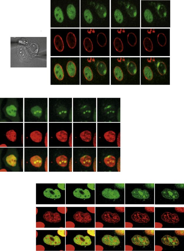
play a similar role in prophase when condensation engages              the nucleoplasm and 9.5% ± 3% were at the NE, in prophase
chromatin domains associated to the NE? Intriguingly, it has           cells, 78.4% ± 3.3% were in the nucleoplasm and 21.6% ±
been shown that ATR contains many HEAT repeats (Perry and              3.4% were at the NE.
Kleckner, 2003) that can behave as elastic connectors (Grinthal           Because ATR and Chk1 have been found associated with cen-
et al., 2010), suggesting that ATR might be influenced by              trosomes (Krämer et al., 2004; Zhang et al., 2007a), we costained
mechanical forces.                                                     prophase cells with Abs against ATR and a-tubulin (Figure 2A).
  We therefore investigated whether ATR responds to the                The ATR fraction associated with centrosomes was lower
mechanical stimuli deriving from chromosomal dynamics. We              compared to the one bound to the NE and partially overlapped
found that a fraction of human and mouse ATR localizes at the          in places in which centrosomes contact NE. Colcemid (a micro-
NE during S phase, particularly under conditions of enhanced           tubule-depolymerizing drug) treatment, did not affect ATR-
replication stress, and in prophase of unperturbed cell cycles.        NE association (Figure 2A). Hence, the NE-bound ATR in
Osmotic stress or mechanical stimulation of the plasma mem-            prophase unlikely depends on centrosomes and microtubule
brane cause relocalization of ATR to the inner and outer nuclear       polymerization.
membranes, independently of the cell-cycle stage and of RPA or            Next, we examined whether the NE distribution of ATR de-
DNA damage. Thus, ATR responds to mechanical forces at the             pended on its kinase activity and whether other checkpoint fac-
NE. Our observations suggest that ATR mediates a mechanical            tors exhibited a similar localization. We did not observe changes
response to membrane stress that could be caused by chro-              in the NE distribution of ATR following treatment with an ATR in-
matin dynamics and is important for genome integrity.                  hibitor (ATRi) (Toledo et al., 2011) (Figure 2B). We also found that
                                                                       a fraction of ATR-interacting protein (ATRIP) was localized at the
RESULTS                                                                NE along with Nup153 (Figure 2C). Under unperturbed condi-
                                                                       tions, Chk1 is phosphorylated at serine 345 during mitosis, and
A Fraction of ATR Localizes at the NE                                  this event is essential for viability (Wilsker et al., 2008). We found
DNA torsional stress generates mechanical strain and arises            a prophase-specific NE localization of p-Chk1 that was inhibited
during chromatin condensation, when the DNA packaging rea-             by ATRi treatment (Figure 2D). The specificity of the anti-
ches the maximal complexity and, transiently, during S phase           phospho-Ser 345-Chk1 Ab was confirmed in HU-treated cells
(Wang, 2002). Based on our previous findings (Bermejo et al.,          (Figure S2). Hence, fractions of ATR, ATRIP, and p-Chk1 localize
2011), we reasoned that lamin-associated chromatin might               at the NE during the cell cycle, under physiological conditions;
mediate the transfer of mechanical forces resulting from DNA           ATR association with the NE does not depend on its kinase
torsional stress to the NE. Given the data on ATR discussed            activity. We also found that ATR and p-Chk1 distributed around
above from our laboratories and others’, we tested whether             the NE following aphidicoline- (Figure 2E) and hydroxyurea- (HU)
ATR localizes at the NE. We examined ATR localization by indi-         (data not shown) induced replication stress. Altogether, these re-
rect immunofluorescence (IF), using an anti-ATR antibody (Ab),         sults are consistent with the initial hypothesis that topological
the specificity of which was validated in HeLa cells depleted          stress accumulating during chromatin condensation in prophase
for ATR (Figure S1A available online) and in ATR-defective             and during chromatin replication following replication stress
human Seckel fibroblasts (Figure S1B). In asynchronous HeLa            evokes a response at the NE that leads to ATR recruitment and
cells, ATR localized mostly in the nucleus in interphase and           activation.
S phase cells (Figure 1A). In prophase, part of ATR exhibited a
perinuclear distribution, which was lost later in mitosis, following   Experimentally Induced Mechanical Stress Triggers the
NE breakdown (NEBD) (Figure 1A). Using structured illumination         NE ATR Response Independent of DNA Damage
super resolution microscopy (SR-SIM), we analyzed ATR and              To more specifically address whether the NE ATR response is
Nup153 colocalization in prophase cells. Both proteins localized       triggered by the NE undergoing mechanical stress, we adopted
at the NE (Figure 1B), exhibiting 70% colocalization. Consis-          a variety of cell manipulation assays that are routinely used
tently, several nucleoporins, including Nup153, are targeted by        to induce mechanical stress at cell membranes, including
ATR/Chk1 (Blasius et al., 2011; Matsuoka et al., 2007). Notably,       osmotic stress, patch-clamp-induced cell stretching, and cell
ATR was prominently enriched at NE invaginations (Figure S1C),         compression.
which often mediate the mechanical attachment of nucleoli and
spindles (Beaudouin et al., 2002; Bourgeois et al., 1979). The         Osmotic Stress Leads to ATR Relocalization at the NE
observations described above were confirmed in primary human           and Chk1 Activation
IMR90 and murine NIH 3T3 fibroblasts (Figure S1C). In leptomy-         We first analyzed ATR distribution in response to osmotic stress,
cin B (a nuclear export inhibitor; Kudo et al., 1998)-treated          a potent inducer of mechanotransduction at cell membranes
prophase cells, ATR intracellular distribution did not change (Fig-    (Martins et al., 2012). Under hyperosmotic conditions, the cell
ure S1D), thus arguing against a contribution of nuclear export        and the nucleus shrink, causing NE ruffling and chromatin
to ATR localization. Cellular distribution of ATR was further          condensation (Martins et al., 2012). Hypotonic stress causes
analyzed by electron microscopy (EM). Gold ATR immunostain-            cell swelling and induces membrane tension but also alters the
ing revealed a nuclear and nonnuclear distribution of ATR parti-       chromatin structure, apparently without causing DNA breaks
cles (Figure 1C). A fraction of these particles localized at the NE,   (Bakkenist and Kastan, 2003).
particularly in prophase. Indeed, whereas in interphase, among           Hypertonic and hypotonic conditions caused Chk1 phos-
the ATR particles visible in the nucleus, 90.5% ± 3% were in           phorylation in an ATR-dependent manner (Figure 3A). By IF,

634 Cell 158, 633–646, July 31, 2014 ª2014 The Authors
A                                                                                                                                   100             Interphase

                                                                                                                           % fluorescence intensity
                                                                                                                                                               Prophase
                                                                                                                                                      80
            Interphase

                                                                                                                                                      60

                                                                                                                                                      40

                                                                                                                                                      20
                                                                                                                                                      0
                                                                                                                                                           C    N    NE
            Mitosis

            B

           C
                                        plasma membrane

                                                    1
                                                                                         2
                             nuclear envelope
                                                                                                     nucleus
                                                                          plasma membrane

                                    Interphase                                  Prophase
                         (Immuno Cryo-EM ATR-nano gold)               (Immuno CLEM ATR-nano gold)

                         1                                        2

                                                                                       ATR

Figure 1. A Fraction of ATR Localizes at the NE during the Cell Cycle
(A) Confocal images of cells stained with anti-ATR Ab (green), DAPI (blue), EdU (red), or p-H3 (red). The graph shows the percent fluorescence intensity in the
cytoplasm (C), nucleus (N), and nuclear envelope (NE) on a set of cells (n = 15).
(B) Superresolution image of a prophase cell stained with anti-ATR (green) and -NUP153 (red) Abs. A magnification is shown.
(C) Left: cryosection-based immuno-EM of an interphase cell stained with gold-tagged ATR Ab. A magnification of the selected area one (rectangle) highlights
sparse ATR staining at the NE. Right: correlative light EM images of a prophase cell stained with gold-tagged ATR Ab. A magnification of the selected area two
(rectangle) highlights the prominent localization of ATR at the NE. The prophase cell used to perform immuno-CLEM is shown in the lower panel, stained with anti-
ATR (green) Ab.
See also Figure S1.

                                                                                             Cell 158, 633–646, July 31, 2014 ª2014 The Authors 635
A                                                                                            Figure 2. Checkpoint Proteins Localizing at
                                                                                             the NE
                                                                                             (A) Confocal images of cells in prophase stained
                                                                                             with anti-ATR, -a-tubulin Abs, and DAPI ± colce-
                                                                                             mid (100 nM; 2 hr).
                                                                                             (B) Confocal images of prophase cell stained with
                                                                                             DAPI and anti-ATR, -p-histone H3 (S10) Abs ±
                                                                                             ATRi (2 mM; 2 hr). The graphs show the percent
                                                                                             fluorescence intensity on a set of cells (n = 10).
                                                                                             (C) Prophase cell stained with anti-ATRIP,
B                                                                                            -NUP153, -p-histone H3 (S10) Abs, and DAPI. A
                                                                                             magnification of the dotted merged area is shown.
                                                                                             (D) Confocal images of NIH3T3 prophase cells
                                                                                             stained with DAPI and anti-p-Chk1 (S345) ± ATRi
                                                                                             (3 ± mM; 1 hr).
                                                                                             (E) HeLa cells were treated with aphidicholine
                                                                                             (3 mM; 16 hr) and stained with anti-ATR, -p-Chk1
                                                                                             Abs, and DAPI.
                                                                                             See also Figure S2.

C

                                                                                              using other hyperosmotic conditions
                                                                                              (0.4 M NaCl) (Figures S3B and S3D).
                                                                                                 Hypotonic stress relocalized ATR at
                                                                                              certain regions on the NE and in the
                                                                                              nucleoli (visualized by anti-TPR Ab and
                                                                                              by RFP-Nucleophosmin, respectively)
D                                         E                                                   (Figure 3E). Because mechanical signals
                                                                                              at the cytoplasmic level can also be expe-
                                                                                              rienced in the nucleolus (Hu et al., 2005;
                                                                                              Wang et al., 2009), and the nucleolus in-
                                                                                              teracts with the NE (Bourgeois et al.,
                                                                                              1979), ATR relocation at the nucleolus in
                                                                                              response to hypotonic conditions likely
                                                                                              reflects a response to mechanical forces,
                                                                                              perhaps mediated by the NE and cyto-
                                                                                              skeleton (Hu et al., 2005; Maniotis et al.,
                                                                                              1997). Given that ATR responds to DNA
we then compared the ATR and chromatin cellular distribution         damage, we also determined whether hypertonic conditions
in cells growing under normal conditions or treated with sorbitol    induced DNA damage. We did not observe accumulation of
(Figure 3B). Compared to untreated conditions, hyperosmotic          gH2AX signals in sorbitol-treated cells, compared to untreated
stress caused a redistribution of ATR at the NE that did             conditions (Figure S3D). However, gH2AX signals increased in
not correlate with changes in chromatin localization (Fig-           cells treated with high NaCl doses (Figure S3D), consistent
ure S3A). We observed ATR redistribution at the NE in                with previous findings showing that high NaCl causes DNA dam-
response to sorbitol treatment in U2OS and IMR90 cells as            age (Kültz and Chakravarty, 2001). Leptomycin-B treatment of
well (Figure S3B). By time-lapse analysis in HeLa cells, we          cells exposed to hyperosmotic stress did not influence ATR
found that GFP-ATR relocalized on the NE within 30 seconds           localization at the NE (Figure S3E).
(s) following hypertonic stress (data not shown). By EM, we             ATR activation by DNA damage is influenced by RPA-ssDNA
further investigated ATR association to the NE in cells treated      nucleofilaments (Zou and Elledge, 2003). We compared ATR
with sorbitol. Whereas in isotonic conditions ATR particles          and RPA localization, in cells treated with or without sorbitol.
accumulated mostly within the karyoplasm, in response to hy-         RPA32 and ATR exhibited intranuclear staining (Figure 4A).
pertonic stress, a significant fraction of ATR particles accumu-     Certain cells undergoing DNA replication (visualized by EdU) ex-
lated at the NE (Figure 3C). ATR nanogold particles localize at      hibited a perinuclear staining of RPA32 (Dimitrova et al., 1999;
both the inner and the outer membranes. ATRIP and p-Chk1             Vassin et al., 2009), but not of ATR. In sorbitol-treated cells,
also localized, in part, at the NE in response to sorbitol treat-    whereas ATR relocalized at the NE, RPA32 remained intranu-
ment (Figures 3D and S3C) in an ATR-dependent manner (Fig-           clear. A small subset of S phase cells showed perinuclear staining
ure 3D). Hence, the NE is a site for ATR activation, following hy-   of RPA32 (Figure S4A). To firmly address whether the NE translo-
perosmotic stress. We obtained analogous results in different        cation of ATR under hypertonic stress was RPA independent, we
cell types, both cancer cell lines and normal diploid cells, and     examined ATR and p-Chk1 localization in RPA70-depleted cells

636 Cell 158, 633–646, July 31, 2014 ª2014 The Authors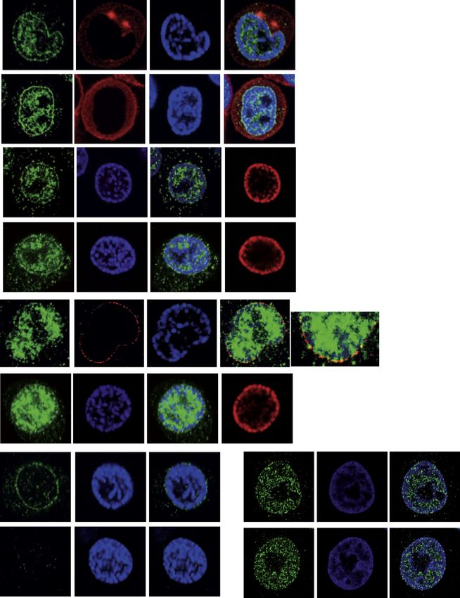
(Figures 4B and 4C). Although RPA70 depletion affected Chk1           ATR redistribution are within the physiological range of forces
phosphorylation in response to HU treatment (data not shown;          experienced by cells in soft tissues and whether the ATR
(Zou and Elledge, 2003), under hyperosmotic stress it neither         mechanical response is reversible, we used the compressive-
influenced Chk1 phosphorylation (data not shown) nor influenced       load system (CLS) (to exert forces in the approximate range of
ATR and p-Chk1 levels at the NE (Figures 4B and 4C). Analo-           1 to 1,000 nN (Figure 5C). Using CLS, which is based on weights
gously, Rad17 and TopBP1, which have also been implicated in          that impose deformations on cells by axial compression, it is
ATR activation in response to DNA damage (Duursma et al.,             possible to approximately estimate the magnitude of the forces
2013; Wang et al., 2006), were dispensable for ATR and p-Chk1         applied to induce the ATR response and to study the recovery
localization to the NE, in response to hyperosmotic stress (Fig-      from compression, after unloading the weights. When we
ures S4B–S4D; data not shown). Hence, hyperosmotic stress-            applied CLS on HeLa cells cotransfected with GFP-ATR and
induced ATR relocalization and activation on the NE is RPA,           m-cherry H2B (Figure 5C), GFP-ATR redistributed at perinuclear
Rad17, Tobp1, and DNA damage independent.                             regions and in the nucleolus. The ATR localization in the nucle-
                                                                      olus anticipated the perinuclear redistribution of ATR. Approxi-
Whole-Cell Mechanostimulation Targets ATR to the NE                   mately 15–30 nN forces were needed to visualize ATR relocation.
We then used an alternative method to induce mechanical stimu-        When we unloaded the weights, both the nucleolus and the
lation at cell membranes. In this case, a patch-clamp-automa-         nuclear envelope rapidly lost the GFP-ATR signal, and ATR
tized platform was employed to stretch the cell using two glass       assumed the typical distribution of unstressed cells (Figure S5B).
pipettes placed on the plasma membrane at opposite poles              Recovery of ATR distribution paralleled chromatin decompac-
in cell-attached configuration (Figure 5A). We reasoned that          tion (Figure S5B). Hence, ATR responds to mechanical stimula-
plasma membrane stretching propagates the mechanical stimu-           tion of the plasma membrane and to cell compression. Following
lation to the NE promoting structural rearrangements within the       mechanical stress, the ATR redistribution to the nucleolus pre-
nucleus (Wang et al., 2009). By moving the pipettes apart, we         cedes the ATR localization to the nuclear envelope. This perhaps
induced in HeLa cells a mechanical stretch of the plasma mem-         reflects a differential viscoelastic response of these two cellular
brane and followed the distribution of GFP-ATR (Figure 5A; Movie      compartments. The ATR response to mechanical stress occurs
S1). ATR rapidly redistributed at perinuclear regions overlapping     at physiological range of forces and is fully reversible. Moreover,
with the lamin A signal. ATR perinuclear localization initiated at    the changes in ATR cellular distribution, following activation and
the focal points of the mechanical tension and progressively          recovery upon mechanical stress, parallels chromatin compac-
distributed along adjacent NE areas. We observed an accum-            tion and decompaction, respectively.
ulation of GFP-ATR in the nucleolus as well (Figure 5A). We
addressed whether the pipette-mediated cell deformation               ATR Influences the Coordination between Chromatin
caused chromatin condensation as in the case of nuclear defor-        Condensation and NEBD
mation (Shivashankar, 2011) (Figure 5B). We will refer to this        The above data show that mechanical stress induces an ATR NE
phenomenon as force-induced chromatin compaction, to distin-          response, suggesting a role for ATR in coordinating chromatin
guish it from the chromatin condensation process taking place         dynamics and NE metabolism in response to mechanical stimu-
in prophase. Concomitantly with the accumulation of GFP-ATR           lations. To address this intriguing concept in a physiological sce-
in the nucleolus, 30 s after pipette mechanostimulation, chromatin    nario, we next monitored the kinetics of chromatin condensation
compaction was evident, as shown by the m-cherry H2B distribu-        and NEBD in HeLa cells, cotransfected with GFP-H2B and RFP-
tion. Subsequently, GFP-ATR localizes at the nuclear periphery.       Lamin A, prearrested in G2 with a CDK1 inhibitor, and released
We consistently observed that mechano-induced GFP-ATR                 into mitosis with or without active ATR (Figure 6A). In cells
redistribution at the nucleolus and/or at the NE paralleled chro-     released into mitosis with a functional ATR, chromatin conden-
matin compaction and that the association of ATR to the nucleolus     sation began at 20 min, was nearly completed by 30 min, and
anticipated the ATR localization at the NE (data not shown). Again,   reached the typical metaphase configuration at 70 min. At
by comparing the distribution of ATR and chromatin following cell     20 min a fraction of lamin A was still tethered to the partially
stretching, we observed no spatial correlation between chromatin      condensed chromatin. By 30 min, NEBD was completed and
movement and ATR relocalization (Figure 5B). Mechanical forces        lamin A was totally dissolved. In cells entering mitosis with phar-
can be transferred by transmembrane receptors that convert            macologically inactived ATR, the onset and the completion of
mechanical stimuli into biochemical signals to modulate various       chromatin condensation were significantly delayed: it started
intracellular functions (Mammoto et al., 2012). In particular, cal-   at 75 min and completed around 115 min. A fraction of lamin A
cium ions are the most rapid mediators of mechanical stimuli.         was still visible and attached to the condensed chromatin until
However, in the case of ATR, extra/intracellular calcium chelation    85 min. NEBD and lamin A disintegration were observed, but
using BAPTA did not affect GFP-ATR cellular redistribution            after 115 min. To rule out that the uncoordinated chromatin
following mechanical stress (data not shown). Mechanostimula-         condensation and NEBD observed in ATR-inhibited cells was
tion by pipettes did not cause accumulation of DNA damage as          due to DNA damage left unresolved because of the absence of
assessed by examining g-H2AX foci (Figure S5A).                       ATR activity, we analyzed g-H2AX foci by IF (Figure S6). We
    The procedure described above has some limitations because        observed comparable g-H2AX foci numbers in control and
it is based on glass pipette-attached cell stretching and does not    ATR-inhibited cells in mitosis. To further confirm the above ob-
allow us to study the recovery from the mechanostimulation.           servations, we used human primary fibroblasts from Seckel
Therefore, to determine whether the mechanical forces inducing        patients, who carry hypomorphic mutations in the ATR gene

                                                                               Cell 158, 633–646, July 31, 2014 ª2014 The Authors 637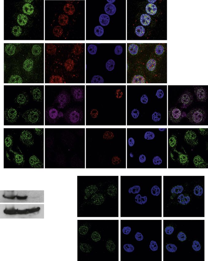
A HU Hyper Hypo B 100 Isotonic
% fluorescence intensity
- - ATRi - ATRi - ATRi Hypertonic
Exponential
80
p-Chk1
60
Chk1 ATR DAPI Merge
40
Exponential
+ Sorbitol
20
0
ATR DAPI Merge C N NE
C ISOTONIC HYPERTONIC Isotonic
100 Hypertonic
N
% Gold Particles
80
N
60
C 40
nuclear envelope 20
nuclear envelope
C 0
ATR -nano gold ATR -nano gold N NE N NE
D
p-Chk1 fluorescence intensity
80
+ Sorbitol
60
p-Chk1 DAPI p-Chk1/ DAPI
40
ATRi + Sorbitol
20
0
p-Chk1 DAPI p-Chk1/ DAPI - + ATRi
E 1 1
Hypotonic Stress
ATR TPR DAPI Merge
ATR RFP-NMP DAPI Merge
Figure 3. Osmotic Stress Induces ATR Relocalization at the NE
(A) HeLa cells were incubated with DMSO or ATRi (3 mM; 2 hr) and treated with HU (10 mM; 1 hr) and then hypertonic media (40 min) or hypotonic media (40 min),
respectively. Cells were examined by western blot using anti-p-Chk1 and Chk1 Abs.
(B) HeLa cells were exposed to mock or hypertonic medium (0.5 M sorbitol; 20 min) and stained with anti-ATR Ab (green) and DAPI (blue). The graphs show the
percent fluorescence intensity on a set of cells (n = 50).
(C) Immuno-cryo-EM images of HeLa cells stained with gold-tagged anti-ATR Ab under normal or hypertonic conditions. White arrows highlight ATR gold
particles. The graph represents the percentage of ATR-related nanogold particles in isotonic or hypertonic conditions in the nucleoplasm or at the nuclear
envelope.
(legend continued on next page)
638 Cell 158, 633–646, July 31, 2014 ª2014 The Authors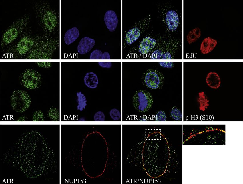
A Figure 4. ATR Activation at the NE Does Not
Depend on RPA
Exponential
(A) Confocal images of HeLa cells exposed to
normal or hypertonic medium (0.5 M sorbitol;
ATR RPA 32 DAPI Merge 20 min). Samples were stained with anti-ATR
(green), -RPA32 (red) Abs, and DAPI.
(B) HeLa cells were transfected with empty vector
or RPA70 shRNA and selected with 3 mg/ml
Exponential
+ Sorbitol
puromycin (72 hr). Cells were incubated with
hypertonic medium containing 0.5 M sorbitol for
ATR RPA 32 DAPI Merge
20 min and stained with anti-ATR (green), -RPA70
B (magenta) Abs, S-phase-specific EdU (red), and
60 control DAPI (blue). RPA70 protein levels were analyzed
+ Sorbitol
Control
shRPA70
%ATR fluor. intensity
by western blot using anti-RPA70 Ab and anti-
40
tubulin as a loading control (lower panel). The
ATR RPA 70 EdU DAPI ATR / RPA 70 graph shows the percent ATR fluorescence in-
tensity in different cellular compartments on a set
20
RPA 70 shRNA
of cells (n = 20).
+ Sorbitol
(C) Puromycin-selected RPA70 shRNA-trans-
0
C NE N fected HeLa cells were incubated with hypertonic
ATR RPA 70 EdU DAPI ATR / RPA 70 medium containing 0.5 M sorbitol for 30 min and
stained with p-Chk1 (green) and DAPI (blue).
Scramble
RPA 70
shRNA
Vector
C Mean fluorescence intensity (AU) Graphs show the integrated p-Chk1 fluorescence
4000 intensity on a set of cells (n = 20). Quantitative
+ Sorbitol
RPA70 analysis of P-Chk1 levels is shown.
Control
3000
See also Figure S4.
Tubulin
p-Chk1 DAPI Merge 2000
1000
RPA 70 shRNA
+ Sorbitol
0
DISCUSSION
Scrambled
shRNA
shRNA
RPA
p-Chk1 DAPI Merge
In this study, we showed that in human
and mouse cells grown under unper-
turbed conditions, a fraction of ATR,
(O’Driscoll et al., 2003) (Figure 6B). As above, control and ATRIP, and pChk1, key components of the ATR signaling
Seckel cells were prearrested with a CDK1 inhibitor and released cascade, accumulate at the NE. Our working model is that
into mitosis. In control cells, chromatin condensation and ATR responds to chromatin dynamics in S phase (particularly un-
NEBD were evident already at 15 min and completed by der replication stress) and prophase, when chromatin replication
30 min; metaphase cells were observed at 75 min. In contrast, and condensation generate topological stress that is converted
in Seckel cells, chromatin condensation and NEBD were into mechanical stimuli. Whereas our previous findings in yeast
delayed; at 30 min, intact NEs were still observed; at 45 min, indicated replication stress as a major cause of mechanical
cells exhibiting condensed chromatin attached to large por- stress on the nuclear membrane (Bermejo et al., 2011), our cur-
tions of lamin A were detected; and complete NEBD and rent data in mammalian cells may suggest that ATR plays a more
chromatin condensation were visualized only at 75 min. Analo- prominent role at the NE in prophase when chromatin condensa-
gously, when we used mouse embryonic fibroblasts derived tion occurs. This might reflect the magnitude of forces acting
from humanized ATR hypomorphic Seckel mice (Murga et al., during chromatin condensation/decondensation (Mazumder
2009), under the same experimental conditions, we observed a et al., 2008), which is greater than the forces acting during
30 min delay in chromatin condensation and NEBD (data not DNA synthesis and/or important differences between chromatin
shown). condensation and chromatin replication from the topological
Thus, in ATR-defective cells, the initiation and completion of point of view. During prophase chromatin condensation causes
chromatin condensation and NEBD are delayed; moreover, torsional stress, whereas during an unperturbed S phase, mov-
portions of lamin A remain associated with condensed chro- ing replication forks are assisted by DNA topoisomerases that
matin even after the NEBD, suggesting a defect in detaching travel with the forks (Bermejo et al., 2007) and prevent the accu-
condensed chromatin from the NE. Similar defects were also mulation of torsional stress. However, given our previous results
observed in Seckel cells (Alderton et al., 2004). during S phase in yeast, it is possible that transient mechanical
(D) HeLa cells were incubated with DMSO or ATRi (ATRi), followed by treatment with hypertonic medium (0.5 M sorbitol; 20–30 min) and stained with anti-p-Chk1
Ab (green) or DAPI (blue). The graphs show the mean fluorescence intensity on a set of cells (n = 10).
(E) Top: HeLa cells were incubated in hypotonic medium and stained with anti-ATR Ab (green), anti-TPR Ab (red), and DAPI (blue). A magnification of the merged
ATR, TPR, and DAPI is shown. Bottom: RFP-NPM-transfected cells were treated as in the top panel and stained with anti-ATR Ab and DAPI.
See also Figure S3.
Cell 158, 633–646, July 31, 2014 ª2014 The Authors 639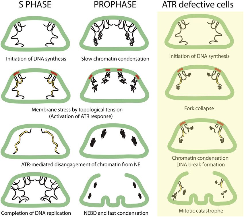
A Mechanical stretch
Glass pipette 0 sec. 90 sec. 120 sec. 180 sec.
GFP-ATR GFP-ATR GFP-ATR GFP-ATR
Glass pipette
RFP-Lamin A RFP-Lamin A RFP-Lamin A RFP-Lamin A
Merge Merge Merge Merge
B Mechanical stretch
1 ATR
2
distribution (AU)
DAPI
distribution (AU)
100 100
% Maximal IF
% Maximal IF
80 80
60 60
40 40
20 20
0 0
Distance Distance
GFP-ATR GFP-ATR GFP-ATR GFP-ATR GFP-ATR
3 4
distribution (AU)
distribution (AU)
100 100
% Maximal IF
% Maximal IF
80 80
60 60
40 40
20 20
0 0
Distance Distance
m-cherry-H2B m-cherry-H2B m-cherry-H2B m-cherry-H2B m-cherry-H2B
1 5
1 2 3 4 5
distribution (AU)
100
% Maximal IF
80
60
40
20
0
Distance
Merge Merge Merge Merge Merge
C cover slip Before Load Cover slip (5nN) (30 nN) (30 nN) (30 nN)
weight 10 min. 20 min. 30 min.
glass bottom
culture dish
cells
oil immersion GFP-ATR GFP-ATR GFP-ATR GFP-ATR GFP-ATR
objective
m-cherry-H2B m-cherry-H2B m-cherry-H2B m-cherry-H2B m-cherry-H2B
Merge Merge Merge Merge Merge
Figure 5. ATR Responds to Mechanical Forces Acting on Membranes
(A) Schematic representation of two-patch-clamp pipettes positioned on the two opposite sides of the plasma membrane. The corresponding microscope image
is on the left. The white arrows indicate the points of focal tension induced by the mechanical stretching. Two patch pipettes were attached to the plasma
membrane of a HeLa cell coexpressing GFP-ATR (green) and RFP-Lamin A (red) and stretched to induce mechanical stress on the membranes. The intracellular
changes in GFP-ATR localization was examined in real time with a gap of 30 s.
(legend continued on next page)
640 Cell 158, 633–646, July 31, 2014 ª2014 The AuthorsA + ATR inhibitor
0 min 20 min 25 min 30 min 35 min 70 min
0 min 70 min 75 min 80 min 85 min 115 min
B RO 3306 15’ release 30’ release 45’ release 75’ release
IMR 90
Lamin A/DAPI Lamin A/DAPI Lamin A/DAPI Lamin A/DAPI Lamin A/DAPI
RO 3306 15’ release 30’ release 45’ release 75’ release
ATR Seckel
Lamin A/DAPI Lamin A/DAPI Lamin A/DAPI Lamin A/DAPI Lamin A/DAPI
Figure 6. ATR Coordinates Chromatin Condensation and NEBD
(A) RFP-Lamin-A-transfected GFP-H2B HeLa cells were arrested in G2 with the R3306 Cdk1 inhibitor (10 mM, 16 hr), followed by incubation with DMSO or ATRi
(3 mM, 1 hr). Later, the cells were released into mitosis ± ATRi, and cells entry into prophase and metaphase was examined. A representative image of n = 10 is
shown.
(B) Same as in (A), IMR90 and ATRs/s human fibroblasts were arrested in G2 with Cdk1 inhibitor R3306 (10 mM, 16 hr), followed by release in mitosis. Cells were
fixed and stained for anti-Lamin A (red) and DAPI (blue) at represented time intervals postrelease.
See also Figure S6.
signals are generated when forks approach LADs. Although we two adjacent LADs may act as mechanosensors of local chro-
did not observe association of ATR to the NE during unperturbed mosomal forces and undergo changes in membrane fluidity
S phase in mammalian cells, there might be a transient relocali- and/or curvature. Changes in membrane fluidity might muffle
zation spread over the several hours that normal S phase lasts. mechanical stress and eventually transduce the signal to ATR.
Perturbing S phase with, aphidicolin- or HU-induced replication This is consistent with the observations that membrane fluidity
stress, which would stall replication forks and generate a persis- is modulated in response to a variety of stress conditions (Aguilar
tent mechanical signal at the NE, did indeed trigger accumula- and de Mendoza, 2006; Vigh et al., 2007), including HU treat-
tion of ATR at the NE. ment (Fujikawa-Yamamoto and Odashima, 1989). The relative
Further work will be required to determine how mechanosens- abundance of polyunsaturated fatty acids influences nuclear
ing influencing ATR localization occurs. NE portions connecting membrane fluidity and ATR activation (Zhang et al., 2007b).
(B) Same as in (A), two patch pipettes were attached to the plasma membrane of a HeLa cell coexpressing GFP-ATR (green) and m-cherry H2B (red) and
stretched to induce mechanical stress on the membranes. The intracellular changes in GFP-ATR and chromatin-associated H2B localization were examined in
real time. The graphs illustrate the quantification in arbitrary units (a.u.) of the distribution of GFP-ATR and m-cherry-H2B by IF along the lines shown in the merged
panels.
(C) Schematic representation of the setup used to perform compressive load experiments (left panel). GFP-ATR and m-cherry-H2B-transfected cells were
compressed with 30 nN forces using weights, and the intracellular changes in GFP-ATR localization and chromatin architecture (m-cherry H2B) were examined in
real time with a gap of 10 min. A representative image of n = 10 is shown.
See also Figure S5 and Movie S1.
Cell 158, 633–646, July 31, 2014 ª2014 The Authors 641chromatin from the NE in S phase (Bermejo et al., 2011), we pro-
pose that, during unperturbed cell cycles, the recruitment of
ATRIP-ATR at the NE contributes to coordinate chromatin and
NE dynamics, in S phase, as well as in prophase (Figure 7). Chro-
mosome replication and condensation, besides being topologi-
cally complex, are influenced by the NE. However, there are key
evolutionary differences between Saccharomyces cerevisiae
and higher eukaryotes that may explain certain species-specific
aspects of the ATR pathway. In yeast, the NE does not break-
down and chromatin condensation never reaches the level of
complexity seen in mammalian cells. In fact, the yeast Mec1/
ATR pathway is prominently involved in the coordination of
S phase events through Rad53, whereas Chk1 plays a minor
role. However, certain mec1 alleles exhibit the typical features
of ATR-deficient cells (Cha and Kleckner, 2002). In human cells,
chromatin condensation begins slowly at 30 min before NEBD
and proceeds more rapidly following the envelope breakdown
(Beaudouin et al., 2002). Thus, in mammals, the coordination be-
tween chromatin condensation and NE breakdown likely plays a
key role in preventing aberrant chromosomal dynamics. Accord-
ingly, Chk1 plays a pivotal function in human cells, and inactiva-
Figure 7. A Model Describing ATR-Mediated Events in S Phase and
tion of ATR causes aberrant chromatin condensation and NEBD
Prophase at the Nuclear Envelope
The model integrates the observations deriving from yeast studies (Bermejo
(Nghiem et al., 2001). Hence, whereas in mammals ATR might be
et al., 2011) focused on the S phase function of the ATR pathway, with the mainly involved in coordinating the topological complexity of
findings described in this paper. The nuclear envelope is shown in green; chromatin condensation with NE dynamics (while still performing
the chromosomes are shown in black; and the replicons are shown in yellow. important functions in S phase, particularly under enhanced
The red bars at the envelope indicate the nuclear membrane areas exposed to replication stress), in yeast it might be primarily required to
mechanical stress. Briefly, nuclear envelope-associated chromatin transfers deal with the topological problems arising at perinuclear chro-
the mechanical forces arising from the topological transitions of replicating or
matin in S phase (Bermejo et al., 2011). The fact that chromatin
condensing chromosomes to the nuclear envelope. Local nuclear membrane
stress recruits ATR. The ATR response then coordinates chromosome repli- condensation starts at a low rate, while the NE is still intact,
cation and condensation with the nuclear envelope by modulating chromatin may reflect the topological complexity of condensing the chro-
and nuclear envelope association. In ATR-defective cells, the inability to matin fiber, where there are still chromatin regions bound to
coordinate replication with the nuclear envelope at those chromatin regions the envelope and that may be the reason why, following
associated to the lamin, causes fork collapse. The partially replicated chro- NEBD, condensation is faster. We propose that ATR responds
matin remains in part associated to the NE when condensation begins, thus
to the topological tension when condensation begins, binds
leading to progressive chromatin fragmentation and mitotic catastrophe.
the NE, and facilitates the condensation process by progres-
sively resolving the LADs. This is consistent with our finding
Membrane fluidity can influence its curvature (McMahon and that ATR-inhibited/mutated cells are delayed in the processes,
Gallop, 2005). Notably, we observed that ATR is enriched at leading to chromatin condensation and NEBD (Figure 6) and
NE invaginations, structures that are known to arise in response with the observation that Seckel cells exhibit nuclear fragmenta-
to mechanical forces (Beaudouin et al., 2002). tion, while the chromatin is still attached to the NE (Alderton
ATR recruitment to the NE in response to membrane stress et al., 2004). Moreover, ATR has been implicated in the phos-
does not require its kinase activity. We speculate that, in phorylation of condensin subunits, as well as of NE proteins
response to mechanical stimuli, ATR is part of a multistep activa- (Matsuoka et al., 2007). According to the scenario we propose,
tion process involving (1) changes in nuclear membrane fluidity the inability to regulate the association between the chromatin
and/or curvature, (2) ATRIP and ATR recruitment at the NE, and the NE in response to mechanical stimuli arising from topo-
and (3) ATR kinase activation in an unorthodox manner indepen- logical transitions might have the following consequences: in S
dent of RPA and DNA damage. The observation that the N termi- phase cells, the chromatin fiber would remain in part attached
nus of ATR exhibits elastic properties (Grinthal et al., 2010; Perry to the envelope, thus causing fork collapse. Attempts to conden-
and Kleckner, 2003), whereas the kinase domain represents a sate a partially replicated chromatin, still bound to the NE, would
small portion of the C terminus, makes ATR an ideal module to generate DNA breaks and aberrant condensation events (Fig-
respond to mechanical stimuli, perhaps through a mechanism ure 7). Indeed, ATR-deficient cells exhibit some of these defects
mediated by its elastic domain. Interestingly, TORC2 has been (Brown and Baltimore, 2003; Cha and Kleckner, 2002; Nghiem
implicated in the cellular response to membrane stress (Berch- et al., 2001). Intriguingly, recent observations showed that fragile
told et al., 2012) and shares homology with ATR (Perry and site expression in mec1 mutants is mediated by condensins and
Kleckner, 2003). DNA topoisomerase II (Hashash et al., 2012). The nuclear frag-
Based on our present data in mammalian cells and on previous mentation phenotype of Seckel cells (Alderton et al., 2004;
findings implicating the yeast ATR ortholog Mec1 in detaching O’Driscoll et al., 2003) can be expressed only if cells have
642 Cell 158, 633–646, July 31, 2014 ª2014 The Authorspreviously undergone fork collapse and occurs when the NE is and Millipore; and Chk1 was obtained from Leica. EdU (click-IT), RPA70-,
still intact. In our experimental conditions (Figure 6), we did not TopBP1-, and RAD17-small hairpin RNAs (shRNAs) were obtained from
Invitrogen and Origene Technologies, respectively. ATR-specific inhibitor
observe nuclear fragmentation as cells passaged through the
and ATR shRNA were from Dr. Oscar Capetillo (Centro Nacional de Oncologia
preceding S phase with a functional ATR. [CNIO]) (Toledo et al., 2011); the GFP-ATR plasmid was from Dr. Randal
One intriguing aspect of our present data set is the result that Tibbetts (Tibbetts et al., 2000); RFP-Lamin was from Prof. Howard J. Worman
the classical DNA damage-induced ATR activation pathway, (Columbia University) (Ostlund et al., 2006); and RFP-Nucleophosmin was a
which is influenced by RPA, TopBP1, and the 9-1-1 complex, gift by Dr. Michelle Hill (University of Queensland). GFP-H2B and m-cherry-
is genetically uncoupled from the ATR response to mechanical H2B were from IFOM. Colchicine, a-tubulin Ab, Leptomycin B, Hydroxy
Urea, sorbitol, Aphidicholine, and R3306 CDK1-specific inhibitor were ob-
stress. Given the large number of nuclear and nonnuclear targets
tained from Sigma.
modulated by ATR, it is reasonable to think that ATR might be
engaged in at least partly different pathways in response to Cell Culture, Transfection, Treatments, and Cell Lysis
different stimuli. One possibility is that the RPA-ssDNA-medi- HeLa, U2OS, NIH 3T3, and mouse embryonic fibroblasts cells were main-
ated signaling generates the context for engaging ATR when tained in complete medium as described (Kumar et al., 2006). Media were
cells experience genotoxic insults, whereas the membrane- supplemented with 0.5 M sorbitol (or 0.4 M NaCl) for hypertonic conditions
dependent checkpoint activation may be more confined to a or diluted 1:5 with MQ water for hypotonic conditions. IMR90 and Human
Seckel cells (Coriell Cell Repository) were grown in Eagle’s minimum essen-
physiological context to sense subtle chromatin dynamics. How-
tial medium (MEM; GIBCO-BRL), supplemented with nonessential amino
ever, certain insults, such as replication stress, might trigger both acids, 10% (vol/vol) fetal bovine serum (not activated), 2 mM glutamine,
ATR responses, by accumulating single stranded DNA at forks 10 mM HEPES, 100 U/ml penicillin, and 100 mg/ml streptomycin in a
(Toledo et al., 2013) and topological stress at perinuclear chro- humidified atmosphere (5% CO2, 37 C). We used lipofectamine 2000
matin (Bermejo et al., 2011). (Invitrogen) for transfection. Cells transfected with ATR shRNA or RPA70
Given that partially condensed silenced chromatin has a ten- shRNA were selected for 72 hr in medium plus 3 mg/ml puromycin. For hy-
perosmotic stress experiments, exponentially growing cells were seeded
dency to localize at the nuclear periphery (Shevelyov and Nur-
on dishes and after 24 hr, the medium was replaced with hypertonic medium
minsky, 2012), an ATR response regulating chromatin-lamin
and incubated for 20–30 min for ATR and for 30–40 min for p-Chk1 ex-
association in response to membrane stress might have impor- periments, before harvesting or fixing the cells. For hypotonic stress con-
tant epigenetic implications. ditions, cells were exposed to hypertonic medium for 2–5 min before
During S phase and prophase, the ATR relocation at the NE fixation. Total cell lysates were prepared in RIPA lysis buffer (Marqués
likely reflects a physiological response triggered by the topolog- et al., 2009). To arrest cells in G2 phase, GFP-H2B and RFP-Lamin-A-
ical stress arising from chromatin dynamics and transmitted to transfected HeLa cells were treated with 10 mM R3306 for 16 hr and were
later washed three times with complete medium followed by release in
the NE through LADs. Conversely, osmotic stress and mechan-
mitosis ± ATR inhibitor.
ical stimuli may lead to more dramatic cell deformation, mem-
brane stress, and chromatin rearrangements. Under osmotic Immunofluorescence
stress conditions, the response is likely triggered by membrane For IF assays, cells were processed as previously described (Kumar et al.,
stress directly. The pipette-induced mechanical stimulation of 2010, 2011). In brief, cells were fixed with 4% formaldehyde (10 min),
the plasma membrane might be instead transduced to the NE blocked, and permeabilized with 0.3% TX-100 PBS (20 min) and incubated
by the cytoskeleton (Wang et al., 2009). Finally, CLS-induced with primary antibodies (ATR and p-Chk1 1:50, TPR, p-histone H3 and
NUP153 1:500, p-H2AX, RPA32, ATRIP, and RPA70 1:200, and lamin A/C
cell deformation mimics the context of interstitial migration in
1:200) in blocking buffer (1 hr, room temperature [RT]), followed by three
which the nucleus has to squeeze through pores. CLS and inter- washes. Species-specific secondary antibodies were added to samples
stitial migration cause mechanical stress at the NE and chro- and incubated (1 hr, RT), followed by three washes with blocking buffer.
matin condensation (Friedl et al., 2011; Gerlitz and Bustin, Samples were mounted on coverslips with mounting medium containing
2010; Wolf and Friedl, 2009). DAPI (VectaShield); cells were then visualized and captured using Leica
Altogether, our observations suggest that ATR is needed to TCS SP2 confocal scanning microscope, equipped with a 633/1.4 NA objec-
tive. Single optical sections of the images were used to represent the staining
protect the perinuclear chromatin from mechanical insults
with different antibodies, and images were processed using ImageJ and
induced by a variety of mechanical stimuli. This might be smoothed to reduce the background noise. For SR-SIM superresolution
achieved by controlling the association of chromatin to the lamin microscopy, cells were grown on glass coverslips and fixed with 4% formal-
but also by controlling cytoskeleton organization as also sug- dehyde (10 min.). IF staining was performed as described above, with pri-
gested by recent observations (Enserink et al., 2006; Kremer mary antibodies (NUP153 1:500, ATR 1:50) incubated for 16 hr at 4 C.
et al., 2007). The ATR pathway may therefore rapidly connect Samples were mounted by mounting medium with DAPI (VectaShiled) and
covered by coverslips. Images were acquired using Zeiss Axioimager Z.1
external signals with epigenetic modifications in the nucleus,
platform equipped with the Elyra PS.1 superresolution system for SR SIM
thereby contributing to coordination of membrane signaling using Zeiss objectives Alpha Plan Apochromat 633/1.40 NA oil objective
with gene expression, and protecting the chromatin and genome (total magnification 1,0083) and Plan Apochromat 1003/1.46 NA oil objec-
integrity. tive (total magnification 1,6003) with appropriate oil (Immersol 518F). Images
were captured with an EM-CCD camera (Andor iXON EM+; 1004 3 1002 px,
cooled at 64 C, 16-bit) at typical exposure times varying between
EXPERIMENTAL PROCEDURES 80–200 ms and with gain values between 20–25. SR-SIM setup included
five rotations and five phases of the grated pattern for each image layer.
Reagents and cDNAs The z stacks were acquired in 110 nm sections for 633/1.40 NA, 101 nm
ATR, p-CHK1 (S345), RPA70, and p-histone H3 (Ser10) antibodies were ob- for 1003/1.46 NA objectives. All acquisitions, SIM calculations and measure-
tained from Cell Signaling; ATRIP, NUP153, and RPA32 were obtained from ments of colocalization were performed in the Zeiss Zen software (v. 11) (Zen
Abcam; TPR, gH2AX (S139), and lamin A Abs were obtained from Sigma Blue version, Carl Zeiss Microscopy).
Cell 158, 633–646, July 31, 2014 ª2014 The Authors 643Fluorescence Intensity Quantification ACKNOWLEDGMENTS
We used ImageJ to quantify the integrated fluorescence intensity (FI) of the
8-bit images. A cell was divided in three compartments: nucleus, nuclear We thank Nancy Kleckner (Harvard University) for stimulating discussions,
envelope (including inner and outer nuclear membranes), and cytoplasm. Karlene Cimprich (Stanford University), Oscar F. Capetillo (CNIO) for reagents
The nuclear area was defined using NUP153 as nucleus boundary. The and suggestions, Gururaj Kidiyoor (IFOM) and Radhakrishnan (MBI-NUS) for
NUP153-stained region was considered the nuclear envelope. We calculated assistance, the imaging department of CEN and IFOM for support, and
the integrated intensity of the whole cell, the nucleus (N), and nucleus including Michele Giannattasio, Dana Branzei, and our groups for discussions. A.K.
out nuclear envelope (NIO). The nuclear envelope-associated fluorescence was supported by a long-term European Molecular Biology Organization
was determined by subtracting the FI in nucleus from FI in NIO. To measure fellowship, a Marie Curie Intra-European (under grant agreement 274093)
the cytoplasm-linked FI, NIO-associated FI was subtracted from the whole- fellowship, and a Fondazione Umberto Veronesi postdoctoral fellowship.
cell FI. The percent FI in each compartment was calculated as a fraction of This work was supported by grants from Associazione Italiana per la Ricerca
whole-cell FI. To compare the movement (redistribution) of ATR and chro- sul Cancro, Association for International Cancer Research, Telethon-Italy,
matin, following isotonic/hypertonic stress or cell stretching, a line was drawn CEN-Italy, Ministero dell’Istruzione Universitaria e della Ricerca, the Danish
across the cell in a region of interest (where significant ATR localization was Cancer Society, the Lundbeck Foundation, the Danish Council for Indepen-
evident) and the relative fluorescence signals (ATR and DAPI or GFP-ATR dent Research, and the European Commission (projects DDResponse,
and m-cherry H2B) were measured along the lines using plot profile (ImageJ). aDDRess, Biomedreg). G.V.S. is funded by the Mechanobiology Institute,
The graphs were plotted as the percent of maximal intensity of the correspond- NUS, Singapore.
ing fluorescence signals along the region of interest.
Received: November 23, 2013
Electrophysiology Revised: April 14, 2014
The two-electrode patch-clamp technique has been used to mechanically and Accepted: May 28, 2014
electrically manipulate the cell. Glass patch pipettes were prepared using a Published: July 31, 2014
Flaming/Brown P-97 micropipette puller (Sutter Instrument) and fire polished
to a tip diameter of 1–2 mM in diameter. The electrodes were filled with a REFERENCES
solution containing (in mM) 140 KCl, 10 NaCl, 2.5 CaCl2, 1 MgCl2, 10 HEPES,
10 Glucose (pH 7.4). Current and voltage were monitored using a 700B Axon Aguilar, P.S., and de Mendoza, D. (2006). Control of fatty acid desaturation: a
amplifier (Molecular Device) and elaborated with PClamp 10 software (Molec- mechanism conserved from bacteria to humans. Mol. Microbiol. 62, 1507–
ular Device). Cells were approached using two motorized micromanipulators 1514.
(Luigs and Neumann) mounted on the microscope stage. Immediately after in-
Alderton, G.K., Joenje, H., Varon, R., Børglum, A.D., Jeggo, P.A., and
duction of mechanical stretch, repeated images (we acquired z stacks time-
O’Driscoll, M. (2004). Seckel syndrome exhibits cellular features demon-
lapses in most cases, and not single plane time-lapses) of the same field
strating defects in the ATR-signalling pathway. Hum. Mol. Genet. 13, 3127–
were acquired on a UltraVIEW VoX spinning-disc confocal unit (PerkinElmer),
3138.
equipped with an Eclipse Ti inverted microscope (Nikon) and a C9100-50 elec-
tron-multiplying CCD (charge-coupled device) camera (Hamamatsu). We used Bakkenist, C.J., and Kastan, M.B. (2003). DNA damage activates ATM through
a Nikon objective Plan Fluor 403/1.30 NA. All components were controlled by intermolecular autophosphorylation and dimer dissociation. Nature 421,
Volocity software (PerkinElmer). Time-lapses were recorded with a time frame 499–506.
of 30 s per z stack for double patch experiments. The images were processed Beaudouin, J., Gerlich, D., Daigle, N., Eils, R., and Ellenberg, J. (2002). Nuclear
using ImageJ and smoothed to reduce the background noise. envelope breakdown proceeds by microtubule-induced tearing of the lamina.
Cell 108, 83–96.
Electron Microscope Imaging Berchtold, D., Piccolis, M., Chiaruttini, N., Riezman, I., Riezman, H., Roux, A.,
Routine electron microscopic examination and immuno-EM labeling based on Walther, T.C., and Loewith, R. (2012). Plasma membrane stress induces reloc-
pre-embeding, cryosectioning, and correlative light-electron microscopy were alization of Slm proteins and activation of TORC2 to promote sphingolipid syn-
performed as previously described (Kweon et al., 2004; Mironov and Beznous- thesis. Nat. Cell Biol. 14, 542–547.
senko, 2009, 2012; Polishchuk et al., 1999). Cells (5–8) were counted for esti- Bermejo, R., Kumar, A., and Foiani, M. (2012a). Preserving the genome by
mating the number of ATR-tagged gold particles in different compartments of regulating chromatin association with the nuclear envelope. Trends Cell Biol.
the nuclei including nuclear envelope. Percentage of labeling on the nuclear 22, 465–473.
envelope and nucleoplasm was estimated in the following way: gold particles
Bermejo, R., Lai, M.S., and Foiani, M. (2012b). Preventing replication stress to
localized within 20 nm from the nuclear envelope were considered as being on
maintain genome stability: resolving conflicts between replication and tran-
the nuclear envelope. All other particles were considered as localized within
scription. Mol. Cell 45, 710–718.
the nucleoplasm.
Bermejo, R., Doksani, Y., Capra, T., Katou, Y.M., Tanaka, H., Shirahige, K., and
Compressive Load System Foiani, M. (2007). Top1- and Top2-mediated topological transitions at replica-
To apply quantitative forces on cells, different weights (2–4 g) were put on the tion forks ensure fork progression and stability and prevent DNA damage
coverslip mounted on the cells. The approximate magnitude of force/cell was checkpoint activation. Genes Dev. 21, 1921–1936.
calculated considering the total area of the coverslip, number of cells seeded, Bermejo, R., Capra, T., Jossen, R., Colosio, A., Frattini, C., Carotenuto, W.,
and the weight applied on the coverslip. Cocito, A., Doksani, Y., Klein, H., Gómez-González, B., et al. (2011). The repli-
cation checkpoint protects fork stability by releasing transcribed genes from
Force=cell = Pressure under coverslip nuclear pores. Cell 146, 233–246.
3 Total area of cells N=Total number of cells under coverslip
Blasius, M., Forment, J.V., Thakkar, N., Wagner, S.A., Choudhary, C., and
Jackson, S.P. (2011). A phospho-proteomic screen identifies substrates of
2
Pressure = Weight applied 3 9:8 N=m =Area of coverslip the checkpoint kinase Chk1. Genome Biol. 12, R78.
Bourgeois, C.A., Hemon, D., and Bouteille, M. (1979). Structural relationship
SUPPLEMENTAL INFORMATION between the nucleolus and the nuclear envelope. J. Ultrastruct. Res. 68,
328–340.
Supplemental Information includes six figures and one movie and can be Brown, E.J., and Baltimore, D. (2003). Essential and dispensable roles of ATR
found with this article online at http://dx.doi.org/10.1016/j.cell.2014.05.046. in cell cycle arrest and genome maintenance. Genes Dev. 17, 615–628.
644 Cell 158, 633–646, July 31, 2014 ª2014 The AuthorsCasper, A.M., Nghiem, P., Arlt, M.F., and Glover, T.W. (2002). ATR regulates (2004). Golgi enzymes are enriched in perforated zones of golgi cisternae but
fragile site stability. Cell 111, 779–789. are depleted in COPI vesicles. Mol. Biol. Cell 15, 4710–4724.
Cha, R.S., and Kleckner, N. (2002). ATR homolog Mec1 promotes fork pro- Mammoto, A., Mammoto, T., and Ingber, D.E. (2012). Mechanosensitive
gression, thus averting breaks in replication slow zones. Science 297, mechanisms in transcriptional regulation. J. Cell Sci. 125, 3061–3073.
602–606. Maniotis, A.J., Chen, C.S., and Ingber, D.E. (1997). Demonstration of mechan-
Comings, D.E. (1980). Arrangement of chromatin in the nucleus. Hum. Genet. ical connections between integrins, cytoskeletal filaments, and nucleoplasm
53, 131–143. that stabilize nuclear structure. Proc. Natl. Acad. Sci. USA 94, 849–854.
Dimitrova, D.S., and Gilbert, D.M. (1999). The spatial position and replication Martins, R.P., Finan, J.D., Guilak, F., and Lee, D.A. (2012). Mechanical regula-
timing of chromosomal domains are both established in early G1 phase. tion of nuclear structure and function. Annu. Rev. Biomed. Eng. 14, 431–455.
Mol. Cell 4, 983–993. Marqués, M., Kumar, A., Poveda, A.M., Zuluaga, S., Hernandez, C., Jackson,
Dimitrova, D.S., Todorov, I.T., Melendy, T., and Gilbert, D.M. (1999). Mcm2, S., Pasero, P., and Carrera, A.C. (2009). Specific function of phosphoinositide
but not RPA, is a component of the mammalian early G1-phase prereplication 3-kinase beta in the control of DNA replication. Proc. Natl. Acad. Sci. USA 106,
complex. J. Cell Biol. 146, 709–722. 7525–7530.
Duursma, A.M., Driscoll, R., Elias, J.E., and Cimprich, K.A. (2013). A role for the Matsuoka, S., Ballif, B.A., Smogorzewska, A., McDonald, E.R., 3rd, Hurov,
MRN complex in ATR activation via TOPBP1 recruitment. Mol. Cell 50, K.E., Luo, J., Bakalarski, C.E., Zhao, Z., Solimini, N., Lerenthal, Y., et al.
116–122. (2007). ATM and ATR substrate analysis reveals extensive protein networks
responsive to DNA damage. Science 316, 1160–1166.
Enserink, J.M., Smolka, M.B., Zhou, H., and Kolodner, R.D. (2006). Checkpoint
proteins control morphogenetic events during DNA replication stress in Mazumder, A., Roopa, T., Basu, A., Mahadevan, L., and Shivashankar, G.V.
Saccharomyces cerevisiae. J. Cell Biol. 175, 729–741. (2008). Dynamics of chromatin decondensation reveals the structural integrity
of a mechanically prestressed nucleus. Biophys. J. 95, 3028–3035.
Friedl, P., Wolf, K., and Lammerding, J. (2011). Nuclear mechanics during cell
McMahon, H.T., and Gallop, J.L. (2005). Membrane curvature and mecha-
migration. Curr. Opin. Cell Biol. 23, 55–64.
nisms of dynamic cell membrane remodelling. Nature 438, 590–596.
Fujikawa-Yamamoto, K., and Odashima, S. (1989). Effects of hydroxyurea on
Mekhail, K., and Moazed, D. (2010). The nuclear envelope in genome organi-
the phagocytosis of microspheres in V79 cells. Cell Struct. Funct. 14, 485–493.
zation, expression and stability. Nat. Rev. Mol. Cell Biol. 11, 317–328.
Gerlitz, G., and Bustin, M. (2010). Efficient cell migration requires global chro-
Mironov, A.A., and Beznoussenko, G.V. (2009). Correlative microscopy: a
matin condensation. J. Cell Sci. 123, 2207–2217.
potent tool for the study of rare or unique cellular and tissue events.
Grinthal, A., Adamovic, I., Weiner, B., Karplus, M., and Kleckner, N. (2010). J. Microsc. 235, 308–321.
PR65, the HEAT-repeat scaffold of phosphatase PP2A, is an elastic connector
Mironov, A.A., and Beznoussenko, G.V. (2012). Correlative light-electron
that links force and catalysis. Proc. Natl. Acad. Sci. USA 107, 2467–2472.
microscopy a potent tool for the imaging of rare or unique cellular and tissue
Hashash, N., Johnson, A.L., and Cha, R.S. (2012). Topoisomerase II- and con- events and structures. Methods Enzymol. 504, 201–219.
densin-dependent breakage of MEC1ATR-sensitive fragile sites occurs inde- Murga, M., Bunting, S., Montaña, M.F., Soria, R., Mulero, F., Cañamero, M.,
pendently of spindle tension, anaphase, or cytokinesis. PLoS Genet. 8, Lee, Y., McKinnon, P.J., Nussenzweig, A., and Fernandez-Capetillo, O.
e1002978. (2009). A mouse model of ATR-Seckel shows embryonic replicative stress
Hu, S.H., Chen, J.X., Butler, J.P., and Wang, N. (2005). Prestress mediates and accelerated aging. Nat. Genet. 41, 891–898.
force propagation into the nucleus. Biochem. Biophys. Res. Commun. 329, Nghiem, P., Park, P.K., Kim, Y., Vaziri, C., and Schreiber, S.L. (2001). ATR in-
423–428. hibition selectively sensitizes G1 checkpoint-deficient cells to lethal premature
Jackson, S.P., and Bartek, J. (2009). The DNA-damage response in human chromatin condensation. Proc. Natl. Acad. Sci. USA 98, 9092–9097.
biology and disease. Nature 461, 1071–1078. O’Driscoll, M., Ruiz-Perez, V.L., Woods, C.G., Jeggo, P.A., and Goodship, J.A.
Krämer, A., Mailand, N., Lukas, C., Syljuåsen, R.G., Wilkinson, C.J., Nigg, E.A., (2003). A splicing mutation affecting expression of ataxia-telangiectasia and
Bartek, J., and Lukas, J. (2004). Centrosome-associated Chk1 prevents pre- Rad3-related protein (ATR) results in Seckel syndrome. Nat. Genet. 33,
mature activation of cyclin-B-Cdk1 kinase. Nat. Cell Biol. 6, 884–891. 497–501.
Kremer, B.E., Adang, L.A., and Macara, I.G. (2007). Septins regulate actin Ostlund, C., Sullivan, T., Stewart, C.L., and Worman, H.J. (2006). Dependence
organization and cell-cycle arrest through nuclear accumulation of NCK medi- of diffusional mobility of integral inner nuclear membrane proteins on A-type
ated by SOCS7. Cell 130, 837–850. lamins. Biochemistry 45, 1374–1382.
Kudo, N., Wolff, B., Sekimoto, T., Schreiner, E.P., Yoneda, Y., Yanagida, M., Perry, J., and Kleckner, N. (2003). The ATRs, ATMs, and TORs are giant HEAT
Horinouchi, S., and Yoshida, M. (1998). Leptomycin B inhibition of signal- repeat proteins. Cell 112, 151–155.
mediated nuclear export by direct binding to CRM1. Exp. Cell Res. 242, Polishchuk, R.S., Polishchuk, E.V., and Mironov, A.A. (1999). Coalescence of
540–547. Golgi fragments in microtubule-deprived living cells. Eur. J. Cell Biol. 78,
Kültz, D., and Chakravarty, D. (2001). Hyperosmolality in the form of elevated 170–185.
NaCl but not urea causes DNA damage in murine kidney cells. Proc. Natl. Rowat, A.C., Lammerding, J., and Ipsen, J.H. (2006). Mechanical properties of
Acad. Sci. USA 98, 1999–2004. the cell nucleus and the effect of emerin deficiency. Biophys. J. 91, 4649–4664.
Kumar, A., Marqués, M., and Carrera, A.C. (2006). Phosphoinositide 3-kinase Shevelyov, Y.Y., and Nurminsky, D.I. (2012). The nuclear lamina as a gene-
activation in late G1 is required for c-Myc stabilization and S phase entry. Mol. silencing hub. Curr. Issues Mol. Biol. 14, 27–38.
Cell. Biol. 26, 9116–9125. Shivashankar, G.V. (2011). Mechanosignaling to the cell nucleus and gene
Kumar, A., Fernandez-Capetillo, O., and Carrera, A.C. (2010). Nuclear phos- regulation. Annu. Rev. Biophys. 40, 361–378.
phoinositide 3-kinase beta controls double-strand break DNA repair. Proc. Simon, D.N., and Wilson, K.L. (2011). The nucleoskeleton as a genome-asso-
Natl. Acad. Sci. USA 107, 7491–7496. ciated dynamic ‘network of networks’. Nat. Rev. Mol. Cell Biol. 12, 695–708.
Kumar, A., Redondo-Muñoz, J., Perez-Garcı́a, V., Cortes, I., Chagoyen, M., Sogo, J.M., Lopes, M., and Foiani, M. (2002). Fork reversal and ssDNA accu-
and Carrera, A.C. (2011). Nuclear but not cytosolic phosphoinositide 3-kinase mulation at stalled replication forks owing to checkpoint defects. Science 297,
beta has an essential function in cell survival. Mol. Cell. Biol. 31, 2122–2133. 599–602.
Kweon, H.S., Beznoussenko, G.V., Micaroni, M., Polishchuk, R.S., Trucco, A., Tibbetts, R.S., Cortez, D., Brumbaugh, K.M., Scully, R., Livingston, D., Elledge,
Martella, O., Di Giandomenico, D., Marra, P., Fusella, A., Di Pentima, A., et al. S.J., and Abraham, R.T. (2000). Functional interactions between BRCA1 and
Cell 158, 633–646, July 31, 2014 ª2014 The Authors 645You can also read