Beyond the Lactate Paradox: How Lactate and Acidity Impact T Cell Therapies against Cancer
←
→
Page content transcription
If your browser does not render page correctly, please read the page content below
antibodies
Review
Beyond the Lactate Paradox: How Lactate and Acidity Impact T
Cell Therapies against Cancer
Violet Y. Tu 1,2 , Asma Ayari 3 and Roddy S. O’Connor 1,4, *

                                          1   Center for Cellular Immunotherapies, Perelman School of Medicine of the University of Pennsylvania,
                                              Philadelphia, PA 19104, USA; yitaotu@sas.upenn.edu
                                          2   Department of Biological Physics, University of Pennsylvania, Philadelphia, PA 19104, USA
                                          3   Nucleus Biologics, LLC., San Diego, CA 92127, USA; aayari@nucleusbiologics.com
                                          4   Department of Pathology and Laboratory Medicine, Perelman School of Medicine of the University of
                                              Pennsylvania, Philadelphia, PA 19104, USA
                                          *   Correspondence: oconnorr@pennmedicine.upenn.edu

                                          Abstract: T cell therapies, including CAR T cells, have proven more effective in hematologic malig-
                                          nancies than solid tumors, where the local metabolic environment is distinctly immunosuppressive.
                                          In particular, the acidic and hypoxic features of the tumor microenvironment (TME) present a unique
                                          challenge for T cells. Local metabolism is an important consideration for activated T cells as they un-
                                          dergo bursts of migration, proliferation and differentiation in hostile soil. Tumor cells and activated
                                          T cells both produce lactic acid at high rates. The role of lactic acid in T cell biology is complex, as
                                          lactate is an often-neglected carbon source that can fuel TCA anaplerosis. Circulating lactate is also an
                                          important means to regulate redox balance. In hypoxic tumors, lactate is immune-suppressive. Here,
                                          we discuss how intrinsic- (T cells) as well as extrinsic (tumor cells and micro-environmental)-derived

                                   metabolic factors, including lactate, suppress the ability of antigen-specific T cells to eradicate tumors.
                                          Finally, we introduce recent discoveries that target the TME in order to potentiate T cell-based
Citation: Tu, V.Y.; Ayari, A.;
O’Connor, R.S. Beyond the Lactate
                                          therapies against cancer.
Paradox: How Lactate and Acidity
Impact T Cell Therapies against           Keywords: lactic acid; lactate; acidosis; acidic; TME; CAR T-cells; LDHA; VISTA
Cancer. Antibodies 2021, 10, 25.
https://doi.org/10.3390/
antib10030025
                                          1. Introduction
Academic Editor: Christian Kellner              The tumor microenvironment (TME) consists of driver tumor cells, along with a
                                          heterogeneous array of other cell types, including fibroblasts, T cells, macrophages, myeloid
Received: 21 April 2021
                                          cells, adipocytes and endothelial cells. These cells are embedded in a matrix that undergoes
Accepted: 22 June 2021
                                          dynamic remodeling during tumorigenesis [1]. As these diverse cells undergo proliferation
Published: 28 June 2021
                                          and differentiation, their metabolic activities create a distinct extracellular milieu. This can
                                          hinder the efficacy of the endogenous sentinels-T cells on patrol for neo-antigens.
Publisher’s Note: MDPI stays neutral
                                                In 1889, Dr. Stephen Paget developed the “seed and soil” theory to explain how the
with regard to jurisdictional claims in
                                          TME supports tumorigenesis in breast cancer. In this analogy, the TME (soil) provides a
published maps and institutional affil-
                                          rich niche to support the development and growth of tumor cells. As the TME adapts to
iations.
                                          support cancer progression, it becomes an increasingly important determinant of outcome
                                          and disease severity [2,3]. A number of follow-up studies supported Paget’s theory in
                                          several tumor lineages. The use of orthotopic models with tumor-derived cell lines, and in
                                          some cases patient cell xenografts provided conclusive evidence that the growth of cancer
Copyright: © 2021 by the authors.
                                          cells is supported by specific microenvironmental factors found in localised niches [4,5].
Licensee MDPI, Basel, Switzerland.
                                          Complementary studies implicated the TME in the development of resistance to standard
This article is an open access article
                                          therapies, including anti-tumor immune responses [6]. These initial findings triggered
distributed under the terms and
                                          a series of studies to delineate the composition of the TME, and understand how micro-
conditions of the Creative Commons
                                          environmental factors suppress T cell function. Such studies unearthed an unequivocal
Attribution (CC BY) license (https://
creativecommons.org/licenses/by/
                                          connection between the TME and local cell metabolism.
4.0/).

Antibodies 2021, 10, 25. https://doi.org/10.3390/antib10030025                                            https://www.mdpi.com/journal/antibodies
Antibodies 2021, 10, 25 2 of 12
2. Dichotomous Roles for Lactate: Important Energy Source Versus Oncometabolite
2.1. Lactate Replenishes NAD+ for Glycolysis
Tumor cells, like all cells in the human body, require ATP to support their growth.
Consistent with its role as the universal energy source, glucose metabolism supports the
biogenesis, progression and evolution of cancer [7]. Glucose crosses the cell surface by
facilitative transport, which is a saturable process. Regardless of ambient oxygen levels,
tumor cells metabolise glucose by “aerobic” glycolysis. This process involves a series
of enzyme-catalysed reactions that generate precursors for macromolecular biosynthesis,
ATP to support the energy demands of proliferation, and cofactors to regulate redox
balance [8]. The end-product of glycolysis, pyruvate, has alternate fates. After entering
the mitochondria, pyruvate can be completely oxidised to carbon dioxide and water in a
process known as oxidative phosphorylation. This process results in 36 molecules of ATP.
Alternatively, pyruvate can be reduced to lactic acid in the cytoplasm in a reaction catalysed
by Lactate dehydrogenase (LDH). Restricting glucose metabolism to the cytoplasm yields
a net gain of two molecules of ATP per glucose molecule. There are several reasons why
tumor cells may switch to an energy pathway with less inherent capacity to support ATP
replenishment. The final step of glycolysis (an LDH-catalysed reduction of pyruvate
to lactate) facilitates the intracellular replenishment of NAD+ , which is an important
co-factor supporting redox balance; and co-substrate for anabolic reactions underlying
proliferation and differentiation. The oxygen “cost” or requirements to metabolise glucose
by oxidative phosphorylation are obviously higher, and this critical metabolite may be
limiting in some tumors. Distinguishing between bioenergetic capacity and efficiency is
an important principle that is often misunderstood. As glycolysis supports higher rates
of ATP production, it is a more efficient means to support the energy demands of clonal
expansion and migration.
The biology of lactate metabolism is highly complex, and its immune-suppressive
attributes may be limited to hypoxic conditions. As a fuel, lactate oxidation supports
ATP replenishment in neurons [9], skeletal myofibers [10] and some tumor cells [11]. Lac-
tate has important roles in metabolism beyond energy production. Circulating lactate
provides an important mechanism to regulate redox balance in the kidney, liver, spleen
and pancreas [12]. In the Crypts of Lieberkuhn, lactate supports the clonal expansion
and differentiation of LGR5+ stem cells [13]. A recent study showed that lactate supports
gluconeogenesis and tricarboxylic acid (TCA) cycle function in regulatory T cells, a unique
lymphocyte subset with distinct oxidative attributes [14]. Lactate supports TCA cycle
anaplerosis in effector T cells [15]. Embedded within this paradox is an important con-
sequence of lactic acid production and secretion: acidification of the extracellular milieu.
How this feature of lactate biology impacts T cell function is an active area of research
and increasing evidence suggests its immune-suppressive attributes may be limited to
hypoxic conditions.
2.2. Lactic Acid as an Oncometabolite
Otto Warburg proposed that cancer cells rely on glycolysis rather than oxidative
phosphorylation due to intrinsic impairments in their mitochondrial function [16]. Indeed,
cancer cells do rely on glycolysis to channel intermediates into anabolic reactions sup-
porting nucleotide, as well as amino acid biosynthesis. However, the theory that cancer
cells have dysfunctional mitochondria was later disproven. Oxidative phosphorylation is
upregulated in several cancers, including leukemia, lymphoma and pancreatic ductal ade-
nocarcinoma [17]. Tumor cells rely on oxidative phosphorylation for several reasons. The
glycolytic enzyme Hexokinase is localised to mitochondrial membranes, and relies on ATP
produced via oxidative phosphorylation to initiate glycolysis. Mitochondrial metabolism
also supports the anaplerotic replenishment of citrate in the tricarboxylic acid (TCA) cycle.
Heightened tumor cell metabolism and dysregulated perfusion create a unique
metabolic milieu in tumors. As glucose levels diminish, interstitial lactate levels rise [18].
Lactic acid accumulation is often viewed as a metabolic checkpoint that provides a barrierAntibodies 2021, 10, 25 3 of 12
to adoptive immunotherapies. Lactate production negatively correlates with T cell infiltra-
tion and overall response to T cell immunotherapies in melanoma and lung cancer [19];
additionally, lactate accumulation has been associated with poor outcome, metastasis, and
relapse in cervical cancer [20]. Mechanistically, lactate production is catalysed by LDH.
Several isoforms of LDH exist. LDHA and LDHB are often described as cytosolic forms,
contributing to NAD+ replenishment during glycolysis. In contrast, LDHD is localised to
the mitochondria. Interestingly, nuclear LDHA translocation has been observed in cervical
cancer cells. In the nucleus, LDHA has a promiscuous role, and its specificity is not limited
to oxidation-reduction reactions involving lactate and pyruvate. Noncanonical LDHA
activation catalyses α-hydroxybutyrate (α-HB) production in the nucleus. LDHA promoted
H3K79 hypermethylation at the loci of several antioxidant genes, including SOD1 and CAT,
as well as genes involved in Wnt signaling [21]. Dysregulated LDHA levels have been
implicated in the progression several cancers including pancreatic, gastric, bladder and
endometrial [22]. LDHB expression levels in oral squamous cell carcinoma correlated with
poor treatment outcome and response to neoadjuvant chemotherapy [22]. The expression
profile of LDH, combined with its intrinsic catalytic activity relative to PDH, as well as
the abundance of MCT proteins in tumor cells, facilitate the production, secretion and
accumulation of extracellular lactate. How this end-product of glucose metabolism impacts
T cell function is an important focus of our discussion below.
Colegio et al., identified a novel signaling role for lactate, independent of acidity, in
the tumor environment [23]. Beyond its role in energy metabolism, lactate provides an
important signal to bone marrow-derived macrophages undergoing differentiation in solid
tumors. They showed that activation of the lactate/HIF-1α/VEGF signaling axis promotes
neovascularisation in hypoxic tumors. Lactate induced a broad array of other genes,
including ARG1, Fizz1, Mgl1 and Mgl2; collectively, this cohort supports the polarisation
of macrophages to an M2 tumor-promoting state.
Cross talk between metabolic pathways and epigenetic reprogramming via the “hi-
stone code” is an important aspect of gene regulation in mammalian cells. During this
process, the tail ends of core histone proteins are post-translationally modified with func-
tional groups that are replenished by metabolic intermediates and cofactors. Epigenetic
remodeling can influence chromatin accessibility, and ultimately, transcriptional status. A
number of acyl-CoA metabolites, including acetyl-CoA, propionyl-CoA and succinyl-CoA,
can actively mark transcribing regions at promoters and enhancer sites within various
genes. The nature and consequence of histone posttranslational modifications are highly
specific: H3K27 acetylation within intronic regions is a hallmark of exhausted T cells [24];
higher levels of acetylation at the IFN-γ promoter distinguish memory and effector T
cells from their naïve counterparts [25]. Histone lactylation has been recently added to
this growing list of epigenetic posttranslational modifications, revealing an important
link between lactate biology and gene regulation. 28 lactylation sites on core histone
proteins have been identified to date. It was recently shown that lysine lactylation sup-
ported ARG1 expression during later stages of M1 macrophage polarisation following LPS
stimulation [26]. Epigenetic gene regulation in pancreatic ductal adenocarcinoma is also
influenced by lactate metabolism [27]. Given the potential of small molecules targeting
lactate production (inhibiting LDH) or secretion (inhibiting MCT) in cancer treatment,
future studies are needed to understand their impact on epigenetic remodeling on other
immune cells, especially T cells, within the TME.
2.3. Challenges in Measuring Local Metabolic Turnover in Tumors
With the renewed interest in understanding how metabolism regulates T cell function,
more sensitive approaches to measure local metabolic effects in tumors are being developed.
Using intra-tumoral microdialysis, Roslin and colleagues established baseline measure-
ments of several metabolites, including glucose and lactate, in high-grade astrocytomas.
Positioning catheters in the belly of the tumor revealed increased glucose consumption
relative to peri-tumoral regions; an effect exemplifying the use of glucose as a substrate toAntibodies 2021, 10, 25 4 of 12
fuel the energy demands of tumor growth. Reciprocal increases in lactate were measured
peri-tumorally providing further evidence that tumor cell glucose metabolism culminates
in lactic acid production and secretion [28]. Similar findings were demonstrated in malig-
nant gliomas and squamous cell carcinomas [29,30]. These shared metabolic features across
a number of tumors suggest that high rates of glucose consumption and lactate secretion
are evolutionarily beneficial to cancer cell survival [31].
3. Metabolic Consequences of Lactic Acid on T Cell Function
3.1. T Cell-Therapies against Cancer Are Gaining Widespread Approval
Specificity is an important aspect of targeted approaches against cancer; sparing
healthy cells which would otherwise be vulnerable to standard-of-care approaches such as
chemotherapy. There has also been a movement towards the use of cell -based therapies to
selectively target and eradicate tumor cells. This led to the resurgence of T cells as “living
drugs” against cancer. Several iterations of this theme have culminated in the use of genet-
ically engineered receptors (CARs) to redirect T cell specificity in an MHC-independent
manner, the use of transgenic T cell receptors (TCR) to recognise distinct neo-peptides,
and the use checkpoint blockade to re-invigorate hypofunctional T cells against cancer.
Following tumor eradication, a subset of cytolytic effector T cells transition to memory T
cells, providing long-lasting immune surveillance. The potential of CAR T-cell therapies
to provide durable remissions led to three successive US Food and Drug Administra-
tion (FDA) approvals, including Kymriah™ (Tisagenlecleucel) for pediatric patients and
young adults with relapsed or refractory B-cell acute lymphoblastic leukemia (R/R B-ALL),
Yescarta™ (Axicabtagene ciloleucel) and Breyanzi™ (lisocabtagene maraleucel) for patients
with diffuse large B-cell lymphoma (DLBCL) and Tecartus™ (Brexucabtagene autoleucel)
for adults with mantle cell lymphoma (MCL) [32,33].
In contrast to their unprecedented impact in hematologic malignancies, the efficacy
of antigen-specific T cells in solid tumors has been limited, in part due to the complex
metabolic nature of the TME. [34]. Reduced glucose availability impedes the efficacy of
adoptively transferred T cells. In murine models of melanoma, extracellular glucose levels
can reach a critical threshold that inhibits intracellular glycolytic flux. Decreased levels of
the glycolytic intermediate phosphoenolpyruvate (PEP) have multiple consequences on T
cell function, including decreased NFAT-mediated transcription, reduced T cell activation
and impaired anti-tumor immunity [18].
3.2. Contexts Where Acidity Is an Important Checkpoint Limiting T Cell Activation
In 2019, Wu and colleagues identified an important physiologic context where aci-
dosis protects T cell clonal integrity through inhibitory mechanisms. Much like tumor
environments, pro-inflammatory states are characterised by increasing acidosis, lactate
accumulation, and in some instances hypoxia [35]. Using a Seahorse extracellular flux
analyser, Wu et al. quantified the extracellular acidification rate (ECAR), proton production
rate (PPR) and glycolytic profile in activated T cells [36]. The group altered the media for-
mulation to adjust intracellular (pHi) and extracellular pH (pHe), and observed a coupling
action between the two metrics. In general, proton flux is regulated by monocarboxylate
co-transporters (MCTs). The MCT family regulates carbohydrate, amino acid and fatty
acid metabolism by catalysing the proton-linked transport of monocarboxylates, including
pyruvate, lactate and ketone bodies, across the plasma membrane [36]. In particular, MCT1
and MCT4 prevent intracellular acidification by co-exporting lactate and hydrogen ions
across the cell surface. This paper showed that MCT1 and MCT4 are dynamically regulated,
highly active at physiologic pH levels and inhibited by ambient acidity. When MCT iso-
forms are inactivated, H+ ions and lactate accumulate intracellularly and impair glycolysis
by end-point inhibition. These findings have important implications for understanding
how T cell activation is tempered by the acidic state of in the lymph node. Maintaining an
acidic rather an alkaline state raises the threshold at which inflammatory cascades could
potentially trigger an autoimmune response [36].Antibodies 2021, 10, 25 5 of 12
Metabolic conditions specific to lymph nodes—particularly their location within
adipose depots—may support the homeostatic replenishment of memory T cells [37].
The physiologic context of T cell activation may be an important determinant of overall
efficacy. In contrast to T cells activated through their endogenous TCR in lymph nodes, T
cells genetically engineered with CARs undergo constitutive activation in hostile tumor
environments where low pH is inherently immunosuppressive.
3.3. Lactate Versus Acidity: Identifying the Bonafide Barrier to T Cell Function
Relapse following allogeneic hematopoietic cell transplantation (HCT) is often at-
tributed to a progressive loss of function in donor T cells, culminating in diminishing graft
versus leukemia response. In a recent study, Uhl and colleagues provided insight into the
mechanisms underlying relapse following allogeneic HCT in AML [38]. In the context
of “allogeneic immune pressure”, leukemic cells evade T cell lysis by creating a hostile
microenvironment enriched with immune suppressive metabolites. Metabolomic screening
revealed lactic acid as a top candidate in conditioned media derived from cultured AML
cells. Complementary analyses showed elevated lactic acid levels in the plasma of patients
following a relapse. Using CFSE dilution assays, they showed that leukemia-derived lactic
acid impeded T cell proliferation in vitro. The inhibitory effects of H+ La− were reversed
by conditioning T cells with sodium bicarbonate. Similarly, cytokine production could be
rescued by including sodium bicarbonate as a neutralising buffer. As increasing lactic acid
levels have been implicated in cancer, it is important to consider which component, when
dissociated (H+ or La− ), is the errant factor [39]. Interestingly, the immune-suppressive
of lactic acid effects were not attributed to acidity alone, as T cells tolerated a disrupted
pH balance (established by the addition of HCL) in the absence of lactate. Using 13 C3
lactic acid and LC/MS approaches, lactic acid is consumed by proliferating T cells, and is
integrated into the TCA cycle where it supports the anaplerotic replenishment of several
intermediates, including citrate, succinate, fumarate and malate. These data are interesting,
as they suggest that there is no bottleneck on lactate accumulation in activated T cells.
As the pKa of lactic acid is 3.8, it likely dissociates into lactate anion and hydrogen ion
constituents in physiologic and pathologic contexts (extracellular pHs of 7.2–7.4 and 6.2–6.9,
respectively). Both H+ and La− are co-transported in an equimolar manner (1:1) across
the cell membrane by monocarboxylate transporters. The authors provide pharmacologic
evidence that MCT-1 is involved in this process. Given the important role of lactate as a fuel
in other cell types, it is often challenging to explain how lactic acid selectively suppresses
T cell function, independent of acidity. One possible scenario is that the accumulation
of lactate impairs glycolysis by end-point inhibition, i.e., providing a negative feedback
and causing a glycolytic shutdown. Consequently, an inability to recirculate cytoplasmic
NAD+ and sustain glycolysis impairs ATP production, nucleotide biosynthesis and overall
proliferation. Quinn and colleagues showed that exposure to high-lactate, low-glucose
environments induced such reductive stress, culminating in decreased glycolysis and
reduced serine production, in effector as well as regulatory T cells [40]. This may explain
why (in the aforementioned study by Uhl and coworkers) the addition of oligomycin to
the glycolytic stress test assay led to a decrease instead of an expected increase in ECAR
in some assays. The cells may have an increased reliance on oxidative phosphorylation
to support NAD+ production via the electron transport chain. However, rates of oxygen
consumption (OCR) were also suppressed in lactic acid-treated T cells, so future work is
necessary to explain these findings. Regardless, their data hints that sustained exposure to
elevated levels of lactic acid above a critical threshold of 10 mM (which does not suppress
metabolic function) and reaching 15 mM (led to metabolic crisis) suppresses T cell function.
As described above, T cell function was rescued by using sodium bicarbonate (NaBi)
as a biological buffer [38]. How NaBi neutralises lactic acid in this scenario was not the
focus of this paper. When T cells were preconditioned with lactic acid and NaBi for 48 h
and then transferred to fresh Seahorse medium prior to analysis, there was no evidence of
additional buffering during standard glycolytic stress test assays. These findings suggestAntibodies 2021, 10, 25 6 of 12
that NaBi is not transported intracellularly. It is possible that 15 mM NaBi neutralises
15 mM lactic acid extracellularly, preventing its transport across the plasma membrane.
Also interesting was that the addition of lactic acid and NaBi as conditioning factors led
to superior glycolytic capacity in mouse T cells (a finding not observed in human T cells).
How the experimental design influences this is unknown, but it is important to note that
enhanced glycolytic capacity was measured after a 48-h conditioning regimen following
a starvation period of up to 20 h in glucose free medium. This was then followed by
exposure to RMPI medium (containing 11 mM glucose) and a further 15 mM glucose
through the injection port. It is unlikely that these conditions giving rise to the metabolic
phenotype would be encountered in any physiologic/pathologic context. These data were
also generated using CD8+ T cells exclusively, and it would be interesting to see how
AML-derived lactic acid influences CD4+ metabolic function or impacts the differentiation
of CD4+ T cells to the regulatory (Tregs) or Th17 lineages. Although not considered in this
study, NaBi can act as a precursor for important carboxylase reactions that are a part of key
steps in metabolism [38].
As a buffering agent, NaBi activates carbonic anhydrase enzymes that convert acid into
carbon dioxide and water. Clinically, NaBi has been used to treat tumor lysis syndrome [41].
It will be exciting to see if NaBi is used in conjunction with immunotherapy to boost T cell
metabolic function in solid TMEs. Although speculative, NaBi may promote the expansion
of T cells with enhanced quality during ex-vivo culture. Activated T cells undergo high
rates of Warburg metabolism, and including 15 mM NaBi to medium formulations may
give rise to T cell progeny with enhanced functional competence.
4. VISTA Is a pH-Sensitive Metabolic Checkpoint in the TME
Two interconnected features of the solid tumor environment, hypoxia and acidity,
create an important barrier to adoptive immunotherapies. Hypoxia accentuates the reliance
on glycolysis, culminating in lactic acid production, and the extrusion of protons into the
extracellular space. Consequently, intratumoral pH levels can decline from 7.4 to 5.85 [42].
VISTA is a novel co-inhibitory ligand belonging to the B7 family of Immunoglobulins
(Ig) [43]. VISTA is a homologue of PD-1, and also functions as an immune checkpoint
molecule [44]. VISTA is highly expressed in tumor-infiltrating myeloid cells. Once activated,
VISTA suppresses T cell proliferation and cytokine production. Structurally, VISTA contains
a variable immunoglobulin domain containing several histidine residues in the extracellular
chain [45]. At acidic pH, the imidazole ring becomes protonated, polar and hydrophilic.
Johnston and coworkers developed an innovative screening assay to distinguish candidate
receptors that bind to VISTA a pH-dependent manner [46]. They demonstrated that VISTA
binds to PSGL-1, a glycoprotein that regulate extravasation from the blood to tissues
(using selectins as their corresponding ligands) [46]. At low pH, histidine protonation
promotes an interaction with negatively charged sulfated tyrosine, as well as glutamic acid
residues found on PSGL-1. Such structural insights led to a model where VISTA acts as a
pH-sensitive switch that dials down T cell activity [46]. Replacing three critical histidine
residues of H153, H154, and H155 in the extracellular chain with negatively charged
aspartic acid abrogated the interaction of VISTA with PSGL-1; moreover, substituting H98,
H100, H153, H154 and H155 with a positively charged amino acid (arginine) restored
VISTA:PSGL-1 binding, but pH selectivity was lost.
Several studies have implicated a role for VISTA in immune suppression in tumors.
In prostate cancer, VISTA has emerged as an important factor underlying immune evasion
following ipilimumab treatment [47]. In an adaptive response to ipilimumab treatment,
macrophages induce VISTA forming an important immune inhibitory pathway within
the prostate tumor microenvironment. Enhanced VISTA expression in hematopoietic cells
that infiltrate metastatic melanomas is also correlated with disease severity and worsened
prognosis [48]. Targeted inhibition of VISTA has emerged as an important strategy to
overcome immune resistance and bolster the pre-existing immune response against cancer.
Le Mercier and colleagues showed that disrupting VISTA:PSGL-1 interactions with mon-Antibodies 2021, 10, 25 7 of 12
oclonal antibodies enhanced the anti-tumor function of adoptively transferred T cells in
mouse models of colorectal adenocarcinoma and melanoma [44]. As the VISTA:PSGL-1
interaction is enhanced by the acidic TME, combination therapy with pH regulators and
VISTA checkpoint monoclonal antibodies may have additive benefits for next-generation
immunotherapies. Taken together, these studies identify a novel checkpoint inhibitor,
induced in acidic, hypoxic tumors—further exemplifying the hostile soil within tumors
that antigen-specific T cells must operate. Increasing evidence suggests that VISTA ex-
pression is permissive for the select expansion of tumor cells treated with checkpoint
blockers. Future studies will likely reveal how the disrupted pH balance that facilitates the
immune-inhibitory role of VISTA influences the hierarchy of substrate metabolism in this
competitive microenvironment. Importantly, acidosis can impede other aspects of T cell
function, including cytolytic activity [49].
5. Targeting MCTs to Remodel the TME
Immune and metabolic checkpoints can limit T cell effector function and persistence
following adoptive transfer (Figure 1). Although therapies with immune checkpoint
inhibitors offer a solution to restore function in exhausted T cells, such therapies have
Antibodies 2021, 10, x FOR PEER REVIEW 8 of 12
limited applicability and durability [50]. Targeting the immune-suppressive effects of
acidity in the TME has attracted considerable interest, and warrants further discussion.
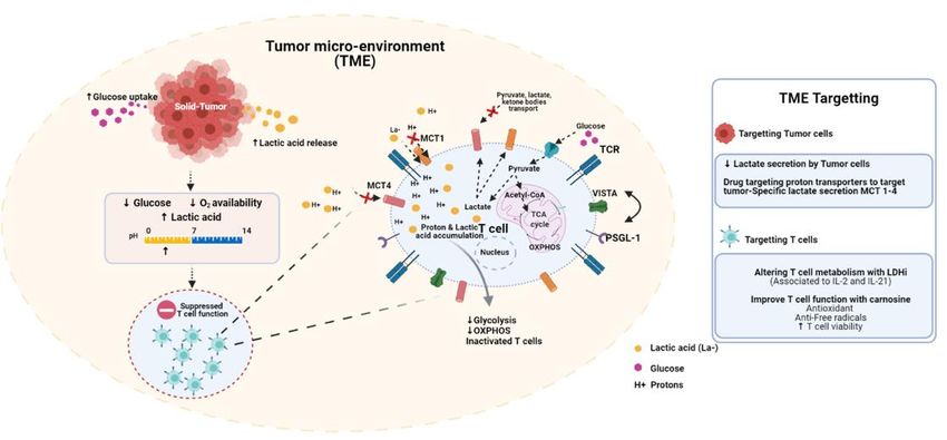
Figure1.1.The
Figure Thetumor
tumor microenvironment
microenvironment(TME)(TME)isisacidic
acidicand
and immune-suppressive.
immune-suppressive. Tumor Tumorproliferation
proliferationisisdistinguished
distinguishedby by
enhanced
enhancedglucose
glucoseconsumption
consumptionand andlactic
lacticacid
acidproduction.
production.InIn
response
response to to
increased tumor
increased tumor cellcell
metabolism,
metabolism,thethe
surrounding
surround-
ing milieu
milieu becomes
becomes increasingly
increasingly hypoglycemic,
hypoglycemic, hypoxic
hypoxic and
and acidic.
acidic. Theseconditions
These conditionsare
areinherently
inherentlyimmune-suppressive.
immune-suppressive.
Lowglucose
Low glucoseandandoxygen
oxygenlevels
levelsimpair
impairTTcell
cellmetabolic
metabolicactivity.
activity.Acidosis
Acidosisinhibits
inhibitsglycolysis
glycolysisand anddownstream
downstreamoxidative
oxidative
phosphorylation.Low
phosphorylation. LowpH pHinduces
inducesV-domain
V-domainIg Igsuppressor
suppressorof ofTTcell
cellactivation
activation(VISTA),
(VISTA),ananimmune-inhibitory
immune-inhibitorycheckpoint
checkpoint
molecule. Several
molecule. Several strategies
strategies have
have been
been developed
developed to tocounter
counterthese
theseeffects.
effects.Nonsteroidal
Nonsteroidalanti-inflammatories
anti-inflammatories (NSAIDS),
(NSAIDS),
including diclofenac, can selectively inhibit MCT1-4 on cancer cells. Ex-vivo conditioning strategies, including the use of
including diclofenac, can selectively inhibit MCT1-4 on cancer cells. Ex-vivo conditioning strategies, including the use of
LDH inhibitors, can limit T cell terminal differentiation prior to adoptive transfer. Treating T cells with carnosine ex-vivo
LDH inhibitors, can limit T cell terminal differentiation prior to adoptive transfer. Treating T cells with carnosine ex-vivo
can shift their metabolic state from glycolytic/acidic to oxidative.
can shift their metabolic state from glycolytic/acidic to oxidative.
6. Altering T Cell Metabolism with LDHi
Strategies to limit lactate secretion by targeting MCT isoforms have been proposed
Interleukin-2
as a potential (IL-2)
adjuvant toand Interleukin-21
T cell (IL-21)However,
adoptive transfer. are two closely related
T cells share families
many of sig-
metabolic
naling molecules
features with tumor that arose
cells, from gene
including duplication.
an induction Both following
of MCT4 cytokinesTCRare pleiotropic
engagementmole-and
cules that
CD28 support theand
co-stimulation, maturation and differentiation
thus compounds designed to of CD8+
inhibiteffector T cells [53].
MCT isoforms mayIL-2 has
prove
been widelytoused
antagonistic in adoptiveresponses.
T cell-mediated immunotherapy as aissoluble
Diclofenac growth factor
a monocarboxylic acidthat
withsupports
known
the expansion of T properties
anti-inflammatory cells ex-vivo, and
[51]. the growth
Diclofenac canand
alsodifferentiation
suppress tumor of adoptively trans-
cell metabolism
ferred tumor-reactive T cells in vivo [54]. However, IL-2 can promote the development of
T cells into terminally differentiated effector cells, which limits durable efficacy following
adoptive transfer. IL-2 can also trigger the development of regulatory T cells. IL-21 is an
important factor that supports the development of Th17 cells, a subset with enhanced anti-Antibodies 2021, 10, 25 8 of 12
and proliferation by inhibiting MCT4 [51]. Remarkably, diclofenac has minimal impact
on T cell cytokine production, as measured by the levels of IFN-γ and tumor necrosis
factor (TNF) in vitro [52]. These findings position diclofenac as a potential therapeutic
to selectively disable tumor cell metabolism in the TME. In T cell:tumor cell co-cultures,
diclofenac enhanced T cell cytolytic responses. These findings highlight the potential
benefit of targeting tumor-specific lactate secretion using diclofenac and its derivatives in
the TME.
6. Altering T Cell Metabolism with LDHi
Interleukin-2 (IL-2) and Interleukin-21 (IL-21) are two closely related families of
signaling molecules that arose from gene duplication. Both cytokines are pleiotropic
molecules that support the maturation and differentiation of CD8+ effector T cells [53].
IL-2 has been widely used in adoptive immunotherapy as a soluble growth factor that
supports the expansion of T cells ex-vivo, and the growth and differentiation of adoptively
transferred tumor-reactive T cells in vivo [54]. However, IL-2 can promote the development
of T cells into terminally differentiated effector cells, which limits durable efficacy following
adoptive transfer. IL-2 can also trigger the development of regulatory T cells. IL-21 is an
important factor that supports the development of Th17 cells, a subset with enhanced anti-
tumor function [55]. Metabolically, IL-2 and IL-21 are diametrically opposed: IL-2 drives
aerobic glycolysis in a PI3K-AKT-dependent manner while IL-21 activates STAT5 signal
transduction; culminating in an oxidative rather than glycolytic state [56]. The metabolic
phenotypes conferred by IL-2 and IL-21 have been proposed to be a key determinant
underlying their differential antitumor activity.
In a recent study, Hermans and coworkers compared the metabolic state of IL-2
versus IL-21 primed T cells. Murine T cells were activated with dynabeads containing
immobilised antibodies against CD3 and CD28 for two days, and then supplemented with
either IL-2 or IL-21 for a further 48 h. Metabolically, IL-21 retained metabolic properties
of quiescent T cells, as shown by extracellular flux analyses. In contrast, the energy cost
to sustain IL-2 cultures was larger, exemplified by increased rates of basal and maximal
oxygen consumption rates. Increased metabolic demands at rest, and following stimulation,
led to a reduced overall energy generating capacity. A deficit in the contingency energy
reserve may impede the persistence and anti-tumor function of adoptively transferred T
cells, especially in hostile environments. Importantly, IL-2 and IL-21-treated cells show a
difference in glucose consumption rates, as well as the expression of certain metabolites
including lactate and pyruvate [57]. The lactate/pyruvate ratio, an important biomarker
for glycolytic activity, was increased in IL-2 relative to IL-21 cultures. Attempts to revert IL-
2-primed T cells to an oxidative state or accentuate substrate oxidation in IL-21-stimulated
cultures relied on the use of a small molecule inhibitor against LDH.
LDH catalyses the reduction of pyruvate to lactic acid. Ldh mRNA expression is higher
in IL-2 relative to IL-21-treated cultures. IL-2-treated cells secrete more lactate and protons,
as measured by extracellular acidification rates in a Seahorse Assay. IL-2-treated cells also
express more of the phosphorylated, inactive form of pyruvate dehydrogenase (PDH).
PDH controls the metabolic fate of pyruvate, driving it towards cytoplasmic rather than
mitochondrial metabolism [57]. This explains how IL-2 limits the oxidation of pyruvate for
energy and TCA anaplerosis. LDHi resulted in a substantial decrease in lactate secretion
and glucose consumption in both IL-2- and IL-21-treated cells [57]. In IL-2-treated cells,
LDHi increased the intracellular pyruvate/lactate and NADH/NAD+ ratio significantly.
Conceptually, increasing pyruvate availability by LDHi facilitates its entry and oxidation
into the mitochondria by mass action. In their 13 C6 glucose tracer assays, there was limited
evidence that pyruvate metabolism in the TCA cycle was dramatically increased by LDHi.
Follow-up studies are necessary to tease out the mechanisms by which LDHi treatment
ex-vivo led to improved anti-tumor function following infusion, especially as the com-
pound targets A and B isoforms of LDH. Comparatively, eliminating LDHA at the level of
the germline gives rise to T cell progeny with limited anti-tumor function suggesting theAntibodies 2021, 10, 25 9 of 12
temporal regulation of LDH activity is an essential consideration for adoptive immunother-
apies. Overall, their promising data support a model in which Ldhi during ex-vivo culture
confers unique metabolic benefits. In contrast, LDHA is indispensable to tumor-reactive T
cells following adoptive transfer [57].
7. Carnosine Buffers TME and Improves T Cell Function
An important aspect of adoptive immunotherapy is the isolation and expansion
of T lymphocytes ex-vivo to generate increased numbers of “sentinels” that recognise
and target cancer cells following reinfusion. Understanding how medium formulations
impact the effectiveness of T cell therapies is an exciting new area of research. Recently, we
showed how a unique medium formulation, that was custom adjusted to mimic physiologic
microenvironments, gave rise to CAR T-cell progeny with enhanced anti-tumor function in
human xenograft models of neuroblastoma [58].
Using metabolomics, we screened a blood-based growth factor concentrate “Physi-
ologix”, and identified increased levels of the biogenic amine carnosine relative to human
serum. Carnosine is an L-histidine/beta-alanine dipeptide, abundantly found in skeletal
muscle. It is well established that carnosine is an important physiochemical buffer to
reduce muscle fatigue and lactic acid buildup during high-energy activities [59]. Carnosine
is also an effective antioxidant and free-radical scavenger and has been shown to limit
DNA damage and cellular stress [60]. These observed properties positioned carnosine
as an excellent candidate to study in adoptive immunotherapy. Future efforts will likely
determine how carnosine influences T cell viability and clonal integrity during extended
cultures at normoxia and hypoxia.
Remarkably, we showed that increased carnosine availability increased lentiviral
transduction; an effect accentuated at low MOIs using VSV-pseudotyped lentivirus. VSV-
pseudotyped virus enter cells via the LDH receptor, which becomes embedded in clathrin-
coated pits. Following endocytosis, there is a pH-dependent release/fusion of virus
particles prior to nuclear import and reverse transcription. The specific mechanisms by
which carnosine enhances lentiviral-mediated transduction is unknown, but its intracellular
accumulation in T cells [58], and the unique pKa of L-histidine in its dipeptide form, likely
plays an important role.
In Seahorse assays, carnosine treatment significantly decreased ECAR. The intrinsic
ability of carnosine to neutralise an increase in proton load turn shifted the metabolic
preference of T cells to oxidative phosphorylation. As extracellular acidosis is a feature
of hypoxic tumor environments, as well as a barrier to effective immunotherapies, our
findings highlight carnosine as a novel factor to enhance the quality of T cells expanded
ex-vivo for adoptive immunotherapies.
8. Conclusions
Mitigating the immunosuppressive effects of the TME is necessary to improve the
efficacy of cancer treatments. The TME is distinctly acidic due to high rates of glucose
metabolism in cancer cells, culminating in excess lactic acid production and secretion.
The corresponding decrease in extracellular pH inhibits glycolysis in T cells by negative
feedback, induces reductive stress at hypoxia, and activates inhibitory immune checkpoint
molecules such as VISTA. Overcoming these metabolic effects will require innovative
strategies to selectively target the tumor cells themselves, thereby indirectly supporting T
cell function, or using combinatorial approaches to addresses both factors synergistically. It
is clear from the rigorous examination of the studies described above that regulating pH to
restore T cell functional competence in hostile soils will enhance the therapeutic potential
of adoptive immunotherapies against cancer.
Author Contributions: Conceptualization: V.Y.T. and R.S.O.; validation: V.Y.T., A.A. and R.S.O.;
Resources: R.S.O., Writing-original draft preparation: V.Y.T., A.A. and R.S.O.; Writing-review and
editing: V.Y.T. and R.S.O.; Supervision: R.S.O.; Funding Acquisition: R.S.O. All authors have read
and agreed to the published version of the manuscript.Antibodies 2021, 10, 25 10 of 12
Funding: ROC is supported by NIH, grant number RO1CA226983-04.
Institutional Review Board Statement: Not applicable.
Informed Consent Statement: Not applicable.
Data Availability Statement: Not applicable.
Conflicts of Interest: The authors declare no conflict of interest.
References
1. Whiteside, T.L. The tumor microenvironment and its role in promoting tumor growth. Oncogene 2008, 27, 5904–5912. [CrossRef]
2. Langley, R.R.; Fidler, I.J. The seed and soil hypothesis revisited-The role of tumor-stroma interactions in metastasis to different
organs. Int. J. Cancer 2011, 128, 2527–2535. [CrossRef]
3. Paget, S. The distribution of secondary growths in cancer of the breast. Cancer Metastasis Rev. 1989, 8, 98–101. [CrossRef]
4. Giavazzi, R.; Campbell, E.D.; Jessup, J.M.; Cleary, K.; Fidler, I.J. Metastatic behavior of tumor cells isolated from primary and
metastatic human colorectal carcinomas implanted into different sites in nude mice. Cancer Res. 1986, 46, 1928–1933.
5. Naito, S.; Von Eschenbach, A.C.; Giavazzi, R.; Fidler, I.J. Growth and metastasis of tumor cells isolated from a human renal cell
carcinoma implanted into different organs of nude mice. Cancer Res. 1986, 46, 4109–4115. [PubMed]
6. Klein, G.; Sjögren, H.O.; Klein, E.; Hellström, K.E. Demonstration of resistance against methylcholanthrene-induced sarcomas in
the primary autochthonous host. Cancer Res. 1960, 20, 1561–1572. [PubMed]
7. Liberti, M.V.; Locasale, J.W. The Warburg Effect: How Does it Benefit Cancer Cells? Trends Biochem. Sci. 2016, 41, 211–218.
[CrossRef]
8. Crabtree, H.G. Observations on the carbohydrate metabolism of tumours. Biochem. J. 1929, 23, 536–545. [CrossRef] [PubMed]
9. Dienel, G.A. Brain lactate metabolism: The discoveries and the controversies. J. Cereb. Blood Flow Metab. 2012, 32, 1107–1138.
[CrossRef]
10. Brooks, G.A.; Arevalo, J.A.; Osmond, A.D.; Leija, R.G.; Curl, C.C.; Tovar, A.P. Lactate in contemporary biology: A phoenix risen. J.
Physiol. 2021. [CrossRef]
11. Sonveaux, P.; Vegran, F.; Schroeder, T.; Wergin, M.; Verrax, J.; Rabbani, Z.N.; De Saedeleer, C.J.; Kennedy, K.M.; Diepart, C.;
Jordan, B.F.; et al. Targeting lactate-fueled respiration selectively kills hypoxic tumor cells in mice. J. Clin. Investig. 2008,
118, 3930–3942. [CrossRef]
12. Hui, S.; Ghergurovich, J.M.; Morscher, R.J.; Jang, C.; Teng, X.; Lu, W.; Esparza, L.A.; Reya, T.; Zhan, L.; Guo, J.Y.; et al. Glucose
feeds the TCA cycle via circulating lactate. Nat. Cell Biol. 2017, 551, 115–118. [CrossRef]
13. Rodríguez-Colman, M.J.; Schewe, M.; Meerlo, M.; Stigter, E.; Gerrits, J.; Pras-Raves, M.; Sacchetti, A.; Hornsveld, M.; Oost, K.C.;
Snippert, H.J.; et al. Interplay between metabolic identities in the intestinal crypt supports stem cell function. Nat. Cell Biol. 2017,
543, 424–427. [CrossRef]
14. Watson, M.J.; Vignali, P.D.A.; Mullett, S.J.; Overacre-Delgoffe, A.E.; Peralta, R.M.; Grebinoski, S.; Menk, A.V.; Rittenhouse, N.L.;
DePeaux, K.; Whetstone, R.D.; et al. Metabolic support of tumour-infiltrating regulatory T cells by lactic acid. Nat. Cell Biol. 2021,
591, 645–651. [CrossRef]
15. Angelin, A.; Gil-De-Gómez, L.; Dahiya, S.; Jiao, J.; Guo, L.; Levine, M.H.; Wang, Z.; Quinn, W.J.; Kopinski, P.K.; Wang, L.; et al.
Foxp3 Reprograms T Cell Metabolism to Function in Low-Glucose, High-Lactate Environments. Cell Metab. 2017, 25, 1282–1293.e7.
[CrossRef] [PubMed]
16. Warburg, O. The Metabolism of Carcinoma Cells. J. Cancer Res. 1925, 9, 148–163. [CrossRef]
17. Ashton, T.M.; McKenna, W.G.; Kunz-Schughart, L.A.; Higgins, G.S. Oxidative Phosphorylation as an Emerging Target in Cancer
Therapy. Clin. Cancer Res. 2018, 24, 2482–2490. [CrossRef] [PubMed]
18. Ho, P.-C.; Bihuniak, J.D.; Macintyre, A.; Staron, M.; Liu, X.; Amezquita, R.; Tsui, Y.-C.; Cui, G.; Micevic, G.; Perales, J.C.; et al.
Phosphoenolpyruvate Is a Metabolic Checkpoint of Anti-tumor T Cell Responses. Cell 2015, 162, 1217–1228. [CrossRef] [PubMed]
19. Cascone, T.; McKenzie, J.A.; Mbofung, R.; Punt, S.; Wang, Z.; Xu, C.; Williams, L.J.; Wang, Z.; Bristow, C.A.; Carugo, A.; et al.
Increased Tumor Glycolysis Characterizes Immune Resistance to Adoptive T Cell Therapy. Cell Metab. 2018, 27, 977–987.e4.
[CrossRef]
20. Walenta, S.; Wetterling, M.; Lehrke, M.; Schwickert, G.; Sundfør, K.; Rofstad, E.K.; Mueller-Klieser, W. High lactate levels
predict likelihood of metastases, tumor recurrence, and restricted patient survival in human cervical cancers. Cancer Res. 2000,
60, 916–921.
21. Liu, Y.; Guo, J.-Z.; Liu, Y.; Wang, K.; Ding, W.; Wang, H.; Liu, X.; Zhou, S.; Lu, X.-C.; Yang, H.-B.; et al. Nuclear lactate
dehydrogenase A senses ROS to produce α-hydroxybutyrate for HPV-induced cervical tumor growth. Nat. Commun. 2018,
9, 1–16. [CrossRef]
22. Mishra, D.; Banerjee, D. Lactate Dehydrogenases as Metabolic Links between Tumor and Stroma in the Tumor Microenvironment.
Cancers 2019, 11, 750. [CrossRef]
23. Colegio, O.R.; Chu, N.-Q.; Szabo, A.L.; Chu, T.; Rhebergen, A.M.; Jairam, V.; Cyrus, N.; Brokowski, C.E.; Eisenbarth, S.C.;
Phillips, G.M.; et al. Functional polarization of tumour-associated macrophages by tumour-derived lactic acid. Nature 2014,
513, 559–563. [CrossRef] [PubMed]Antibodies 2021, 10, 25 11 of 12
24. Kim, C.G.; Jang, M.; Kim, Y.; Leem, G.; Kim, K.H.; Lee, H.; Kim, T.-S.; Choi, S.J.; Kim, H.-D.; Han, J.W.; et al. VEGF-A drives
TOX-dependent T cell exhaustion in anti–PD-1–resistant microsatellite stable colorectal cancers. Sci. Immunol. 2019, 4, eaay0555.
[CrossRef]
25. Northrop, J.K.; Thomas, R.M.; Wells, A.D.; Shen, H. Epigenetic Remodeling of the IL-2 and IFN-γ Loci in Memory CD8 T Cells Is
Influenced by CD4 T Cells. J. Immunol. 2006, 177, 1062–1069. [CrossRef]
26. Zhang, D.; Tang, Z.; Huang, H.; Zhou, G.; Cui, C.; Weng, Y.; Liu, W.; Kim, S.; Lee, S.; Perez-Neut, M.; et al. Metabolic regulation of
gene expression by histone lactylation. Nat. Cell Biol. 2019, 574, 575–580. [CrossRef] [PubMed]
27. Bhagat, T.D.; Von Ahrens, D.; Dawlaty, M.; Zou, Y.; Baddour, J.; Achreja, A.; Zhao, H.; Yang, L.; Patel, B.; Kwak, C.; et al.
Lactate-mediated epigenetic reprogramming regulates formation of human pancreatic cancer-associated fibroblasts. eLife 2019,
8, e50663. [CrossRef]
28. Roslin, M.; Henriksson, R.; Bergström, P.; Ungerstedt, U.; Bergenheim, A.T. Baseline levels of glucose metabolites, glutamate and
glycerol in malignant glioma assessed by stereotactic microdialysis. J. Neuro-Oncol. 2003, 61, 151–160. [CrossRef]
29. Celis, J.E.; Gromov, P.; Cabezón, T.; Moreira, J.; Ambartsumian, N.; Sandelin, K.; Rank, F.; Gromova, I. Proteomic Characterization
of the Interstitial Fluid Perfusing the Breast Tumor Microenvironment. Mol. Cell. Proteom. 2004, 3, 327–344. [CrossRef] [PubMed]
30. Tabatabaei, P.; Bergström, P.; Henriksson, R.; Bergenheim, A.T. Glucose metabolites, glutamate and glycerol in malignant glioma
tumours during radiotherapy. J. Neuro-Oncol. 2008, 90, 35–39. [CrossRef]
31. Chang, C.-H.; Qiu, J.; O’Sullivan, D.; Buck, M.; Noguchi, T.; Curtis, J.D.; Chen, Q.; Gindin, M.; Gubin, M.M.; Van Der Windt, G.J.; et al.
Metabolic Competition in the Tumor Microenvironment Is a Driver of Cancer Progression. Cell 2015, 162, 1229–1241. [CrossRef]
32. Boroojerdi, M.H.; Rahbarizadeh, F.; Kozani, P.S.; Kamali, E.; Kozani, P.S. Strategies for having a more effective and less toxic CAR
T-cell therapy for acute lymphoblastic leukemia. Med. Oncol. 2020, 37, 1–15. [CrossRef]
33. Mullard, A. FDA approves fourth CAR-T cell therapy. Nat. Rev. Drug Discov. 2021, 20, 166. [CrossRef]
34. Kouidhi, S.; Elgaaied, A.B.; Chouaib, S. Impact of Metabolism in on T-Cell Differentiation and Function and Cross Talk with
Tumor Microenvironment. Front. Immunol. 2017, 8, 270. [CrossRef]
35. Certo, M.; Tsai, C.-H.; Pucino, V.; Ho, P.-C.; Mauro, C. Lactate modulation of immune responses in inflammatory versus tumour
microenvironments. Nat. Rev. Immunol. 2021, 21, 151–161. [CrossRef]
36. Wu, H.; Estrella, V.; Beatty, M.; Abrahams, D.; El-Kenawi, A.; Russell, S.; Ibrahim-Hashim, A.; Longo, D.L.; Reshetnyak, Y.K.;
Moshnikova, A.; et al. T-cells produce acidic niches in lymph nodes to suppress their own effector functions. Nat. Commun. 2020,
11, 1–13. [CrossRef]
37. Calder, P.C.; Dimitriadis, G.; Newsholme, P. Glucose metabolism in lymphoid and inflammatory cells and tissues. Curr. Opin.
Clin. Nutr. Metab. Care 2007, 10, 531–540. [CrossRef]
38. Uhl, F.M.; Chen, S.; O’Sullivan, D.; Edwards-Hicks, J.; Richter, G.; Haring, E.; Andrieux, G.; Halbach, S.; Apostolova, P.;
Büscher, J.; et al. Metabolic reprogramming of donor T cells enhances graft-versus-leukemia effects in mice and humans. Sci.
Transl. Med. 2020, 12, eabb8969. [CrossRef]
39. Yokota, H.; Guo, J.; Matoba, M.; Higashi, K.; Tonami, H.; Nagao, Y. Lactate, choline, and creatine levels measured by vitro1H-MRS
as prognostic parameters in patients with non-small-cell lung cancer. J. Magn. Reson. Imaging 2007, 25, 992–999. [CrossRef]
40. Quinn, W.J.; Jiao, J.; TeSlaa, T.; Stadanlick, J.; Wang, Z.; Wang, L.; Akimova, T.; Angelin, A.; Schäfer, P.M.; Cully, M.D.; et al. Lactate
Limits T Cell Proliferation via the NAD(H) Redox State. Cell Rep. 2020, 33, 108500. [CrossRef]
41. Rampello, E.; Fricia, T.; Malaguarnera, M. The management of tumor lysis syndrome. Nat. Clin. Pr. Oncol. 2006, 3, 438–447.
[CrossRef] [PubMed]
42. Wike-Hooley, J.; Haveman, J.; Reinhold, H. The relevance of tumour pH to the treatment of malignant disease. Radiother. Oncol.
1984, 2, 343–366. [CrossRef]
43. Wang, L.; Rubinstein, R.; Lines, J.L.; Wasiuk, A.; Ahonen, C.; Guo, Y.; Lu, L.-F.; Gondek, D.; Wang, Y.; Fava, R.A.; et al. VISTA, a
novel mouse Ig superfamily ligand that negatively regulates T cell responses. J. Exp. Med. 2011, 208, 577–592. [CrossRef]
44. Le Mercier, I.; Chen, W.; Lines, J.L.; Day, M.; Li, J.; Sergent, P.; Noelle, R.J.; Wang, L. VISTA Regulates the Development of
Protective Antitumor Immunity. Cancer Res. 2014, 74, 1933–1944. [CrossRef]
45. Yuan, L.; Tatineni, J.; Mahoney, K.M.; Freeman, G.J. VISTA: A Mediator of Quiescence and a Promising Target in Cancer
Immunotherapy. Trends Immunol. 2021, 42, 209–227. [CrossRef] [PubMed]
46. Johnston, R.J.; Su, L.J.; Pinckney, J.; Critton, D.; Boyer, E.; Krishnakumar, A.; Corbett, M.; Rankin, A.L.; DiBella, R.;
Campbell, L.; et al. VISTA is an acidic pH-selective ligand for PSGL. Nature 2019, 574, 565–570. [CrossRef]
47. Gao, J.; Ward, J.F.; Pettaway, A.C.; Shi, L.Z.; Subudhi, S.K.; Vence, L.M.; Zhao, H.; Chen, J.; Chen, H.; Efstathiou, E.; et al. VISTA is
an inhibitory immune checkpoint that is increased after ipilimumab therapy in patients with prostate cancer. Nat. Med. 2017,
23, 551–555. [CrossRef]
48. Kakavand, H.; Jackett, A.L.; Menzies, A.; Gide, T.; Carlino, M.S.; Saw, R.P.M.; Thompson, J.; Wilmott, J.; Long, G.; Scolyer, A.R.
Negative immune checkpoint regulation by VISTA: A mechanism of acquired resistance to anti-PD-1 therapy in metastatic
melanoma patients. Mod. Pathol. 2017, 30, 1666–1676. [CrossRef]
49. Nakagawa, Y.; Negishi, Y.; Shimizu, M.; Takahashi, M.; Ichikawa, M.; Takahashi, H. Effects of extracellular pH and hypoxia on
the function and development of antigen-specific cytotoxic T lymphocytes. Immunol. Lett. 2015, 167, 72–86. [CrossRef]
50. Balar, A.V.; Weber, J.S. PD-1 and PD-L1 antibodies in cancer: Current status and future directions. Cancer Immunol. Immunother.
2017, 66, 551–564. [CrossRef]Antibodies 2021, 10, 25 12 of 12
51. Moreno-Sanchez, R.; Bravo, C.; Vásquez, C.; Ayala, G.; Silveira, L.H.; Martínez-Lavín, M. Inhibition and uncoupling of oxidative
phosphorylation by nonsteroidal anti-inflammatory drugs. Biochem. Pharmacol. 1999, 57, 743–752. [CrossRef]
52. Renner, K.; Bruss, C.; Schnell, A.; Koehl, G.; Becker, H.M.; Fante, M.; Menevse, A.-N.; Kauer, N.; Blazquez, R.; Hacker, L.; et al.
Restricting Glycolysis Preserves T Cell Effector Functions and Augments Checkpoint Therapy. Cell Rep. 2019, 29, 135–150.e9.
[CrossRef]
53. Brocker, C.; Thompson, D.; Matsumoto, A.; Nebert, D.W.; Vasiliou, V. Evolutionary divergence and functions of the human
interleukin (IL) gene family. Hum. Genom. 2010, 5, 30–55. [CrossRef]
54. Overwijk, W.W.; Tagliaferri, M.A.; Zalevsky, J. Engineering IL-2 to Give New Life to T Cell Immunotherapy. Annu. Rev. Med.
2021, 72, 281–311. [CrossRef]
55. Yang, L.; Anderson, D.E.; Baecher-Allan, C.; Hastings, W.D.; Bettelli, E.; Oukka, M.; Kuchroo, V.K.; Hafler, D.A. IL-21 and TGF-β
are required for differentiation of human TH17 cells. Nat. Cell Biol. 2008, 454, 350–352. [CrossRef]
56. Loschinski, R.; Böttcher, M.; Stoll, A.; Bruns, H.; Mackensen, A.; Mougiakakos, D. IL-21 modulates memory and exhaustion
phenotype of T-cells in a fatty acid oxidation-dependent manner. Oncotarget 2018, 9, 13125–13138. [CrossRef]
57. Hermans, D.; Gautam, S.; García-Cañaveras, J.C.; Gromer, D.; Mitra, S.; Spolski, R.; Li, P.; Christensen, S.; Nguyen, R.;
Lin, J.-X.; et al. Lactate dehydrogenase inhibition synergizes with IL-21 to promote CD8+T cell stemness and antitumor immunity.
Proc. Natl. Acad. Sci. USA 2020, 117, 6047–6055. [CrossRef] [PubMed]
58. Ghassemi, S.; Martinez-Becerra, F.J.; Master, A.M.; Richman, S.A.; Heo, D.; Leferovich, J.; Tu, Y.; García-Cañaveras, J.C.; Ayari, A.;
Lu, Y.; et al. Enhancing Chimeric Antigen Receptor T Cell Anti-tumor Function through Advanced Media Design. Mol. Ther.
Methods Clin. Dev. 2020, 18, 595–606. [CrossRef] [PubMed]
59. Mannion, A.F.; Jakeman, P.M.; Dunnett, M.; Harris, R.C.; Willan, P.L.T. Carnosine and anserine concentrations in the quadriceps
femoris muscle of healthy humans. Graefe’s Arch. Clin. Exp. Ophthalmol. 1992, 64, 47–50. [CrossRef]
60. Hyland, P.; Duggan, O.; Hipkiss, A.; Barnett, C.; Barnett, Y. The effects of carnosine on oxidative DNA damage levels and in vitro
lifespan in human peripheral blood derived CD4+T cell clones. Mech. Ageing Dev. 2001, 121, 203–215. [CrossRef]You can also read