Hedgehog signaling enables repair of ribosomal DNA double-strand breaks
←
→
Page content transcription
If your browser does not render page correctly, please read the page content below
10342–10352 Nucleic Acids Research, 2020, Vol. 48, No. 18                                                                 Published online 7 September 2020
doi: 10.1093/nar/gkaa733

Hedgehog signaling enables repair of ribosomal DNA
double-strand breaks
Tshering D. Lama-Sherpa 1,† , Victor T.G. Lin 2,3,† , Brandon J. Metge1 , Shannon E. Weeks1 ,
Dongquan Chen3,4 , Rajeev S. Samant 1,3,5 and Lalita A. Shevde 1,3,*
1
  Department of Pathology, The University of Alabama at Birmingham, Birmingham, AL, USA, 2 Division of
Hematology and Oncology, Department of Medicine, The University of Alabama at Birmingham, Birmingham, AL,
USA, 3 O’Neal Comprehensive Cancer Center, The University of Alabama at Birmingham, Birmingham, AL, USA,
4
  Division of Preventative Medicine, Department of Medicine, The University of Alabama at Birmingham, Birmingham,

                                                                                                                                                                  Downloaded from https://academic.oup.com/nar/article/48/18/10342/5902436 by guest on 28 October 2020
AL, USA and 5 Birmingham VA Medical Center, Birmingham, AL, USA

Received March 03, 2020; Revised August 20, 2020; Editorial Decision August 21, 2020; Accepted September 04, 2020

ABSTRACT                                                                                 uploidy, and ultimately mitotic catastrophe and cell death
                                                                                         (5–7).
Ribosomal DNA (rDNA) consists of highly repeated                                            Cells have two major repair mechanisms in place
sequences that are prone to incurring damage. De-                                        to resolve DSBs: homologous recombination (HR) and
lays or failure of rDNA double-strand break (DSB) re-                                    non-homologous end joining (NHEJ). HR is cell cycle-
pair are deleterious, and can lead to rDNA transcrip-                                    dependent and results in high-fidelity repair of DSBs,
tional arrest, chromosomal translocations, genomic                                       whereas NHEJ is cell cycle-independent and comprises
losses, and cell death. Here, we show that the zinc-                                     blunt end-joining that is more immediate but more error-
finger transcription factor GLI1, a terminal effector                                    prone (8). NHEJ is the preferred repair pathway for rDNA
of the Hedgehog (Hh) pathway, is required for the re-                                    DSBs (9), though they may also be resolved by HR machin-
pair of rDNA DSBs. We found that GLI1 is activated in                                    ery independent of the cell cycle as a contingency (10). Er-
triple-negative breast cancer cells in response to ion-                                  rors in repair can lead to loss of repeats and chromosomal
                                                                                         translocations involving 45S rDNA loci, which are found
izing radiation (IR) and localizes to rDNA sequences
                                                                                         on five different chromosomes (4). Delays in the resolu-
in response to both global DSBs generated by IR and                                      tion of rDNA DSBs result in the arrest of rDNA transcrip-
site-specific DSBs in rDNA. Inhibiting GLI1 interferes                                   tion by RNA polymerase I (Pol I) via an ATM-dependent
with rDNA DSB repair and impacts RNA polymerase                                          mechanism (9). Unrepaired rDNA damage, especially of
I activity and cell viability. Our findings tie Hh sig-                                  45S rDNA, is deleterious (5). Thus, rDNA repair is an at-
naling to rDNA repair and this heretofore unknown                                        tractive target for novel cancer therapeutics.
function may be critically important in proliferating                                       We sought to investigate the role of Hedgehog (Hh) sig-
cancer cells.                                                                            naling in DNA repair. Previous work in our laboratory con-
                                                                                         nected GLI1, a zinc-finger transcription factor that is a ter-
INTRODUCTION                                                                             minal effector of the Hh pathway, to the DNA single-strand
                                                                                         break (SSB) repair via the nucleotide and base excision re-
Ribosomal DNA (rDNA) comprises repeated sequences                                        pair pathways (NER and BER). We showed that GLI1 up-
encoding the 45S and 5S ribosomal RNAs (rRNAs) that                                      regulates expression of NER and BER genes in response
form the basis of ribosome structure and function (1). These                             to SSBs to facilitate their repair (11). Hh inhibition is also
regions of chromatin are open and highly transcribed in                                  known to sensitize cancer cells to agents that induce DSBs,
actively proliferating cells in order to match their exten-                              but the mechanisms by which Hh influences DSB repair are
sive metabolic demands, making them prone to incurring                                   poorly understood (12,13). We hypothesized that induction
damage (2–4). Genome-wide mapping of DSBs shows a                                        of DSBs by ionizing radiation (IR) would result in cistromic
predilection for rDNA that leads to genomic rearrange-                                   changes in GLI1 in order to orchestrate the activation of
ments (2). Thus, chromosomal translocations in rDNA are                                  DNA repair programs. Unexpectedly, we found that GLI1
among the most common genomic alterations in adult solid                                 occupancy of rDNA loci is markedly enriched in response
tumors (4). Delayed, erroneous, or incomplete repair of                                  to IR and that GLI1 is required for timely repair of rDNA
double-strand breaks (DSBs) in these loci can lead to delete-                            DSBs.
rious chromosomal translocations, genomic instability, ane-

* To   whom correspondence should be addressed. Tel: +1 205 975 6261; Fax: +1 205 975 6615; Email: lalitasamant@uabmc.edu
†
    The authors wish it to be known that, in their opinion, the first two authors should be regarded as Joint First Authors.

C The Author(s) 2020. Published by Oxford University Press on behalf of Nucleic Acids Research.
This is an Open Access article distributed under the terms of the Creative Commons Attribution Non-Commercial License
(http://creativecommons.org/licenses/by-nc/4.0/), which permits non-commercial re-use, distribution, and reproduction in any medium, provided the original work
is properly cited. For commercial re-use, please contact journals.permissions@oup.com
Nucleic Acids Research, 2020, Vol. 48, No. 18 10343
MATERIALS AND METHODS hg19 genome using BWA Aligner (15). Peak calling was
done using MACS2 (16) (https://github.com/taoliu/MACS/
Cell culture
tree/master/MACS2) and filtered for peaks of interest be-
SUM1315 cells were cultured in DMEM/F12 (Gibco) con- tween −5000 and +2000 of transcriptional start sites. ChIP-
taining 5% heat-inactivated FBS (Gibco), 10 g/ml in- Seq peaks were visualized using the Integrative Genomics
sulin (Sigma), and 25 ng/ml EGF (Sigma). MDA-MB-468 Viewer (17).
cells were cultured in DMEM/F12 containing 10% FBS.
SUM159 cells were cultured in DMEM/F12 containing Immunoblotting
5% FBS, 10 g/ml insulin, and 1 g/ml hydrocortisone
Cells were lysed in RIPA buffer (Millipore) containing
(Sigma). Culture media were all free of antibiotics and an-
HALT protease and phosphatase inhibitor cocktail (Ther-
timycotics unless otherwise stated. Cells were maintained at
moScientific) and sonicated to complete lysis. Lysates were
37◦ C in a humidified environment containing 5% CO2 .
clarified by centrifugation before protein concentrations
Downloaded from https://academic.oup.com/nar/article/48/18/10342/5902436 by guest on 28 October 2020
were assayed using the Precision Red assay (Cytoskeleton).
Hh/GLI1 inhibition Equal masses were electrophoresed by SDS-PAGE and wet
transferred to PVDF membranes (BioRad). When prob-
Cells were treated with 10 M vismodegib (SelleckChem)
ing for proteins with molecular weights over 200 kDa, pre-
or 10 M GANT61 (Tocris) solubilized in DMSO (Fisher)
cast gradient gels were used (Invitrogen and BioRad) and
as stated in each figure. Unless otherwise noted, cells were
wet transfers were done at 30 V for 16 h. Primary an-
treated with an inhibitor or vehicle control for 24 h prior to
tibodies against ␥ -H2AX (Cell Signaling, 9718S), GLI1
irradiation. Where noted, SUM1315 cells were stably trans-
(Cell Signaling, 2643S), fibrillarin (Cell Signaling, 2639S),
fected with either a non-targeted plasmid or shRNA di-
I-SceI (Abcam, ab216263), 53BP1 (Cell Signaling, 4937S),
rected against GLI1 as previously described (14) and main-
phospho-53BP1 S1778 (Cell Signaling, 2675S), ATM (Cell
tained in selection with 300 g/ml G418 (Gibco).
Signaling, 2873S), phospho-ATM S1981 (Cell Signaling,
5883S), NBS1 (Cell Signaling, 3002S), phospho-NBS1 S343
Ionizing radiation (Cell Signaling, 3001S), and -actin (Sigma) were used,
as well as secondary HRP-conjugated antibodies against
Cells were irradiated with the indicated doses using the X-
mouse and rabbit IgG (GE) when appropriate. Chemilumi-
RAD 320 (Precision) X-ray irradiator with exposures quan-
nescence images were captured using the Imager 600 (Amer-
tified using the UNIDOS E dosimeter (PTW).
sham). Densitometry was calculated using ImageJ software
(NIH).
Chromatin immunoprecipitation and next-generation se-
quencing Immunocytochemistry
SUM1315 cells were plated on 100 mm dishes (Corning). Cells were plated on poly-L-lysine coated coverslips (Corn-
At approximately 90% confluence, they were either mock ing) in 35 mm dishes (Corning) at a seeding density of 300
irradiated or irradiated with 4 Gy and crosslinked with 000 cells per dish and were fixed with 4% paraformalde-
1% formaldehyde (Fisher) at room temperature at the in- hyde (Sigma) for 30 min followed with two washes with
dicated times after irradiation. Where indicated, cells were ice-cold PBS (Corning). The fixed cells were permeabilized
pre-treated with DMSO or 10 M GANT61 beginning 24 in 0.1% Triton X-100 (Fisher) in PBS (PBST) for 15 min-
h prior to irradiation. After crosslinking, cells were har- utes followed by blocking in 5% BSA (Fisher) in PBST for
vested for chromatin immunoprecipitation (ChIP) using the 1 hour. Cells were incubated overnight at 4◦ C with 1:200
SimpleChIP Plus Kit with Magnetic Beads (Cell Signaling) GLI1 (Cell Signaling, 2553S), 1:400 ␥ -H2AX (Cell Signal-
according to manufaturer’s protocol. Sonication was done ing, 9718S), 1:400 fibrillarin (Cell Signaling, 2639S), and
with six 10-s pulses at 50% power using a FB-120 sonic 1:200 phospho-53BP1 S1778 (Cell Signaling, 2675S) pri-
dismembrator (Fisher), with 60-s rests on ice in between mary antibodies in 5% BSA. Following three 10-min washes
each pulse. After confirmation of shearing quality using with PBS, cells were incubated in the dark with 1:100 sec-
agarose gel electrophoresis, 10 g of crosslinked, sheared ondary anti-mouse and anti-rabbit IgG antibodies conju-
chromatin was used for each immunoprecipitation reac- gated to Alexa Fluor 488 and 594 (Invitrogen) in 5% BSA,
tion, with 2% input controls saved for comparison. GLI1 followed by three 10-min washes in PBS before mounting
ChIP was performed using 3 g of C-1 antibody (Santa with DAPI using VECTASHIELD (Vector Laboratories).
Cruz). For standard ChIP analysis, equal volumes of eluted Slides were visualized using an Eclipse TE2000-U micro-
DNA were used for quantitative PCR and normalized to scope (Nikon) and representative images for ␥ -H2AX or
CT values of the corresponding 2% input control with the phospho-53BP1 foci were captured at 100X magnification.
following formula: percent input = 2% × 2(CT 2% in- Cells from 10 random fields were counted for analysis of ␥ -
put sample - CT IP sample). Nonspecific signals calcu- H2AX or phospho-53BP1 foci at 30X magnification. Cells
lated from beads only controls were subtracted from each were considered positive for ␥ -H2AX or phospho-53BP1
corresponding IP. Primers used are tabulated in Supple- foci if 10 or more foci were identified. Intensity of GLI1-
mentary Table S1. For ChIP-Seq, 2% input controls and Fibrillarin co-localization was determined using the manu-
eluted DNA from ChIP reactions done in duplicate were facturer’s protocol for calculating binary intersection mean
submitted to an external vendor (GENEWIZ) for next- intensity in NIS-Elements (Nikon) software from five ran-
generation sequencing. ChIP-Seq data was aligned to the dom 100X fields of similar cell density (∼10 cells per field).
10344 Nucleic Acids Research, 2020, Vol. 48, No. 18
Confocal microscopy as noted. The experiment was terminated 6 h later for mea-
suring luciferase activity using the Luciferase Assay System
SUM1315 cells incubated for 1 h at room temperature with
(Promega) and a GloMax 20/20 Luminometer (Promega)
1:200 GLI1 (Cell Signaling, 2553S) and 1:200 phospho-
according to manufacturer’s protocol. Experiments were
53BP1 S1778 (Cell Signaling, 2675S) primary antibodies
performed in triplicate and normalized to total protein con-
in 5% BSA. After washing, cells were incubated in 1:100
tent as measured by Precision Red assay.
secondary anti-mouse and anti-rabbit IgG antibodies con-
jugated to Alexa Fluor 488 and 594 (Invitrogen) in 5%
Endogenous protein co-immunoprecipitation
BSA, followed by three washes in PBS before incuba-
tion for 1 h with 1:50 UBF Antibody (F-9) conjugated SUM1315 cells were raised to confluence and treated as in-
to Alexa Fluor 647 (Santa Cruz, sc-13125 AF647). The dicated. At the listed times, cells were washed in ice cold
cells were stained with DAPI (Fisher) and mounted with PBS and lysed in RIPA buffer containing HALT protease
VECTASHIELD (Vector Laboratories) and analyzed us- and phosphatase inhibitor cocktail. Lysates were kept on ice
ing a Nikon A1R Confocal Microscope at 60X. Intensity
Downloaded from https://academic.oup.com/nar/article/48/18/10342/5902436 by guest on 28 October 2020
for 1 h before being syringe-passed 15 times through a 21 G
of phospho-53BP1-GLI1-UBF1 co-localization was deter- needle and clarified by centrifugation at 10 000 RPM for 10
mined using the manufacturer’s protocol for calculating bi- min at 4◦ C. Protein concentration was measured by Preci-
nary intersection mean intensity in NIS-Elements (Nikon) sion Red and equal amounts of lysate (750 g) were used for
software from five random confocal images. each immunoprecipitation and corresponding non-specific
binding control, with 30 g of whole cell lysate set aside
as an input loading control. Lysates were incubated and ro-
Nucleolar isolation tated with or without ␣-GLI1 (Cell Signaling, 2643S) for 16
Nucleoli were purified using a published protocol (18) with h at 4◦ C before being added to 30 L slurry of Protein A/G
minor adaptation. Cells were plated onto 100 mm tissue cul- PLUS-Agarose beads (Santa Cruz, sc-2003) washed in PBS
ture dishes in complete media. After reaching 90% conflu- prior to use. After 4 h of rotation at 4◦ C, beads were isolated
ence, they were washed three times with cold PBS at pH by centrifugation and washed in RIPA buffer three times be-
7.4 and were collected in a minimal volume of PBS using fore immunoprecipitated protein was released by adding de-
cell scraper. Pooled cells from at least 10 dishes were cen- naturing sample buffer containing fresh -mercaptoethanol
trifuged at 500 g for 5 min. The Reference Volume (RV) was and boiled at 95◦ C for 5 min. Immunoblotting for GLI1 in
then determined by visually estimating the volume of the this experiment was done with an antibody from a different
cell pellet. Cell pellets were resuspended in 15 RV of Nucle- source animal (Cell Signaling, 2553S) to avoid detection of
oli Standard Buffer (NSB) (10 mM Tris–HCl, pH 7.4, 10 potentially confounding IgG peptides.
mM NaCl, 1 mM MgCl2 and HALT protease and phos-
phatase inhibitor cocktail) and incubated on ice for 30 min. RNA isolation
NP-40 (Roche) was then added to a final concentration of Cells were washed in ice cold PBS before RNA was har-
0.3%. The cells were homogenized using a 7 ml Dounce ho- vested using the PureLink RNA Mini Kit (Ambion) accord-
mogenizer (Wheaton). The homogenate was centrifuged at ing to manufacturer instructions. Quality of RNA was con-
1200 g for 10 min. The supernatant containing the cytoplas- firmed by A260:280 ratio and quantitated using the Nan-
mic fraction was removed and the pellet was resuspended in oDrop Lite spectrophotometer (Thermo Fisher).
10 RV of 250 mM sucrose containing 10 mM MgCl2 . Nuclei
were then purified from the homogenate by centrifugation Quantitative PCR
at 1200 g for 10 min through an 880 mM sucrose cushion
containing 5 mM MgCl2 . Purified nuclei were resuspended When appropriate, cDNA was reverse transcribed from
in 10 RV of 340 mM sucrose containing 5 mM MgCl2 and equal amounts (1 g) of isolated RNA using the High
sonicated using several 10-s pulses with 60-s rests on ice Capacity Reverse Transcription Kit (Applied Biosystems).
between pulses. Membrane disruption was confirmed with Quantitative PCR was done using the Step ONE Plus Real
phase contrast microscopy to ensure the absence of intact Time PCR System (Applied Biosystems) with either Taq-
cells and that the nucleoli were devoid of their surrounding Man Fast Advanced Master Mix (Applied Biosystems) or
nuclei. Nucleoli were then purified from the homogenate by Maxima SYBR Green/ROX qPCR Master Mix (Thermo
centrifugation at 2000 g for 20 min at 4◦ C through an 880 Scientific). TaqMan primers (LifeTech) used were GLI1
mM sucrose cushion containing 5 mM MgCl2 . Purified nu- (Hs01110766 m1) and ACTB (Hs99999903 m1). Primers
cleoli were resuspended in 340 mM sucrose containing the used for SYBR Green reactions are tabulated in Supple-
HALT inhibitor cocktail and stored at −80◦ C for later anal- mentary Table S1. For RNA polymerase I activity assays,
ysis. cDNA was diluted 1:50 prior to use. All reactions were done
in triplicate and expression relative to stated controls was
calculated using the CT method unless otherwise noted.
Luciferase reporter assay
Neutral comet assay
Cells were counted by hemocytometer and plated at 25
000 cells per well in a 96-well plate (Corning). After 24 h, Neutral comet assays were done using the CometAssay Kit
each well was transfected with 50 ng of 8XGli-BS-Luc re- (Trevigen) according to manufacturer’s protocol. Following
porter plasmid (19) using FuGENE 6 (Promega). Twenty- staining with SYBR Gold (Invitrogen), 50 cells per slide well
four hours after transfection, they were irradiated with 4 Gy were analyzed using Comet Assay IV software (Instem).
Nucleic Acids Research, 2020, Vol. 48, No. 18 10345
GFP-based NHEJ repair assay replaced with complete media. Genomic DNA was isolated
at the indicated time-points after tamoxifen withdrawal us-
The reporter plasmid and assay have been previously
ing the QIAamp DNA Mini Kit (Qiagen) and assessed for
described (20). SUM1315 and MDA-MB-468 cells were
I-PpoI induced damage at 45S rDNA loci using quantita-
transfected with the pimEJ5-GFP reporter plasmid using
tive PCR to measure an amplicon spanning the restriction
Lipofectamine 2000 (Invitrogen) according to manufac-
site (Supplementary Table S1), such that unrepaired breaks
turer’s protocol and maintained in selection with 2 g/ml
would reduce expression. 5 ng of genomic DNA was used
puromycin (Sigma) for 4 weeks before resistant colonies
for each reaction, which were done in triplicate and normal-
were isolated and further expanded. 80 000 stably trans-
ized to -actin expression.
fected cells were then seeded per well in a 12-well plate
(Corning) in triplicate. After 24 h, each well was transfected
with 2 g of I-SceI plasmid using FuGENE 6 according Three-dimensional (3D) culture
to manufacturer’s protocol. Parallel transfection of GFP
SUM1315 and MDA-MB-468 cells were pre-treated with
was done to estimate transfection efficiency. Media was re-
Downloaded from https://academic.oup.com/nar/article/48/18/10342/5902436 by guest on 28 October 2020
GANT61 for 24 hours and irradiated with 4 Gy. 200 l of
placed with the appropriate complete media containing ei-
Cultrex 3D Culture Matrix Reduced Growth Factor Base-
ther DMSO or 10 M GANT61. Prior to harvest, cells were
ment Membrane Extract (RGF BME) was added to a ster-
stained with 7-aminoactinomycin D (Invitrogen) as a viabil-
ile eight-well chamber (Millipore) and allowed to solid-
ity control. Flow cytometry was done to quantify the per-
ify at 37◦ C for 30 min. Cells were trypsinized, counted by
centage of GFP-expressing cells 72 h after transfection, with
hemocytometer, and diluted in assay medium before seed-
100 000 events collected per sample. Data were analyzed us-
ing to a final density of 5000 cells per well containing the
ing FlowJo (Tree Star).
3D matrix. Assay medium was replaced every 4 days. Cell
growth was captured and analyzed on day 8 and day 16 for
Luciferase-based NHEJ reporter assay SUM1315 and MDA-MB-468 cells, respectively. A Nikon
The reporter assay has been previously described (20,21). Eclipse TE2000-U microscope was used to visualize cells at
pGL3-Control (Promega) plasmid was digested with 30× magnification. ImageJ software was used to measure
HindIIII (Thermo Scientific) to create a double-strand spheroids.
break between the promoter and firefly luciferase coding
sequence. Digested plasmid was then treated with calf in- Colony formation assays
testinal phosphatase (Thermo Scientific) to mitigate spon-
taneous re-ligation. Linearized plasmid was confirmed by SUM1315 cells were treated for 24 h with either DMSO or
electrophoresis and isolated using the Monarch DNA Gel 20 M GANT61. Cells were then mock irradiated or ir-
Extraction Kit (New England Biolabs). Cells were plated radiated with 4 Gy as indicated. Four hours after irradia-
at a density of 25 000 per well in a 96-well plate. After 24 tion, plates were washed with PBS and cells were trypsinized
h, each well was transfected with 50 ng of either linearized for counting by hemocytometer. Cells were seeded in trip-
or uncut plasmid as well as 10 pg of Renilla luciferase vec- licate in six-well plates (Corning) at a density of 1500 cells
tor as a transfection efficiency control using FuGENE 6. per well in either DMSO- or GANT61-containing medium.
Luciferase activity was measured 24 h after transfection us- Twenty-four hours after seeding, media was gently aspi-
ing the Dual Luciferase Reporter Assay System (Promega) rated and replaced with complete medium without drug.
and normalized to the Renilla luciferase signal. Data are re- Alternatively, SUM1315 cells stably transfected with non-
ported as a percentage of the normalized firefly luciferase targeting shRNA or shGLI1 were irradiated and seeded as
activity from cells transfected with uncut pGL3-Control noted in complete medium containing G418 without re-
plasmid. All assays were done in triplicate. placement of media after plating. Ten days after seeding,
plates were washed with PBS and cells were fixed using 4%
PFA before staining with 0.1% crystal violet (Sigma) in 10%
RNA polymerase (Pol) I activity assay ethanol (Pharmco) for counting.
The activity assay has been previously described (22). RNA
Pol I activity was determined by using quantitative PCR to I-PpoI colony formation assay
measure expression of two amplicons within the short-lived
5 external transcribed spacer (ETS) region of the 47S pre- SUM1315 non-targeting (NT) or shGLI1 were transfected
rRNA (Supplementary Table S1). RNA was isolated and with pICE-HA-NLS-I-PpoI or empty vector using Fugene
used to generate cDNA as described above. cDNA was di- 6 as per manufacturer protocol. Briefly, cells were trans-
luted 1:50 prior to quantitative PCR as described above. fected in 60 mm dishes, and 48 hours post transfection cells
were seeded at low density in six-well plates. Colonies were
allowed to form 7–10 days post seeding. Foci were stained
I-PpoI cleavage assay
with 0.1% crystal violet and were counted using ImageJ
Cells were transfected with pBABe-HA-ER-IPpoI (23) us- analysis software. All experiments were done in triplicate
ing FuGENE6 according to manufacturer’s protocol. Six- and represented as percent change of I-PpoI versus vector
teen hours after transfection, cells were plated in 35 mm control. Statistical analysis was performed using a One-Way
dishes and subsequently treated with 1 M tamoxifen ANOVA and p values are indicated. pICE-HA-NLS-I-PpoI
(Sigma) or ethanol vehicle control as noted. Sixteen hours was a gift from Steve Jackson (Addgene plasmid # 46963;
after initiating treatment, tamoxifen-containing media was http://n2t.net/addgene:46963; RRID:Addgene 46963).10346 Nucleic Acids Research, 2020, Vol. 48, No. 18
MTS assay these five sites was induced as early as 1 hour following 4
Gy IR (Figure 1D). Because the 45S rDNA repeats com-
SUM1315 cells stably transfected with non-targeting (NT)
prise the nucleolar organizer regions (NORs), we enriched
shRNA or shGLI1 were seeded in a 96-well plate at a den-
nucleoli through a sucrose cushion and found that the nu-
sity of 5000 cells per well in either plain media or (0.1, 0.5,
cleolar fraction of GLI1 is increased in response to IR (Sup-
1, 2.5, 5, 7.5, 10) M doxorubicin-containing media for 48
plementary Figure S1C). Using immunocytochemistry, we
h. Plates were replenished with medium containing MTS
were able to visualize nucleolar GLI1 in irradiated cells. The
reagent (Promega), incubated for 1hr, and absorbance was
incidence (Figure 1E) and intensity (Figure 1F) of nucleolar
recorded at 490 nm for colorimetric determination of viable
GLI1 as evidenced by its colocalization with fibrillarin was
cells.
significantly elevated in SUM1315 cells following IR com-
pared to non-irradiated controls.
Statistics
Downloaded from https://academic.oup.com/nar/article/48/18/10342/5902436 by guest on 28 October 2020
Prism 8 (GraphPad) was used for data visualization and Hh activity is induced by IR and facilitates resolution of
statistical analyses. Results shown are representative exam- DSBs
ples from at least three independent replicates. All error bars
We observed a significant increase in GLI1 reporter activ-
shown represent the standard error of the mean. Statistical
ity in response to IR as evidenced in three different TNBC
significance was defined as P < 0.05. Details about specific
cell lines (Figure 2A), suggesting an increase in Hh activ-
tests applied are in the respective legends. Unless otherwise
ity. Concordantly, we registered a significant increase in
noted below, statistics were calculated from n = 3 techni-
transcript (Figure 2B) and a concurrent qualitative rise in
cal replicates from an individual experiment. For neutral
protein levels (Figure 2C) of GLI1 in irradiated SUM1315
comet assays, n = 50 cells were counted per condition. For
and MDA-MB-468 cells. This is also evident when cells
␥ -H2AX foci and phospho-53BP1 foci, n was dependent on
are irradiated with low-dose IR (Supplementary Figures
the number of cells counted in 10 random 30× fields. For 3D
S2A, B). Abrogating GLI1 expression (shGLI1) did not in-
cultures, n = 50 spheroids per condition were counted.
duce appreciable dsDNA damage (Supplementary Figure
S2A). Furthermore, even when low dose IR is used to in-
RESULTS duce DNA damage, cells silenced for GLI1 sustained sig-
nificantly increased dsDNA damage (Supplementary Fig-
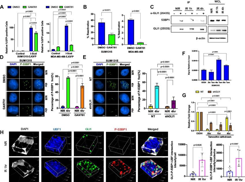
GLI1 localizes to 45S rDNA repeats in response to IR
ures S2A, B). To better understand the relevance of IR-
To explore the hypothesis that DNA damage induces cistro- induced Hh activation, we inhibited the activity of GLI us-
mic changes in GLI1, we undertook an unbiased screen- ing GANT61, a direct GLI1/2 inhibitor (Figure 2D, E).
ing approach in SUM1315 triple-negative breast cancer This resulted in persistence of IR-induced ␥ -H2AX expres-
(TNBC) cells known to have aberrant activation of Hh sig- sion, suggesting delays in DNA repair (Figure 2E). Com-
naling reflected by high endogenous levels of GLI1. GLI1- plementing the outcomes of SUM1315 cells, MDA-MB-468
associated chromatin was immunoprecipitated from cells cells inhibited for Hh/GLI signaling also demonstrated per-
four hours after 4 Gy IR or mock irradiation (Supple- sistence of IR-induced ␥ -H2AX expression, although the
mentary Figure S1A) and evaluated by next-generation kinetics between these two cell systems was markedly dis-
sequencing (ChIP-Seq). We found that GLI1 associates tinct. (Figure 2E and Supplementary Figure S2C). Hh in-
with a novel motif (Figure 1A and Supplementary Fig- hibition using the SMO inhibitor vismodegib (Supplemen-
ure S1B) that is distinct from the previously reported core tary Figures S2D and S2E) or stable transfection of GLI1-
sequence (5 -GACCACCCA-3 ) (24). We identified ChIP- targeting shRNA (shGLI1) yielded similar findings (Sup-
Seq peaks that were specific to the irradiated and nonir- plementary Figure S2F). This corresponded with statisti-
radiated samples (Supplementary Tables S2 and S3). Sur- cally significant delays in resolution of IR-induced ␥ -H2AX
prisingly, unique peaks specific to irradiated cells were foci visualized using immunocytochemistry (Figure 2F). To
markedly enriched for rDNA loci, with approximately a more specifically examine DSBs, we used neutral comet as-
third of the hits associated with RNA5S1–17, RNA45SN1– says to measure average tail moments in SUM1315 and
5, RNA18SN1–5, RNA28SN1–5 and RNA5–8SN1–5 (Fig- MDA-MB-468 cells pretreated with GANT61 and then ex-
ure 1B). We manually searched the 45S rDNA coding se- posed to 4 Gy IR (Figure 2G). As anticipated, the aver-
quence for putative GLI1 interacting sequences based ei- age tail moment increased in irradiated cells with evidence
ther on the previously reported core sequence or the motif of resolution over time in vehicle-treated cells. However,
calculated from our ChIP-Seq data. Using the Integrated GANT61-pretreated cells were unable to recover. Hh inhi-
Genomics Viewer (17) to visualize peaks at these potential bition using stable transfection of shGLI1 yielded similar
sites, we focused on five putative GLI1 binding sites with results compared to non-targeting shRNA control (Sup-
associated peaks that were enhanced after irradiation, rul- plementary Figure S2G). Collectively, these findings sug-
ing out candidates where no peak was seen (Figure 1C). Site gest that inhibiting Hh signaling delays IR-induced DSB
A is based on the previously reported consensus and is lo- repair.
cated in the 5 external transcribed spacer (ETS). Sites B–E
are based on our novel predicted binding sequence and clus-
Hh signaling is required for efficient NHEJ
ter in the 28S region. To validate results of the ChIP-Seq
screen, we performed quantitative PCR of immunoprecipi- Recalling our initial finding that GLI1 is enriched at
tated chromatin and discovered that GLI1 association with rDNA loci following IR, we conjectured that GLI1 pro-Nucleic Acids Research, 2020, Vol. 48, No. 18 10347
Downloaded from https://academic.oup.com/nar/article/48/18/10342/5902436 by guest on 28 October 2020
Figure 1. Ionizing radiation induces GLI1 binding to novel sequences in rDNA. (A) ChIP-Seq data was used to predict a novel GLI1-associated sequence
after IR. (B) Unique ChIP-Seq peaks following IR were enriched in rDNA loci compared to NIR. (C) GLI1 association was increased in the 28S region
of 45S rDNA after IR at sites A-E. (D) Five putative GLI1 binding sites (A–E) show increase in GLI1 occupancy after IR and were validated with ChIP-
qPCR. Statistical significance was determined with a one-way ANOVA and Dunnett’s multiple comparison test, comparisons between NIR- and IR-treated
cells are shown. (E) Immunocytochemistry demonstrates higher GLI1 presence in the fibrillarin-positive nucleoli 1hr after IR, quantitated as a percentage
of nucleoli per cell or absolute number of nucleoli. (F) Fibrillarin and GLI1 co-localization, depicted in yellow, increases after IR, normalized either to the
total number of cells or nucleoli using NIS binary intersection mean intensity (n = 5). Significance was determined by using t-test. All error bars depict the
SEM.
motes rDNA repair, which preferentially utilizes NHEJ (9). BRCA1-mutated SUM1315 cells demonstrated a smaller
SUM1315 and MDA-MB-468 cells were stably transfected magnitude of HR activity, this was unchanged in presence
with the pimEJ5-GFP reporter plasmid, a construct used to of GANT61. In MDA-MB-468 cells, GANT61 reduced
assay NHEJ activity. After site-specific DSBs were induced HR-mediated repair, albeit the overall magnitude of HR ac-
by the restriction enzyme I-SceI (Supplementary Figure tivity was very small (Supplementary Figures S3B, C).
S3A), GFP-expressing cells that had undergone successful Prior work has suggested that GLI1 may interact with the
NHEJ-mediated repair were quantified by flow cytometry. DSB-sensing proteins MRE11 and RAD50 (25). Using en-
Hh inhibition by GANT61 significantly impaired NHEJ in dogenous protein co-immunoprecipitation, we found that
both cell lines (Figure 3A). In an independent verification, GLI1 interacts with 53BP1, a protein critical for dictating
we linearized pGL3-Control with the restriction enzyme repair pathway choice towards NHEJ (26) (Figure 3C).
HindIII, disconnecting the promoter from the firefly lu- The interaction of endogenous GLI1 with 53BP1 is ac-
ciferase coding sequence. Cells transfected with HindIII-cut companied by active P-53BP1 foci at DSB sites (Figure 3D,
pGL3-Control will only express firefly luciferase if NHEJ E). This suggests that the interaction between GLI1 and
has resolved the break. Again, GANT61 significantly re- 53BP1 is likely important for the rapid NHEJ repair re-
duced NHEJ-mediated repair (Figure 3B). We also assessed sponse and P-53BP1 foci formation at DSB sites.
the possible role of HR in IR-induced dsDNA damage Furthermore, IR-induced expression of P-53BP1 and
using the DR-GFP luciferase reporter construct. While abundance of P-53BP1 foci were reduced when Hh was in-10348 Nucleic Acids Research, 2020, Vol. 48, No. 18
Downloaded from https://academic.oup.com/nar/article/48/18/10342/5902436 by guest on 28 October 2020
Figure 2. Inhibition of Hh/GLI signaling delays ionizing radiation-induced DSB repair in TNBC cells. (A) 8X-GBS-luciferase reporter assays show Hh
activation in multiple TNBC cell lines after IR, confirmed by GLI1 (B) mRNA and (C) protein expression using qPCR and immunoblotting, respectively
in SUM1315 and MDA-MB-468 cell lines. Statistical significance was determined with a multiple t test and a one-way ANOVA with Dunnett’s multiple
comparison test. For one-way ANOVA, comparisons between NIR- and IR-treated cells are shown. (D) GANT61 suppresses IR-induced Hh activation.
Statistical significance represents the results of a two-way ANOVA and Tukey multiple comparison test. (E) GANT61 leads to delayed resolution of
IR-induced ␥ -H2AX after IR shown by immunoblot compared to vehicle control. (F) IR-induced ␥ -H2AX foci shown by immunocytochemistry show
similar delay in foci resolution. (G) Neutral comet assays show impaired DSB repair in GANT61-treated cells. Statistical significance was determined with
a two-way ANOVA and Tukey’s multiple comparison test. Comparisons for DMSO treated NIR and DMSO treated IR-4 h for SUM1315 and IR-12 h
for MDA-MB468 are shown. All error bars depict the SEM.
hibited by either GANT61 (Figure 3D) or GLI1 knock- and ATM, which are previously reported indicators of I-
down (Figure 3E, Supplementary Figures S3D, E). PpoI-mediated damage (Supplementary Figure S3H) (23).
Using ChIP, we found that GLI1 localizes to the sites of
I-PpoI-mediated breaks in 45S rDNA (Figure 3F). To eval-
Hh signaling is required for resolution of site-specific rDNA uate the role of GLI1 in the resolution of these breaks, we
DSBs withdrew tamoxifen after overnight treatment to allow for
Because these assays address global DNA repair, we sought repair. The abundance of an amplicon spanning the I-PpoI
to assess how rDNA repair specifically is affected by Hh restriction site was reduced by tamoxifen treatment, indicat-
inhibition. To do so, we transfected cells with tamoxifen- ing the presence of breaks that the polymerase was unable to
inducible I-PpoI (pBABe-HA-ER-IPpoI), a restriction en- read through (Figure 3G). After tamoxifen was withdrawn
zyme with limited recognition sites in the human genome, for four hours, the magnitude of amplicon abundance in-
including one in the 45S rDNA sequence (Supplemen- creased again in control (NT) cells, implying repair of the
tary Figure S3F). Due to the sheer number of 45S rDNA cleavage site. We found that GLI1 knockdown (shGLI1)
repeats found throughout the human genome, I-PpoI- significantly hampered the recovery of amplicon expres-
induced DNA breaks can be considered a surrogate for sion after tamoxifen withdrawal, suggesting that GLI1 is
rDNA-specific damage (23). We first confirmed that tamox- required for the efficient repair of site-specific rDNA DSBs.
ifen treatment of cells transfected with pBABe-HA-ER- Resolution of rDNA breaks requires the recruitment of
IPpoI reduced abundance of an amplicon spanning the I- repair machinery to sites of damage. Using confocal mi-
PpoI cleavage site in the 45S rDNA sequence by qPCR, in- croscopy, we observed the colocalization of GLI1 and P-
dicating breakage (Supplementary Figure S3G). Concomi- 53BP1 in the nucleolus in response to IR (Figure 3H and
tant with this, we also registered phosphorylation of NBS1 Supplementary Figure S3I). We quantified the intersectionNucleic Acids Research, 2020, Vol. 48, No. 18 10349
Downloaded from https://academic.oup.com/nar/article/48/18/10342/5902436 by guest on 28 October 2020
Figure 3. Hh inhibition impairs NHEJ and delays repair of rDNA DSBs. (A) Hh inhibition compromises repair of I-SceI-induced breaks by NHEJ in cells
stably expressing the pimEJ5-GFP reporter. Statistical analysis was performed with a two-way ANOVA and Tukey’s multiple comparison test, p values are
indicated. (B) Impaired NHEJ in Hh-inhibited cells is also evident by diminished reactivation of linearized firefly luciferase plasmid. Statistical analysis was
determined with a t-test. (C) Endogenous protein co-immunoprecipitation of SUM1315 lysates shows that 53BP1 interacts with GLI1. Whole cell lysate
(WCL) input controls are shown to the right. (D, E) P-53BP1 foci are reduced by (D) GANT61 and (E) stable knockdown of GLI1 compared to controls in
SUM1315 cell line at 4 h post-IR. Statistical analysis was performed with a two-way ANOVA and Tukey’s multiple comparison test, p values are indicated.
(F) GLI1 localizes to I-PpoI-induced rDNA breaks as evidenced by greater occupancy at the I-PpoI break-site following tamoxifen treatment shown by
ChIP-qPCR. Statistical significance was determined with a a one-way ANOVA and Dunnett’s multiple comparison test, comparisons between ethanol
treated and tamoxifen withdrawn cells are shown. (G) Tamoxifen treatment induces breaks in SUM1315 NT and shGLI1 cells as measured by reduction
in magnitude of amplicon spanning the I-PpoI restriction site. 4 hr post tamoxifen withdrawal, amplicon levels in NT cells increase but stable knockdown
of GLI1 remains unchanged indicating efficient repair of these sites of damage in NT compared to shGLI1. Statistical significance was determined with a
two-way ANOVA and Tukey’s multiple comparison test for treatment at respective time points. Comparisons for ethanol-treated and tamoxifen withdrawn
cells and comparison at 4 h after tamoxifen withdrawal between NT and shGLI1 cells are shown. (H) 3D confocal images of NIR (top) and IR (bottom)
SUM1315 cells labeled with GLI1 (green), P-53BP1 (red), UBF (blue), and DAPI (gray) depict increased nucleolar localization and interaction of GLI1
and P-53BP1 1 h post IR in the nucleolar compartment. GLI1 and P-53BP1 interaction in the nucleolus was quantified from five NIR and IR cells and
normalized either to the number of cells or nucleoli per field using NIS binary intersection mean intensity (n = 5). All error bars depict the SEM.
of GLI1, P-53BP1, and the nucleolar marker UBF1 at one- during rRNA processing, they have a short half-life and are
hour post-irradiation and noted a substantial increase in ideal surrogates for Pol I activity over time (22). As antici-
their association per cell and per nucleolus (Figure 3H). pated, in control cells treated with vehicle, Pol I activity was
reduced early in response to IR and recovered at later time
points (Figure 4A and B), consistent with the kinetics of
Combining Hh inhibition with IR compromises Pol I activity
repair determined by our earlier experiments. When Hh ac-
and cell viability
tivity was inhibited by GANT61, Pol I activity is reduced by
rDNA DSBs temporarily arrest transcription until repair IR as expected, but does not recover. Hh inhibition by sta-
is complete. To assess how Hh inhibition affects 45S rDNA ble GLI1 knockdown yielded similar results (Supplemen-
transcription by Pol I in response to damage, we used qPCR tary Figures S4A and S4B). Interestingly, the delay in Pol
to quantify expression of two different sites in the 5 ETS. I recovery that we observed with GLI1 inhibition mirrors
Because these regions are removed post-transcriptionally prior findings when NHEJ was directly inhibited instead10350 Nucleic Acids Research, 2020, Vol. 48, No. 18
Downloaded from https://academic.oup.com/nar/article/48/18/10342/5902436 by guest on 28 October 2020
Figure 4. Inhibiting Hh/GLI signaling impairs re-activation of Pol I activity following irradiation-induced DSBs. (A, B) Pol I activity, as quantitated
by qPCR of two different amplicons in the 5’ ETS of 45S rDNA, is reduced by IR and recovers following repair in cells treated with vehicle control
compared to GANT61-treated cells. Statistical significance was determined with a two-way ANOVA and Tukey’s multiple comparison test. Comparisons
for DMSO- and GANT61- NIR group with cells collected at different time points post IR and DMSO- and GANT61-treated cells at 4 and 8 h are shown.
(C, D) Hh inhibition with GANT61 combined with IR almost completely abrogates spheroid growth, compared to more modest reductions with either
modality alone. Growth area was quantified using ImageJ software per 10× field. Statistical significance was determined with a two-way ANOVA and
Tukey’s multiple comparison test for each condition. All error bars depict the SEM.
(9). We then explored whether Hh pathway inhibition would but dramatically impaired spheroid growth when combined
lead to sensitivity due to its role in rDNA repair. In order to with irradiation in both SUM1315 and MDA-MB-468 cells.
specifically induce rDNA breaks, we transfected SUM1315 Similar findings were noted with GLI1 knockdown (Sup-
cells with I-PpoI plasmid or an empty vector control. In plementary Figure S4E). Similarly, colony formation, fol-
order to specifically address the role of Hh/GLI activity, lowing irradiation, was significantly compromised in cells
we queried SUM1315 cells stably silenced for GLI1. We as- deficient for Hh/GLI activity (Supplementary Figures S4F
sessed the effect on cell survival using a colony formation and G). Next, we examined whether Hh activity also im-
assay. Introducing I-PpoI caused an appreciable decrease in pacts survival of breast tumor cells when DNA damage
the number of colonies relative to control plasmid (Supple- is inflicted with the anthracycline doxorubicin, a topoiso-
mentary Figure S4C, D). Interestingly, GLI1-silenced cells merase inhibitor that generates DSBs. We scored cell sur-
transfected with I-PpoI showed a striking decrease in the vival with a colony formation assay using doxorubicin in
number of colonies formed, suggesting that Hh inhibition lieu of IR, and saw that doxorubicin (used at 0.1 M) sig-
further reduces survival of cells inflicted with rDNA breaks nificantly compromised the abundance of colonies formed
with I-PpoI. These findings support that Hh inhibition aug- by GLI1-deficient SUM1315 cells compared to NT shRNA
ments rDNA damage and consequently poses finite lethal- controls (Supplementary Figures S4H and I). We also
ity to TNBC cells independent of global effects on DSB re- scored cell viability using the MTS assay and registered a
pair. significant decrease in cell viability in doxorubicin-treated
To assess the overall outcome of IR-induced DNA dam- SUM1315 shGLI1 cells relative to NT cells (P < 0.01) (Sup-
age in the context of Hh inhibition, we generated spheroids plementary Figure S4J). As such, the data cumulatively in-
in three-dimensional culture from cells pre-treated with dicates that Hh inhibition sensitizes TNBC cells to IR and
either vehicle control or GANT61 (Figure 4C and D). doxorubicin, both of which are clinically relevant agents
GANT61 alone had a modest effect on non-irradiated cells, used to treat TNBC.Nucleic Acids Research, 2020, Vol. 48, No. 18 10351
DISCUSSION intimately involved in orchestrating normal ontogeny and
cancer, it is likely that dysregulated Hh activity may craft
Hh signaling is a classical developmental pathway that is
intersecting and shared programs that enable cells to sur-
aberrantly activated in various cancers (27) and has been
vive erroneous and possibly lethal impediments.
linked to tumor initiation, progression and metastasis in
Interestingly, inhibiting Hh did not impair baseline Pol I
breast cancer. In TNBC patients, high expression of Hh
activity on its own. Instead, its effect is secondary to com-
pathway proteins correlates to poor survival (28,29). Treat-
promised rDNA repair. Thus, GLI1 does not appear to
ment of TNBC relies on DNA-damaging agents, including
directly promote rRNA transcription, but rather helps to
conventional chemotherapy and IR (30). Though inhibit-
maintain the genomic integrity of rDNA loci. These sites
ing Hh has been shown to sensitize cancer cells to genotoxic
may be particularly vulnerable in proliferative and metabol-
therapies, the underlying mechanisms to this point have re-
ically active states where Hh is activated, such as in nor-
mained vague (12,13). Our study has uncovered an unex-
mal development and cancer. In this context, protecting
pected role for Hh signaling in the repair of damaged rDNA
rDNA loci from insults such as replication stress and DNA-
Downloaded from https://academic.oup.com/nar/article/48/18/10342/5902436 by guest on 28 October 2020
that helps to explain this phenomenon.
damaging agents may be a critical new function of Hh sig-
Though we found that GLI1 is required for efficient
naling with potential implications for therapeutic resistance
NHEJ-mediated repair of nonspecific DSBs, we demon-
in cancer. Our findings suggest that further evaluation of Hh
strated a marked enrichment of GLI1 at rDNA loci in re-
inhibitors as potential radiosensitizers or chemosensitizers
sponse to IR using an unbiased ChIP-Seq screen and con-
is warranted.
firmed this relative to nonspecific binding controls with
standard ChIP. Importantly, the precise degree of enrich-
ment at rDNA loci relative to standard genes is unclear. DATA AVAILABILITY
rDNA coding sequences are known to be arranged in tan-
dem arrays of tens to hundreds of repeats, but only 17 5S The datasets used and/or analyzed during the current
rDNA sequences and five 45S rDNA sequences are mapped study have been deposited at the Gene Expression Omnibus
to the current build of the human genome. This was recently (GEO) under the accession number GSE146237.
identified as a critical unmet need in the rDNA field (31).
For this reason, we focused on the defined coding sequences
and not the poorly defined intergenic spacers between re- SUPPLEMENTARY DATA
peats. Though we found more hits were associated with 5S Supplementary Data are available at NAR Online.
rDNA repeats, we elected to study the 45S rDNA both be-
cause of the availability of tools to study its gene expression
(qPCR of the 5 -ETS) and site-specific DSBs (I-PpoI) and ACKNOWLEDGEMENTS
because 45S rDNA breaks have been previously shown to We thank the UAB Animal Resources Program for the use
be more consequential than those in 5S rDNA (5). Even of irradiator facilities; the UAB Comprehensive Flow Cy-
accounting for the likelihood that pulled-down fragments tometry Core for the use of flow cytometry facilities; the
mapped to multiple repeats, our findings may underestimate UAB High Resolution Imaging Facility and Dr R. Grab-
the degree of GLI1 interaction with rDNA given the vast ski for technical assistance; J.M. Stark for providing the
number of repeats known to exist in the human genome. pimEJ5-GFP construct via AddGene; M.B. Kastan for pro-
Notably, we did not observe similar enrichment of GLI1 viding the pBABe-HA-ER-IPpoI construct via AddGene;
at genes associated with NHEJ, including XRCC5, XRCC6, J.A. Bonner for the use of comet analysis facilities and H.Q.
PRKDC, LIG4 and TP53BP1, which encode for Ku70, Trummell for related technical assistance and guidance; and
Ku80, DNA-PKcs, DNA ligase IV, and 53BP1, respectively. E.S. Yang for providing the I-SceI construct and technical
This suggests that the effect of GLI1 on NHEJ is not related expertise.
to its transcription factor activity, despite our finding that
GLI1 expression and activity are increased in response to
IR. Whether the enrichment of GLI1 at rDNA loci is influ- FUNDING
enced by upregulated GLI1 protein expression or shifts in
subcellular localization that we observed in response to IR NCI/NIH [CA183926 to V.T.G.L.]; Merit Review Award
remains an open question. [I01 BX003374] from the U.S. Department of Veterans Af-
Accordingly, we hypothesized that GLI1 instead recruits fairs BLRD service BX003374; NCI/NIH [CA194048 to
NHEJ-associated proteins to the sites of rDNA DSBs. In R.S.S., CA169202 to L.A.S.]; United States Department of
line with this conjecture, we found that GLI1 interacts with Defense [W81XWH-18-1-0036 to L.A.S.]; University of Al-
the NHEJ protein 53BP1. Furthermore, we observed us- abama at Birmingham [AMC21 to L.A.S.]; Breast Cancer
ing confocal microscopy that GLI1 association with both Research Foundation of Alabama [to L.A.S.]. Funding for
P-53BP1 and the rDNA marker UBF1 is triggered by IR. open access charge: Institutional start-up funds.
Inhibition of GLI1 mutes the accumulation of P-53BP1, Conflict of interest statement. None declared.
interferes with repair of site-specific rDNA DSBs, and de-
lays recovery of Pol I activity, an indicator of unrepaired
rDNA DSBs, in response to IR. To our knowledge, this is REFERENCES
the first example of a developmental signaling pathway be- 1. Pelletier,J., Thomas,G. and Volarevic,S. (2018) Ribosome biogenesis
ing directly tied to rDNA repair. Given that Hh signaling is in cancer: new players and therapeutic avenues. Nat. Rev. Cancer, 18,
51–63.10352 Nucleic Acids Research, 2020, Vol. 48, No. 18
2. Tchurikov,N.A., Fedoseeva,D.M., Sosin,D.V., Snezhkina,A.V., 17. Robinson,J.T., Thorvaldsdottir,H., Winckler,W., Guttman,M.,
Melnikova,N.V., Kudryavtseva,A.V., Kravatsky,Y.V. and Lander,E.S., Getz,G. and Mesirov,J.P. (2011) Integrative genomics
Kretova,O.V. (2015) Hot spots of DNA double-strand breaks and viewer. Nat. Biotechnol., 29, 24–26.
genomic contacts of human rDNA units are involved in epigenetic 18. Hacot,S., Coute,Y., Belin,S., Albaret,M.A., Mertani,H.C.,
regulation. J. Mol. Cell Biol., 7, 366–382. Sanchez,J.C., Rosa-Calatrava,M. and Diaz,J.J. (2010) Isolation of
3. Tchurikov,N.A., Yudkin,D.V., Gorbacheva,M.A., Kulemzina,A.I., nucleoli. Curr. Protoc. Cell Biol., 47, 3.36.1–3.36.10.
Grischenko,I.V., Fedoseeva,D.M., Sosin,D.V., Kravatsky,Y.V. and 19. Sasaki,H., Hui,C., Nakafuku,M. and Kondoh,H. (1997) A binding
Kretova,O.V. (2016) Hot spots of DNA double-strand breaks in site for Gli proteins is essential for HNF-3beta floor plate enhancer
human rDNA units are produced in vivo. Sci. Rep., 6, 25866. activity in transgenics and can respond to Shh in vitro. Development,
4. Stults,D.M., Killen,M.W., Williamson,E.P., Hourigan,J.S., 124, 1313–1322.
Vargas,H.D., Arnold,S.M., Moscow,J.A. and Pierce,A.J. (2009) 20. Zhuang,J., Zhang,J., Willers,H., Wang,H., Chung,J.H., van
Human rRNA gene clusters are recombinational hotspots in cancer. Gent,D.C., Hallahan,D.E., Powell,S.N. and Xia,F. (2006) Checkpoint
Cancer Res., 69, 9096–9104. kinase 2-mediated phosphorylation of BRCA1 regulates the fidelity
5. Warmerdam,D.O., van den Berg,J. and Medema,R.H. (2016) Breaks of nonhomologous end-joining. Cancer Res., 66, 1401–1408.
in the 45S rDNA lead to Recombination-Mediated loss of repeats. 21. Sulkowski,P.L., Corso,C.D., Robinson,N.D., Scanlon,S.E.,
Cell Rep., 14, 2519–2527. Purshouse,K.R., Bai,H., Liu,Y., Sundaram,R.K., Hegan,D.C.,
Downloaded from https://academic.oup.com/nar/article/48/18/10342/5902436 by guest on 28 October 2020
6. Roukos,V. and Misteli,T. (2014) The biogenesis of chromosome Fons,N.R. et al. (2017) 2-Hydroxyglutarate produced by neomorphic
translocations. Nat. Cell Biol., 16, 293–300. IDH mutations suppresses homologous recombination and induces
7. Ceccaldi,R., Rondinelli,B. and D’Andrea,A.D. (2016) Repair PARP inhibitor sensitivity. Sci. Transl. Med., 9, eaal2463.
pathway choices and consequences at the double-strand break. 22. Peltonen,K., Colis,L., Liu,H., Trivedi,R., Moubarek,M.S.,
Trends Cell Biol., 26, 52–64. Moore,H.M., Bai,B., Rudek,M.A., Bieberich,C.J. and Laiho,M.
8. Scully,R., Panday,A., Elango,R. and Willis,N.A. (2019) DNA (2014) A targeting modality for destruction of RNA polymerase I
double-strand break repair-pathway choice in somatic mammalian that possesses anticancer activity. Cancer Cell, 25, 77–90.
cells. Nat. Rev. Mol. Cell Biol., 20, 698–714. 23. Berkovich,E., Monnat,R.J. Jr and Kastan,M.B. (2007) Roles of ATM
9. Harding,S.M., Boiarsky,J.A. and Greenberg,R.A. (2015) ATM and NBS1 in chromatin structure modulation and DNA
dependent silencing links nucleolar chromatin reorganization to double-strand break repair. Nat. Cell Biol., 9, 683–690.
DNA damage recognition. Cell Rep., 13, 251–259. 24. Kinzler,K.W. and Vogelstein,B. (1990) The GLI gene encodes a
10. van Sluis,M. and McStay,B. (2015) A localized nucleolar DNA nuclear protein which binds specific sequences in the human genome.
damage response facilitates recruitment of the homology-directed Mol. Cell. Biol., 10, 634–642.
repair machinery independent of cell cycle stage. Genes Dev., 29, 25. Li,X., Wang,W., Wang,J., Malovannaya,A., Xi,Y., Li,W., Guerra,R.,
1151–1163. Hawke,D.H., Qin,J. and Chen,J. (2015) Proteomic analyses reveal
11. Kudo,K., Gavin,E., Das,S., Amable,L., Shevde,L.A. and Reed,E. distinct chromatin-associated and soluble transcription factor
(2012) Inhibition of Gli1 results in altered c-Jun activation, inhibition complexes. Mol. Syst. Biol., 11, 775.
of cisplatin-induced upregulation of ERCC1, XPD and XRCC1, and 26. Panier,S. and Boulton,S.J. (2014) Double-strand break repair: 53BP1
inhibition of platinum-DNA adduct repair. Oncogene, 31, 4718–4724. comes into focus. Nat. Rev. Mol. Cell Biol., 15, 7–18.
12. Mazumdar,T., Devecchio,J., Agyeman,A., Shi,T. and Houghton,J.A. 27. Amakye,D., Jagani,Z. and Dorsch,M. (2013) Unraveling the
(2011) Blocking Hedgehog survival signaling at the level of the GLI therapeutic potential of the Hedgehog pathway in cancer. Nat. Med.,
genes induces DNA damage and extensive cell death in human colon 19, 1410–1422.
carcinoma cells. Cancer Res., 71, 5904–5914. 28. Habib,J.G. and O’Shaughnessy,J.A. (2016) The hedgehog pathway in
13. Teichman,J., Dodbiba,L., Thai,H., Fleet,A., Morey,T., Liu,L., triple-negative breast cancer. Cancer Med., 5, 2989–3006.
McGregor,M., Cheng,D., Chen,Z., Darling,G. et al. (2018) Hedgehog 29. Noman,A.S., Uddin,M., Rahman,M.Z., Nayeem,M.J., Alam,S.S.,
inhibition mediates radiation sensitivity in mouse xenograft models Khatun,Z., Wahiduzzaman,M., Sultana,A., Rahman,M.L., Ali,M.Y
of human esophageal adenocarcinoma. PLoS One, 13, e0194809. et al. (2016) Overexpression of sonic hedgehog in the triple negative
14. Das,S., Harris,L.G., Metge,B.J., Liu,S., Riker,A.I., Samant,R.S. and breast cancer: clinicopathological characteristics of high burden
Shevde,L.A. (2009) The hedgehog pathway transcription factor GLI1 breast cancer patients from Bangladesh. Sci. Rep., 6, 18830.
promotes malignant behavior of cancer cells by up-regulating 30. Liedtke,C., Mazouni,C., Hess,K.R., Andre,F., Tordai,A., Mejia,J.A.,
osteopontin. J. Biol. Chem., 284, 22888–22897. Symmans,W.F., Gonzalez-Angulo,A.M., Hennessy,B., Green,M.
15. Li,H. and Durbin,R. (2009) Fast and accurate short read alignment et al. (2008) Response to neoadjuvant therapy and long-term survival
with Burrows-Wheeler transform. Bioinformatics, 25, 1754–1760. in patients with triple-negative breast cancer. J. Clin. Oncol., 26,
16. Zhang,Y., Liu,T., Meyer,C.A., Eeckhoute,J., Johnson,D.S., 1275–1281.
Bernstein,B.E., Nusbaum,C., Myers,R.M., Brown,M., Li,W. et al. 31. Baserga,S.J., DiMario,P.J. and Duncan,F.E. (2020) Emerging roles for
(2008) Model-based analysis of ChIP-Seq (MACS). Genome Biol., 9, the nucleolus 2019. J. Biol. Chem., 295, 5535–5537.
R137.You can also read