Immunomodulatory potential of anticancer therapy composed of methotrexate nanoconjugate and dendritic cell based vaccines in murine colon carcinoma
←
→
Page content transcription
If your browser does not render page correctly, please read the page content below
ONCOLOGY REPORTS 45: 945-962, 2021

            Immunomodulatory potential of anticancer therapy
          composed of methotrexate nanoconjugate and dendritic
              cell‑based vaccines in murine colon carcinoma
               AGNIESZKA SZCZYGIEŁ, NATALIA ANGER‑GÓRA, KATARZYNA WĘGIEREK‑CIURA,
                 JAGODA MIERZEJEWSKA, JOANNA ROSSOWSKA, TOMASZ M. GOSZCZYŃSKI,
                          MARTA ŚWITALSKA and ELŻBIETA PAJTASZ‑PIASECKA

 Ludwik Hirszfeld Institute of Immunology and Experimental Therapy, Polish Academy of Sciences, 53‑114 Wroclaw, Poland

                                   Received August 27, 2020; Accepted November 30, 2020

                                                   DOI: 10.3892/or.2021.7930

Abstract. Chemotherapy with low‑molecular weight                   Introduction
compounds, despite elimination of cancer cells, entails adverse
effects. To overcome this disadvantage, innovative drug            For many years, chemotherapy has been one of the most
delivery systems are being developed, including conjugation        frequently chosen types of anticancer treatment. However, its
of macromolecular carriers with therapeutics, e.g. a nanocon-      cure rate still remains unsatisfactory and additionally severe
jugate of hydroxyethyl starch and methotrexate (HES‑MTX).          side effects are observed (1,2). Therefore, the development of
The purpose of the present study was to determine whether          effective anticancer therapy is still a challenge, mainly due
HES‑MTX, applied as a chemotherapeutic, is able to modulate        to the complex nature of tumors (3). During cancer growth, a
the immune response and support the antitumor response             specific niche‑the tumor microenvironment (TME)‑is created,
generated by dendritic cells (DCs) used subsequently as immu-      consisting mainly of proliferating tumor cells, extracellular
notherapeutic vaccines. Therefore, MTX or HES‑MTX was              matrix, stromal cells and infiltrating immune cells (3‑6).
administered, as sole treatment or combined with DC‑based          Among the latter, high influx of regulatory T cells (Tregs),
vaccines, to MC38 colon carcinoma tumor‑bearing mice.              tumor‑associated macrophages (TAMs) and myeloid derived
Alterations in antitumor immune response were evaluated by         suppressor cells (MDSCs), which promote tumor progres-
multiparameter flow cytometry analyses and functional assays.      sion and suppress the antitumor immune response, are
The results demonstrated that the nanoconjugate possesses          observed (5‑7). On the other hand, immune cells, such as effector
greater immunomodulatory potential than MTX as reflected           T cells, natural killer (NK) cells, M1‑type macrophages and
by changes in the landscape of immune cells infiltrating the       dendritic cells (DCs) infiltrating the tumor, can be activated
tumor and increased cytotoxicity of splenic lymphocytes. In        in situ in order to inhibit tumor growth and prevent immune
contrast to MTX, therapy with HES‑MTX as sole treatment            evasion and expansion of the disease (5,7). Increasing evidence
or combined with DC‑based vaccines, contributed to signifi-        indicates that the fate of tumor progression is highly correlated
cant tumor growth inhibition. However, only treatment with         with the specific TME, whose composition is a predominant
HES‑MTX and DC‑based vaccines activated the systemic               factor in prognosis and efficacy of chemotherapy (3).
specific antitumor response. In conclusion, due to its immu-           DCs as professional antigen‑presenting cells are potent
nomodulatory properties, the HES‑MTX nanoconjugate could           inducers of a T cell response, and are considered an essential
become a potent anticancer agent used in both chemo‑ and           component of antitumor immunity (8). However, despite high
chemoimmunotherapeutic treatment schemes.                          potential in promoting the antitumor response, the proper func-
                                                                   tion of DCs present in the TME may be impaired, mainly due
                                                                   to the abundance of immunosuppressive factors and aforemen-
                                                                   tioned cells with suppressor activity. DCs under the influence
                                                                   of a hostile tumor milieu become ineffective in their differ-
                                                                   entiation and activation, and in turn are weak stimulators of
Correspondence to: Mrs. Agnieszka Szczygieł, Ludwik Hirszfeld      immune responses (8,9). For this reason, great efforts are made
Institute of Immunology and Experimental Therapy, Polish Academy   to design therapeutic strategies able to overcome the negative
of Sciences, 12 Rudolf Weigl Street, 53‑114 Wroclaw, Poland        impact of TME on endogenous DCs. One of the strategies is
E‑mail: agnieszka.szczygiel@hirszfeld.pl                           ex vivo generation and maturation of DCs for their admin-
                                                                   istration as cellular‑based antitumor vaccines (10‑12). Other
Key words: methotrexate, nanoconjugate, dendritic cells, colon     strategies involve, not only developing new chemotherapeutics
carcinoma, MC38, immunotherapy, chemotherapy                       or innovative solutions for targeted drug delivery, but also
                                                                   combining different types of anticancer therapy‑for example
                                                                   chemotherapy and immunotherapy with DC‑based cellular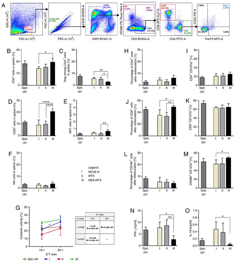
946         SZCZYGIEŁ et al: IMMUNOMODULATORY POTENTIAL OF MTX NANOCONJUGATE AND DC-BASED VACCINES

vaccines. This latter strategy has several immune‑potentiating     of Tregs from the TME (36‑40). Utilization of the HES‑MTX
effects, such as increasing the susceptibility of tumor cells to   nanoconjugate in a murine MC38 colon carcinoma model and
the activity of cytotoxic T lymphocytes (CTLs). Furthermore,       supplementing such anticancer therapy with DC‑based immu-
by depleting certain population of immune cells, e.g. MDSCs        notherapy, as well determination of its immunomodulatory
or Tregs, chemotherapy creates a cytokine milieu for optimal       effect on generation of an antitumor immune response, has not
expansion of effector cells and facilitates the generation of a    been investigated to date. For this reason, the main objective
specific antitumor immune response by DCs (13). However,           of our study was to determine the modulation of the immune
major problems in anticancer chemotherapy with low‑molec-          response after HES‑MTX administration to tumor‑bearing
ular weight compounds are their fast metabolism and excretion      mice and how those changes affect the activity of DC‑based
from an organism, as well as unfavorable biodistribution and       vaccines injected after chemotherapy. The gathered data indi-
low specificity (14). To overcome these disadvantages, many        cate that chemotherapy with HES‑MTX applied in treatment
different drug delivery systems, including micelles (15,16),       of mice with a subcutaneously growing MC38 tumor affected,
dendrimers (17,18), nanocapsules (19) and nanoconjugates           more strongly than MTX, the TME by increasing the influx
with a macromolecular carrier (14,20) have been developed.         of CTLs and NK cells and eliminating certain cells with
The nanoconjugates were designed to enhance delivery and           suppressor activity. In addition, the enlargement of T‑helper
to improve the selectivity and pharmacological properties of       (Th), CTL and natural killer T (NKT) cell percentages among
both conventional and innovative drugs (14). One of these          splenic leukocytes accompanied by a decrease in Tregs was
innovative formulations is a nanoconjugate of hydroxyethyl         found. Moreover, therapy with HES‑MTX resulted in increased
starch (HES) and methotrexate (MTX). The HES‑MTX nano-             cytotoxic activity of splenic lymphocytes. All these factors
conjugate was obtained by covalent coupling of well‑known          led to the creation of a favorable niche necessary to promote
therapeutic compounds‑methotrexate as an anticancer agent          the development of an efficient antitumor immune response
and hydroxyethyl starch as a high‑molecular carrier (20,21).       by DCs used in immunotherapy. The combined therapy with
MTX is one of the oldest antifolate drugs widely used in the       HES‑MTX and DC‑based cellular vaccines contributed to
treatment of autoimmune disorders as well as in anticancer         enhanced influx of effector lymphocytes into tumor tissue and
therapy‑in solid tumors and hematologic malignancies (22).         reduced infiltration of immune cells with suppressor activity.
However, therapy with MTX is often associated with severe          The above changes together with activation of systemic
systemic toxicity, bone marrow suppression, and drug resis-        specific antitumor response resulted in a significant delay in
tance (15,23,24). HES is an amylopectin‑based modified             tumor growth.
polymer used as colloidal plasma volume expanders (20). The
structural similarity of amylopectin to glycogen ensures lack      Materials and methods
of immunogenicity (25). Moreover, unfavorable accumula-
tion in the liver or spleen was not observed (26). The mean        Mice. Female C57BL/6 mice (total number of animals,
hydrodynamic diameter of the HES‑MTX nanoconjugate is              55 mice; initial weight, 20‑22 g) were obtained from the
15.2±6.2 nm, thus HES‑MTX meets the criterion for inclusion        Center of Experimental Medicine of the Medical University
in nanoparticles (20). The main advantage of the HES‑MTX           of Białystok (Białystok, Poland). Mice were kept in a room
nanoconjugate over MTX in free form is the prolonged               with a standard light/dark cycle, with a constant temperature
half‑time in plasma and specific biodistribution. Methotrexate     (22±2˚C), air humidity (55±10%) and access to food and
enters cells via folate receptors (FRs) overexpressed on cancer    water ad libitum. All experiments were performed in accor-
cells or through the ubiquitously expressed reduced folate         dance with EU Directive 2010/63/EU for animal experiments
carriers (RFCs), to which MTX has a low and high affinity,         and were approved by the 1st Local Ethics Committee for
respectively (27). However, when multiple MTX molecules are        Experiments with the Use of Laboratory Animals, Wrocław,
covalently conjugated to a macromolecular carrier (e.g. HES),      Poland (authorization no. 31/2016). After the experiments,
transport through RFCs does not occur. This is possible due to     mice were sacrificed by cervical dislocation.
acquisition of the polyvalence feature as a consequence of the
conjugation process (17,18,28,29). We postulate that the inter-    Cell culture. The in vivo growing MC38 murine colon
nalization of HES‑MTX nanoconjugate in tumors is achieved          carcinoma from the Tumor Bank of the TNA Radiobiology
mainly by its interaction with FRα, or through an enhanced         Institute (Rijswijk, The Netherlands) was adapted to in vitro
vascular permeability and retention effect (EPR). EPR is           conditions as described by Pajtasz‑Piasecka et al (41). The
related to the capacity of macromolecules larger than 40 kDa       culture of MC38/0 (named here MC38) cells was maintained
(hydrodynamic diameter above 10 nm) for selective leakage          in RPMI‑1640 (Gibco; Thermo Fisher Scientific, Inc.) supple-
from tumor vessels and accumulation in tumor tissue (2,29‑32).     mented with 100 U/ml penicillin, 100 mg/ml streptomycin,
Complete physicochemical characteristics of the novel form of      0.5% sodium pyruvate, 0.05 mM 2‑mercaptoethanol and 5%
HES‑MTX nanoconjugate as well as its antitumor activity in         fetal bovine serum (FBS; all reagents from Sigma‑Aldrich;
murine P388 leukemia and human MV‑4‑11 leukemia models             Merck KGaA). Tumor antigen (TAg) was prepared by repeated
have been described (20).                                          freezing and thawing of an MC38 cell suspension (5×10 6
    Recently considerable attention has been focused on the        MC38 cells/ml), which was followed by sonication. DCs
immunomodulatory properties of certain chemotherapeutic            for in vivo experiments were generated from bone marrow
agents, including MTX (1,19), which can act not only as            isolated from femurs and tibias of healthy C57BL/6 mice
modulators of immune cell phenotype (33‑35), but also              according to the protocol described in our previous publica-
through stimulation of effector immune cells and elimination       tion (42). The cells (named here DCs) were cultured in RPMI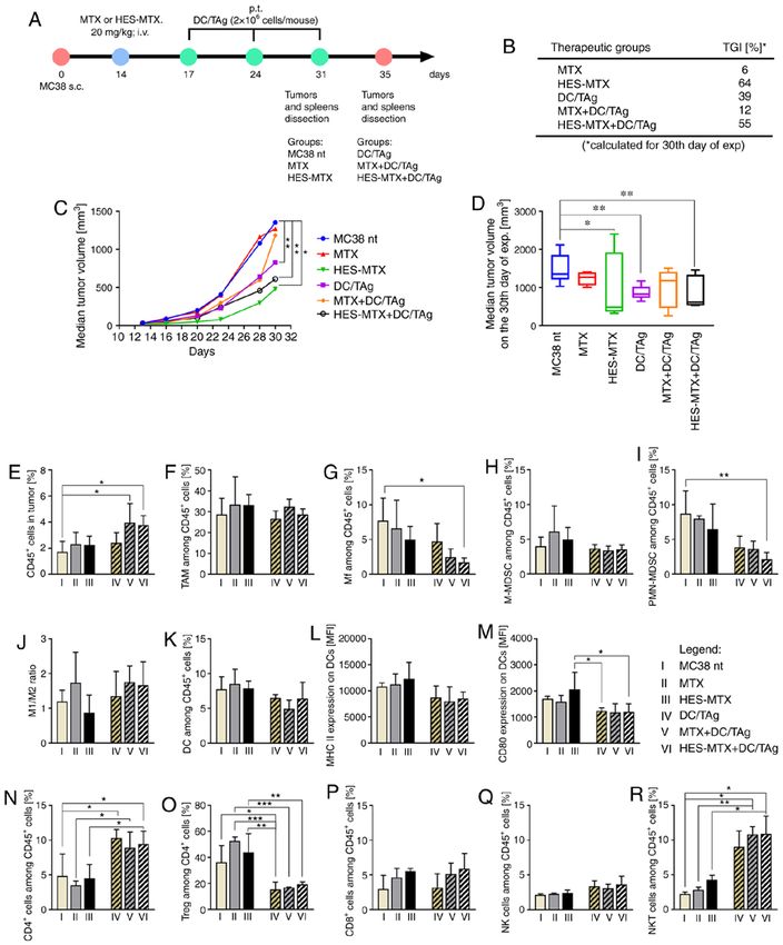
ONCOLOGY REPORTS 45: 945-962, 2021                                                947

supplemented with 10% FBS in the presence of recombinant           water). Absorbance at 570 nm was determined using a Thermo
murine (rm)GM‑CSF (ImmunoTools, 40 ng/ml) and rmIL‑4               Labsystems Multiskan RC microplate reader (Thermo Fisher
(ImmunoTools, 10 ng/ml). After 6 days the loosely attached         Scientific, Inc.) with Genesis Lite 3.05 Software (Thermo Life
immature DCs were stimulated with tumor antigens (10% v/v)         Sciences) and the IC50 value was calculated.
and applied to mice as antitumor vaccines.
                                                                   Interaction of nanoconjugate with MC38 cells and DCs. The
Nanoconjugate preparation. FITC‑HES was synthesized                interaction of FITC‑conjugated compounds (FITC‑HES‑MTX
using a modification of methods previously described (43).         and FITC‑HES) with MC38 cells and DCs was evaluated by
Briefly, FITC‑HES was prepared by the addition of 30 mg of         flow cytometry. The MC38 cells were placed in 24‑well plates
fluorescein isothiocyanate (FITC, isomer I, Sigma‑Aldrich;         (0.2×10⁶ cells/well), immature DCs were placed in 12‑well
Merck KGaA) dissolved in 5 ml of DMSO to a solution that           plates (0.5×10⁶ cells/well) and after 24 h FITC‑HES‑MTX or
contained hydroxyethyl starch (1.2 g in 20 ml of solution          FITC‑HES (10 µg/ml) was added and incubated for the next
containing 150 mM NaCl and 50 mM Na2CO3). This mixture             24 h. After this time, cells were harvested and washed, and
was stirred for 48 h at room temperature (RT). Next, the           dead cells were stained with DAPI dye. The analysis was
mixture was cooled down to 4˚C and precipitated with cold          performed using FACS Fortessa with Diva software (Becton
acetone (100 ml). The crude product was solubilized in water       Dickinson).
and dialyzed against ultrapure water for 5 h at a flow rate of
30 ml/min (Pellicon XL with Ultracel‑10 PLCGC membrane,            Determination of FRα expression. The expression of FRα was
Millipore). Finally, the conjugate of FITC and HES containing      measured by real‑time PCR. Total RNA was isolated using
3.0×10‑3 covalently bound FITC residues per anhydroglucose         a NucleoSpin RNA kit (Macherey‑Nagel) and reverse‑tran-
unit was obtained.                                                 scribed with a First Strand cDNA Synthesis Kit (Thermo
    HES‑MTX nanoconjugate and FITC‑HES‑MTX were                    Fisher Scientific, Inc.). Real‑time PCR was performed using
synthesized using HES 130/0.4 (Voluven, Fresenius Kabi) or         TaqMan Universal PCR Master Mix and TaqMan Gene
FITC‑HES and activated MTX (EBEWE Pharma) according                Expression Assay primers for FRα in reference to the HPRT
to previously described methods (20,44). The following             gene. The analyses were performed using the ViiA7 Real‑Time
absorption coefficients were used: 8,571 M‑1 cm‑1 (372 nm),        PCR System (Applied Biosystem).
70,000 M‑1 cm‑1 (494 nm) for MTX and FITC, respectively.
Eventually, the following conjugates were obtained: HES‑MTX        Surface plasmon resonance spectroscopy. Surface plasmon
containing 52×10‑3 covalently bound MTX residues per anhy-         resonance (SPR) experiments were conducted in a Biacore
droglucose unit and FITC‑HES‑MTX containing 53×10 ‑3               T200 instrument (GE Healthcare Life Sciences). During
MTX and 2.9×10‑3 FITC residues per anhydroglucose unit. In         measurements, the flow buffer HBS‑N was used (10 mM
this study, the presented MTX concentration referring to the       HEPES, pH 7.4 with 150 mM NaCl). Immobilization of bovine
HES‑MTX conjugate was based on the total contents of the           folate binding protein (FBP, Sigma‑Aldrich) was carried out
covalently bound MTX in conjugate. The analysis and charac-        at 25˚C by an EDC‑based amide coupling method using
terization of conjugates were performed using a combination        standard Biacore reagents (GE Healthcare Life Sciences).
of spectrophotometric, chromatographic and light scattering        FBP‑presenting chips (CM5) were prepared with protein
methods based on previously published procedures (20,45).          density at 9.03 FBP ng/mm2. SPR experiments were performed
Hydrodynamic parameters of HES and HES‑MTX were                    by injection of a ligand solution, HES or HES‑MTX nanocon-
characterized by dynamic light scattering (DLS). The sample        jugate, each prepared in HBS‑N buffer (concentrations were
solution was illuminated by a 633‑nm laser, and the light inten-   presented as MTX‑equiv), at a flow rate of 40 µl/min. The
sity scattered at an angle of 173˚ was measured. At least six      conjugates were injected over a reference channel and over
consecutive measurements of each sample were carried out.          a channel with immobilized FBP for 200 sec. Each analysis
All samples were measured at 25˚C using a Zetasizer Nano           cycle consisted of a 60 sec initial period, in which the stability
ZS (Malvern Instruments, UK) in a 12‑µl quartz cuvette (size       of the baseline was monitored. The injection of buffer was
measurement) and folded capillary cells (zeta potential). HES      performed between each analysis cycle for a double refer-
and HES‑MTX conjugate concentration was 5.5 mM (AGU).              ence. At the end of each dissociation phase, the chip surface
DLS data were analyzed using the dts 6.10 software (Malvern        was treated with 10 µl of 10 mM glycine‑HCl (pH 2.5) for
Instruments, UK). The intensity particle size distributions        surface regeneration. Sensorgrams for the reference channel
were obtained using the General Purpose algorithm included         were subtracted from sensorgrams for the channel with FBP.
in the DTS software.                                               Subsequently, sensorgrams of buffer were subtracted from
                                                                   sensorgrams of the HES‑MTX conjugates.
MTT assay. To calculate the half maximal inhibitory concen-
tration (IC50) value, MTT assays were performed. The MC38          Annexin V binding assay. To evaluate apoptosis in MC38
cells were placed in 96‑well plates (0.005×10⁶ cells/well) and     cells after 72 h incubation with MTX or HES‑MTX the
after 24 h MTX or HES‑MTX in various concentrations was            Annexin V binding assay was performed. Briefly, the MC38
added (in the range from 0.001 to 1,000 ng/ml) and incubated       cells were placed in 24‑well plates (0.1×10⁶ cells/well) and
for 72 h. After this time, MTT dye (3‑(4,5‑dimethylthi-            after 24 h MTX or HES‑MTX was added (500 ng/ml). Next,
azol‑2‑yl)‑2,5‑diphenyltetrazolium bromide; 5 mg/ml) was           harvested cells were suspended in binding buffer and stained
added for 4 h. Next, cells were lysed overnight in lysis buffer    with Annexin V protein conjugated with APC fluorochrome
(N,N‑dimethylmethanamide, sodium dodecyl sulfate and               (Thermo Fisher Scientific, Inc.) (15 min, RT). To determine the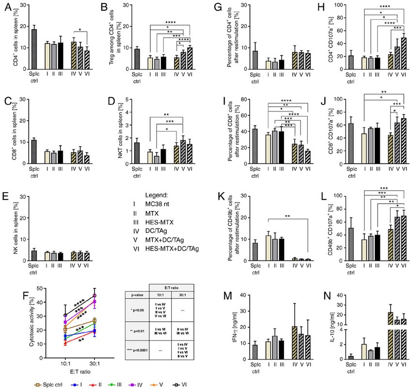
948         SZCZYGIEŁ et al: IMMUNOMODULATORY POTENTIAL OF MTX NANOCONJUGATE AND DC-BASED VACCINES

percentage of dead cells, propidium iodide (PI) was applied         groups (DC/TAg, MTX+DC/TAg, HES‑MTX+DC/TAg)
(10 µg/ml, Thermo Fisher Scientific, Inc.) and the percentage       tumor nodules and spleens were dissected on the 35th day of
of Annexin V+ MC38 cells was analyzed using FACSCalibur             the experiment (3‑5 mice per group). Then tumors and spleens
with CellQuest 3.3 Software (Becton Dickinson).                     were homogenized and stored in liquid nitrogen for further
                                                                    analyses. The health of the mice was monitored during the
Modulation of maturation and phenotype of DCs generated             experiments (weight loss, bristling hair, lethargy) and tumors
in the presence of metabolites released by MC38 cells after         were measured by using a calliper two times a week. Mice
MTX or HES‑MTX treatment. The conditioned medium (CM)               were sacrificed when the tumor volume was >2 cm 3. The
necessary to assess modulation of the DC phenotype gener-           procedure of tumor growth monitoring was presented by
ated in the presence of metabolites of tumor cells treated with     Rossowska et al (42). The therapeutic effect of the treatment
MTX or HES‑MTX was freshly prepared before each test.               was evaluated using tumor growth inhibition (TGI), calculated
For this purpose, the MC38 cells were placed in 6‑well plates       according to the formula: TGI [%]=100‑(TVt ⁄TVnt ×100), where
(1.15×10⁶ cells/well) and 24 h later MTX or HES‑MTX was             TVt is the median tumor volume in the treated group of mice
added (500 ng/ml). Additionally, culture medium containing          and TVnt is the median tumor volume in the non‑treated group
MTX or HES‑MTX without any cells was also prepared.                 of mice.
After 72 h of incubation, CM from the treated MC38 cells
was collected, cellular debris was removed by centrifuga-           Analysis of myeloid cells and lymphocytes in tumors and
tion (15 min, 2,000 × g) and the obtained CM was used in            spleens of mice after the therapy. Tumor cells and spleen cells
differentiating culture of DCs from bone marrow. Medium             isolated from mice were thawed and stained for identification
containing MTX or HES‑MTX maintained without cells                  of myeloid or lymphoid cell subpopulations according to the
was prepared according to the same procedure. Bone marrow           procedure described previously (46). Briefly, tumor single‑cell
cells (0.5×10⁶ cells/well, 12‑well plates) were suspended in a      suspensions were stained with the LIVE/DEAD Fixable
mixture of culture medium (including cytokines necessary            Violet Dead Staining Kit (Thermo Fisher Scientific, Inc.)
for DC generation) and conditioned medium (or medium with           and then labelled with cocktails of fluorochrome‑conjugated
MTX or HES‑MTX) in a 50:50 ratio. After the first 48 h of           monoclonal antibodies: anti‑CD3 PE‑CF594, anti‑CD19
DC generation, the mixture of culture medium and CM or              PE‑CF594, anti‑CD49b PE‑CF594 (all from BD Biosciences),
medium containing MTX or HES‑MTX without cells was                  anti‑CD45 BV605, anti‑CD11b PerCP‑Cy5.5, anti‑CD11c
replaced with RPMI supplemented with 10% FBS, 40 ng/ml              BV650, anti‑F4/80 Alexa Fluor 700, anti‑Ly6C PE, anti‑Ly6G
rmGM‑CSF and 10 ng/ml rmIL‑4. Further DC culture was                APC‑Cy7, anti‑MHC II FITC, anti‑CD80 PE‑Cy7 (all from
conducted according to the protocol in our previous publica-        BioLegend) for myeloid cell identification, and anti‑CD45
tion (42). After 6 days, the loosely attached immature DCs          BV605, anti‑CD3 BV650, anti‑CD4 FITC, anti‑CD8
were collected and then stimulated for 24 h with tumor anti-        APC/Fire 750, anti‑CD25 PE (all from BioLegend) for
gens as described above. The phenotype of mature DCs was            lymphocyte identification. Then, the cells were fixed using the
analyzed. For this purpose, DCs were stained with anti‑CD11c        Foxp3/Transcription Factor Staining Buffer Set (eBioscience).
BV650, anti‑MHC II FITC, anti‑CD40 PE, anti‑CD80 APC                Cells stained with myeloid or lymphocyte cocktail were
(all from BioLegend) and anti‑CD11c BV650 (BioLegend) with          additionally incubated with anti‑CD206 APC (BioLegend)
anti‑CD86 PE (BD Biosciences). The analysis was performed           or anti‑FoxP3 APC (eBioscience) antibodies, respectively. In
using FACS Fortessa with Diva software (Becton Dickinson).          spleen single cell suspension only the lymphocyte identifica-
                                                                    tion was performed according to the procedure described
Therapeutic treatment schedule. Eight‑to 10‑week old female         above. The analysis was performed using a FACS Fortessa
C57BL/6 mice (45 mice) were subcutaneously (s.c.) inocu-            flow cytometer with Diva software (Becton Dickinson).
lated in the right flank with MC38 cells (1.1×10⁶ cells/0.2 ml
NaCl 0.9%/mouse). The mice were treated according to                Analysis of antitumor response of effector spleen cells. Spleen
the schemes presented in Figs. 3A and 5A. In the course of          cells obtained from non‑treated or treated tumor‑bearing
the chemotherapeutic treatment scheme (results presented            mice were cocultured with mitomycin C‑treated MC38 cells
in Figs. 3 and 4), on the 14th day of the experiment, mice          (50 mg mitomycin C/3×106 cells, 30 min., 37˚C) in the pres-
received intravenously (in tail vein, intravenously; i.v.) MTX or   ence of recombinant human IL‑2 (200 U/ml). After 5 days of
HES‑MTX (20 mg/kg body weight) and three days later (17th           restimulation, supernatants were collected and stored at 4˚C
day of experiment) 5 mice from each group were sacrificed           until ELISA was performed. Cytotoxic activity of cells stained
and tumor nodules and spleens were dissected, homogenized           with DiO lipophilic dye (Molecular Probes) was analyzed
and stored in liquid nitrogen for further analyses. In the          according to a previously described procedure (47). Two E:T
chemoimmunotherapeutic treatment scheme (results presented          (effector to target) ratios were investigated: 10:1 and 30:1.
in Figs. 5 and 6) mice received chemotherapy on the 14th day        The percentage of dead double positive (DiO+PI+) MC38 cells
of the experiment and on the 17, 24th and 31st day of the           was determined after analysis using a FACSCalibur with
experiment tumor antigen‑stimulated DC‑based vaccines were          CellQuest 3.3 software (Becton Dickinson). In order to deter-
applied peritumorally (p.t.) (DC/TAg, 2×10⁶ cells/0.2 ml NaCl       mine the percentage of CD107a+ cells, restimulated spleen
0.9%/mouse/p.t. injection). In the group of mice from non‑treated   cells were incubated for 2 h with MC38 cells in the presence
and chemotherapy‑receiving groups (MC38 control, MTX and            of monoclonal anti‑CD107a antibody conjugated with APC
HES‑MTX) tumor nodules and spleens were dissected on                (BioLegend). Afterwards, cells were stained with anti‑CD45
the 31st day of the experiment, and from DC/TAg‑receiving           V500, anti‑CD4 FITC, anti‑CD8 PE‑Cy7 and anti‑CD49b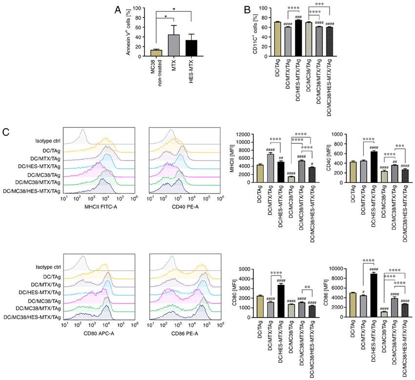
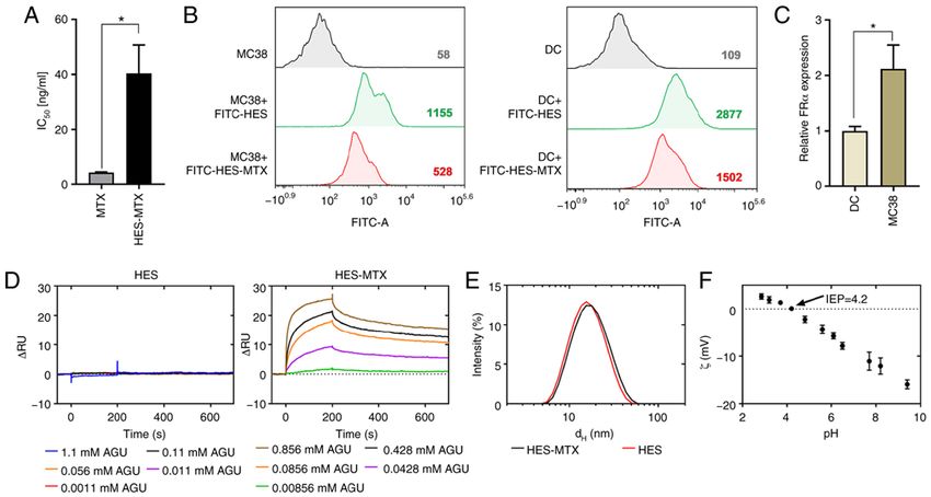
ONCOLOGY REPORTS 45: 945-962, 2021                                                             949

Figure 1. (A) Antiproliferative activity of MTX and HES‑MTX against MC38 cells after a 72‑h treatment was measured by MTT assay and the IC50 value was
calculated. (B) Representative histogram showing the interaction of FITC‑conjugated HES‑MTX and HES with MC38 cells and DCs after a 24‑h treatment
measured by flow cytometry. The numbers presented on the histograms represent mean fluorescence intensity (MFI) values. (C) Relative expression of folate
receptor α (FRα) measured by real‑time PCR in murine DCs and MC38 cells. (D) Representative sensorgrams from SPR measurements showing the associa-
tion and dissociation phase of HES and HES‑MTX conjugate from FBP immobilized on the surface of the CM5 chip. Sensorgrams are shown after subtraction
for reference channel and blank measurements. Concentrations for both HES and HES‑MTX are shown as anhydroglucose unit (AGU). (E) Size distributions
are shown according to intensity measured by dynamic light scattering technique. d H, mean hydrodynamic diameter. (F) Zeta (ζ) potential of HES‑MTX as a
function of pH measured by dynamic light scattering technique. The points denote experimental values determined for ionic strength 0.15 mM; IEP, isoelectric
point. Results (A and C) are expressed as the mean ± SD calculated for at least three independent experiments. Differences between the groups were estimated
by (A) Mann‑Whitney test (*P
950         SZCZYGIEŁ et al: IMMUNOMODULATORY POTENTIAL OF MTX NANOCONJUGATE AND DC-BASED VACCINES

MC38 cells showed significantly higher expression of FRα                 The phenotype analysis of mature DCs showed that in
(Fig. 1C). The affinity of the HES and the HES‑MTX nano-             comparison to the non‑treated DCs (DC/TAg group), the pres-
conjugate to folate binding protein (FBP) was investigated           ence of tumor metabolites (DC/MC38/TAg) did not influence
using SPR. HES (a negative control without MTX molecules             the percentage of CD11c+ DCs, but considerably affected the
attached) did not show any significant response over the             maturation of these cells (Fig. 2B and C). DCs generated in CM
studied concentration range (1.1‑0.0011 mM AGU), indicating          harvested from MC38 cells (DC/MC38/TAg) were character-
a lack of affinity to the FBP surface (Fig. 1D). In contrast,        ized by statistically significantly lower expression of MHCII and
the HES‑MTX showed an increasing concentration‑dependent             CD40, CD80 and CD86 co‑stimulatory molecules compared
response. The shape of the sensorgrams indicated a fast and          to the non‑treated DC/TAg group. However, this effect was
strong association of the HES‑MTX on the FBP presenting              partially restored when MTX or HES‑MTX was used. When
surface. Dissociation curves for HES‑MTX indicated that the          CM from above MC38 cells treated with MTX or HES‑MTX
conjugate dissociates with complex kinetics, initially at a fast     was used in generation of DCs (DC/MC38/MTX/TAg and
and subsequently at slower rates. At the end of analysis, the        DC/MC38/HES‑MTX/TAg), those cells were characterized
dissociation phase was still incomplete. This suggests a high        by a statistically significantly lower percentage of CD11c+ cells
affinity of the nanoconjugate to FBP. Characterization of HES        compared to DC/TAg and DC/MC38/TAg (Fig. 2B). When
and HES‑MTX by light scattering technique revealed that              we applied medium from the above MC38 cells treated with
HES‑MTX represents a typical batch of HES polymers with a            MTX (DC/MC38/MTX/TAg), expression of the analyzed
mean hydrodynamic diameter of about 15 nm when compared              antigens (Fig. 2C) was significantly higher than that observed
to the initial (unmodified) polymer (~14 nm) (Fig. 1E). The          after using HES‑MTX (DC/MC38/HES‑MTX/TAg). We
surface of HES‑MTX nanoconjugate has a negative zeta ζ               postulate that this effect was related to the lower antiprolif-
potential [about‑10 mV at pH=7.4, isoelectric point (IEP)=4.2]       erative activity of HES‑MTX than MTX against tumor cells.
(Fig. 1F). The gathered data demonstrated that the HES‑MTX           In comparison to MTX (DC/MTX/TAg), the nanoconjugate
nanoconjugate has weaker antiproliferative activity against          itself has an immunomodulatory effect on the phenotype of
tumor cells than the free form of MTX. Moreover, HES                 DCs (DC/HES‑MTX/TAg), which was observed especially
after conjugation with MTX gains a high affinity to FBP              with a statistically significantly greater percentage of CD11c+
and is a good candidate as a folate targeting macromolecule          cells and higher expression of co‑stimulatory molecules.
(an FR‑targeted chemotherapeutic). We assume that, due                   Summarizing these results, in comparison to the
to the acquired properties of HES‑MTX, it should act more            nanoconjugate, the treatment of MC38 cells with MTX
specifically towards tumor cells and have greater potential as a     (DC/MC38/MTX/TAg) resulted in more efficient abolition of
chemotherapeutic agent.                                              the negative effect of tumor cells on DC generation and matu-
                                                                     ration. However, when considering the influence of MTX or
Modulation of maturation and phenotype of DCs gener‑                 HES‑MTX on the DC phenotype and their response to stimu-
ated in the presence of metabolites released by MC38 cells           lation with TAg, the nanoconjugate (DC/HES‑MTX/TAg)
after MTX or HES‑MTX treatment. It was found that certain            was a more efficient immunomodulator of DC maturation.
chemotherapeutics, including MTX, used in appropriate
concentrations, can act as immunomodulators and contribute           Influence of nanoconjugate administration on activation of
to an increase in DC maturation (33,34). To investigate the          local and systemic antitumor response in the MC38‑bearing
impact of the nanoconjugate on DC generation and phenotypic          mice. To answer the question whether therapy with the novel
changes as well as to assess whether the nanoconjugate could         MTX conjugate modulates the local and systemic antitumor
reverse the inhibitory effect of MC38 cells on maturation of         response and how it would affect the efficacy of DC‑based
DCs, in vitro studies were conducted. For this purpose, the          vaccines administered after chemotherapy, in vivo experi-
percentage of Annexin V+ MC38 cells previously treated with          ments were conducted. In our previous chemoimmunotherapy
MTX or HES‑MTX was determined (Fig. 2A). Similar to the              schedules in the MC38‑tumor model, DC‑based vaccines
observation made in the MTT assay and calculated IC50 value,         were applied three days after cyclophosphamide administra-
the Annexin V binding assay revealed that the HES‑MTX                tion (39,40,46). Therefore, in the present experiment, we
nanoconjugate was less effective in induction of apoptosis than      aimed to determine what changes in the tumor and spleen
MTX. To reflect the changes in the TME occurring after nano-         would occur as a result of the application of MTX or the
conjugate treatment and the potential effect on DC maturation,       nanoconjugate HES‑MTX. For this purpose, mice with subcu-
the MC38 cells were treated with MTX or HES‑MTX for 72 h.            taneously growing MC38 tumor received MTX or HES‑MTX
Subsequently, the conditioned medium (CM) harvested from             i.v. and three days later the tumor nodules and spleens were
treated MC38 cells was used in ex vivo generation of DCs             dissected for further analyses (Fig. 3A). Flow cytometric
(named hereafter as DC/MC38/TAg, DC/MC38/MTX/TAg                     analyses allowing the simultaneous identification of multiple
and DC/MC38/HES‑MTX/TAg). Due to the immunomodula-                   immune cell subpopulations in tumor and spleen tissues were
tory effect of the nanoconjugate and MTX on DC phenotype,            performed (Fig. 3B for tumors and Fig. 4A for spleens).
the culture medium containing MTX or HES‑MTX without                      In the tumor tissue, we examined the percentage of CD45+
any cells was incubated for 72 h and then used in in vitro studies   cells (leukocytes) and among them we identified myeloid
(named hereafter as DC/MTX/TAg and DC/HES‑MTX/TAg).                  cells, TAMs (CD11b + CD11c+F4/80 +), macrophages (Mfs)
Next, in order to obtain the mature DCs, immature DCs                (CD11b+CD11c‑F4/80+), subpopulations of MDSCs [monocytic
were stimulated with tumor antigens (TAg) and phenotype              (M‑)MDSCs (CD11b+CD11c‑F4/80‑Ly6C+Ly6G‑), polymorpho-
alterations of mature DCs were evaluated by flow cytometry.          nuclear (PMN‑)MDSCs (CD11b+CD11c‑F4/80‑Ly6CintLy6G+)],
ONCOLOGY REPORTS 45: 945-962, 2021 951 Figure 2. Modulation phenotype of mature DCs generated in the presence of metabolites released by MC38 cells after MTX or HES‑MTX treatment. (A) Percentage of Annexin V+ MC38 cells after a 72‑h treatment with MTX or HES‑MTX as determined by Annexin V binding assay; (B) Bar plots showing the percentage of CD11c+ mature DCs (stimulated with TAg) (C) Bar plots and representative histograms showing the expression of MHC II and costimu- latory molecules on mature DCs. Results are expressed as mean ± SD calculated for three independent experiments. Differences between groups were calculated using (A) the Brown‑Forsythe and Welch ANOVA test followed by Dunnett's T3 multiple comparisons post‑hoc test or (B and C) one‑way ANOVA followed by Tukey's multiple comparison post‑hoc test. The asterisks (*) presented in the graphs indicate statistically significant differences between the given groups; a hashtag (#) above a bar indicates a statistically significant difference between the given group and the control group‑DC/TAg (*/#P
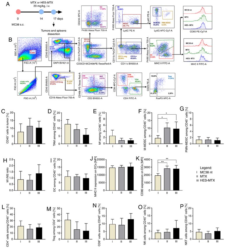
952           SZCZYGIEŁ et al: IMMUNOMODULATORY POTENTIAL OF MTX NANOCONJUGATE AND DC-BASED VACCINES

Figure 3. Impact of chemotherapy on infiltration of MC38 tumor nodules with immune cells. (A) Scheme of treatment. (B) Schemes of multiparameter flow
cytometry analyses showing the method of distinguishing myeloid or lymphoid cell subpopulation in tumors dissected from MC38 tumor‑bearing mice treated
according to the scheme presented in A. (C) Percentage of CD45+ cells in tumor nodules. (D‑G) Percentage of myeloid cell subpopulations among CD45+
cells in tumors. (H) M1/M2 ratio showing changes in polarization of tumor‑infiltrating macrophages after therapy. (I‑K) Percentage of DCs infiltrating into
tumor tissue and expression of MHC II and CD80 molecules on the surface of DCs. (L‑P) Percentage of lymphoid cell subpopulations among CD45+ cells in
tumors. Results are expressed as mean ± SD (5 mice per group were analyzed from one experiment). In all presented data the differences between groups were
calculated using the one‑way ANOVA followed by Tukey's multiple comparison post‑hoc test (*P
ONCOLOGY REPORTS 45: 945-962, 2021                                                                953

Figure 4. Effect of applied chemotherapy on induction of systemic antitumor response. (A) Scheme of multiparameter flow cytometry analyses showing the
method of distinguishing lymphoid cell subpopulation in spleens dissected from MC38 tumor‑bearing mice treated according to the scheme presented in
Fig. 3A. (B‑F) Percentage of effector and suppressor lymphoid cell subpopulations in the spleens. (G) Cytotoxic activity of splenocytes (effector cells) against
DiO+ MC38 cells (target cells). Asterisk above the line indicates statistical significance between different E:T ratios within a given group, while statistical
significance between groups within a given E:T ratio is presented in the table. (H‑M) Percentage of Th, CTL and NK cells (CD49b+) among splenocytes after
restimulation of spleen cells with MC38 cells and the percentage of CD107a+ among CD4+, CD8+ and CD49b+ cells measured by CD107a degranulation assay.
(N and O) IFN‑γ and IL‑10 concentration in supernatants after restimulation. Results are expressed as mean ± SD (5 mice per group were analyzed from one
experiment). Splc ctrl, splenocytes isolated from spleen derived from healthy mice (i.e. without MC38‑tumor). Differences between groups were calculated
using: (B‑F and H‑M) one‑way ANOVA followed by Tukey's multiple comparison post‑hoc test (N) nonparametric Kruskal‑Wallis test followed by Dunn's
multiple comparison test; (O) Brown‑Forsythe and Welch ANOVA test followed by Dunnett's T3 multiple comparisons post‑hoc test; or (G) two‑way ANOVA
followed by Tukey's multiple comparison post‑hoc test (*P
954         SZCZYGIEŁ et al: IMMUNOMODULATORY POTENTIAL OF MTX NANOCONJUGATE AND DC-BASED VACCINES

application of HES‑MTX was an increase in the percentage of         Antitumor activity of therapy composed of HES‑MTX and
CTL and NK cells noted.                                             DC‑based vaccines and its influence on local and systemic
    With the use of the multiparametric flow cytometry              antitumor immune response. Taking into consideration the
analysis protocol presented in Fig. 4A, the percentages of          immunomodulatory activity of HES‑MTX, in the next step of
CTL, Th and Treg cells among spleen cells were estimated. In        the research, the nanoconjugate was applied in combination
contrast to the effects caused by MTX application, the use of       with DC‑based vaccines according to the scheme presented
HES‑MTX induced significant changes in the percentage of            in Fig. 5A. The tumors and spleens were dissected at two time
lymphocytes among spleen cells (Fig. 4B‑F). The application         points: From non‑treated (MC38 nt), MTX and HES‑MTX
of HES‑MTX contributed to a significantly higher percentage         groups on the 31st day, and from immunotherapy receiving
of CD4+ and CD8+ T cells compared to the non‑treated group          groups (DC/TAg, MTX+DC/TAg, HES‑MTX+DC/TAg)
and, in the latter subpopulation, to the MTX‑treated group. It      on the 35th day of the experiment. The time discrepancies
should be noted that after HES‑MTX application a statistically      were caused by different tumor growth rates, especially in
significant reduction in the Treg percentage among spleen           the non‑treated group and MTX group. Thus, in the course
CD4+ T cells was observed. Also, among splenocytes from             of the experiment, we decided to separate the dissections‑the
the HES‑MTX‑treated group the highest NK and NKT cell               major reason for this decision was prolonged observation of
percentage was observed. The use of nanoconjugate resulted in       tumor growth after the last injection of DC‑based vaccines and
the restoration of the size of these lymphocyte populations to      maintaining the interval between organ dissections as short as
the level typical for healthy mice (splc control) or even higher.   possible. The tumor growth inhibition (TGI) calculated on the
Analysis of the activation status of splenic CD4 + and CD8+         30th day of the experiment for all groups of mice indicated the
T cells revealed that after therapy with the nanoconjugate the      strongest influence of HES‑MTX on tumor growth (the TGI in
percentage of memory CD4+ T cells was significantly higher,         HES‑MTX group was 64% in relation to the MC38 nt group),
than after therapy with MTX (Fig. S1D‑F). In order to esti-         while MTX exhibited only 6% inhibition of tumor growth
mate the ability of activated splenic lymphocytes to induce a       (Fig. 5B). A moderate effect was observed after application of
systemic antitumor response after treatment with HES‑MTX,           the DC‑based vaccines as sole therapy (DC/TAg; TGI 39%).
the spleen cells were restimulated ex vivo with MC38 cells.         However, when compared to the HES‑MTX, group combining
After five days of co‑culture of spleen cells with mitomycin        the chemotherapy with DC/TAg‑based vaccines did not
C‑treated MC38 cells, their cytotoxic activity towards MC38         contribute to the enhancement of tumor growth inhibition
cells as well as their phenotype, CD107a degranulation and          as we expected. In the HES‑MTX+DC/TAg group the TGI
cytokine production were evaluated. Determination of cyto-          was 55%, whereas in the MTX+DC/TAg group the inhibition
toxic activity of splenocytes against MC38 cells through direct     of tumor growth was negligible (TGI 12%). The kinetics of
contact revealed statistically significantly higher cytotoxicity    MC38 tumor growth and median tumor volume on the 30th
in the HES‑MTX‑treated group compared to the other groups,          day of the experiment are shown in Fig. 5C and D.
especially in the 10:1 E:T ratio (Fig. 4G). Similar to observa-         To compare the effect of multiple injections of DC‑based
tions made during splenocyte phenotype analysis, the elevated       vaccines, the TGI for the 35th day of the experiment was deter-
percentages of CD4 + and CD8 + cells in the HES‑MTX                 mined in relation to the DC/TAg group (Fig. S2A). TGI for
group were maintained after restimulation, and in the latter        the MTX+DC/TAg group was negative and was ‑14%; hence
cell population this difference was statistically significant. It   application of MTX prior to DC‑based vaccines probably
was accompanied by a slight, but not statistically significant,     reduced the effectiveness of the immunotherapy. On the other
decrease in the percentage of CD49b+ cells (Fig. 4H, J and L).      hand, in the HES‑MTX+DC/TAg group, TGI for the 35th day
However, the evaluation of immune cell antitumor activity by        of the experiment was 22%, which indicates that HES‑MTX
CD107a degranulation assay did not show a statistically signif-     application prior to the start of immunotherapy enhanced
icant increase in the percentage of CD4+CD107a+ cells in the        tumor growth delay compared to the DC/TAg group, although
HES‑MTX group, while the percentage of CD8+CD107a+ cells            these differences were not statistically significant. This was
remained unchanged in all groups. It should be highlighted that     also reflected in the median tumor volume on the 35th day
therapy with the nanoconjugate caused a significant increase        (Fig. S2B).
in the percentage of CD49b+CD107a+ cells (Fig. 4I, K and M).            The multiparameter flow cytometry analyses of tumor
Moreover, in the HES‑MTX group a significant decrease in            tissue (according to the scheme presented in Fig. S3A)
IFN‑γ and IL‑10 production after restimulation was observed         showed the overall increase in the percentage of leukocytes
(Fig. 4N and O).                                                    in all treated groups of mice (Fig. 5E). Although after mono-
    The results presented above suggest that three days after       therapy (MTX, HES‑MTX, DC/TAg groups) the percentage
chemotherapy application, especially when HES‑MTX was               of CD45+ cells was slightly higher than in the non‑treated
used, the modulation of antitumor response occurred. It was         group, application of combined therapy, both MTX+DC/TAg
reflected in a reduction in the size of the population of immune    and HES‑MTX+DC/TAg, caused the most intensive influx
cell with suppressor activity (Tregs, TAMs, Mfs and M2‑type         of the cells and observed changes were statistically signifi-
macrophages) in tumors, as well as in an expansion in the           cant in comparison to the MC38 nt group. One of the most
population of immune cells with cytotoxic activity (CD8+            numerous cell populations among myeloid cells found in the
T cells, NK, NKT cells) in spleen and tumor tissues. Moreover,      tumor tissue was tumor‑associated macrophages (TAMs),
generation of cytotoxic activity of spleen lymphocytes against      which accounted for approximately 30% of all leukocytes
tumor cells indicated that HES‑MTX contributed to activation        in tumors from the non‑treated group of mice (Fig. 5F). The
of the systemic antitumor immune response.                          applied therapy, both chemo‑ and chemoimmunotherapy, did
ONCOLOGY REPORTS 45: 945-962, 2021 955 Figure 5. Impact of combined therapy on tumor growth and infiltration of MC38 tumor nodules with immune cells. (A) Scheme of treatment. (B) Table presenting MC38 tumor growth inhibition (TGI) calculated on 30th day of experiment in relation to the MC38 nt group; (C) Graph presenting median tumor volume after chemoimmunotherapy. (D) Box graph presenting median tumor volume, calculated on the 30th day of the experiment. (E) Percentage of CD45+ cells in tumor nodules. (F‑I) Percentage of myeloid cell subpopulations among CD45+ cells in tumors. (J) M1/M2 ratio showing changes in polarization of tumor‑infiltrating macrophages after therapy. (K‑M) Percentage of DCs infiltrating into tumor tissue and expression of MHC II and CD80 molecules on their surface. (N‑R) Percentage of lymphoid cell subpopulations among CD45+ cells in tumors. Scheme of multiparameter flow cytometry analyses showing the method of distinguishing myeloid or lymphoid cell subpopulation in tumors is presented in Fig. S3A. Results are expressed as mean ± SD (3‑5 mice per group were analyzed from one experiment). The differences between groups were calculated by (C and D) two‑way ANOVA followed by Bonferroni's multiple comparisons test, (E‑Q) one‑way ANOVA followed by Tukey's multiple comparison post‑hoc test or (R) Brown‑Forsythe and Welch ANOVA test followed by Dunnett's T3 multiple comparisons post‑hoc test (*P
956         SZCZYGIEŁ et al: IMMUNOMODULATORY POTENTIAL OF MTX NANOCONJUGATE AND DC-BASED VACCINES

was about four times lower than that noted in the MC38              statistically significant. The highest percentage of CD8+ T cells
non‑treated group and three times lower than in the DC/TAg          was noted in the HES‑MTX‑receiving groups (HES‑MTX and
group (Fig. 5G). Although there were no statistically signifi-      HES‑MTX+DC/TAg), which is consistent with our previous
cant changes in size of the M‑MDSC population‑an increase           observation that HES‑MTX affects the enhanced influx of
in the M‑MDSC percentage occurred only when MTX or                  CD8+ T cells into tumor tissue. Furthermore, in comparison
HES‑MTX was applied as monotherapy; significant changes             to the control or DC/TAg group, a significant increase in
in the PMN‑MDSC percentage in tumor tissue were observed            the percentage of NKT cells was observed when MTX or
(Fig. 5H and I). After application of each type of therapy, a       HES‑MTX was used (as mono‑ and combined therapy)
reduced population of PMN‑MDSCs was found. Although                 (Fig. 5R). Analysis of the activation status of CD4+ T cells
application of chemotherapy alone caused a moderate decrease        infiltrating into tumor tissue showed that compared to the
of PMN‑MDSC percentage (especially after HES‑MTX),                  non‑treated and chemotherapy‑receiving groups (MC38
the use of immunotherapy induced a significant reduction in         control, MTX and HES‑MTX), the use of DC/TAg‑based
the PMN‑MDSC percentage. The lowest percentage of these             vaccines resulted in a significantly higher percentage of
cells was noted after combined therapy with HES‑MTX and             effector CD4 + T cells, while percentage of memory CD4 +
DC‑based vaccines. Considering the influence of applied             T cells was decreased. Moreover, therapy consisting of
therapies on the stage of macrophage polarization, the M1/M2        HES‑MTX and DC/TAg caused a significant increase in the
ratio was not significantly increased after application of MTX      percentage of effector CD8+ T cells infiltrating into the tumor
or combined therapy (MTX+DC/TAg, HES‑MTX+DC/TAg                     tissue (Fig. S4B and C).
group), while it decreased after HES‑MTX treatment (Fig. 5J).           The obtained results indicated that supplementing the
In comparison to non‑treated and chemotherapy groups,               chemotherapy with DC‑based vaccines contributed to an
DC‑based vaccines caused a slight, but not statistically signifi-   enhanced influx of leukocytes into the tumor tissue, especially
cant reduction in the percentage of DCs infiltrating tumor          CTL and NKT cells, and reduced the population of immune
tissue, which was accompanied by decreased expression of            cells with suppressor activity, such as Mfs, PMN‑MDSCs and
MHC II and CD80 molecules on their surface (Fig. 5K‑M).             Tregs.
Despite the fact that the lowest percentage of DCs was found            The estimation of the percentage of CTLs, Th and Tregs
in the MTX+DC/TAg group, the expression of MHC II and               among spleen cells (according to the scheme presented in
CD80 antigens did not change and remained at the same               Fig. S3B) revealed that significant changes in the lymphoid
level as in the DC/TAg‑receiving group. It should be noted          cell population occurred only when DC‑based vaccines were
that in the HES‑MTX group, tumor‑infiltrating DCs were              used (Fig. 6A‑E). In comparison to the non‑treated group, the
characterized by the highest expression of MHC II and CD80          application of MTX or HES‑MTX as monotherapy did not
molecules, which is consistent with our observations about the      contribute to significant alterations among CTLs, Th or Tregs,
modulatory potential of the nanoconjugate for the DC pheno-         like those observed on the third day after chemotherapy. When
type, and observed changes in the expression of CD80 antigen        DC‑based vaccines were applied alone, we did not observe
were statistically significant.                                     significant changes in the population size of the mentioned
    The changes occurring in the myeloid populations were           lymphocytes in the spleens. However, therapy consisting of
accompanied by modifications in the percentage of lymphoid          HES‑MTX and DC/TAg was found to cause a decrease in
cell infiltrating tumor nodules. When compared to the               the percentage of CD4 + and CD8+ T cells compared to the
MC38 nt group, the use of cytostatics alone did not cause           non‑treated and DC/TAg groups. This effect was accompanied
statistically significant changes in the percentage of CD4 +        by a significant increase in the Treg percentage in this group.
cells infiltrating tumor tissue, unlike in other lymphoid cell      Similar tendencies were observed in the MTX+DC/TAg
subpopulations. Application of DC‑based vaccines resulted           group. Among the DC/TAg‑receiving groups, the highest
in statistically significant enlargement of CD4+ T cells, NKT       percentage of splenic effector CD4+ T cells was observed in
cell percentage and a decrease in the percentage of Tregs,          the HES‑MTX+DC/TAg group and this change was statisti-
while the increase of NK cells percentage was not statistically     cally significant compared to the DC/TAg and MTX+DC/TAg
significant (Fig. 5N, O, Q and R). Trends in changes in the         group. Moreover, in the case of memory CD8+ T cells found
percentage of Tregs suggest that application of chemothera-         in the spleen, after use of the DC/TAg‑based vaccines the
peutics alone was not sufficient to maintain the size of the        percentage of these was significantly lower than observed in
tumor‑infiltrating Treg population at a low level, like those       the non‑treated and HES‑MTX groups (Fig. S4E and F).
observed on the third day after chemotherapy in the previous            It should be noted that assessment of cytotoxic activity of
experiment. In comparison to the control group, the percentage      restimulated spleen cells towards MC38 tumor cells revealed
of Tregs cells was much higher, especially in the MTX               that immunotherapy generated more efficient cytotoxic activity
group (Fig. 5O). Multiple application of DC‑based vaccines          than chemotherapy applied alone (Fig. 6F). Moreover, the
was found to cause a statistically significant strong reduc-        highest cytotoxic activity was noted in the HES‑MTX+DC/TAg
tion in the Treg percentage, but in the HES‑MTX+DC/TAg              group in both E:T ratios. The restimulation of splenocytes by
group the size of the Treg population was slightly greater          MC38 cells confirmed the impact of DC‑based vaccines on the
than that noted in the other DC/TAg‑receiving groups. It is         percentage of CD4+, CD8+ and CD49b+ cells, while chemo-
noteworthy that an increase in CD8+ T cells was noted when          therapy applied alone did not cause any significant changes
chemotherapy was applied alone (MTX, HES‑MTX groups)                in these subpopulations (Fig. 6G, I and K). Despite the lack
or in combination with DC‑based vaccines (MTX+DC/TAg,               of alterations in the CD4+ T cell percentage after restimula-
HES‑MTX+DC/TAg) (Fig. 5P), but these changes were not               tion between groups receiving DC‑based vaccines, a strong
ONCOLOGY REPORTS 45: 945-962, 2021 957 Figure 6. Effect of applied chemoimmunotherapy on induction of systemic antitumor response. (A‑E) Percentage of effector and suppressor lymphoid cell subpopulations in spleens of MC38 tumor‑bearing mice treated according to the scheme presented in Fig. 5A. (F) Cytotoxic activity of splenocytes (effector cells) against DiO+ MC38 cells (target cells). Asterisks above or under the lines indicate statistical significance between different E:T ratios within a given group, while statistical significance between groups within a given E:T ratio is presented in the table. (G‑L) Percentage of Th, CTL and B NK cells (CD49b+) among splenocytes after restimulation of spleen cells with MC38 cells and the percentage of CD107a+ among CD4+, and cytotoxic CD8+ and CD49b+ cells measured by CD107a degranulation assay. (M and N) IFN‑γ and IL‑10 concentration in supernatants after restimulation. Scheme of multiparameter flow cytometry analyses showing the method of distinguishing lymphoid cell subpopulation in spleens is presented in Fig. S3B. Results are expressed as mean ± SD (3‑5 mice per group were analyzed from one experiment). Splc ctrl, splenocytes isolated from spleen derived from healthy mice (i.e. without MC38‑tumor). Differences between groups were calculated using: (A‑E, G‑J and L) one‑way ANOVA followed by Tukey's multiple comparison post‑hoc test (K, M and N) the nonparametric Kruskal‑Wallis test followed by Dunn's multiple comparison test; or (F) two‑way ANOVA followed by Tukey's multiple comparison post‑hoc test (*P
958          SZCZYGIEŁ et al: IMMUNOMODULATORY POTENTIAL OF MTX NANOCONJUGATE AND DC-BASED VACCINES

and DC‑based vaccines contributed to generation of a specific        with vascular permeability (49,50). Moreover, the use of a
antitumor response. It was confirmed by increased ability of         carrier reduces the toxicity of therapy, since the EPR effect for
spleen cells to secrete cytolytic granules and enhanced cyto-        drug delivery does not occur in normal tissue.
toxic activity as a result of secondary contact with the tumor           The main objective of the present study was to determine
antigen. All of these factors caused the statistically significant   whether the HES‑MTX nanoconjugate, applied as chemo-
delay of tumor growth after therapy with HES‑MTX and                 therapy, modulates the systemic antitumor immune response,
DC‑based vaccines.                                                   and affects changes in the landscape of immune cells infil-
                                                                     trating tumor tissue. This, in turn, should support the generation
Discussion                                                           of a proper immune response against a growing tumor by
                                                                     DC‑based vaccines injected peritumorally after chemotherapy
In the present work, we demonstrated for the first time that an      administration. Furthermore, there are reports confirming that
innovative drug delivery system‑in the form of a nanoconju-          certain cytostatics, including methotrexate, used at appropriate
gate of well‑known therapeutic compounds, i.e. methotrexate          doses, can act as modulators of the DC phenotype and func-
(MTX) as an anticancer agent and hydroxyethyl starch (HES)           tion (33,34), thus DCs reinforced in this way should generate
as a high‑molecular carrier‑was able to modulate the anti-           an efficient antitumor immune response (51).
tumor immune response. Moreover, we were the first to apply              Taking all the above into account, we designed in vitro
chemotherapy with the HES‑MTX nanoconjugate together                 studies in which we found that the antiproliferative activity
with DC‑based cellular vaccines in a murine MC38 colon               of the HES‑MTX nanoconjugate against MC38 colon carci-
carcinoma model.                                                     noma cells was considerably lower than that of the free form
     According to Goszczyński et al the mean hydrodynamic            of MTX. Previous results reported by Goszczyński et al
diameter of HES‑MTX is 15.2±6.2 nm (20); therefore undoubt-          revealed that HES‑MTX possessed approximately 10‑fold
edly this type of conjugate can be defined as a nanoconjugate.       weaker antiproliferative activity towards human (MV4‑11)
It is noteworthy that conjugation of MTX with HES is achieved        and murine (P388) leukemia cell lines than MTX alone (20),
by esterification of HES's hydroxyl groups, but the linker           which is consistent with our observations. Nevertheless, weak
between the carrier and MTX is glutamic acid‑an integral part        in vitro efficiency of the conjugates does not necessarily
of the MTX molecule‑ and hence no other additional linking           predict diminished in vivo activity, as it has been shown for
substances are needed (20). MTX release from the nanoconju-          the fibrinogen‑MTX conjugate or dextran‑MTX conjugate
gate occurs as a result of chemical and enzymatic hydrolysis         used in a P388 mouse leukemia model by Nevozhay et al (44)
by esterases or amylases. Enzymatic degradation of HES leads         or Goszczyński et al (14), respectively. Furthermore, we
to the release of glucose derivatives only, allowing for easy        confirmed the interaction of HES‑MTX labelled with FITC
elimination of these derivatives from the body. At the begin-        dye with MC38 cells and dendritic cells (DCs). In comparison
ning of the research on the anticancer potential of HES‑MTX,         to MC38 cells, DCs interacted more strongly with HES and
the key issue was to determine what the main advantage of            HES‑MTX. It was reflected in greater MFI values for DCs, but
HES‑MTX over the free form of MTX is. In fact, chemotherapy          it may be associated with increased antigen uptake capacity,
in its conventional form, including methotrexate application, is     which is typical for this type of cell. We also confirmed a
related to overall toxicity to healthy cells, rapid elimination of   high affinity of nanoconjugate to folate binding protein
chemotherapeutics from the body and low specificity towards          and we verified the overexpression of FRα in MC38 cells
target cancer cells (23,48). Thus, it was challenging to design      in comparison to DCs ex vivo generated from murine bone
a chemotherapeutic‑carrier system overcoming these difficul-         marrow precursors.
ties. It is well known that MTX enters the cell mainly through           In the next step in the in vitro research, we estimated
the ubiquitously expressed reduced folate carrier (RFC) to           the influence of metabolites released by MC38 cells treated
which MTX has high affinity. MTX can also enter cells via            with nanoconjugate on the generation and maturation
folate receptors (FRs) overexpressed on cancer cells, although       of DCs. It is well known that in the presence of the tumor
with low affinity (27). As a result of conjugation of one mole-      microenvironment, DC functions are hindered and thereby
cule of hydroxyethyl starch with 50 molecules of MTX, the            creation of an efficient antitumor immune response by DCs
nanoconjugate becomes polyvalent (17,28). This polyvalence           is impaired (8,9,52). It has also been confirmed by us that
allows interactions of the nanoconjugate with folate receptor        in comparison to untreated DCs, murine bone marrow DC
alpha (FRα) with a much higher binding constant than free            precursors cultured in the presence of CM harvested from
MTX, and therefore we postulate that HES‑MTX interacts               above MC38 cells responded more weakly to stimulation with
more strongly with the tumor cells overexpressing FRα than           TAg (DC/MC38/TAg), which was demonstrated in statistically
normal cells. Another important advantage of HES‑MTX is              significantly lower expression of surface molecules necessary
its biodistribution, which is attained not only, although mainly,    for efficient antigen presentation. Using the Annexin V binding
by interaction of HES‑MTX with FRs on target cells but also          assay, we confirmed that HES‑MTX induced weaker apop-
by an enhanced vascular permeability and retention effect            tosis of MC38 cells than MTX. DC precursors cultured in CM
(EPR). This phenomenon is considered as an ability of macro-         harvested from above MC38 cells treated with HES‑MTX and
molecules larger than 40 kDa (hydrodynamic diameter above            stimulated with TAg (DC/MC38/HES‑MTX/TAg) exhibited
10 nm) to selectively leak from tumor vessels and accumulate         modest changes in expression of DC antigens in comparison
in tumor tissue (2,30‑32). EPR is often observed in solid            to DC/MC38/MTX/TAg. It was associated with lower toxicity
tumors due to extensive angiogenesis, malfunctional vascular         of HES‑MTX, than MTX, towards tumor cells. Therefore,
architecture and increased expression of proteins associated         in relation to DC/MC38/MTX/TAg, only partial abolition of
You can also read