Diversity of adult neural stem and progenitor cells in physiology and disease
←
→
Page content transcription
If your browser does not render page correctly, please read the page content below
Preprints (www.preprints.org) | NOT PEER-REVIEWED | Posted: 16 June 2021                              doi:10.20944/preprints202106.0449.v1

          Review

          Diversity of adult neural stem and progenitor cells in physiol-
          ogy and disease
          Zachary Finkel 1, Fatima Esteban 1, Brianna Rodriguez 1, Tianyue Fu 1, Xin Ai 1, Li Cai 1

                                       1   Department of Biomedical Engineering, Rutgers University; lcai@rutgers.edu

                                       Abstract: Adult neural stem and progenitor cells (NSPCs) contribute to learning, memory, mainte-
                                       nance of homeostasis, energy metabolism and many other essential processes. They are highly het-
                                       erogeneous populations that require input from a regionally distinct microenvironment including
                                       a mix of neurons, oligodendrocytes, astrocytes, ependymal cells, NG2+ glia, vasculature, cerebro-
                                       spinal fluid (CSF), and others. The diversity of NSPCs is present in all three major parts of the CNS,
                                       i.e., the brain, spinal cord, and retina. Intrinsic and extrinsic signals, e.g., neurotrophic and growth
                                       factors, master transcription factors, and mechanical properties of the extracellular matrix (ECM),
                                       collectively regulate activities and characteristics of NSPCs: quiescence/survival, proliferation, mi-
                                       gration, differentiation, and integration. This review discusses the heterogeneous NSPC popula-
                                       tions in the normal physiology and highlights their potentials and roles in injured/diseased states
                                       for regenerative medicine.

                                       Keywords: Central Nervous System; Ependymal Cells; Neural Stem and Progenitor Cells; NG2+
                                       Cells; Regenerative Medicine; Retina Injury; Spinal Cord Injury; Traumatic Brain Injury.

                                       1. Introduction
                                            During development, neural stem cells (NSCs) are responsible for the formation of
                                       the central and peripheral nervous systems. Initially NSCs, also called radial glial cells,
                                       proliferate into pools of neural progenitor cells (NPCs). These progenitors then migrate
                                       and differentiate into highly specified networks of neurons, oligodendrocytes, and astro-
                                       cytes via neurogenesis [1]. The term NSC refers to an uncommitted cell with differentia-
                                       tion potential into the neurons and glia of the CNS and is defined by two essential char-
                                       acteristics: self-renewal and multipotency [2] (Figure 1). The term NPC refers to a cell type
                                       generated in later stages of neurogenesis associated with a committed lineage. NSPC de-
                                       scribes both populations and is established as the only self-renewing cell type in the adult
                                       CNS. Thus, NSPCs are a major research thrust in the field of regenerative medicine. Ex-
                                       trinsic and intrinsic factors such as neurotrophic/growth factors, transcription factors, and
                                       canonical pathways guide neurogenesis during development and adulthood.
                                            For the past 50 years, the topic of endogenous adult neurogenesis has been highly
                                       debated. This began with the initial discovery of adult mammalian neurogenesis in 1962
                                       by Joseph Altman and has continued with noteworthy publications supporting the exist-
                                       ence or non-existence of adult neurogenesis in mammals [3]. In the adult CNS, neurogen-
                                       esis plays a primary role in essential processes such as learning, memory, maintenance of
                                       tissue homeostasis, and many others.
                                            Heterogeneous populations of NSPCs exist in the neurogenic niches of the brain, spi-
                                       nal cord, and retina. Primary NSPCs are found in the subventricular zone (SVZ) and sub-
                                       granular zone (SGZ) of the brain and include radial glial-like cells, NG2+/oligodendrocyte
                                       progenitor cells (OPCs), and Foxj1+ ependymal cells. Both OPCs and ependymal cell

            © 2021 by the author(s). Distributed under a Creative Commons CC BY license.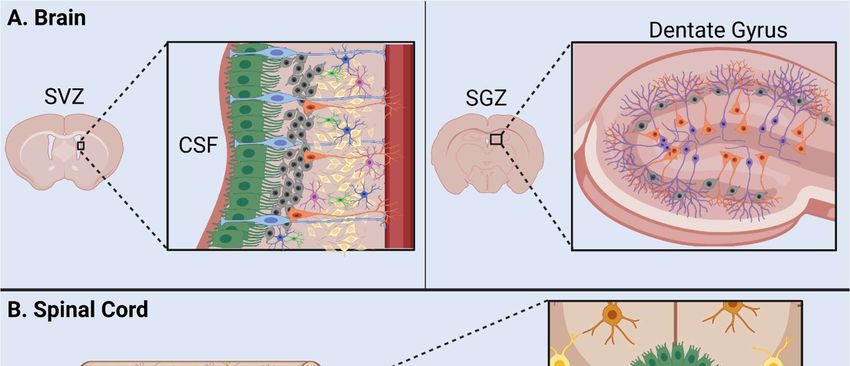
Preprints (www.preprints.org) | NOT PEER-REVIEWED | Posted: 16 June 2021                    doi:10.20944/preprints202106.0449.v1

                                       populations can be found in the spinal cord. In the adult retina, potential sources of NSPCs
                                       include Müller glia cells and the ciliary epithelium (CE).
                                            NSPC response to CNS injury is extraordinarily complex and dependent upon the
                                       extent and location of injury. Injuries are most often contusion or blunt force-based and
                                       primarily result from sporting or vehicular accidents. Traumatic brain injuries habitually
                                       damage two central niches: SGZ of the hippocampus and SVZ of the lateral ventricles.
                                       Damage to these regions can result in consequences including aberrant migration of NSPC
                                       progeny cells, incorrect dendritic branching, enhanced progenitor cell proliferation, inef-
                                       fective integration of cells into networks of tissue, and many others. Spinal cord injury
                                       may affect the neurogenic niche of the central canal resulting in differing contributions of
                                       NSPC populations to the glial scar. In addition, large differences in injury pathophysiol-
                                       ogy occur as a direct result of injury-mediated proliferation and altered differentiation.

          Figure 1. NSPC characteristics in adult mammals. (A) Self renewal requires input via extrinsic and intrinsic factors.
          These include signaling pathways Notch, Wnt, and Shh, and transcription factors Sox2, Ascl1, Bmi1, Tlx, and neuro-
          transmitters and neurotrophic/trophic growth factors. (B) Multipotency allows NSPCs to differentiate into a variety of
          cell fates such as Neurons, Astrocytes, and Oligodendrocytes. Adapted from Navarro Quiroz et al, 2018 [4].

                                             In the eye, retinal injury results from chemical or mechanical damage and is highly
                                       dependent on NSPC activity. Traumatic mechanical injury of the eye results in severe
                                       morphological and functional changes in the eye structure including retinal detachment
                                       [5]. Common retinal degenerative diseases include retinitis pigmentosa (RP), age-related
                                       macular degeneration (AMD) and glaucoma. Retinal degeneration affects photoreceptors,
                                       retinal ganglion cells and retinal pigment epithelium (RPE) to cause vision loss at varying
                                       degrees and eventual blindness.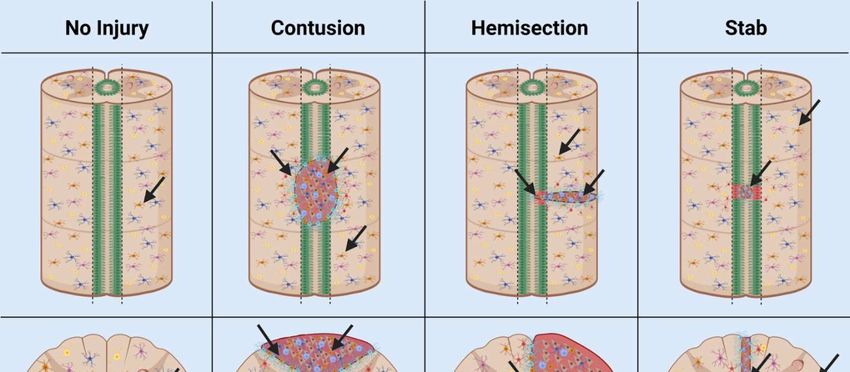
Preprints (www.preprints.org) | NOT PEER-REVIEWED | Posted: 16 June 2021                   doi:10.20944/preprints202106.0449.v1

                                         Neurogenic activity of the brain, spinal cord, and retina may facilitate the generation
                                    of functional networks of integrated tissue in damaged or diseased areas. The activity of
                                    NSPCs has been shown to affect neurodegenerative, inflammatory, and demyelinating
                                    conditions of the CNS as well as traumatic injury [6]. Overall, NSPCs play an essential
                                    role in injuries or degenerative disorders that are largely affected by neurogenesis and
                                    disruptions in cell behavior such as traumatic brain injury (TBI), spinal cord injury (SCI),
                                    retinal injury, multiple sclerosis (MS), and schizophrenia [7-9]. Thus, an in-depth under-
                                    standing of neurogenesis throughout the CNS will facilitate effective stem cell oriented
                                    therapeutic development.

                                    2. Neural Stem/Progenitor Cells
                                          The adult NSPCs (e.g., progenitor cells, neuroblasts, ependymal cells, NG2+ glia) are
                                    present in the stem cell niches of the brain, spinal cord, and retina. Major cell types present
                                    in the general NSPC niche include neurons, oligodendrocytes, astrocytes, pericytes, and
                                    endothelial cells.
                                          Additional contributors to the microenvironment of NSPCs in CNS niches include
                                    cerebrospinal fluid (CSF), the extracellular matrix (ECM), and vasculature. The CSF con-
                                    sists of neurotrophic/growth factors, transcription factors, and ECM molecules required
                                    for NSPC guidance and is important for cell migration, morphogenesis, growth, and de-
                                    velopment [10]. The ECM provides mechanical support and regulates extracellular signal-
                                    ing environments. Moreover, proteoglycan and glycoprotein composition varies to influ-
                                    ence signaling and bioavailability, motivating NSPC behavior within the stem cell niche
                                    [11].
                                          Cellular cross talk between the stem cells and specified cell types contribute to the
                                    symphony of cascading signals regulating NSPC behavior. NSPC populations in the stem
                                    cell niche are highly regulated to produce neuronal or glial lineage cell types [12]. The
                                    vasculature also regulates neurogenesis in the adult CNS by transport of infiltrating bio-
                                    chemical signals to interact with NSPCs [13]. In this way, intrinsic and extrinsic signals
                                    regulate neurogenesis, generated via cross talk with cells, vasculature, ECM via external
                                    forces, and CSF in the neural niche. Intrinsic signals include master transcription factors
                                    such as Sox2 and REST [14]. Extrinsic signals include neurotrophic/trophic and growth
                                    factors, neurotransmitters, and signaling pathways such as Wnt and Notch.
                                          When networks of neural cell types responsible for a regulated signaling microenvi-
                                    ronment are damaged, NSPCs exhibit extreme behavior [15]. This is due to distinctly dif-
                                    ferent signals or lack of signals required to regulate pools of active or quiescent NSPCs.
                                    Traumatic injury stimulates NSPCs to proliferate rapidly and produce cells which con-
                                    tribute to a glial scar border and upregulate angiogenesis in addition to neurogenic activ-
                                    ities [9]. Preferential survival of transplanted NSCs was observed in geographical areas of
                                    high-density vasculature, which is said to play an essential role in the survival and mainte-
                                    nance of NSPCs in the injured spinal cord [13].
                                          NSPCs often generate new non-functional networks of cells in response to injury
                                    which inhibits neural regeneration [16]. Altered niche activity may contribute to segrega-
                                    tion of the injury, but does not lead to regeneration of functional tissue. Differences in
                                    traumatic injury type and grade in the CNS result in significant changes in neurogenesis
                                    in one or more niches [15]. The heterogeneity of cell populations affected by traumatic
                                    injury result in clinical inconsistencies between cases. Further, the neurogenic niches of
                                    the brain, spinal cord, or retina exhibit regionally distinct niche composition before and
                                    after traumatic injury (Figure 2).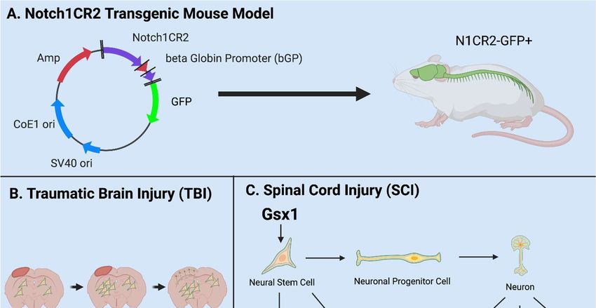
Preprints (www.preprints.org) | NOT PEER-REVIEWED | Posted: 16 June 2021                      doi:10.20944/preprints202106.0449.v1

          Figure 2. NSPC Niche in mammals: the SVZ and SGZ in the brain (A); the ependymal cells and NG2 cells in the spinal
          cord (B); and the base of the optic nerve, the Müller glia, and the pigment epithelium in the retina (C). AC, anterior
          chamber; CSF, cerebrospinal fluid; PC, posterior chamber; SVZ, subventricular zone; SGZ, subgranular zone. Adapted
          from Cutler and Kokovay, 2020 [17] (A); Sabelström et al, 2014 [18], Andreotti et al, 2019; Picoli et al, 2019 [19,20] (B);
          Yoshida et al, 2000 [21](C).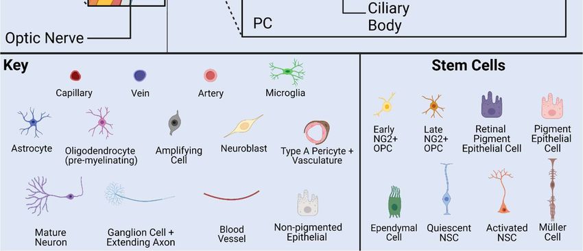
Preprints (www.preprints.org) | NOT PEER-REVIEWED | Posted: 16 June 2021                  doi:10.20944/preprints202106.0449.v1

                                    2.1. Adult NSPCs in the Brain
                                          The mammalian brain contains two primary neurogenic niches, i.e., the SGZ of the
                                    hippocampus and the SVZ of the lateral ventricles [22]. The hypothalamus serves as a
                                    third neurogenic niche conserved in some species, but is nonexistent in humans [23]. Each
                                    distinct niche contains specific populations of NSPCs and differing functions.
                                          The hippocampal neurogenic niche is present at the base of the hippocampus within
                                    the dentate gyrus (DG) in the SGZ (Figure 2A). In this niche, NSPCs are required for
                                    maintenance of the hippocampal tissue homeostasis, learning, and memory. Major stem
                                    cells in this neurogenic niche are radial glial-like cells (RGLs) which maintain neurogenic
                                    activity into adulthood [24]. Key cell types include OPCs, neuroblasts, immature/mature
                                    neurons, and oligodendrocytes. As a note, OPC populations in this niche include NG2+
                                    cells.
                                          The neurogenic niche along the walls of the lateral ventricles is located in the SVZ
                                    (Figure 2A). The lateral ventricle niche can be separated into two different geographical
                                    regions in the tissue: 1. Dorsal, 2. Lateral. Both dorsal and lateral components are in direct
                                    contact with pools of CSF, where the ependymal cell layer serves as a border between CSF
                                    and niche NSPCs [25]. This allows regulated contact between the ventricular cavities and
                                    undifferentiated progeny. Internal mechanisms direct NSPC behavior via fluid flow of
                                    CSF in the lateral ventricles [10]. Cell types include neuronal and glial subtypes which can
                                    be subdivided into further populations based on gene mapping analysis in both domains.
                                    Transcriptional patterning in temporal and spatial arrangements shows distinct NSPC
                                    populations [26]. Differential gene expression is driven by cell niche based signaling. Ma-
                                    jor signals include the Wnt/B-catenin and sonic hedgehog (Shh) pathway and are im-
                                    portant to maintain regulatory behavior [27].
                                          Adult neurogenesis in the hippocampus is dictated by intrinsic and extrinsic cues
                                    [28]. Signaling is initiated by surrounding cell types and vasculature in addition to master
                                    transcription factors Oct4, Sox2, and CREB. Signals from the Notch, Wnt, Shh, and other
                                    pathways direct neurogenesis in the SGZ.
                                          The rostral migratory stream is a migration pathway for neuroblasts from the SVZ to
                                    the olfactory bulb and is present in some mammalian species. Conserved signaling path-
                                    ways direct differentiation and integration of specified neurons and glia into the olfactory
                                    bulb. However, this is present to a lesser extent in larger mammalian species such as hu-
                                    mans. Migrating neuroblasts from the hippocampal niche have been documented in ro-
                                    dent models to contribute to olfactory bulb mature cell types [29]. However, in human
                                    and primate models these cells are instead generated in the striatum. Damage to the neu-
                                    ral niche of the hippocampus has been associated with cognitive deficits in learning and
                                    memory.
                                          The hypothalamus neurogenic niche is located near the lateral ventricles below the
                                    SVZ, also called the periventricular zone [21]. Major cell types in this niche include hypo-
                                    thalamic ribbon cells lining the outer wall and monocytes which may present neurogenic
                                    potential. Three populations of NSCs have been found in the hypothalamus of animal
                                    models including mouse, rat, and monkey including tanycytes, ependymal cells, and
                                    small stellate cells [15]. These populations generate neurons and glia throughout life in
                                    the hypothalamic parenchyma. Neurogenesis in this region occurs at a lesser incidence in
                                    comparison with the two classic niches, hippocampal SGZ and lateral ventricles SVZ. This
                                    may translate into functional significance in murine models via control of energy metab-
                                    olism [22].
                                          In the injured brain, specific regulation of quiescence/survival in NSPCs has been
                                    attributed to the small glycoprotein lactadherin, growth factors vascular endothelial
                                    growth factor (VEGF), fibroblast growth factor-2 (FGF2), and Notch and Wnt pathways
                                    [27,30,31]. Proliferation is regulated by lactadherin, amyloid precursor protein, neu-
                                    rotrophic factor Tumor necrosis factor alpha (TNFa), growth factors FGF2, and VEGF,
                                    chemokine CX3CL1, and pathways Shh, Notch, and Wnt [32]. Migration is regulated by
                                    growth factor VEGF, chemokines CCR2 and CX3CL1, and the Wnt pathway [31].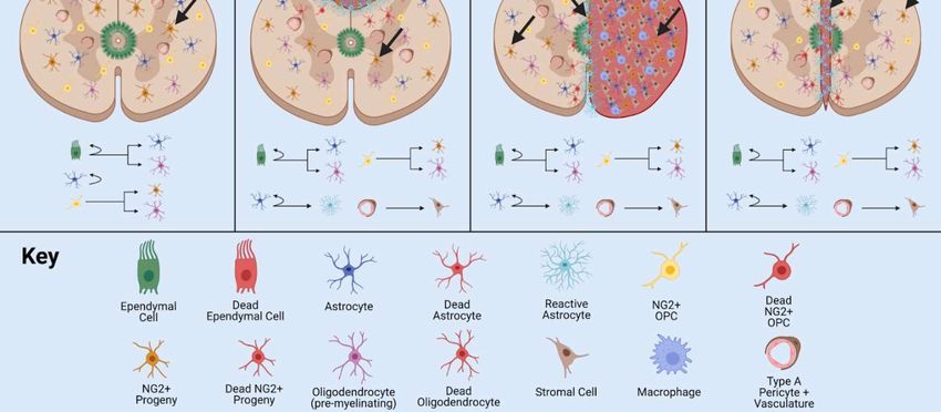
Preprints (www.preprints.org) | NOT PEER-REVIEWED | Posted: 16 June 2021                  doi:10.20944/preprints202106.0449.v1

                                    Differentiation is regulated by growth factors FGF2 and VEGF, chemokines CCR2 and
                                    CX3CL1, as well as Notch and Shh pathways. Integration is regulated by growth factor
                                    VEGF and chemokine CX3CL1 [30]. Injury-induced or altered signals contribute to the
                                    enhanced proliferation, aberrant progenitor migration, ineffective integration, and re-
                                    duced dendritic branching observed in TBI and SCI.
                                          Using a combination of transgenic mouse model and single-cell RNA-seq analysis,
                                    distinct adult NSPC populations were identified in the SVZ [33]. In this study, GFP+ cells
                                    represent Nestin+ stem cell populations in the adult. Four groups of NSCs and three
                                    groups of progenitor cells were characterized with in vivo and in vitro RNA-seq studies of
                                    the SVZ neurogenic niche [33]. Immunostaining and imaging analysis revealed distinct
                                    subgroups of cells separated by signal intensity: high GFP, low GFP and no GFP, and co-
                                    labeled with specific markers such as DCX and GLAST. Further, RNA-seq analysis iso-
                                    lated cells into profiles of quiescent and active stem cells in addition to stem cell markers,
                                    e.g., Sox2, Ascl1, and DCX. Groups of cells are also separated anatomically, further sup-
                                    porting the existence of distinct populations. NSPC heterogeneity has also been demon-
                                    strated using stem cell markers including Gli1 and Ascl1 in both dividing and nondivid-
                                    ing NSPCs [34]. The utility of these NSPC populations is unknown, but clear differences
                                    exist in gene expression profile.

                                    2.2. Adult NPSCs in the Spinal Cord
                                          The mammalian spinal cord contains one neurogenic niche in the ependyma of the
                                    central canal in which stem cells are present in an undifferentiated and self-renewable
                                    state (Figure 2B). The central canal serves as a continuation of the lateral ventricles into
                                    the spinal cord, while the ependymal cells serve as the bridge and a major regulatory ele-
                                    ment between the CSF and the stem cell niche [35]. The central canal neurogenic niche is
                                    lined with multiple populations of ependymal cells and CSF contacting neurons [36]. Ep-
                                    endymal cell populations can be further characterized into cells with short basal processes
                                    and cells with long extended processes. Other major components of the niche include
                                    NG2+ cells, vasculature, astroglial cells, and oligodendrocytes. Populations of progenitors
                                    in the spinal cord are indicated by markers Olig2, PDGFRa, and NG2 [37]. In addition, the
                                    ependymal cell layer is surrounded by supporting mature cell types, while the layer itself
                                    contains astroglial cells, NG2+ cells, and Nestin+ undifferentiated stem cells [36]. In nor-
                                    mal physiology, NSPC proliferation is observed in this stem cell niche, indicated by Ki67
                                    antibody staining in numerous studies [38,39].
                                         Extrinsic signals guiding adult neurogenesis in the spinal cord include connexin,
                                    Notch and Wnt signaling pathways [40,41]. Intrinsic signals include neural progenitor
                                    transcription factors Nkx6.1, Pax6, and Olig6 [39,42,43]. These signals cohesively create an
                                    environment to control NSPC activity and maintain normal pools of immature and ma-
                                    ture cell types in quiescent or active states. During injury or disease, NSPCs are subject to
                                    altered specific niche-based signals and exhibit skewed behavior. Thus, the neural niche
                                    in the central canal of the spinal cord is incredibly unique and maintained by a delicate
                                    balance of intrinsic and extrinsic signals.
                                         SCI affects the NSPC stem cell niche in models of contusive, surgical stab, and slice
                                    injury at any anatomical level of the spinal cord [41]. Common clinical SCI damages dis-
                                    turbs the niche due to equidistant dorsal and ventral positioning of the central canal [15].
                                    NSPCs proliferate after injury and interact with inflammatory signals to produce the glial
                                    scar border, a chemical/physical barrier which segregates the injury and prevents addi-
                                    tional damage [44]. However, this scar also prevents axonal outgrowth into the site of
                                    injury and generation of new cell types within the neural lesion. NSPCs proliferate and
                                    differentiate into reactive astrocytes in the injured spinal cord and contribute to the glial
                                    scar border. In addition to newly generated progeny, resident astrocytes transition to re-
                                    active gliosis state and are recruited to the site of injury, lengthen their processes, and
                                    fatten to become the scar border [45]. A multitude of NSPCs in the spinal cord produce
                                    progeny of differing lineages to contribute to the glial scar after SCI and TBI.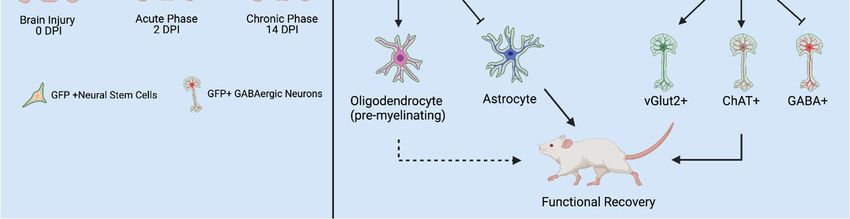
Preprints (www.preprints.org) | NOT PEER-REVIEWED | Posted: 16 June 2021                  doi:10.20944/preprints202106.0449.v1

                                         Two major cell types have been controversially implicated in the NSPC response to
                                    injury and pose high therapeutic potential: NG2+ and ependymal cells. Many published
                                    studies are in support of the stem-like character or non-stem-like character of these cells.
                                    Both NG2+ cells and ependymal cells have been reported to contribute to the formation
                                    of the scar border. More recently, NG2+ cells have shown to contribute to the generation
                                    of neurons in the injured spinal cord [44,46]. We will discuss the heterogeneity of NSPCs
                                    after injury with a focus on the activity of NG2+ and ependymal cells in Section 4.

                                    2.3. Adult Retinal Stem Cells
                                          Cells from regions of the adult retina such as the retinal pigment epithelium (RPE)
                                    [47,48], CE [49-53], Müller glia cells [51,54-56], iris pigment epithelium [57,58] and optic
                                    nerve [48] show stem cell characteristics to varying degrees (Figure 2C). Among them, the
                                    CE and Müller glia are identified as two main retinal stem cell sources.
                                          A subpopulation of adult human RPE cells is capable of being activated to become
                                    RPE retinal stem cells in vitro and differentiated into multipotent stable RPE or mesenchy-
                                    mal lineages [47]. The optic nerve lamina region (ONLR) in both humans and mice con-
                                    tains a retinal NPC niche [48]. Adult NPCs in the ONLR exhibit multipotency and gener-
                                    ate two types of glia: astrocytes and oligodendrocytes. These populations contribute to
                                    enable glial replacement and remyelination in adulthood [48]. The derived adult rat iris
                                    pigment epithelium (IPE) cells have NSPC differentiation ability to differentiate into rod
                                    photoreceptor cells under CRX transfer [58]. NeuroD induces human iris cells into rod
                                    photoreceptor cells. Moreover, Yuko et al. observed the combination of CRX, RX and Neu-
                                    roD induces the generation of photoreceptor cells from the derived human IPE cells [57].
                                          Non-pigmented CE cells show stem cell markers and actively proliferate after pho-
                                    toreceptor cell degeneration or retinal ganglion cell injury in the mouse model [49,59]. In
                                    the human CE, non-pigmented CE cells are labeled with stem cell markers, e.g., Sox2,
                                    Chx10 and Notch1. Non-pigmented CE cells showed proliferative ability under epidermal
                                    growth factor (EGF) induction using explants of the human retina [50]. CE cells including
                                    the pigmented cells and non-pigmented cells from human and mouse express NSPC cell
                                    markers and characteristics in vitro study [51]. CE cells can be induced into photoreceptor
                                    cells, bipolar cells, retinal ganglion cells and Müller glia cells in the mouse model [52]. In
                                    addition, human CE cells can be induced in many types of retinal cells in vitro [53].
                                          Müller glial cells are also considered as a primary source of retinal stem cells. Bhatia
                                    et al. concluded that retinal Müller glia may perform similar functions ascribed to astro-
                                    cytes, ependymal cells and oligodendrocytes in other regions of the CNS [51]. Das et al.
                                    also stated that Müller glia are the NSCs of the adult retina [54]. They demonstrated that
                                    rat Müller glia have potential to generate retinal neurons in vitro and in vivo. Moreover,
                                    they proved the role of Notch and Wnt pathways in regulating this activity. Similarly, in
                                    mouse models, Müller glia can be reprogrammed into photoreceptors and retinal gan-
                                    glion cells under certain culture conditions [55]. In adult human eyes, no evidence has
                                    been found to suggest that Müller glia possess the retinal neuronal regeneration ability in
                                    vivo. However, in vitro, these progenitor-type glia can be induced to proliferate and dif-
                                    ferentiate into retinal neurons and RPE cells [56]. Human Müller glia-derived stem cells
                                    can be differentiated towards the fate of retinal ganglion cell (RGC) precursors using FGF-
                                    2 and Notch inhibition [56]. In summary, Müller glia-derived stem cells can function as
                                    NSCs and serve as a potential target of therapy for retinal degenerative disease.
                                          Common retinal diseases/injuries such as retinitis pigmentosa (RP) and age-related
                                    macular degeneration (AMD) cause the photoreceptor cell loss and damaged RPE. How-
                                    ever, no enhanced differentiation or proliferation was observed after injury [52]. Damaged
                                    cells release growth factors and cytokines which cause the Müller glia cell to differentiate,
                                    proliferate and express progenitor cell markers [60]. The ability of these proliferating Mül-
                                    ler cells to regenerate new neurons and repair the injured retina appears to be extremely
                                    limited. Regardless, the multipotent stem cells may generate more functional
Preprints (www.preprints.org) | NOT PEER-REVIEWED | Posted: 16 June 2021                  doi:10.20944/preprints202106.0449.v1

                                    photoreceptor cells and help with the recovery of vision loss in the RP and AMD via trans-
                                    plantation method [61].

                                    2.4. Heterogeneity Between CNS Niches
                                          The perivascular stem cell niche is not technically a NSPC niche, but it interacts with
                                    cell types and influences NSPC behavior in all niches, thus contributing to the diversity
                                    of NSPC behavior observed in the mammalian CNS. In particular, the retina contains
                                    sources of NSPCs such as Müller glia and CE. Major factors unique to the retinal niche
                                    include CRX, RX and NeuroD. Interestingly, the retina does not contain ependymal cells,
                                    a major controversial stem type cell in the brain and spinal cord. However, NG2+ cells can
                                    be found in the retina [62]. The brain contains NSPC populations such as radial glial-like
                                    cells, OPCs, and ependymal cells. However, these populations and their characteristics
                                    vary throughout distinct NSPC niches. Major signals unique to the SGZ and SVZ include
                                    Shh pathway and transcription factors CREB and Oct4 [63]. The spinal cord stem cell niche
                                    contains both ependymal cells and NG2+ cells. Signals unique to the spinal cord include
                                    connexin signaling. The activity and consistency of NG2+ populations vary significantly
                                    between the niches of the brain, spinal cord, and retina. Specifically, NG2+ cells in the
                                    brain and spinal cord generate oligodendrocyte cell types and consist of glia and pericytes
                                    [64]. However, NG2+ cells in the retina consist of microglia and pericytes [62]. Ependymal
                                    cells also exhibit a variety of diverse behaviors in neurogenic niches of the brain and spinal
                                    cord. These controversial stem-like cells will be discussed in the following sections.
                                          Understanding the heterogeneity of these stem cell populations and neurogenic
                                    niches is necessary to effectively design therapeutics for SCI, TBI, mechanical/chemical
                                    injury, and diseased states such as Glaucoma, Retinitis Pigmentosa, and inflammatory
                                    conditions.

                                    3. Notch1CR2-GFP+ NSPCs in Development and Injury
                                          The canonical Notch signaling pathway is required to regulate the quiescence, pro-
                                    liferation, and differentiation of NSPCs in the CNS [43,65-67]. The Cai lab identified a 399-
                                    bp cis-element in the second intron of the Notch1 locus (CR2) [68]. In the Notch1CR2 -GFP
                                    transgenic mouse, CR2 directs the reporter GFP expression in the interneuron progenitor
                                    cells. The activities of Notch pathway and NSPCs can be traced by the reporter GFP ex-
                                    pression (Figure 3A). The cell fate of GFP tagged interneuron progenitors have been char-
                                    acterized in both normal development and neurological disease/injury conditions, which
                                    facilitate the study of the potentials of NSPCs in regenerative medicine [43,66,68]. In these
                                    studies, the Cai lab has demonstrated that GFP+ NSPCs preferentially differentiate into
                                    interneurons of the brain and spinal cord during embryonic development and in adult-
                                    hood [43,67]. Injury increased the number of GFP+ NSPCs and interneurons at the injury
                                    site in a closed head injury model [67]. These results demonstrate that the endogenous
                                    NSPCs in the brain proliferate after injury and differentiate into specific cell fates (Figure
                                    3B).
                                           In a more recent study, virus-mediated Gsx1 expression in NSPCs displayed an in-
                                    creased rate of cell proliferation with increased number of GFP+ NSPCs. Gsx1 further pro-
                                    moted neuronal differentiation over glial lineage in the injured spinal cord (Figure 3C).
                                    This resulted in an increased number of neurons, reduced reactive astrocytes and glial
                                    scar formation, and improved functional recovery [66].
                                          The transgenic animal model serves as a valuable tool to study endogenous NSPCs
                                    following traumatic CNS injury, and further NSPCs represent important cell source for
                                    neural regeneration in the adult CNS [44,69,70]. For this reason, diversity of NSPC popu-
                                    lations (e.g., Nestin+, Notch1+, NG2+, Foxj1+ cells) have become an intensely focused area
                                    of research in regenerative medicine for CNS diseases and injuries. Several controversial
                                    issues arise and are discussed in the next section.
Preprints (www.preprints.org) | NOT PEER-REVIEWED | Posted: 16 June 2021 doi:10.20944/preprints202106.0449.v1
Figure 3. Utilities of the Notch1CR2-GFP transgenic mouse line in SCI and TBI models. (A) Notch1CR2-GFP transgenic
mouse model labels NSPCs in the CNS. (B) Adult NSPCs in the brain proliferate in the acute phase of TBI and differen-
tiate into neurons in the chronic phase of TBI. (C) In the injured spinal cord, Gsx1 expression promotes adult NSPC
proliferation and preferential differentiation into excitatory interneurons and inhibits astrocytes and glial scar formation
after injury. Adapted from Tzatzalos, 2012 [68] (A), Anderson et al, 2020 [67] (B) and Patel et al, 2021 [66] (C).
4. Controversial NSPC Populations in Injury/Disease
Adult NSPC populations are composed of diverse cell types in the mammalian CNS,
which contribute to growth and regeneration after injury and disease. Several cell types,
e.g., ependymal and NG2+ cells have been controversially proposed as stem cells. The
stem cell behavior of these populations has been implicated in the injury response and
neurodegenerative/demyelinating disorders [71]. These populations are primarily glial
producing cells; however, extrinsic and intrinsic factors have been used to modulate the
glial fate to neuronal fate for functional recovery after traumatic injury [46,67]. Both pop-
ulations have also been polemically associated with the glial scar formation after TBI and
SCI in mammals [72,73]. In the following sections, we will explore the stemness of these
populations in normal physiology and the utility of these populations as a treatment for
CNS injury/disease. To appropriately assess ependymal and NG2+ cells as a stem cell, we
will use the basic criteria: self-renewal and multipotency [2,74] (Figure 1).Preprints (www.preprints.org) | NOT PEER-REVIEWED | Posted: 16 June 2021 doi:10.20944/preprints202106.0449.v1
Figure 4. Behavior of ependymal cells and NG2+ cells in animal models of SCI. In normal physiology, the ependymal
cells lining the wall of the central canal are largely quiescent, while NG2+ cells are ubiquitously distributed throughout
the grey and white matter of the spinal cord. In contusion SCI, the ependymal cell layer is not damaged, but may in-
crease proliferation and differentiation potential. In the hemisection model, the ependymal cell layer is damaged and
ependymal cells/NG2+ cells are activated by injury. In stab SCI, the ependymal cell layer is damaged and contributes
greatly to glial scar formation. Adapted from Sabelström et al, 2014 [18], Hackett et al, 2016 [75], and Picoli et al, 2019
[20].
4.1. Ependymal Cells
Ependymal cells are neuroglia which line the central canal of the spinal cord and the
lateral ventricles of the brain. The epithelial layer (ependyma) acts as a barrier between
the CSF and stem cell niche and regulates CSF balance and NSPC activity in central CNS
niches [36]. In this layer, ependymal cells project differing length cilia into the CSF and
aid in motility, production, and absorption of the CSF [76]. Ependymal cells have been
controversially proposed as stem cells in the brain and spinal cord. Contradicting results
have been reported regarding the appropriate contribution of ependymal cells to the path-
ophysiology of SCI and TBI [38,72,77,78]. This discrepancy is attributed to major differ-
ences in CNS injury models, animal models, and quantification techniques [77,79]. Differ-
ences in injury models range from damage to the ependymal layer, gray, and white matterPreprints (www.preprints.org) | NOT PEER-REVIEWED | Posted: 16 June 2021 doi:10.20944/preprints202106.0449.v1
to no damage to the ependymal layer but exposure to injury mediators such as glutamate.
Further, age of animals used in research may also account for discrepancies in reported
ependymal cell ability, as younger animals maintain populations with increased neuro-
genic activity in comparison with older animals [77].
During development, the ependymal and neuronal cell fates are decided by numer-
ous transcription factors e.g., Ascl1, Sox10. Non-differentiated pools of progenitors gen-
erated by NSCs produce ependymal and neural cell types in a finely tuned spatiotemporal
manner [80]. By embryonic day 15.5 (E15.5), the ependymal cell populations can be fully
distinguished [81]. In the adult, ependymal cells express Foxj1 and proliferate actively to
produce multipotent glial fated cell types such as astrocytes and oligodendrocytes [72].
This does not occur at a high rate or contribute to tumorigenesis in the spinal cord or brain
[82]. Pools of ependymal cells also have displayed self-renewal capability, but this capa-
bility decreases with age of animal [77].
Extrinsic factors guide ependymal cell activity in the adult including neurotrophic
factors and Notch and Wnt signaling pathways. Intrinsic factors include transcription fac-
tor Foxj1 and nuclear factor IX (NFIX) [76]. Quiescence/survival is regulated by DNA-
binding protein inhibitor (Id3) and HES family transcription factor 5 (Hes5) [79]. Prolifer-
ation is regulated by Wnt signaling and growth factors [83].Differentiation is directed by
the Geminin superfamily, an antagonist of DNA replication, and NFIX [84,85]. Migration
is regulated by NFIX and non-muscle myosin II [35]. Ependymal cells also secrete factors
to produce chemical gradients promoting migration of neuroblasts in the SVZ of the lat-
eral ventricles [86] (Table 1). Ependymal cell activity is consistent with the consensus that
adult neurogenesis is present in the adult mammalian CNS but decreases with age and
development. In addition, the neurogenic activity in many stem-like cell types decreases
with age [33].
After CNS injury, NSPCs become activated and display enhanced proliferation and
differentiation potential outside of the normal lineage programming [78]. Ependymal cell
activity also varies significantly between major SCI models such as contusion model, hem-
isection/transection model, and stab model. (Figure 4).
In the stab SCI model, a thin blade penetrates the central canal, disturbing the epen-
dymal cell layer and damaging ependymal cells. This results in high proliferation and
contribution to both the glial scar content and lesion by ependymal progeny [82,87]. Im-
ages and quantification of this scar border directly within the NSC niche of the central
canal may have overinflated the regenerative capacity of ependymal cells in adult mam-
mals. Due to the location of the injury, the segregation of the injured cells and formation
of scar tissue consists highly of ependymal cell progeny which can easily integrate into
the network of scar tissue in their immediate vicinity. In addition, a greater percentage of
damaged ependymal cells in this injury model result in increased proliferation. As a note,
the little migration necessary from the NSPC niche to the entire lesion primarily differen-
tiates this injury model from many others and results in high ependymal contribution.
In the dorsal hemisection or full transection SCI model, a surgical blade is used to
slice half, or the entire spinal cord and the central canal ependymal cell populations are
damaged. However, these cells still contribute little to the glial scar border due to the lim-
ited migratory capacity of ependymal progeny following injury [88]. Resident astrocytes
and NG2+ cells in these models contribute to glial scar border, with lesser ependymal
progeny recruitment to the injury [89]. The anatomical location of the damaged cells in
this model explains the small ependymal contribution to the scar border and neural lesion.
Populations primarily line the central canal, and the slice injury minimizes cellular/niche
damage. Thus, consistency of damaged or injury-stimulated cell types is little in compar-
ison with stab model (Figure 4).
In the contusion SCI model, the most clinically relevant injury model, a pneumati-
cally driven rod is dropped onto the cord and raised immediately to create the injury. The
ependymal cell layer is disturbed, but not penetrated and thus minimal ependymal cell
progeny migrate to the site of injury and contribute to the glial scar [90] (Figure 4). While
ependymal cells are multipotent and produce oligodendrocyte and astrocyte fated cells,Preprints (www.preprints.org) | NOT PEER-REVIEWED | Posted: 16 June 2021 doi:10.20944/preprints202106.0449.v1
the limited migratory capacity of ependymal cells after injury results in little contribution
to the injury itself. The contusion injury does not directly damage ependymal cells but
does damage tissue in proximity with the NSPC niche of the central canal.
Ependymal contribution to glial scar has been associated with age, as younger epen-
dymal cells in the spinal cord retain more proliferative and migratory capacity, thus in-
creased contribution to glial scarring [82]. This is significant in clinically relevant contu-
sion SCI model, where the lesion is not in direct contact with the central canal ependymal
layer but may be subject to molecular signals from damaged cells.
The ependymal cell populations in the brain and spinal cord are highly heterogene-
ous and can generate neuroblasts and glia in response to stroke, elicit aberrant NSPC ac-
tivity following SCI and TBI. Within ependymal populations, subpopulations have been
identified by gene expression studies [91]. However, the function of these subpopulations
is still under investigation, but a thorough understanding will support the development
of treatments for stroke, TBI, SCI, schizophrenia, and many other injured or diseased
states.
Ependymal cells in the adult mammalian CNS are stem-like cells, as demonstrated
by their ability of self-renewal and multipotency. They contribute to glial populations in
the normal physiology and injury. In the stab SCI model, a blade penetrates the ependy-
mal cell layer in the central canal. In this case, ependymal cells contribute greatly to glial
scar border formation and migrate into the neural lesion. However, in all other validated
SCI models these cells do not contribute a significant number of astroglial progeny to the
glial scar. These cells serve as a major source of NSPCs in the spinal cord but do not pro-
vide a suitable therapeutic target in contusion SCI. Regardless, these cells may serve as a
viable therapeutic target for regeneration in stab wound type clinical SCI and TBI.
Table 1. Ependymal and NG2+ Cell Activity: Normal Physiology vs Injury
Ependymal No Injury Contusion Hemisection Stab
No: Ependyma
not injured [72]
Yes, Medium Yes [77]
[84] Yes, High Yes, Low:
Proliferation
[92] Yes, Medium Ependyma
Yes [82] [82] injured [72]
Yes [93]
Yes [77] Yes, Low [72]
Differentiation No [82] Yes [92]
Yes [82] Yes [93]
Yes, Low [72]
Migration Yes, Low [84] Yes, Low [92] Yes, High [77]
Yes [93]
Yes [94]
Quiescence No No No
Yes [82]
Glial Scar No Yes: Ependyma
N/A Yes [92]
Formation [82] injuredPreprints (www.preprints.org) | NOT PEER-REVIEWED | Posted: 16 June 2021 doi:10.20944/preprints202106.0449.v1
[72]
Neural Lesion N/A Yes [92] Yes [82] Yes [72]
NG2 No Injury Contusion Hemisection Stab
Yes, Low [92]
Yes, Gradual
Yes, High [44]
Decline [95]
Proliferation Yes, High [44] Yes, High [98]
Yes, High [97]
Yes, High [94]
Yes, High [96]
Yes, Medium
[99] Yes, Low [89]
Yes, Medium Yes, Medium
Differentiation
[95] [89]
Yes, Medium Yes [98]
[44]
Yes [99]
Yes, Medium
Migration Yes [96] Yes [44]
[95]
Yes [98]
Yes, Low [99]
Decrease
Quiescence No [96] No [97]
[100]
Possibly [12]
Yes [44] Yes [99] Yes, 5-8% [45]
Glial Scar
N/A
Formation
Yes, 25% [45] Yes, 5% [45] Yes [98]
Yes, Delayed
Yes [101]
increase [101]
Neural Lesion N/A Yes, High [96]
Yes [98]
Yes [97]
1Ependymal and NG2+ cell stem-like behaviors in the normal physiology and after different types
of SCI.
4.2. NG2+ Cells
The NG2 is a type I transmembrane glycoprotein also called chondroitin sulfate pro-
teoglycan 4 or nerve glial antigen-2 [102]. The NG2+ cells are heterogeneous populations
composed of glia, pericytes and macrophages of vasculature, also regarded as polyden-
drocytes. Cell morphology varies throughout life and subpopulation, but generally can
be characterized by soma with long extended or short processes. A major percentage of
the NG2+ glia population are oligodendrocyte progenitor cells (OPCs) which actively con-
tribute to the oligodendrocyte population [99]. Populations of OPCs have been harvested
and purified in vitro and approximately 95% are positive for NG2 marker [37]. NG2+ cells
have been controversially proposed as stem-like cells in the literature [45]. However, sev-
eral studies contradict each other regarding this cell type classification and contribution
to the pathophysiology of SCI, TBI, and various diseased states. These discrepancies are
largely due to differences in recombinant genetic mouse lines, animal models, injury mod-
els, and quantification techniques. Transgenic mouse lines have been established to target
NG2+ populations using promoters from PDGFRa, Olig2, and Sox10 genes; commonly
expressed in NG2+ populations [69]. Approximately 80% or more cells in NG2+Preprints (www.preprints.org) | NOT PEER-REVIEWED | Posted: 16 June 2021 doi:10.20944/preprints202106.0449.v1
populations are targeted by these factors, however in varying ratio [94]. This fact contrib-
utes to inconsistencies between published results on NG2+ populations and NSPC char-
acterization. Targeted populations may not truly represent the NG2+ cells but extend to a
variety of other cell types as well [94].In addition, major differences in injury models and
glial scar properties contributes to controversial NG2+ stem-like nature [69]. Issues may
also result from astrocyte identification methods within the glial scar border, as GFAP is
the most common astrocyte marker used but is not expressed by all astrocyte subtypes.
During development, the NG2 marker exists in three major populations of self-re-
newing cells: oligodendrocyte lineage, NG2 glia, and astrogenic glia. Within early to mid-
stages of development, NG2+ cells have high differentiation potential and supply progeny
to populations such as astrocytes and oligodendrocytes [103]. In the adult, there are three
well-established glial populations, i.e., astrocytes, microglia, and oligodendrocytes, and
NG2+ cells make up the fourth major glial population [37,70,104]. NG2+ polydendrocytes
are characterized as highly proliferative [89]. NG2+ cells are evenly distributed through-
out brain and spinal cord, often described as a checker pattern in tissue sections of the
spinal cord. These cells also interact uniquely with neurons and glia, receiving both inhib-
itory and excitatory signals from areas throughout the brain indicating the diverse func-
tionality of the NG2+ cell populations [105]. Interestingly, NG2+ cells generate white mat-
ter mature oligodendrocyte cells at a faster rate than grey matter mature oligodendrocyte
cells. In addition, NG2+ cells in white and grey matter have been shown to exhibit differ-
ing morphological and electrophysiological characteristics such as the ability to generate
mature action potential spikes in white matter NG2+ cells [106].
NG2+ cells isolated from the rat optic nerve exhibited multipotency in vitro as they
differentiated into oligodendrocyte or astrocyte cell fates [107]. However, this capacity is
limited in vivo, as NG2+ contribute primarily to oligodendrocyte populations [64,108]. In-
terestingly, ectopic expression of Sox2 has been shown to restore multipotent lineage ca-
pacity in the adult [46]. Self-renewal has also been observed in NG2+ cell populations in
vivo [109]. Distinct heterogeneous populations of NG2+ cells exist in the adult CNS and
may contribute to a complex variety of essential activities, e.g., maintenance of homeosta-
sis, glutamate signaling [102,110].
Extrinsic and intrinsic factors guide NG2+ cell activity in the adult, including ciliary
neurotrophic factor (CNF), brain derived neurotrophic factor (BDNF), astrocyte derived
growth factors, neurotransmitters, cytokines, Notch and Wnt signaling pathways, and
transcription factors Olig2 and Sox10 [69,89,111]. The interaction between these signals
maintains the delicate balance to maintain the NSPC behavior of NG2+ populations.
Quiescence/survival of these populations is regulated by chemokine CXCL12 and CXCR4
[112]. Proliferation is directed by intrinsic gene Ascl1 [113]. Differentiation is directed by
Shh signaling in oligodendrocyte populations in the dorsal SVZ, but not the ventral SVZ,
indicating regionally distinct NG2+ populations in the SVZ (Table 1). In addition, oli-
godendrocyte lineage differentiation is directed by Wnt signaling and the kon-tiki gene.
Migration is directed by NG2 interaction, as shown in stab injuries to orient the NG2+ cells
toward the injury site [114]. Integration is directed by neurotransmitter receptor activation
and synaptic input from glutamatergic and GABAergic neurons.
NG2+ cell populations react to traumatic injury and neurodegenerative/demyelinat-
ing conditions. Specifically, traumatic injury reactivates the differentiation potential in
populations of the brain and spinal cord. NG2+ cells transition to a stem-like state and are
assumed to contribute to the injury in two segregated zones, i.e., necrotic core and glial
scar. The ratio of infiltrating cells and percentage NG2+ contribution to total scar border
varies significantly between injury types. For example, in the contusion SCI model, 25%
NG2+ progeny cells were reported in the glial scar border [44]. This may be due to in-
creased inflammation and extent of injury resulting in greater differentiation potential. In
the dorsal hemisection model, 5% NG2+ astrocyte progeny content has been reported in
the glial scar border [82]. In the cortical stab model, 8% NG2+ cell progeny content has
been reported in the glial scar border [45]. Thus, the highest NG2+ contribution to the glialPreprints (www.preprints.org) | NOT PEER-REVIEWED | Posted: 16 June 2021 doi:10.20944/preprints202106.0449.v1
scar border occurs in the contusion injury model (Figure 4). This may be due to the size
and depth of lesion formed in clinically relevant contusion SCI.
In acute injury, NG2+ cells migrate into the injury site and contribute to the formation
of the glial scar border. Specifically, the migratory capacity of NG2+ cells increase with
traumatic injury in both the brain and spinal cord [115,116]. This may be due to signals
directing aberrant migratory activity or regenerative abilities of NG2+ cells in the spinal
cord. In the formation of the glial scar border, Wnt signaling controls multipotent differ-
entiation into astrocyte and oligodendrocyte cell types [117]. In addition, NG2+ progeny
cells in the spinal cord preferentially differentiate into reactive astrocytes via Shh signal-
ing.
NG2+ cells are a potential target for SCI, TBI, MS, and other demyelinating and de-
generative conditions due to the consistency of NG2+ cells throughout the neurogenic
niches of the CNS. NSPC genes such as Sox2, Olig2, Pax6, and PDGFRa have been force-
fully overexpressed in NG2+ populations and resulted in increases in neurogenesis and
reactivation of stem-like characteristics [103]. The clinical relevance of this approach has
recently been demonstrated, i.e., targeted overexpression of Sox2 in NG2+ populations
resulted in improved functional recovery after SCI [46]. Neurogenesis in the post injury
CNS is altered significantly leading to ineffective maintenance of homeostasis, learning,
memory, and generation of nonfunctional neuronal networks.
Overall, although the NG2+ cells are not inherently stem-like cells, they are self-re-
newing during development and into adulthood. In addition, NG2+ cells are widely
known to actively proliferate and contribute to oligodendrocyte glial cells in the mamma-
lian CNS. NG2+ cells are not multipotent in nature, however, the multilineage potential is
reactivated by injury and mediators such as transcription factors. This capability of NG2
cells to acquire multipotency has a great potential to contribute to neural regeneration.
Thus, these cells present a viable therapeutic route to modulate the glial scar formation in
SCI and TBI. Gene or cell therapy targeted for NG2+ cells may use the acquired self-re-
newal capability of these populations to regenerate tissue in the CNS. The broad distribu-
tion of these cells throughout the CNS may increase the application and ability of targeted
stem cell activity to treat injured and diseased states. Other feasible therapeutic ap-
proaches include applying neurotrophic, growth, and anti-inflammatory factors to guide
the activity of NG2+ cells after injury.
Table 2. Literature Supporting or Refuting NG2+ and Ependymal Cells as Stem Cells
Ependymal For Against
Capable of Division or
[77,106,118,119] [79,120]
Self - Renewal
Capable of Giving Rise to
[77,91,118,119] [72,79]
Specialized Cells
Expression of Stem Cell
[79,91,118] [46]
Markers
NG2 For Against
Caple of Division or
[89,94,108] [37]
Self - Renewal
Capable of Giving Rise to
[46,89,94,108] N/A
Specialized Cells
Expression of Stem Cell
[44,46] [37]
Markers
2 References in support and against NG2+ cells and Ependymal cells as stem cells in the CNS.Preprints (www.preprints.org) | NOT PEER-REVIEWED | Posted: 16 June 2021 doi:10.20944/preprints202106.0449.v1
5. Conclusions
A wide variety of NSPCs reside within the adult CNS. This diversity contributes to
the complex pathophysiology of clinical injured and diseased states of the CNS such as
SCI, TBI, and retinal degeneration. Adult neurogenesis in the brain, spinal cord, and retina
is necessary for maintenance of homeostasis, learning, memory, and energy metabolism.
Interestingly, variability exists between major neurogenic niches of the CNS in the retina,
brain, and spinal cord. Differences in specification of these cells, ratio of cells, and exist-
ence at all or some of these cell types in the neurogenic individual niches varies greatly
with anatomical location [19].
NG2+ and ependymal cells are heterogeneous populations distributed distinctively
in the CNS neural niches. During development, NG2+ cells are multipotent and self-re-
newing, contributing to glial cell populations including astrocytes and oligodendrocytes.
Multipotency is lost in postnatal stages but is acquired following traumatic injury and by
manipulation of gene expression, thus these cells present a highly viable target for trau-
matic CNS injury. The relative percentage of NG2+ cells contributing to the glial scar is
highest in the contusion SCI model, lesser in the cortical stab SCI model, and the least in
the hemisection SCI model. During development, ependymal cells proliferate and con-
tribute to multipotent cell types. In adults, ependymal cells maintain multipotency and
activation, but to a lesser degree than in embryonic development. After contusion and
hemisection injury, ependymal cells contribute minimally to glial scar formation due to
lack of direct damage to the central canal of the spinal cord. However, stab injury damages
only the central canal, leading to scar border formation directly inside of the ependymal
cell layer. Injured ependymal cells proliferate actively and produce multipotent progeny
to contribute to glial scar border and the neural lesion extensively. However, migratory
capacity is seemingly unaffected by injury. Thus, these cells present a therapeutic target
for specific injury types which exclusively damage ependymal cell layer. As another ther-
apeutic route, the migratory capacity of ependymal progeny can be stimulated to contrib-
ute to the site of injury. Examples include stimulation of migration via neu-
rotrophic/growth factors and gene/cell therapy.
Interestingly, ependymal cells exhibit inverse behavior to NG2+ cells after traumatic
injury (Figure 4), contributing most to the glial scar in stab SCI model and the least in
contusion SCI model. This may be due to the anatomical location of these cell populations
and limited migratory capacity of ependymal progeny. Thus, targeting ependymal and
NG2+ cells for CNS regeneration, as well as a variety of other proliferating cell types such
as sub-populations of astrocytes, represent potential therapeutic strategies for regenera-
tive medicine. However, this will depend on the extent, anatomical location, and type of
injury or disease.
Future research directions for NG2+ and ependymal cell populations include a
deeper mechanistic understanding of progeny differentiation fates, migratory ability, and
functionality of subpopulations of these cells in the normal physiology and traumatic in-
jury. Applicable fields include developmental biology, tissue engineering and regenera-
tive medicine. Application of these cell types as target cells may hold the key to treatments
for SCI, TBI, retinal mechanical damage, and degenerative diseases of the CNS.
Author Contributions: L.C. and Z.F. conceptualized and wrote the paper. F.E. and B.R. contributed
to the brain and spinal cord portion of the review, and made the figures and tables; T.F. and X.A.
contributed to the retina portion of the review. All authors revised the manuscript and agreed to the
published version of the manuscript.
References
1. Eze, U.C.; Bhaduri, A.; Haeussler, M.; Nowakowski, T.J.; Kriegstein, A.R. Single-cell atlas of early human brain development
highlights heterogeneity of human neuroepithelial cells and early radial glia. Nat Neurosci 2021, 24, 584-594,
doi:10.1038/s41593-020-00794-1.Preprints (www.preprints.org) | NOT PEER-REVIEWED | Posted: 16 June 2021 doi:10.20944/preprints202106.0449.v1
2. Molofsky, A.V.; Pardal, R.; Iwashita, T.; Park, I.K.; Clarke, M.F.; Morrison, S.J. Bmi-1 dependence distinguishes neural stem
cell self-renewal from progenitor proliferation. Nature 2003, 425, 962-967, doi:10.1038/nature02060.
3. Altman, J. Are new neurons formed in the brains of adult mammals? Science 1962, 135, 1127-1128,
doi:10.1126/science.135.3509.1127.
4. Navarro Quiroz, E.; Navarro Quiroz, R.; Ahmad, M.; Gomez Escorcia, L.; Villarreal, J.L.; Fernandez Ponce, C.; Aroca
Martinez, G. Cell Signaling in Neuronal Stem Cells. Cells 2018, 7, doi:10.3390/cells7070075.
5. Potockova, A.; Strmen, P.; Krasnik, V.; Olah, Z. Mechanical injuries of the eye. Bratisl Lek Listy 2010, 111, 329-335.
6. Beyer, F.; Jadasz, J.; Samper Agrelo, I.; Schira-Heinen, J.; Groh, J.; Manousi, A.; Butermann, C.; Estrada, V.; Reiche, L.;
Cantone, M.; et al. Heterogeneous fate choice of genetically modulated adult neural stem cells in gray and white matter of
the central nervous system. Glia 2020, 68, 393-406, doi:10.1002/glia.23724.
7. Park, I.H.; Arora, N.; Huo, H.; Maherali, N.; Ahfeldt, T.; Shimamura, A.; Lensch, M.W.; Cowan, C.; Hochedlinger, K.; Daley,
G.Q. Disease-specific induced pluripotent stem cells. Cell 2008, 134, 877-886, doi:10.1016/j.cell.2008.07.041.
8. Akkermann, R.; Beyer, F.; Kury, P. Heterogeneous populations of neural stem cells contribute to myelin repair. Neural Regen
Res 2017, 12, 509-517, doi:10.4103/1673-5374.204999.
9. Li, Y.; Chang, S.; Li, W.; Tang, G.; Ma, Y.; Liu, Y.; Yuan, F.; Zhang, Z.; Yang, G.Y.; Wang, Y. cxcl12-engineered endothelial
progenitor cells enhance neurogenesis and angiogenesis after ischemic brain injury in mice. Stem Cell Res Ther 2018, 9, 139,
doi:10.1186/s13287-018-0865-6.
10. Alonso, M.I.; Gato, A. Cerebrospinal fluid and neural stem cell niche control. Neural Regen Res 2018, 13, 1546-1547,
doi:10.4103/1673-5374.237114.
11. Ahmed, M.; Ffrench-Constant, C. Extracellular Matrix Regulation of Stem Cell Behavior. Curr Stem Cell Rep 2016, 2, 197-206,
doi:10.1007/s40778-016-0056-2.
12. Spitzer, S.O.; Sitnikov, S.; Kamen, Y.; Evans, K.A.; Kronenberg-Versteeg, D.; Dietmann, S.; de Faria, O., Jr.; Agathou, S.;
Karadottir, R.T. Oligodendrocyte Progenitor Cells Become Regionally Diverse and Heterogeneous with Age. Neuron 2019,
101, 459-471 e455, doi:10.1016/j.neuron.2018.12.020.
13. Karakatsani, A.; Shah, B.; Ruiz de Almodovar, C. Blood Vessels as Regulators of Neural Stem Cell Properties. Front Mol
Neurosci 2019, 12, 85, doi:10.3389/fnmol.2019.00085.
14. Surzenko, N.; Crowl, T.; Bachleda, A.; Langer, L.; Pevny, L. SOX2 maintains the quiescent progenitor cell state of postnatal
retinal Muller glia. Development 2013, 140, 1445-1456, doi:10.1242/dev.071878.
15. Falnikar, A.; Stratton, J.; Lin, R.; Andrews, C.E.; Tyburski, A.; Trovillion, V.A.; Gottschalk, C.; Ghosh, B.; Iacovitti, L.; Elliott,
M.B.; et al. Differential Response in Novel Stem Cell Niches of the Brain after Cervical Spinal Cord Injury and Traumatic
Brain Injury. J Neurotrauma 2018, 35, 2195-2207, doi:10.1089/neu.2017.5497.
16. Griffin, J.M.; Bradke, F. Therapeutic repair for spinal cord injury: combinatory approaches to address a multifaceted problem.
EMBO Mol Med 2020, 12, e11505, doi:10.15252/emmm.201911505.
17. Cutler, R.R.; Kokovay, E. Rejuvenating subventricular zone neurogenesis in the aging brain. Curr Opin Pharmacol 2020, 50,
1-8, doi:10.1016/j.coph.2019.10.005.
18. Sabelstrom, H.; Stenudd, M.; Frisen, J. Neural stem cells in the adult spinal cord. Exp Neurol 2014, 260, 44-49,
doi:10.1016/j.expneurol.2013.01.026.
19. Andreotti, J.P.; Silva, W.N.; Costa, A.C.; Picoli, C.C.; Bitencourt, F.C.O.; Coimbra-Campos, L.M.C.; Resende, R.R.; Magno,
L.A.V.; Romano-Silva, M.A.; Mintz, A.; et al. Neural stem cell niche heterogeneity. Semin Cell Dev Biol 2019, 95, 42-53,
doi:10.1016/j.semcdb.2019.01.005.
20. Picoli, C.C.; Coimbra-Campos, L.M.C.; Guerra, D.A.P.; Silva, W.N.; Prazeres, P.; Costa, A.C.; Magno, L.A.V.; Romano-Silva,
M.A.; Mintz, A.; Birbrair, A. Pericytes Act as Key Players in Spinal Cord Injury. Am J Pathol 2019, 189, 1327-1337,
doi:10.1016/j.ajpath.2019.03.008.You can also read