Epigenetics in Prader-Willi Syndrome - Frontiers
←
→
Page content transcription
If your browser does not render page correctly, please read the page content below
REVIEW
                                                                                                                                         published: 15 February 2021
                                                                                                                                   doi: 10.3389/fgene.2021.624581

                                              Epigenetics in Prader-Willi Syndrome
                                              Aron Judd P. Mendiola and Janine M. LaSalle *

                                              Department of Medical Microbiology and Immunology, Genome Center, MIND Institute, University of California, Davis,
                                              Davis, CA, United States

                                              Prader-Willi Syndrome (PWS) is a rare neurodevelopmental disorder that affects
                                              approximately 1 in 20,000 individuals worldwide. Symptom progression in PWS is
                                              classically characterized by two nutritional stages. Stage 1 is hypotonia characterized by
                                              poor muscle tone that leads to poor feeding behavior causing failure to thrive in early
                                              neonatal life. Stage 2 is followed by the development of extreme hyperphagia, also known
                                              as insatiable eating and fixation on food that often leads to obesity in early childhood.
                                              Other major features of PWS include obsessive-compulsive and hoarding behaviors,
                                              intellectual disability, and sleep abnormalities. PWS is genetic disorder mapping to
                                              imprinted 15q11.2-q13.3 locus, specifically at the paternally expressed SNORD116 locus
                                              of small nucleolar RNAs and noncoding host gene transcripts. SNORD116 is processed
                                              into several noncoding components and is hypothesized to orchestrate diurnal changes
                                              in metabolism through epigenetics, according to functional studies. Here, we review the
                          Edited by:
                                              current status of epigenetic mechanisms in PWS, with an emphasis on an emerging role
                     Mojgan Rastegar,
      University of Manitoba, Canada          for SNORD116 in circadian and sleep phenotypes. We also summarize current ongoing
                        Reviewed by:          therapeutic strategies, as well as potential implications for more common human metabolic
                      Merlin G. Butler,       and psychiatric disorders.
 University of Kansas Medical Center,
                         United States        Keywords: epigenetic, imprinting, neurodevelopment, metabolic, circadian, diurnal, genetic, obesity
                   Christian P. Schaaf,
      Heidelberg University, Germany
                        Marc Lalande,
     University of Connecticut Health         INTRODUCTION
                 Center, United States

                  *Correspondence:            Clinical Features and Metabolic Phases of PWS
                     Janine M. LaSalle        Prader-Willi Syndrome (PWS) is initially characterized by infantile hypotonia, failure to thrive
                jmlasalle@ucdavis.edu         due to poor suck, small hands and feet, and hypogonadism due to growth hormone deficiencies
                                              (Holm et al., 1993; Cassidy et al., 2012; Butler, 2020). During childhood, the development of
                   Specialty section:         extreme hyperphagia leads to obesity if not controlled is a major clinical feature of PWS.
         This article was submitted to        Other PWS features include obsessive-compulsive disorders, behavioral difficulties, intellectual
        Epigenomics and Epigenetics,
                                              disability, and sleep abnormalities.
               a section of the journal
                  Frontiers in Genetics
                                                 PWS clinical characteristics are classically divided into two nutritional stages; however, it
                                              was recently identified that the stages are more complex and can be subdivided into five
         Received: 31 October 2020
                                              stages as described in Table 1 (Miller et al., 2011; Butler et al., 2019b). The first stage (phase 0)
         Accepted: 18 January 2021
        Published: 15 February 2021           occurs in utero, characterized by decreased movement in the womb and a low birth weight
                                              and size. Generally undiagnosed until birth, infants are assessed for PWS through a series of
                             Citation:
Mendiola AJP and LaSalle JM (2021)
                                              physical tests that determine the state of reflex and musculature (Holm et al., 1993; Miller
Epigenetics in Prader-Willi Syndrome.         et al., 2011; Cassidy et al., 2012). The next stage (phase 1a) of PWS is characterized by
            Front. Genet. 12:624581.          hypotonia, which leads to poor feeding and a resultant failure to thrive. Eventually, feeding
    doi: 10.3389/fgene.2021.624581            normalizes entering phase 1b, but difficulty in feeding remains, and PWS infants often lag in

Frontiers in Genetics | www.frontiersin.org                                        1                                       February 2021 | Volume 12 | Article 624581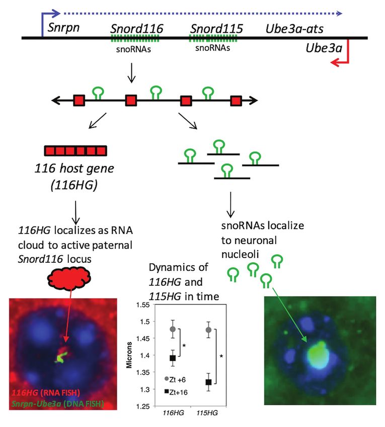
Mendiola and LaSalle                                                                                                       Epigenetics in Prader-Willi Syndrome

TABLE 1 | Clinical characteristics of nutritional phases.                             Abnormal sleep patterns have been well-established in PWS,
                                                                                      however, the molecular outcomes and downstream effects are
Phase 0        Decreased fetal movement and growth                In utero
               restriction
                                                                                      not well understood. In this article, we will review what is
                                                                                      known, delve into promising research findings, as well as discuss
               Infant becomes hypotonic and can develop                               some therapeutic strategies for PWS that are either encouraging
Phase 1a       failure to thrive                                ~0–9 months           or controversial.
               Infant begins to feed and grows steadily along
Phase 1b       a growth curve                                   ~9–25 months
               Weight increase, without significant change in   ~2–4 years of
Phase 2a       appetite or caloric intake                       age                   Molecular Genetics of PWS
               Continuous weight gain with increased food       ~4–8 years of         PWS is both a genetic and epigenetic disorder, mapping the
Phase 2b       interest                                         age                   imprinted chromosomal domain of 15q11.2-13.3. Common
               Development of hyperphagia, increased food
                                                                                      to all cases of PWS is the absence of an expressed paternal
Phase 3        seeking, and lack of satiety                     ~8 years of age
Phase 4        Loss of insatiable appetite and can feel full    Adulthood             copy of the SNORD116 locus. Due to parental imprinting of
                                                                                      the locus, outlined in more detail in the next section, loss
Miller et al. (2011) and Butler et al. (2019b).                                       of SNORD116 can occur through deletion, uniparental disomy,
                                                                                      or imprinting error. Most cases of PWS are caused by a large
meeting standard developmental milestones. In the more severe                         6 Mb deletion of the entire 15q11.2-q13.3 locus (Holm et al.,
cases of PWS, cranial and skeletal features are also apparent                         1993; Cassidy et al., 2012). Two major large deletion classes
(Kindler et al., 2015). Although development is altered and                           include those with breakpoints at BP1 vs. BP2 combined with
delayed at infancy, patients feeding normalizes resulting in a                        the downstream BP3 common deletion (Butler, 2020). However,
steady increase in weight. However, stage 2 of nutritional                            microdeletions of the imprinting control region upstream of
development persists through early childhood, characterized                           SNRPN (Figure 1) also result in loss of expression of SNORD116
by extreme fixation on food and development of hyperphagia                            due to loss of the promoter. Rare microdeletions that only
(Holm et al., 1993; Cassidy and Driscoll, 2009; Miller et al.,                        encompass SNORD116, but not SNRPN or SNORD115, have
2011). Stage 2 is divided into two phases in which phase 2a                           also been found in patients with PWS (Sahoo et al., 2008;
is an increase in weight that occurs without changes in appetite                      de Smith et al., 2009; Duker et al., 2010). Approximately
or feeding followed by phase 2b, characterized by fixation on                         60% of patients have paternal deletions, 36% are a result of
food leading to phase 3, hyperphagia. In PWS, hyperphagia                             maternal uniparental disomy, 4% are due to imprinting mutations
is developed at 2 years of age on average, and the severity of                        that lead to a maternal imprinting status, and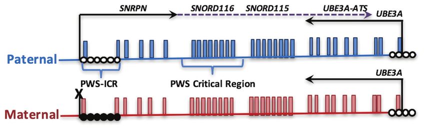
Mendiola and LaSalle                                                                                                                  Epigenetics in Prader-Willi Syndrome

detail, as well as the cluster of biallelically expressed GABAA                         SNORD115 cluster does not lead to the PWS phenotype in
receptor genes (GABRB3, GABRA5, and GABRG3), which are                                  humans (Runte et al., 2005). To date, the precise mechanisms
implicated in some of the neuropsychiatric phenotypes that                              of how Snord116 functions are critical for neurodevelopment
are more severe in the deletion PWS molecular subclass.                                 remain elusive, however, advancements in sequencing technology
   SNORD116 is processed through a long noncoding transcript                            have provided new insights and will be covered in more detail
that initiates at the imprinting control region upstream of                             in the section below. In addition to SNORD116, other genes
SNRPN, followed by two repeat clusters of small nucleolar                               in the 15q11.2-13.3 locus, including NECDIN, MAGEL2, and
RNAs (snoRNAs SNORD116 and SNORD115) and terminating                                    a cluster of GABA receptor genes are implicated in the phenotypes
at the UBE3A antisense transcript (Figure 1; Sutcliffe et al.,                          observed in most cases of PWS.
1994; Buiting et al., 1995; Runte et al., 2001; Landers et al.,                             NECDIN (NDN) is an imprinted gene that is paternally
2004; Vitali et al., 2010; Chamberlain, 2013). In humans,                               expressed and encodes for the protein NECDIN, which belongs
SNORD115, but not SNORD116 or UBE3A-ATS, is exclusively                                 to the melanoma antigen-encoding gene (MAGE) family of
expressed in neurons, while Snord116, Snord115, and Ube3a-ats                           proteins that are enriched in differentiated cells. NDN is one
are all neuron-specific transcripts in mouse. SNORD115 and                              of several protein coding genes deleted from the large 6 Mb
SNORD116 encompass clusters of repeated subunits of sequences                           chromosomal deletion observed in PWS patients and is implicated
encoding a C/D box snoRNAs embedded within intronic regions                             in neuronal maturation (Ren et al., 2003). Other than its role
of the noncoding exons encoding the snoRNA host transcript                              in cellular differentiation and neuronal maturation, NDN is
SNHG14 (Cavaillé et al., 2000; de los Santos et al., 2000;                              also involved in neurite and axonal growth, arborization,
Bortolin-Cavaillé and Cavaillé, 2012; Stanurova et al., 2018).                          migration, and fasciculation, which are important for normal
C/D box snoRNAs have known functions in regulating 2-O                                  neurological signaling and development (MacDonald and
methylation rRNA modifications by recruiting ribonucleoprotein                          Wevrick, 1997; Kuwajima et al., 2006; Davies et al., 2008; Miller
complexes including fibrillarin, which catalyzes methylation                            et al., 2009; Bervini and Herzog, 2013). Mouse models of Ndn
(Dupuis-Sandoval et al., 2015; Bratkovič et al., 2020).                                 deficiency have been instrumental for studying abnormal brain
   SnoRNAs are processed from introns of the SNORD116                                   development and cognitive impairments in PWS. However,
and SNORD115 within the SNHG14 host gene subunits, called                               studies using Ndn deficient mice did not exhibit any
as 116HG and 115HG (Figure 2; Cavaillé et al., 2000; Leung                              morphological differences in brain development, but led to
et al., 2009; Vitali et al., 2010). Unlike other C/D box snoRNAs,                       respiratory failure causing apneas and irregular breathing patterns
SNORD116 and SNORD115 are classified as “orphan snoRNAs”                                that are caused by increased activity in serotonin transporter
because their targets and functions are unknown (Bratkovič                              (SERT/slc6a4; Matarazzo et al., 2017). Furthermore, Ndn knockout
et al., 2020). Previous studies have shown that SNORD116                                mice exhibit a higher pain threshold due to a decrease in
localizes in the nucleolus and may participate in splicing and                          nerve growth factor sensory neurons (Kuwako et al., 2005).
RNA modifications (Bazeley et al., 2008; Leung et al., 2009).                           Respiratory failure and higher pain thresholds are also observed
In contrast, 116HG and 115HG localize in the form of RNA                                in patients with PWS (Rittinger, 2001; Butler et al., 2002;
“clouds” at the site of their own transcription in the nucleus                          Angulo et al., 2015). Specifically, irregularities in breathing
(Figure 2), and dynamically regulate many additional genes                              may be a large proponent to sleep abnormalities in PWS.
across the genome (Powell et al., 2013; Coulson et al., 2018b).                             MAGEL2 is another imprinted gene, that is, paternally
SNORD115 is also shown to be involved in the alternative                                expressed and encodes for the protein MAGEL2 that belongs
splicing specifically of the serotonin receptor 5-HT2C mRNA                             to the MAGE family of proteins. Truncated MAGEL2
(Bazeley et al., 2008; Raabe et al., 2019). Although, both loci                         mutations cause PWS-like phenotypes observed in patients
are potentially implicated in PWS, microdeletion of only the                            (Schaaf et al., 2013; Fountain and Schaaf, 2016), but these

  FIGURE 1 | Parental imprinting in the heart of the Prader-Willi syndrome (PWS) locus. The PWS on human chromosome 15q11.2-q13.3 is shown, depicting
  transcripts specifically expressed from the paternal (blue) or maternal (red) alleles. PWS patients with rare paternal microdeletions have defined the critical region
  over SNORD116. DNA methylation (closed circles) on the maternal allele of the PWS imprinting control region (PWS-ICR) silences the expression of SNRPN (solid
  arrow) and the long noncoding transcript expressed in neurons (dotted arrow) that encompasses repeated snoRNA clusters (including SNORD116 and SNORD115)
  and the antisense transcript to UBE3A (UBE3A-ATS). UBE3A encodes an E3 ubiquitin ligase protein that regulates protein turnover of multiple cytoplasmic and
  nuclear factors. Since the paternal UBE3A allele is silenced by the expression of UBE3A-ATS in neurons, deletion or mutation of the maternal copy of UBE3A
  causes Angelman syndrome.

Frontiers in Genetics | www.frontiersin.org                                         3                                           February 2021 | Volume 12 | Article 624581
Mendiola and LaSalle                                                                                                             Epigenetics in Prader-Willi Syndrome

  FIGURE 2 | PWS noncoding RNA summary. (Top panel) Individual components of the processed PWS snoRNA-lncRNA region between Snrpn and Ube3a. Within
  the Snord116 and Snord115 loci are repeated units of snoRNAs (green), lncRNA exons (red boxes), and introns with G-C skew. Processing results in spliced 116HG
  and 115HG lncRNAs that localize to their sites of transcription, the snoRNAs that localize to nucleoli, and R-loops that displace histones and promote locus
  chromatin decondensation. (Bottom left panel) Seen by RNA-FISH, 116HG forms a large RNA cloud (red) localized to the decondensed paternal allele (green) in
  nuclei (blue), associated with 2,403 genes enriched for metabolic function. 116HG and 115HG RNA clouds are significantly larger at diurnal time ZT6 (sleep) than
  ZT16 (wake), corresponding to gene dysregulation in Snord116+/− specifically at ZT6. (Bottom right panel) Processed Snord116 snoRNAs (green) localize to a
  single nucleolus in mature cortical neurons.

cases have been recently distinguished from PWS in a new                             clock proteins through ubiquitination (Mercer et al., 2009;
classification of Schaaf-Yang syndrome (SYS). SYS shares                             Tacer and Potts, 2017; Vanessa Carias et al., 2020).
phenotypic overlap with PWS, but also exhibit distinct                                  The 15q11-q13 PWS region also contains a cluster of three
behavioral and metabolic phenotypes including autism                                 genes encoding subunits of receptors for the neurotransmitter,
spectrum disorder (Fountain and Schaaf, 2016). In mouse                              GABAA. GABA is the major inhibitory neurotransmitter in
embryogenesis, Magel2 is highly expressed in non-neuronal                            the postnatal brain, so loss of these GABA receptors in the
(placenta, midgut turbucle, and midgut region) and neuronal                          large deletion cases of PWS is expected to be involved in
tissue types (dorsal root ganglia and peripheral neurons                             some of the phenotypes of PWS. 15q11.2-13.3 genes GABRB3,
surrounding limb and trunk muscles (Bervini and Herzog,                              GABRA5, and GABRG3 encode for β3, α5, and γ3 subunits,
2013). In adult mouse brain, Magel2 is highly enriched in                            respectively. GABAA receptors are assembled into hexameric
hypothalamic regions and extends to the superchiasmic                                protein complexes made up of combinations of a1–6, b1–3,
nucleus, specific regions that regulate feeding and circadian                        g1–3, and other subunits, with α5 containing receptors making
rhythms, respectively (Kozlov et al., 2007; Mercer et al.,                           up ~5% of GABAA receptors in human brain (Mohamad
2009). The prevalence of MAGEL2 in the hypothalamus                                  and Has, 2019). Unlike the imprinted genes in the PWS
initially identified it as strong candidate for the hyperphagia                      locus, these 15q11.2-13.3 GABAA receptor genes are biallelically
phenotype of PWS. However, SYS patients and mouse models                             expressed in the brain. However, monoallelic expression and
with MAGEL2 mutations show a lower prevalence of                                     decreased protein expression of each GABAA receptor subunits
overeating and obesity. Instead, it was determined that                              have been observed in autism postmortem brain (Samaco
MAGEL2 functions as a ubiquitin transporter that localizes                           et al., 2005; Hogart et al., 2007). Furthermore, both transcript
in SCN neurons and acts as a direct regulator of circadian                           and protein levels of GABRB3 were not correlated with copy

Frontiers in Genetics | www.frontiersin.org                                      4                                         February 2021 | Volume 12 | Article 624581
Mendiola and LaSalle                                                                                               Epigenetics in Prader-Willi Syndrome

number in an analysis of PWS, AS, and 15q11.2-13.3                          promise for possible epigenetic therapies that will be discussed
duplication syndrome postmortem brain (Scoles et al., 2011).                at the end of this review.
A recent study on phenotypes and gene expression patterns
in a Gabrb3 deletion mouse model is also consistent with
complex gene regulation, as neighboring Oca2 expression                     Epigenetics and Imprinting in PWS and
was reduced and ocular hypopigmentation observed                            Related Human Neurodevelopmental
(Delahanty et al., 2016). Dysregulated gene expression of                   Disorders
the 15q11.2-13.3 GABAA receptors is expected to have                        In addition to PWS, loss of imprinting is involved in related
consequences for the balance of inhibitory and excitatory                   neurodevelopmental disorders: Angelman (AS), 15q duplication
signals that regulate sleep, metabolism, and mood in PWS.                   (Dup15q), Kagami-Ogata (KOS14), and Temple (TS14) syndromes
Recently, it has been shown that levels of GABA metabolites                 (Schanen, 2006; Kagami et al., 2015; Briggs et al., 2016). Unlike
vary between different molecular subclasses of PWS                          the default state of biallelic expression, imprinted genes are
(Lucignani et al., 2004; Rice et al., 2016; Brancaccio et al.,              selectively silenced on either the maternal or paternal allele
2017). Since there are major targets for therapeutic                        by epigenetic differences including DNA methylation and
intervention in multiple neurodevelopmental disorders,                      repressive chromatin modifications. Imprinted genes are clustered
understanding their altered expression in PWS is expected                   in discrete chromosomal loci and are regulated by a central
to be important for the treatment of other neurodevelopmental               imprinting control region (ICR), such as the PWS-ICR, in
disorders (Braat and Kooy, 2015).                                           which methylation is diagnostic for AS, PWS, and Dup15q
                                                                            disorders (Figure 1). Some imprinted genes exhibit tissue-
                                                                            specific or developmental-specific imprinting patterns regulated
EPIGENETIC MECHANISMS IN PWS                                                by long noncoding RNAs. Furthermore, the largest conserved
                                                                            cluster of microRNA (miRNA) in the mammalian genome is
Epigenetic Regulation of the Imprinting                                     found within the KOS14 imprinted locus and is responsible
Control Region in PWS                                                       for regulating neuronal maturation and mTOR growth pathways
As mentioned in the previous section on molecular genetics,                 (Winter, 2015). Experimental evidence is emerging for regulatory
small deletions of the imprinting control region (PWS-ICR)                  cross-talk between different imprinted gene loci (Stelzer et al.,
are sufficient to cause PWS when inherited on the paternal                  2014; Jung and Nolta, 2016; Martinet et al., 2016; Vincent
allele. Interestingly, the ICR at 15q11.2-13.1 is actually bipartite,       et al., 2016; Lopez et al., 2017), but this emerging “imprinted
because maternal microdeletions of a region called as the                   gene network” hypothesis (Fauque et al., 2010; Haga and
AS-ICR are found in rare cases of Angelman syndrome (Buiting                Phinney, 2012; Monnier et al., 2013; Ribarska et al., 2014)
et al., 1995; Smith et al., 2011). Subsequent studies in a                  has been understudied in the context of the developing
variety of mammals have demonstrated that the AS-ICR                        nervous system.
contains alternate 5' noncoding exon for SNRPN that are                         RNA FISH has shown that 116HG localizes in the nucleus,
uniquely expressed in oocytes, but not sperm or other tissues               where it forms an RNA cloud that is absent in Snord116 deletion
(Smith et al., 2011; Lewis et al., 2015, 2019). It is the oocyte-           brain. 116HG was also found to colocalize with metabolic,
specific transcription that leads to methylation and                        circadian, and epigenetic gene loci including Mtor, Clock, Cry1/2,
transcriptional silencing of the maternal allele specifically on            Per1/2/3, Dnmt1/3b, Tet1/2/3, Mecp2, and others at ZT6, the
the maternal but not the paternal allele of the PWS-ICR. A                  time point with the largest effect of Snord116 deletion on
more recent study of individuals with AS imprinting mutations               transcription globally (Powell et al., 2013). Snord116’s involvement
have identified a more common haplotype that deletes a                      in transcriptional regulation, therefore, prompted an investigation
binding site for the transcription factor SOX2 (Beygo et al.,               of epigenetic differences that may explain the interaction of
2020). Together, these studies have demonstrated that this                  Snord116 with diurnal light cycles. Whole genome bisulfite
upstream region, defined as the AS-ICR, is critical for                     sequencing (WGBS) was performed on cortex samples from
establishing silencing of the maternal allele of the imprinted              wild-type (WT) and PWS mice sacrificed every 3 h starting
genes within the PWS locus.                                                 from Zt0–Zt16 and showed that Snord116 is involved in regulating
    In addition to being characterized by allele-specific DNA               a dynamic rhythm of diurnal methylation (Coulson et al., 2018b).
methylation, several additional epigenetic marks are                        Rhythmically methylated CpG dinucleotides were identified (
Mendiola and LaSalle Epigenetics in Prader-Willi Syndrome
TABLE 2 | Examples of Snord116 associated and impacted genes and predicted functions.
Snord116-dependent Snord116-dependent DNA methylation
Category Function Gene name Gene binding transcriptional changeb
to 116HGa changea
Mouse Human
Epigenetic Methyl binding protein critical to Mecp2 Yes Increased at Zt6 No No
neurodevelopment
Binds DNA:RNA hybrids Setx No Increased at Zt6 Yes Yes
DNA demethylases Tet1 No Increased at Zt6 Yes Yes
Tet2 No Increased at Zt6 Yes No
Tet3 No Increased at Zt6 Yes Yes
Histone deacetylases Hdac3 No Increased at Zt6 No No
Hdac4 No Increased at Zt6 Yes Yes
Hdac5 No Increased at Zt6 Yes Yes
DNA methyltransferases Dnmt1 No Increased at Zt6 No Yes
Dnmt3a Yes Increased at Zt6 No Yes
Circadian Establishes phases and periods Per2 No Increased at Zt6 No Yes
Per3 No Increased at Zt6 No No
Arntl Yes Increased at Zt6 Yes Yes
Metabolic Kinase involved in regulating cellular Mtor Yes Increased at Zt6 No Yes
energy homeostasis
Transcription Transcriptional regulator of E-box motif Neurod1 No Increased at Zt6 & Zt16 Yes No
containing genes
Full gene lists are included in Powell et al. (2013).
a
Full gene lists are included in Coulson et al. (2018b).
b
recognizing DNA methylation while other genes are important These environmental and metabolic inputs play an important
transcriptional regulators for development. Further integration role in the synchronization of the core circadian clock with
of promoter methylation and RNA-seq data revealed that genes the rhythmic patterns of many physiological and behavioral
being diurnally dysregulated were central to the body weight, processes in peripheral tissues (Wright et al., 2013; Legates
behavior, and metabolic phenotypes of PWS (Coulson et al., 2018b). et al., 2014; Mukherji et al., 2015; Blasiak et al., 2017). The
The Coulson et al. study also demonstrated a molecular genetically encoded circadian cycle and the environmentally
connection between the 116HG and the KOS14 locus, building regulated diurnal/nocturnal cycle are integrated by a complex
upon a prior study showing a connection between IPW (part regulatory feedback network, which acts at the chromatin,
of the 116HG transcript) and DLK1 regulation at the KOS14/ transcriptional, and translational levels to coordinate biological
TS14 locus in human neuronal culture (Stelzer et al., 2014). and environmental rhythms (Koike et al., 2012; Papazyan
In this case, DNA FISH was used to examine chromosome et al., 2016; Takahashi, 2017). In mammals, the core circadian
decondensation, a measurement of neuronal activation of the clock resides in the suprachiasmatic nucleus of the
paternal allele resulting from histone displacement, at both hypothalamus; however, almost half of all transcripts, both
PWS and TS14 loci in adult mouse brain at six different diurnal protein-coding and non-coding, exhibit diurnal rhythms in
time points. Interestingly, the TS14 locus only showed evidence one or more peripheral tissues (Yan et al., 2008; Zhang R.
of active chromatin decondensation in Snord116 deletion mouse et al., 2014). While most studies on circadian biology focus
cortex. Furthermore, chromatin decondensation at the PWS on the suprachiasmatic nucleus, investigations into diurnal
locus did occur in Snord116 deletion, but the timing was shifted rhythms of cerebral cortex are relevant to the cognitive deficits
from light to dark cycle, similar to the effects observed on in PWS and to energy expenditure. For instance, circadian
DNA methylation. Together, these results suggest that the and metabolic genes showed light-cycle-specific dysregulation
ancestrally older imprinted TS14/KOS14 locus may become in the Snord116del mouse model, corresponding to cyclical
more active as a compensatory mechanism to fill in for loss dynamics of Snord116 expression (Powell et al., 2013). Rhythmic
of Snord116, but this comes at a cost of proper timing of epigenetic dynamics within the cerebral cortex are less well
these epigenetic events. characterized; however, increasing evidence indicates a role
for DNA methylation in these rhythms. Approximately 6%
(25,476) of CpG sites assayed by 450k array are dynamically
Epigenetics and Imprinting of Mammalian regulated throughout diurnal and seasonal cycles in human
Imprinted Loci and the Emerging cortex (Lim et al., 2017). This epigenetic plasticity plays an
Importance in Circadian Rhythmicity and important role in circadian entrainment and the resiliency
Sleep of the circadian clock to changes in the diurnal environment
Daily and seasonal cycles of light, temperature, and feeding (Stevenson and Prendergast, 2013; Azzi et al., 2014;
govern energy and activity of organisms from all branches of life. Lim et al., 2014).
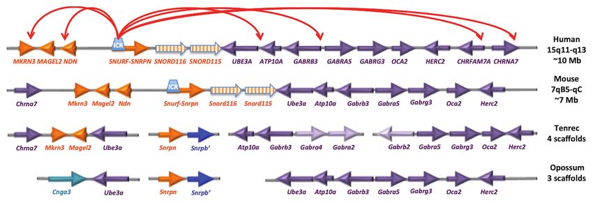
Frontiers in Genetics | www.frontiersin.org 6 February 2021 | Volume 12 | Article 624581Mendiola and LaSalle Epigenetics in Prader-Willi Syndrome The 14q32.2 imprinted locus bears striking similarity to chromosomes (Rapkins et al., 2006; Yasui et al., 2011; the PWS locus, as it encodes the only other repetitive cluster Zhang Y. J. et al., 2014). Humans (and chimps) have 22 of snoRNAs in the mammalian genome (SNORD113 and SNORD116 and 44 SNORD115 copies, while mouse has 27 SNORD114), which are maternally expressed and exhibit detectable Snord116 and 130 Snord115 copies. Potentially allele-specific chromatin decondensation in neurons, similar relevant for the PWS phenotype, tenrecs have a unique to SNORD116 and SNORD115 (Cavaillé et al., 2002; Tierling metabolic and sleep structure among mammals adapted to et al., 2006; Leung et al., 2009). TS and KOS are reciprocally long periods of reduced activity and body temperature called imprinted disorders, with TS caused by maternal uniparental torpor (Lovegrove and Génin, 2008; Lovegrove et al., 2014a,b). disomy 14 [UPD(14)mat], and KOS caused by paternal Non-REM sleep and periods of torpor are thought to uniparental disomy 14 [UPD(14)pat]. Loss of paternal gene be ancestrally adaptive to conserve energy and escape expression at this locus in TS, results in aberrantly high predation. Eutherian mammals have distinct adaptations for expression of maternal non-coding RNAs, including SNORD113 daily sleep and activity patterns based on diet, body size, and SNORD114, whereas KOS results from the loss of maternally and brain size (Siegel, 2005; Gerhart-Hines and Lazar, 2015). expressed, non-coding RNAs and the upregulation of paternally Unlike the PWS/AS locus, the chromosomal arrangement expressed DLK1. Interestingly, TS phenocopies PWS suggesting of the SNORD113/SNORD114 cluster at the KOS/TS locus that these two imprinted loci may perform similar functions is similar in monotremes, marsupials, and placental mammals, and share common pathways (Temple et al., 1991; Hosoki and the miRNAs at this cluster are evolutionarily stable et al., 2009; Kagami et al., 2015). The loss of Snord116 in (Zhang Y. J. et al., 2014). Both imprinted snoRNA loci PWS increases gene expression in the TS locus, indicating exhibit neuron-specific chromatin decondensation (Leung that the two loci may interact through a cross-regulatory et al., 2009) and also show evidence for diurnally expressed network. In support of this hypothesis, IPW from the PWS transcripts, many of which are also dysregulated in Snord116del locus has been shown to regulate the TS locus in an induced mice (Powell et al., 2013; Coulson et al., 2018b). Interestingly, pluripotent stem cell line of PWS (Stelzer et al., 2014). Though circadian rhythmicity of the Dlk1/Dio3 (Labialle et al., both PWS and TS loci show circadian oscillations, the 2008a,b) and Magel2 (Kozlov et al., 2007; Devos et al., 2011; mechanism of this regulation and the impact of circadian Tennese and Wevrick, 2011) loci and cross-regulation between rhythms on their cross-regulation suggests that a balance PWS/AS and DLK1 loci have been described previously between the two loci is critical for sleep and metabolism (Stelzer et al., 2014) but are poorly understood at a (Labialle et al., 2008a,b; Powell et al., 2013). mechanistic level. Most imprinted loci, such as IGF2, PEG1/MEST, and Despite its function being fully known, loss of Snord116 IGR2R, are imprinted in marsupials as well as eutherian in PWS mouse models has been demonstrated to dysregulate (placental) mammals (Figure 3). In contrast, Snrpn and sleep, feeding, and temperature cycles (Lassi et al., 2016a,b). Ube3a are not imprinted in marsupials and are on distinct These studies have demonstrated the importance of chromosomes (Rapkins et al., 2006). Interestingly, the ancestral hypothalamic Snord116 expression on temporally regulated eutherian mammal tenrec (Echinops telfairi) lacks the Snord116 behavior. Interestingly, Snord116 deficient mice exhibited and Snord115 genes and Snrpn and Ube3a are on separate disrupted feeding cues induced by erratic behavior due to FIGURE 3 | The PWS/AS imprinted locus has emerged recently within placental mammals. The gene orientation and linear organization is shown for human and mouse, as well as the earliest placental mammal (tenrec) and marsupial (opossum). The red arrows on top represent results from neuronal 4C analysis of chromatin looping (Yasui et al., 2011). Interestingly, the tenrec arrangement of Chrna7-Mkrn3-Magel2-Ube3a (spanning ~500 kb) is similar to the human 4C long-range interactions spanning ~10 Mb, despite the lack of evidence for Snrpn or Snord clusters at the locus. Humans (and chimps) have 22 SNORD116 and 44 SNORD115 copies, mouse has 27 detectable Snord116 and 130 Snord115 copies. Frontiers in Genetics | www.frontiersin.org 7 February 2021 | Volume 12 | Article 624581
Mendiola and LaSalle                                                                                          Epigenetics in Prader-Willi Syndrome

increased activity prompted by foraging. Reminiscent of                 contribute to the development of therapeutic interventions
humans with PWS, Snord116 deficient mice exhibited a                    that target sleep and metabolism which are critical
strong fixation on food and high food intake irrespective               to development.
of weight gain (Lassi et al., 2016a). Furthermore, Snord116
deficient mice also exhibited a prolonged REM phase that
was uncoupled with normal circadian patterning (Lassi et al.,           PWS Mouse Models for Preclinical Testing
2016b). Together, these studies have demonstrated the                   of Therapeutic Interventions
importance of Snord116 on temporally regulated behaviors                Mouse models of Snord116 deficiency that recapitulate some
including sleep, feeding, foraging, and temperature regulation          features of PWS have been created as useful models for testing
that are consistent with the recent evolutionary selection              possible therapeutic interventions. Like in humans, Snord116
of the imprinted PWS locus in mammalian-specific                        is a maternally imprinted gene in mouse and localizes to a
diurnal cycles.                                                         syntenic loci chromosome 7qC. The first generation of mouse
   Multiple studies have also explored the role of Snord116             models generated were designed with large deletions mimicking
in hypothalamic regulation of hormones linked to diurnal                those observed in humans with PWS (Yang et al., 1998). These
behaviors. Orexin neurons in the hypothalamus facilitate sleep-         mouse models exhibited extreme hypotonia and failure to thrive,
wake cycles by regulating hormones that promote wakefulness             leading to death 1 week after birth. The high lethality rate
(noradrenaline, histamine, and acetylcholine) and rest [melanine-       was caused by the loss of protein coding genes Snrpn and
concentrating hormone (MCH); Pace et al., 2020]. Loss of                Ube3a-ats, which are hypothesized to be important to alternative
orexin neurons are widely implicated in dysregulated sleep in           splicing (Tsai et al., 1999; Bressler et al., 2001; Bervini and
patients with narcolepsy and has been observed in patients              Herzog, 2013). As in humans with PWS, these mouse models
with PWS as well (Vgontzas et al., 1996; Chemelli et al., 1999;         exhibited a dysregulation of major endocrine hormones including
Mignot et al., 2002; Omokawa et al., 2016). However, it was             growth hormone, glucose, and insulin, which are necessary
not until recently that loss of Snord116 was demonstrated to            for cellular homeostasis and proliferation. Disruption of each
decrease orexin neuron levels in the lateral hypothalamus               hormone lead to metabolic dysregulation which results in
without altering levels of MCH and MCH neurons in mice                  extreme hypotonia that leads to the failure to thrive.
(Pace et al., 2020). A decrease in orexin neurons may facilitate           Today, the most commonly used PWS mouse models were
the prolonged REM sleep characteristic of PWS due to the                originally generated by two separate labs using cre-mediated
imbalance in orexin/MCH ratio with a higher MCH concentration           deletion of Snord116 (Skryabin et al., 2007; Ding et al., 2008).
during wake cycles promoting more rest (Pace et al., 2020).             These mouse models were designed by a targeted insertion
This phenomenon is not unique to Snord116 deletion mice,                of loxP cassettes flanking the Snord116 (Ding et al., 2008)
however, as this orexin/MCH imbalance was also observed in              cluster or Snord116 and IPW (Skryabin et al., 2007) through
Magel2 deficient models (Kozlov et al., 2007).                          homologous recombination in embryonic stem (ES) cells
   Patients with PWS are characterized as having reduced                derived from male blastocytes. The 2-loxP ES cells were then
levels of growth hormone, but elevated levels of ghrelin                injected into C57Bl/6J mice that gave birth to male mice
(Tauber et al., 2019). Ghrelin is the endogenous ligand of              with a 2-loxP (+/−) genotype. These mice were mated with
growth hormone secretagogue receptor 1a. Ghrelin is peptide             a transgenic strain expressing Cre recombinase under an ovary
produced by the gut with a diversity of physiological effects,          specific promoter producing 1-loxP mice with a Snord116
including appetite stimulation and lipid accumulation.                  (+/−) genotype (Ding et al., 2008). For ES cells targeted
Subsequent studies have demonstrated that it is actually the            with loxP cassettes flanking Snord116 and IPW, CRE
acylated form of ghrelin that is elevated in PWS children               recombinase were expressed then injected into blastocytes to
and young adults, while nonacylated ghrelin levels are                  produce PWScre(+/−) (Skryabin et al., 2007). These mouse
indistinguishable from controls (Kuppens et al., 2015).                 models have a 150 kb deletion of the Snord116 cluster or a
However, while both growth hormone and ghrelin are known                deletion that encompasses Snord116 and IPW. Like previous
to have clear diurnal patterns of secretion, with nocturnal             models, both mice develop hypotonia and failure to thrive
levels being higher than daytime levels in humans, there                with low to no post-natal lethality. Although these mouse
have been a surprising lack of investigation into the possibility       models do not consistently exhibit the hyperphagia phenotype,
growth hormone abnormalities in PWS may be due to altered               they do exhibit a significant deficiency in cognition and energy
diurnal rhythms (Kyung et al., 2004; Stawerska et al., 2020).           expenditure (Powell et al., 2013; Adhikari et al., 2019) making
   Despite orexin neurons being a critical cell type for                these phenotypes useful in preclinical therapeutic strategies.
Snord116 regulation on hormonal regulation from the                     Furthermore, development of 2-loxp(−/+) and PWScre(+/−)
hypothalamus, loss of Snord116 in other brain regions, such             mice enabled the generation of several new mouse models
as cerebral cortex, also appear to contribute to the proper             that are able to recapitulate the hyperphagia phenotype in
expression of core circadian clock regulators such as Per               adult mice through Cre-mediated and tamoxifen induced
and Bmal genes (Powell et al., 2013; Coulson et al., 2018a).            Snord116 deletion in the hypothalamus (Qi et al., 2016; Purtell
These findings reinforce the role of Snord116 in establishing           et al., 2017; Polex-Wolf et al., 2018) and identified the disrupted
multiple aspects of circadian rhythms that are lost upon                REM sleep phenotypes (Lassi et al., 2016b), respectively.
deletion. Studying Snord116 and identifying its targets can             Previous studies have shown that Snord116 expression in the

Frontiers in Genetics | www.frontiersin.org                         8                                   February 2021 | Volume 12 | Article 624581
Mendiola and LaSalle                                                                                            Epigenetics in Prader-Willi Syndrome

hypothalamus is developmentally regulated and is enriched                 Epigenetic Therapies
postnatally at weaning and early adulthood (Zhang et al.,                 In contrast to gene therapy, epigenetic therapy for PWS has
2012), implicating its involvement in regulating metabolism               a stronger potential for clinical relevance, since PWS is an
and circadian rhythms.                                                    inherently epigenetic disorder. The general strategy for
                                                                          epigenetic strategies for PWS involves de-repressing the maternal
                                                                          silent PWS-ICR to activate SNRPN and Snord116 transcription
Genetic Therapies                                                         (Crunkhorn, 2017; Chung et al., 2020). Recent successes using
While most genetic diseases are amenable to genetic                       high throughput screening of small molecule libraries identified
complementation and standard gene therapy design and delivery,            several inhibitors of EHMT2/G9a, a histone 3 lysine 9
there are unique challenges to gene therapy in PWS because                methyltransferase, that were capable of reactivating the
of the epigenetic and molecular complexities of the SNORD116              expression of paternally expressed SNRPN and SNORD116
locus. In the original characterization of a Snord116 deletion            from the maternal chromosome, both in cultured PWS cell
mouse model of PWS, it was mentioned that a transgene                     lines and in a PWS mouse model (Kim et al., 2017, 2019).
containing a single snoRNA from Snord116 was insufficient                 Similarly, inhibitor of SETDB1 using shRNA knockdown
to rescue the metabolic phenotypes (Ding et al., 2008). Since             resulted in partial reactivation of SNORD116 and 116HG in
it remained possible that the limitations of using either a single        PWS-derived iPSC cell lines and neurons (Cruvinel et al.,
copy and/or an already processed snoRNA were the reason                   2014). The main differences in the epigenetic changes resulting
for the lack of complementation, a new transgenic mouse was               between these two epigenetic therapies was that EHMT2/
created and reported by our group using the Snord(+/−) model              G9a did not alter DNA methylation at the PWS-ICR, while
(Coulson et al., 2018a). This Snord116 transgene contained                SETDB1 did not show a change in H3K9me3 at the PWS-ICR.
the complete subunits of 116HG exons, introns, and snoRNAs                Potentially more completely, the inactivation of ZNF274 using
repeated in a total of 27 copies was expressed broadly at the             CRISPR/Cas9 in PWS-derived iPSC lines resulted in reactivation
transcript level in all tissues, but was only spliced and processed       of both SNRPN and SNORD116 as well as a reduction of
into snoRNAs in brain. The neuron-specific splicing was attributed        H3K9me3 at the PWS-ICR (Langouët et al., 2020). Together,
to the splicing factor RBFOX3, which is also known as the                 these studies suggest that combinations of targeted epigenetic
neuron-specific marker NeuN. In wild-type neurons, the extra              strategies for unsilencing maternal SNORD116 hold promise
copies of Snord116 contributed to the nucleolar accumulation              for future treatments of PWS.
of processed snoRNAs as well as the size of the 116HG RNA
cloud. However, in the Snord116 deletion PWS model, the
Snord116 transgene did not become processed or localized to
these locations, indicating that an active allele was needed for
                                                                          POTENTIAL DEVELOPMENTS:
correct processing and localization. In addition, the body weight         RELEVANCE OF SNORD116-MEDIATED
phenotype of the Snord116 mice was similar to that of the                 EPIGENETIC MECHANISMS TOWARD
Snord116 deletion mouse, and there was no complementation                 COMMON HUMAN DISEASES
of this phenotype in the cross.
    In another study, a mouse model was generated with a                  While this review has focused on the relevance of epigenetic
5’HPRT-LoxP-NeoR insertion upstream of the maternally                     regulation of and by SNORD116 and other genes within the
imprinted Snord116 using the PWScre(+/−) model                            locus to the pathogenesis of PWS, we expect that understanding
(Rozhdestvensky et al., 2016). The cassette insertion did not             the interactions between imprinted genes and metabolism at
affect the PWS imprinting center methylation status, but                  this locus will have relevance to other more common metabolic
disrupted the imprinting effect enabling expression of Snord116           and neuropsychiatric human disorders. Because of the
from the maternal allele, a result that was not observed in               hypothalamic network alterations in PWS associated with satiety
WT and KO mice without the cassette. Like the Coulson                     and food reward systems, this locus is considered to be a model
et al. study in 2018, Snord116 was expressed across all tissue            for understanding food addictions as well as other addictive
types, but in this case, the body weight phenotype was rescued            behaviors (Salles et al., 2020). The molecular mechanisms leading
in KO mice with the cassette insertion. The differences in                hyperphagia and overeating in PWS could be informative for
results may depend on the imprinting mechanism of the                     understanding the intersections of epigenetics, diurnal rhythms,
PWS region as well as the genomic location of Snord116.                   and metabolism in more common causes of overweight and
For instance, when the Snord116 transgene is introduced                   obesity. Food addictions in PWS may be similar in mechanisms
outside of the imprinted region, as would be the case for                 to those establishing other addictions. Interestingly, “morphine
most gene therapy strategies, the ability to complement the               addiction” and “circadian entrainment” were among the gene
missing paternal allele is expected to be challenging. These              pathway terms identified by the unbiased search for gene promoters
results demonstrate the complexities of this locus and suggest            that showed both rhythmic demethylation and increased expression
that gene therapy for PWS using conventional complementation              during sleep in Snord116 deletion mice (Coulson et al., 2018a),
strategies will be problematic. Despite the issues, these results         suggesting that further characterization of these pathways could
also highlight the importance of targeting imprinting regulation          be relevant to improved treatments for opioid use disorders. In
for therapeutic interventions.                                            addition, there are emerging links between circadian disruptions

Frontiers in Genetics | www.frontiersin.org                           9                                   February 2021 | Volume 12 | Article 624581
Mendiola and LaSalle Epigenetics in Prader-Willi Syndrome
and the exacerbation of psychiatric disorders such as bipolar AUTHOR CONTRIBUTIONS
disorder and depression. Chronotherapy involving sleep deprivation
followed by the re-entrainment of diurnal cycles has shown Both authors contributed to the literature review, writing, and
effectiveness in treating these common mood disorders editing of the manuscript. All authors contributed to the article
(Gottlieb et al., 2019; D’Agostino et al., 2020). and approved the submitted version.
In conclusion, the PWS locus epigenetically regulated
SNORD116 transcripts that have evolved to become parentally ACKNOWLEDGMENTS
imprinted within mammals, in turn serve to regulate a large
number of additional genes through the genome that are related We are grateful for the support of research on epigenetics in
to circadian rhythms, metabolic and nutritional cycles, and Prader-Willi syndrome from the NIH/NICHD (R01HD098038)
brain functions. Future studies designed to better understand and the Foundation for Prader-Willi Research. We would like
the genomic impacts of SNORD116 regulation is expected to to thank the home department of Medical Microbiology and
have far-reaching impacts beyond the scope of PWS. Immunology for covering open access fees for this publication.
REFERENCES of the 14q32 imprinted DLK1/MEG3 region. Am. J. Med. Genet. A 170A,
170–175. doi: 10.1002/ajmg.a.37400
Adhikari, A., Copping, N. A., Onaga, B., Pride, M. C., Coulson, R. L., Yang, M., Buiting, K., Saitoh, S., Gross, S., Dittrich, B., Schwartz, S., Nicholls, R. D.,
et al. (2019). Cognitive deficits in the Snord116 deletion mouse model for et al. (1995). Inherited microdeletions in the Angelman and Prader-Willi
Prader-Willi syndrome. Neurobiol. Learn. Mem. 165:106874. doi: 10.1016/j. syndromes define an imprinting centre on human chromosome 15. Nat.
nlm.2018.05.011 Genet. 9, 395–400. doi: 10.1038/ng0495-395
Angulo, M. A., Butler, M. G., and Cataletto, M. E. (2015). Prader-Willi syndrome: Butler, M. G. (2020). Imprinting disorders in humans: a review. Curr. Opin.
a review of clinical, genetic, and endocrine findings. J. Endocrinol. Investig. Pediatr. 32, 719–729. doi: 10.1097/MOP.0000000000000965
38, 1249–1263. doi: 10.1007/s40618-015-0312-9 Butler, M. G., Bittel, D. C., Kibiryeva, N., Talebizadeh, Z., and Thompson, T.
Azzi, A., Dallmann, R., Casserly, A., Rehrauer, H., Patrignani, A., Maier, B., (2004). Behavioral differences among subjects with Prader-Willi syndrome
et al. (2014). Circadian behavior is light-reprogrammed by plastic DNA and type I or type II deletion and maternal disomy. Pediatrics 113, 565–573.
methylation. Nat. Neurosci. 17, 377–382. doi: 10.1038/nn.3651 doi: 10.1542/peds.113.3.565
Bazeley, P. S., Shepelev, V., Talebizadeh, Z., Butler, M. G., Fedorova, L., Filatov, V., Butler, M. G., Hartin, S. N., Hossain, W. A., Manzardo, A. M., Kimonis, V.,
et al. (2008). SnoTARGET shows that human orphan SnoRNA targets locate Dykens, E., et al. (2019a). Molecular genetic classification in Prader-Willi
close to alternative splice junctions. Gene 408, 172–179. doi: 10.1016/j. syndrome: a multisite cohort study. J. Med. Genet. 56, 149–153. doi: 10.1136/
gene.2007.10.037 jmedgenet-2018-105301
Bervini, S., and Herzog, H. (2013). Mouse models of Prader-Willi syndrome: Butler, M. G., Miller, J. L., and Forster, J. L. (2019b). Prader-Willi syndrome—
a systematic review. Front. Neuroendocrinol. 34, 107–119. doi: 10.1016/j. clinical genetics, diagnosis and treatment approaches: an update. Curr. Pediatr.
yfrne.2013.01.002 Rev. 15, 207–244. doi: 10.2174/1573396315666190716120925
Beygo, J., Grosser, C., Kaya, S., Mertel, C., Buiting, K., and Horsthemke, B. Butler, J. V., Whittington, J. E., Holland, A. J., Boer, H., Clarke, D., and Webb, T.
(2020). Common genetic variation in the Angelman syndrome imprinting (2002). Prevalence of, and risk factors for, physical ill-health in people with
centre affects the imprinting of chromosome 15. Eur. J. Hum. Genet. 28, Prader-Willi syndrome: a population-based study. Dev. Med. Child Neurol.
835–839. doi: 10.1038/s41431-020-0595-y 44, 248–255. doi: 10.1017/S001216220100202X
Bieth, E., Eddiry, S., Gaston, V., Lorenzini, F., Buffet, A., Auriol, F. C., et al. Cassidy, S. B., and Driscoll, D. J. (2009). Prader-Willi syndrome. Eur. J. Hum.
(2015). Highly restricted deletion of the SNORD116 region is implicated Genet. 17, 3–13. doi: 10.1038/ejhg.2008.165
in Prader-Willi syndrome. Eur. J. Hum. Genet. 23, 252–255. doi: 10.1038/ Cassidy, S. B., Schwartz, S., Miller, J. L., and Driscoll, D. J. (2012). Prader-Willi
ejhg.2014.103 syndrome. Genet. Med. 14, 10–26. doi: 10.1038/gim.0b013e31822bead0
Blasiak, A., Gundlach, A. L., Hess, G., and Lewandowski, M. H. (2017). Cavaillé, J., Buiting, K., Kiefmann, M., Lalande, M., Brannan, C. I., Horsthemke, B.,
Interactions of circadian rhythmicity, stress and orexigenic neuropeptide et al. (2000). Identification of brain-specific and imprinted small nucleolar
systems: implications for food intake control. Front. Neurosci. 11:127. doi: RNA genes exhibiting an unusual genomic organization. Proc. Natl. Acad.
10.3389/fnins.2017.00127 Sci. U. S. A. 97, 14311–14316. doi: 10.1073/pnas.250426397
Bortolin-Cavaillé, M. L., and Cavaillé, J. (2012). The SNORD115 (H/MBII-52) Cavaillé, J., Seitz, H., Paulsen, M., Ferguson-Smith, A. C., and Bachellerie, J. P.
and SNORD116 (H/MBII-85) gene clusters at the imprinted Prader-Willi (2002). Identification of tandemly-repeated C/D SnoRNA genes at the
locus generate canonical box C/D SnoRNAs. Nucleic Acids Res. 40, 6800–6807. imprinted human 14q32 domain reminiscent of those at the Prader-Willi/
doi: 10.1093/nar/gks321 Angelman syndrome region. Hum. Mol. Genet. 11, 1527–1538. doi: 10.1093/
Braat, S., and Kooy, R. F. (2015). The GABAA receptor as a therapeutic target hmg/11.13.1527
for neurodevelopmental disorders. Neuron 86, 1119–1130. doi: 10.1016/j. Chamberlain, S. J. (2013). RNAs of the human chromosome 15q11-Q13 imprinted
neuron.2015.03.042 region. Wiley Interdiscip. Rev. RNA 4, 155–166. doi: 10.1002/wrna.1150
Brancaccio, M., Patton, A. P., Chesham, J. E., Maywood, E. S., and Hastings, M. H. Chemelli, R. M., Willie, J. T., Sinton, C. M., Elmquist, J. K., Scammell, T.,
(2017). Astrocytes control circadian timekeeping in the suprachiasmatic Lee, C., et al. (1999). Narcolepsy in orexin knockout mice: molecular genetics
nucleus via glutamatergic signaling. Neuron 93, 1420–1435. doi: 10.1016/j. of sleep regulation. Cell 98, 437–451. doi: 10.1016/S0092-8674(00)81973-X
neuron.2017.02.030 Chung, M. S., Langouët, M., Chamberlain, S. J., and Carmichael, G. G. (2020).
Bratkovič, T., Bozič, J., and Rogelj, B. (2020). Functional diversity of small Prader-Willi syndrome: reflections on seminal studies and future therapies.
nucleolar RNAs. Nucleic Acids Res. 48, 1627–1651. doi: 10.1093/nar/gkz1140 Open Biol. 10:200195. doi: 10.1098/rsob.200195
Bressler, J., Tsai, T. F., Wu, M. Y., Tsai, S. F., Ramirez, M. A., Armstrong, D., Coulson, R. L., Powell, W. T., Yasui, D. H., Dileep, G., Resnick, J., and
et al. (2001). The SNRPN promoter is not required for genomic imprinting LaSalle, J. M. (2018a). Prader-Willi locus Snord116 RNA processing requires
of the Prader-Willi/Angelman domain in mice. Nat. Genet. 28, 232–240. an active endogenous allele and neuron-specific splicing by Rbfox3/NeuN.
doi: 10.1038/90067 Hum. Mol. Genet. 27, 4051–4060. doi: 10.1093/hmg/ddy296
Briggs, T. A., Lokulo-Sodipe, K., Chandler, K. E., Mackay, D. J. G., and Karen Coulson, R. L., Yasui, D. H., Dunaway, K. W., Laufer, B. I., Ciernia, A. V.,
Temple, I. (2016). Temple syndrome as a result of isolated hypomethylation Zhu, Y., et al. (2018b). Snord116-dependent diurnal rhythm of DNA
Frontiers in Genetics | www.frontiersin.org 10 February 2021 | Volume 12 | Article 624581Mendiola and LaSalle Epigenetics in Prader-Willi Syndrome methylation in mouse cortex. Nat. Commun. 9:1616. doi: 10.1038/s41467- Holm, V. A., Cassidy, S. B., Butler, M. G., Hanchett, J. M., Greenswag, L. R., 018-03676-0 Whitman, B. Y., et al. (1993). Prader-Willi syndrome: consensus diagnostic Crunkhorn, S. (2017). Steps towards epigenetic therapy for PWS. Nat. Rev. criteria. Pediatrics 91, 398–402. Drug Discov. 16:85. doi: 10.1038/nrd.2017.3 Hosoki, K., Kagami, M., Tanaka, T., Kubota, M., Kurosawa, K., Kato, M., et al. Cruvinel, E., Budinetz, T., Germain, N., Chamberlain, S., Lalande, M., and (2009). Maternal uniparental disomy 14 syndrome demonstrates Prader-Willi Martins-Taylor, K. (2014). Reactivation of maternal SNORD116 cluster via syndrome-like phenotype. J. Pediatr. 155, 900–903. doi: 10.1016/j.jpeds. SETDB1 knockdown in Prader-Willi syndrome IPSCs. Hum. Mol. Genet. 2009.06.045 23, 4674–4685. doi: 10.1093/hmg/ddu187 Jung, Y., and Nolta, J. A. (2016). BMI1 regulation of self-renewal and multipotency D’Agostino, A., Ferrara, P., Terzoni, S., Ostinelli, E. G., Carrara, C., Prunas, C., in human mesenchymal stem cells. Curr. Stem Cell Res. Ther. 11, 131–140. et al. (2020). Efficacy of triple chronotherapy in unipolar and bipolar doi: 10.2174/1574888X1102160107171432 depression: a systematic review of the available evidence. J. Affect. Disord. Kagami, M., Kurosawa, K., Miyazaki, O., Ishino, F., Matsuoka, K., and Ogata, T. 276, 297–304. doi: 10.1016/j.jad.2020.07.026 (2015). Comprehensive clinical studies in 34 patients with molecularly defined Davies, W., Lynn, P. M. Y., Relkovic, D., and Wilkinson, L. S. (2008). Imprinted UPD(14)pat and related conditions (Kagami-Ogata syndrome). Eur. J. Hum. genes and neuroendocrine function. Front. Neuroendocrinol. 29, 413–427. Genet. 23, 1488–1498. doi: 10.1038/ejhg.2015.13 doi: 10.1016/j.yfrne.2007.12.001 Kim, Y., Lee, H. M., Xiong, Y., Sciaky, N., Hulbert, S. W., Cao, X., et al. de los Santos, T., Schweizer, J., Rees, C. A., and Francke, U. (2000). Small (2017). Targeting the histone methyltransferase G9a activates imprinted genes evolutionarily conserved RNA, resembling C/D box small nucleolar RNA, and improves survival of a mouse model of Prader-Willi syndrome. Nat. is transcribed from PWCR1, a novel imprinted gene in the Prader-Willi Med. 23, 213–222. doi: 10.1038/nm.4257 deletion region, which is highly expressed in brain. Am. J. Hum. Genet. Kim, S. J., Miller, J. L., Kuipers, P. J., German, J. R., Beaudet, A. L., Sahoo, T., 67, 1067–1082. doi: 10.1086/303106 et al. (2012). Unique and atypical deletions in Prader-Willi syndrome de Smith, A. J., Purmann, C., Walters, R. G., Ellis, R. J., Holder, S. E., reveal distinct phenotypes. Eur. J. Hum. Genet. 20, 283–290. doi: 10.1038/ Van Haelst, M. M., et al. (2009). A deletion of the HBII-85 class of ejhg.2011.187 small nucleolar RNAs (SnoRNAs) is associated with hyperphagia, obesity Kim, Y., Wang, S. E., and Jiang, Y. -H. (2019). Epigenetic therapy of Prader- and hypogonadism. Hum. Mol. Genet. 18, 3257–3265. doi: 10.1093/hmg/ Willi syndrome. Transl. Res. 205, 105–118. doi: 10.1016/j.trsl.2019.02.012 ddp263 Kindler, J. M., Lewis, R. D., and Hamrick, M. W. (2015). Skeletal muscle and Delahanty, R. J., Zhang, Y., Bichell, T. J., Shen, W., Verdier, K., Macdonald, R. L., pediatric bone development. Curr. Opin. Endocrinol. Diabetes Obes. 22, et al. (2016). Beyond epilepsy and autism: disruption of GABRB3 causes ocular 467–474. doi: 10.1097/MED.0000000000000201 hypopigmentation. Cell Rep. 17, 3115–3124. doi: 10.1016/j.celrep.2016.11.067 Koike, N., Yoo, S. H., Huang, H. C., Kumar, V., Lee, C., Kim, T. K., et al. Devos, J., Weselake, S. V., and Wevrick, R. (2011). Magel2, a Prader-Willi (2012). Transcriptional architecture and chromatin landscape of the core syndrome candidate gene, modulates the activities of circadian rhythm circadian clock in mammals. Science 338, 349–354. doi: 10.1126/ proteins in cultured cells. J. Circadian Rhythms 9:12. doi: 10.1186/1740- science.1226339 3391-9-12 Kozlov, S. V., Bogenpohl, J. W., Howell, M. P., Wevrick, R., Panda, S., Ding, F., Li, H. H., Zhang, S., Solomon, N. M., Camper, S. A., Cohen, P., Hogenesch, J. B., et al. (2007). The imprinted gene Magel2 regulates normal et al. (2008). SnoRNA Snord116 (Pwcr1/MBll-85) deletion causes growth circadian output. Nat. Genet. 39, 1266–1272. doi: 10.1038/ng2114 deficiency and hyperphagia in mice. PLoS One 3:1709. doi: 10.1371/journal. Kuppens, R. J., Diène, G., Bakker, N. E., Molinas, C., Faye, S., Nicolino, M., pone.0001709 et al. (2015). Elevated ratio of acylated to unacylated ghrelin in children Duker, A. L., Ballif, B. C., Bawle, E. V., Person, R. E., Mahadevan, S., Alliman, S., and young adults with Prader-Willi syndrome. Endocrine 50, 633–642. doi: et al. (2010). Paternally inherited microdeletion at 15q11.2 confirms a 10.1007/s12020-015-0614-x significant role for the SNORD116 C/D box SnoRNA cluster in Prader-Willi Kuwajima, T., Nishimura, I., and Yoshikawa, K. (2006). Necdin promotes syndrome. Eur. J. Hum. Genet. 18, 1196–1201. doi: 10.1038/ejhg.2010.102 GABAergic neuron differentiation in cooperation with Dlx homeodomain Dupuis-Sandoval, F., Poirier, M., and Scott, M. S. (2015). The emerging landscape proteins. J. Neurosci. 26, 5383–5392. doi: 10.1523/JNEUROSCI.1262-06.2006 of small nucleolar RNAs in cell biology. Wiley Interdiscip. Rev. RNA 6, Kuwako, K. I., Hosokawa, A., Nishimura, I., Uetsuki, T., Yamada, M., Nada, S., 381–397. doi: 10.1002/wrna.1284 et al. (2005). Disruption of the paternal necdin gene diminishes TrkA Fauque, P., Ripoche, M. A., Tost, J., Journot, L., Gabory, A., Busato, F., et al. signaling for sensory neuron survival. J. Neurosci. 25, 7090–7099. doi: 10.1523/ (2010). Modulation of imprinted gene network in placenta results in normal JNEUROSCI.2083-05.2005 development of in vitro manipulated mouse embryos. Hum. Mol. Genet. Kyung, H. P., Jin, D. K., Sang, Y. S., Ji, E. L., Si, H. K., Seng, M. S., et al. 19, 1779–1790. doi: 10.1093/hmg/ddq059 (2004). Correlation between fasting plasma ghrelin levels and age, body Fountain, M., and Schaaf, C. (2016). Prader-Willi syndrome and Schaaf-Yang mass index (BMI), BMI percentiles, and 24-hour plasma ghrelin profiles syndrome: neurodevelopmental diseases intersecting at the MAGEL2 gene. in Prader-Willi syndrome. J. Clin. Endocrinol. Metab. 89, 3885–3889. doi: Diseases 4:2. doi: 10.3390/diseases4010002 10.1210/jc.2003-032137 Gerhart-Hines, Z., and Lazar, M. A. (2015). Circadian metabolism in the light Labialle, S., Croteau, S., Bélanger, V., McMurray, E. N., Ruan, X., Moussette, S., of evolution. Endocr. Rev. 36, 289–304. doi: 10.1210/er.2015-1007 et al. (2008a). Novel imprinted transcripts from the Dlk1-Gtl2 intergenic Gottlieb, J. F., Benedetti, F., Geoffroy, P. A., Henriksen, T. E. G., Lam, R. W., region, mico1 and mico1os, show circadian oscillations. Epigenetics 3, Murray, G., et al. (2019). The chronotherapeutic treatment of bipolar disorders: 322–329. doi: 10.4161/epi.3.6.7109 a systematic review and practice recommendations from the ISBD task Labialle, S., Yang, L., Ruan, X., Villemain, A., Schmidt, J. V., Hernandez, A., force on chronotherapy and chronobiology. Bipolar Disord. 8, 741–773. doi: et al. (2008b). Coordinated diurnal regulation of genes from the Dlk1-Dio3 10.1111/bdi.12847 imprinted domain: implications for regulation of clusters of non-paralogous Haga, C. L., and Phinney, D. G. (2012). MicroRNAs in the imprinted DLK1- genes. Hum. Mol. Genet. 17, 15–26. doi: 10.1093/hmg/ddm281 DIO3 region repress the epithelial-to-mesenchymal transition by targeting Landers, M., Bancescu, D. L., Le Meur, E., Rougeulle, C., Glatt-Deeley, H., the TWIST1 protein signaling network. J. Biol. Chem. 287, 42695–42707. Brannan, C., et al. (2004). Regulation of the large (∼1000 kb) imprinted doi: 10.1074/jbc.M112.387761 murine Ube3a antisense transcript by alternative exons upstream of Snurf/ Hartley, S. L., MacLean, W. E., Butler, M. G., Zarcone, J., and Thompson, T. Snrpn. Nucleic Acids Res. 32, 3480–3492. doi: 10.1093/nar/gkh670 (2005). Maladaptive behaviors and risk factors among the genetic subtypes Langouët, M., Gorka, D., Orniacki, C., Dupont-Thibert, C. M., Chung, M. S., of Prader-Willi syndrome. Am. J. Med. Genet. 136 A, 140–145. doi: 10.1002/ Glatt-Deeley, H. R., et al. (2020). Specific ZNF274 binding interference at ajmg.a.30771 SNORD116 activates the maternal transcripts in Prader-Willi syndrome Hogart, A., Nagarajan, R. P., Patzel, K. A., Yasui, D. H., and LaSalle, J. M. neurons. Hum. Mol. Genet. 29, 3285–3295. doi: 10.1093/hmg/ddaa210 (2007). 15q11-13 GABAA receptor genes are normally biallelically expressed Lassi, G., Maggi, S., Balzani, E., Cosentini, I., Garcia-Garcia, C., and Tucci, V. in brain yet are subject to epigenetic dysregulation in autism-spectrum (2016a). Working-for-food behaviors: a preclinical study in Prader-Willi disorders. Hum. Mol. Genet. 16, 691–703. doi: 10.1093/hmg/ddm014 mutant mice. Genetics 204, 1129–1138. doi: 10.1534/genetics.116.192286 Frontiers in Genetics | www.frontiersin.org 11 February 2021 | Volume 12 | Article 624581
You can also read