Genetic Imaging of Neuroinflammation in Parkinson's Disease: Recent Advancements - Frontiers
←
→
Page content transcription
If your browser does not render page correctly, please read the page content below
REVIEW
                                                                                                                                              published: 15 July 2021
                                                                                                                                      doi: 10.3389/fcell.2021.655819

                                            Genetic Imaging of
                                            Neuroinflammation in Parkinson’s
                                            Disease: Recent Advancements
                                            Longping Yao 1* , Jiayu Wu 1 , Sumeyye Koc 2 and Guohui Lu 1*
                                            1
                                             Department of Neurosurgery, The First Affiliated Hospital of Nanchang University, Nanchang, China, 2 Department
                                            of Neuroscience, Institute of Health Sciences, Ondokuz Mayıs University, Samsun, Turkey

                                            Parkinson’s disease (PD) is one of the most prevalent neurodegenerative aging
                                            disorders characterized by motor and non-motor symptoms due to the selective loss
                                            of midbrain dopaminergic (DA) neurons. The decreased viability of DA neurons slowly
                                            results in the appearance of motor symptoms such as rigidity, bradykinesia, resting
                                            tremor, and postural instability. These symptoms largely depend on DA nigrostriatal
                                            denervation. Pharmacological and surgical interventions are the main treatment for
                                            improving clinical symptoms, but it has not been possible to cure PD. Furthermore, the
                         Edited by:
                 Chencheng Zhang,
                                            cause of neurodegeneration remains unclear. One of the possible neurodegeneration
 Shanghai Jiao Tong University, China       mechanisms is a chronic inflammation of the central nervous system, which is mediated
                       Reviewed by:         by microglial cells. Impaired or dead DA neurons can directly lead to microglia activation,
               Sachchida Nand Rai,
                                            producing a large number of reactive oxygen species and pro-inflammatory cytokines.
        University of Allahabad, India
                        Huajian Chen,       These cytotoxic factors contribute to the apoptosis and death of DA neurons, and the
   Southern Medical University, China       pathological process of neuroinflammation aggravates the primary morbid process and
                  *Correspondence:          exacerbates ongoing neurodegeneration. Therefore, anti-inflammatory treatment exerts
                        Longping Yao
                 loupe_yao@163.com
                                            a robust neuroprotective effect in a mouse model of PD. Since discovering the first
                           Guohui Lu        mutation in the α-synuclein gene (SNCA), which can cause disease-causing, PD has
                  guohui-lu@163.com
                                            involved many genes and loci such as LRRK2, Parkin, SNCA, and PINK1. In this article,
                    Specialty section:      we summarize the critical descriptions of the genetic factors involved in PD’s occurrence
         This article was submitted to      and development (such as LRRK2, SNCA, Parkin, PINK1, and inflammasome), and
                   Molecular Medicine,
                                            these factors play a crucial role in neuroinflammation. Regulation of these signaling
                a section of the journal
  Frontiers in Cell and Developmental       pathways and molecular factors related to these genetic factors can vastly improve
                                Biology     the neuroinflammation of PD.
         Received: 19 January 2021
           Accepted: 14 June 2021           Keywords: Parkinson’s disease, microglia, genetics, neuroinflamamation, dopaminergic neurons, neurotoxins
           Published: 15 July 2021
                              Citation:
        Yao L, Wu J, Koc S and Lu G
                                           INTRODUCTION
               (2021) Genetic Imaging
  of Neuroinflammation in Parkinson’s
                                           Parkinson’s disease (PD) is an age-related neurodegenerative disease characterized by motor and
     Disease: Recent Advancements.         non-motor symptoms (Brundin et al., 2018). It is the second most common neurodegenerative
      Front. Cell Dev. Biol. 9:655819.     disorder after Alzheimer’s disease, and now, it has become a significant public health problem
      doi: 10.3389/fcell.2021.655819       worldwide (Fan, 2020). One characteristic pathological change is the loss of midbrain dopaminergic

Frontiers in Cell and Developmental Biology | www.frontiersin.org                 1                                              July 2021 | Volume 9 | Article 655819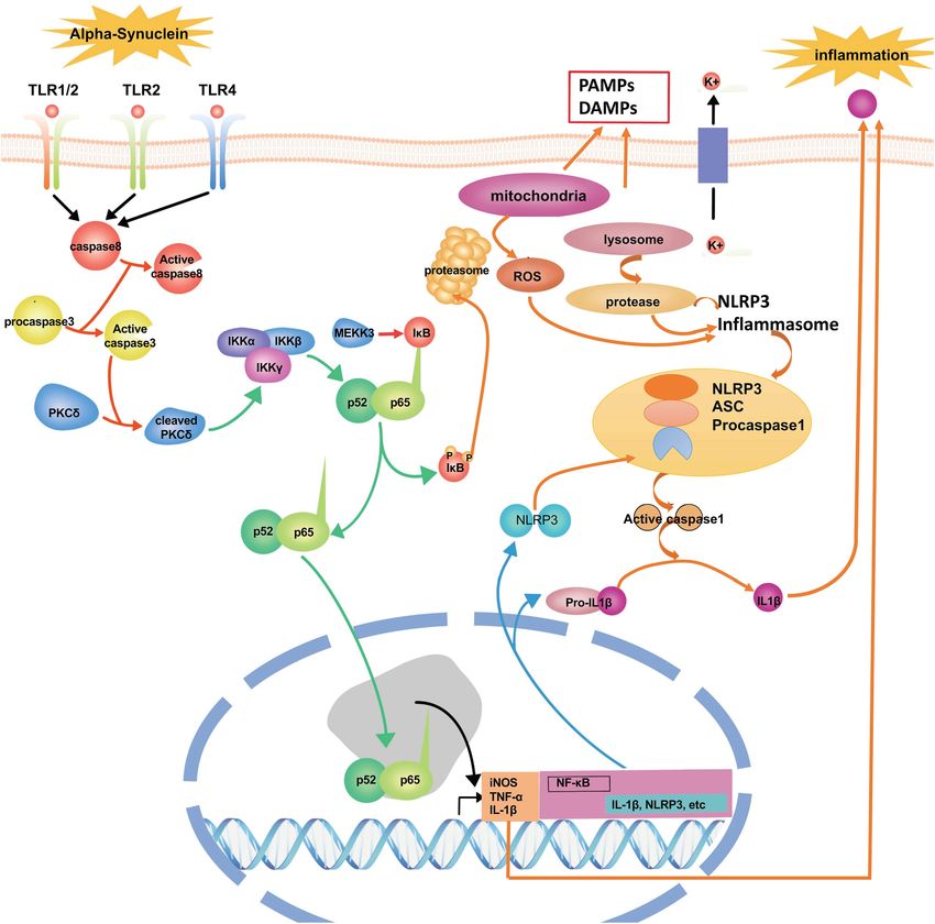
Yao et al.                                                                                                               Neuroinflammation in Parkinson’s Disease

(DA) neurons (Charvin et al., 2018). Parkinson’s disease is usually                     of the nigrostriatal DA, which includes apoptosis or death
characterized by the deposition of protein aggregates containing                        caused by neuroinflammation (Zhang et al., 2018). These factors
α-synuclein (Lewy bodies) in multiple brain regions (Rey et al.,                        are potential targets to interfere with the disease process in
2018). The decreased viability of DA neurons slowly results in                          PD. This review discusses the details of genetic imaging of
the appearance of motor symptoms, which largely depend on                               neuroinflammation in PD.
dopaminergic nigrostriatal denervation. With the progression of
neurodegeneration and advancing disease, people with PD also
experience sleep disturbances, fatigue, altered mood, cognitive                         INFLAMMATION AND PARKINSON’S
changes, autonomic dysfunction, and pain (Schapira et al., 2017).                       DISEASE
These “non-motor” symptoms dominate the clinical picture and
are the main determinants of quality of life. Besides, some                             The presence of activated microglial cells in the substantia
endophenotypes dominated by non-motor symptoms also have                                nigra has been shown in postmortem studies and in the
been reported in recent studies (Ehgoetz Martens and Shine,                             1-methyl-4-pheny-1, 2, 3, 6-tetrahydropyridine (MPTP)-animal
2018). Nowadays, pharmacological and surgical interventions are                         models (both mice and non-human primates) of PD (Zella
the main treatments for improving clinical symptoms. However,                           et al., 2019). Microglia are the macrophages that reside in
preventing or curing the disease is not possible at present                             the central nervous system (CNS) and are the brain’s primary
(Sarrafchi et al., 2016).                                                               innate immune effector cells (Yin et al., 2017). Besides, they
    Although various possible pathogenetic mechanisms have                              are the main generator of inflammatory cytokines and reactive
been proposed in recent years, the cause of neurodegeneration                           oxygen species (ROS), acting as the central active immune
remains unknown. One possible mechanism is inflammation                                 defense under normal conditions (Takeda et al., 2018). In
mediated by microglial cells (Yao et al., 2018). Emerging                               pathological conditions, microglia are activated and releases anti-
evidence indicates that sustained inflammatory stimulation plays                        inflammatory cytokines and neurotrophic factors, contributing
a vital role in the degeneration of DA neurons and is a                                 to tissue repair and the protection of neurons against apoptosis
common feature in both human PD patients and animal models                              or death (Hilla et al., 2017). However, when pathological
of PD (Ikeda-Matsuo et al., 2019). The neuroinflammatory                                factors are continuously present, the number of noxious
response may also lead to a cascade of events leading to                                phenotypes of microglia increase and release a large amount of
neuronal degeneration. Anti-inflammatory treatment showed                               ROS, proteinases, and inflammatory cytokines contributing to
beneficial effects in preventing neurodegeneration mediated by                          neuronal damage. In PD, the inflammatory mediators such as
inflammatory damage (Singh et al., 2020). For example, ursolic                          tumor-α (TNF-α), interleukin-1β (IL-1β), inducible nitric oxide
acid acted as a therapeutic drug targeted for protecting from                           synthase (iNOS), and interleukin-6 (IL-6) have been found to
neuroinflammation-induced neurodegeneration (Rai et al., 2019;                          regulate the progression of PD (Yao et al., 2019b). These pro-
Singh et al., 2020; Zahra et al., 2020). Besides, the supplement                        inflammatory mediators have been found to increase significantly
of tyrosine hydroxylase in MPTP-intoxicated mice could protect                          in the midbrain of PD patients and animal models. Furthermore,
dopaminergic neurons by suppressing neuroinflammation (Birla                            numerous studies have shown that impaired or dead DA neurons
et al., 2019). Thus, regulating neuroinflammation is a therapeutic                      can directly induce the activation of microglia, increasing the
strategy by reducing oxidative stress (Rai et al., 2017; Yadav et al.,                  production of ROS and pro-inflammatory cytokines. Therefore,
2017; Singh et al., 2018). In recent years, research has shown                          as mentioned above, the activation of microglia and DA neuronal
that many genetic factors are associated with neurodegeneration                         damage form a self-propelled degeneration cycle in PD; thus,
                                                                                        microglia are more likely to play critical roles in establishing and
Abbreviations: 6-mer, Six monosaccharides; 6-OHDA, 6-hydroxydopamine;                   maintaining inflammatory responses in PD (Figure 1).
AIM2, Absent in melanoma 2; ATP, Adenosine triphosphate; BHB, β-hydroxy
butyrate; c-Abl, c-Abelson murine leukemia viral oncogene homolog; CNS,
                                                                                            The anti-inflammatory treatment has been found to exert
Central nervous system; COX2, Cyclooxygenase 2; DA, Dopaminergic;                       a strong neuroprotective effect in a mouse model of PD. For
DAMPs, damage associated proteins; EAE, Experimental autoimmune                         example, specially designed liposomes targeted for the CD163
encephalomyelitis; FAK, Focal adhesion kinase; GFAP, Glial fibrillary acidic            receptor were loaded with glucocorticoids involved in PD-
protein; IL-1β, Interleukin-1β; IL-6, Interleukin-6; iNOS, Inducible nitric oxide
synthase; IRFs, Interferon regulatory factors; JAK/STAT, Janus kinase/signal            like neurodegeneration. The results showed that glucocorticoids
transducers and activators of transcription; LPC, Lysophosphatidylcholine;              protect DA neurons in the 6-hydroxydopamine (6-OHDA)
LPS, Lipopolysaccharide; LRRK2, Leucine-rich repeat kinase 2; MAPK-NF-κB,               PD model via anti-inflammatory modulation (Tentillier et al.,
p38 mitogen-activated protein kinase-NF-κB; MPTP, 1-methyl-4-pheny-1, 2, 3,
6-tetrahydropyridine; MSU, Monosodium urate; NADPH, Nicotinamide adenine
                                                                                        2016). APSE-Aq (fraction isolated from A. pyrifolium seeds)
dinucleotide phosphate; NFA, Nuclear factor of activated T cell; NF-κB, Nuclear         may have antioxidant and anti-inflammatory properties, which
factor-kappa B; NLRC4, Nod-like receptor family CARD domain containing                  may offer neuroprotection in a model of PD (de Araujo et al.,
protein 4; NO, Nitric oxide; Nrf2/HO-1, Nuclear factor erythroid 2-related              2018). Tetramethylpyrazine exhibits its anti-apoptotic, anti-
factor 2/heme oxygenase-1; PAMPs, Pathogen specific proteins; PD, Parkinson’s
disease; p-ERK, Phosphorylated extracellular signal-regulated kinase; PINK1,            inflammatory, and antioxidant actions via attenuating rotenone-
PTEN-induced putative kinase 1; PKA, Protein kinase A; PPAR-γ, Peroxisome               induced upregulation of the transcription factor nuclear factor
proliferator-activated receptor γ; PRGF, Plasma rich in growth factors; PRRs,           erythroid 2-related factor 2/heme oxygenase-1 (Nrf2/HO-1)
Pattern recognition receptors; ROS, Reactive oxygen species; ROT, LPS-primed
                                                                                        pathway and inflammation markers: nuclear factor-kappa B (NF-
rotenone; SNCA, α-synuclein gene; SNpc, Substantia nigra pars compacta; TNF-α,
Tumor necrosis factor-α; TrxR, Thioredoxin reductase; UBL, Ubiquitin-like;              κB), iNOS, cyclooxygenase 2 (COX2), and glial fibrillary acidic
α-syn, Alpha-synuclein.                                                                 protein (GFAP) expression (Lu et al., 2014; Michel et al., 2017).

Frontiers in Cell and Developmental Biology | www.frontiersin.org                   2                                        July 2021 | Volume 9 | Article 655819
Yao et al.                                                                                                                     Neuroinflammation in Parkinson’s Disease

  FIGURE 1 | A self-propelled degeneration cycle in PD. In PD’s pathological conditions, microglia are activated and release anti-inflammatory cytokines to repair the
  tissues, protecting neurons against apoptosis or death. However, the continuous stimulation of pathological factors increases the number of toxic phenotypes of
  microglia, releasing a large number of inflammatory cytokines, such as TNF-α, IL-1β, iNOS, IL-6, and ROS, which contribute to neuronal damage. Besides, the
  impaired or dead DA neurons can directly induce microglial activation, increasing ROS and pro-inflammatory cytokines. Thus, the activation of microglia and DA
  neuronal damage form a self-propelled degeneration cycle in PD. PD, Parkinson’s disease; DA, dopaminergic; ROS, reactive oxygen species; iNOS, inducible nitric
  oxide synthase; IL-6, interleukin-6; TNF-α, tumor necrosis factor-α; IL-1β, interleukin-1β.

The isothiocyanate is mainly found in Brassica vegetables                               to the inhibition of glial activation and the subsequent
(Brassicaceae) and Moringaceae plants. It shows potent anti-                            generation of pro-inflammatory factors via inhibiting the
inflammatory activity in the treatment of murine subacute                               p38 mitogen-activated protein kinase-NF-κB (MAPK-NF-κB)
PD and promises compounds against oxidative stress and                                  pathway (Leonoudakis et al., 2017). A novel compound (VSC2)
neuroinflammation (Sita et al., 2016; Giacoppo et al., 2017).                           has anti-inflammatory and antioxidant properties in microglia
The neuroprotective properties of securinine may be due                                 and in an animal model of PD by preventing NF-κB activation

Frontiers in Cell and Developmental Biology | www.frontiersin.org                   3                                               July 2021 | Volume 9 | Article 655819
Yao et al.                                                                                                Neuroinflammation in Parkinson’s Disease

and the production of TNF-α, iNOS, IL-1β, NO, and COX-2                   c-Rel, NF-κB1/p50, and NF-κB2/p52 (Sokolova and Naumann,
(Lee et al., 2015). Morin effectively prevents MPTP-induced PD-           2017). The transcription factors can form homodimers and
like pathologies in mice and protects primary neurons against             heterodimers to regulate the expression of genes. The abundant
MPP + −induced toxicity by ameliorating oxidative stress and              basal expression of NF-κB in the brain is much higher than in
inflammation (Zhang et al., 2010; Lee et al., 2016). The anti-            peripheral tissues (Shih et al., 2015). NF-κB is largely involved
inflammatory effect of β-Hydroxybutyric acid on microglia is              in the progression of PD. For example, it reveals that NF-
mediated by G-protein-coupled receptor 109A. This process                 κB is activated in an MPTP model of PD (Dehmer et al.,
involves the NF-κB signaling pathway in both in vivo and                  2004), following the microglia activation (Aoki et al., 2009). One
in vitro PD models, causing the inhibition of the production              research study showed that NF-κB increased more than 70-fold
of pro-inflammatory enzymes (iNOS and COX-2) and pro-                     in the brain tissue of PD patients and exhibited strong nuclear
inflammatory cytokines (TNF-α, IL-1β, and IL-6) (Fu et al.,               p65 immunoreactivity of DA neurons in the substantia nigra
2015). The synthesis and biological evaluation of clovamide               (Mattson and Camandola, 2001). The activated NF-κB leading to
analogs show that they have potent anti-neuroinflammatory                 DA neuron degeneration has been demonstrated in PD (Phani
effects by reducing the expression of GFAP in a model of                  et al., 2012). Selective inhibition NF-kB has been found to protect
PD (Hu et al., 2018). Compound 21 is obtained from 2,4,6-                 against DA neurons’ death from MPTP toxicity in a PD model
trimethoxybenzaldehyde by adding amines or alkyl (methyl or               (Bassani et al., 2015).
ethyl) ester of the amino acid hydrochloride salts. Compound                  In neuroinflammation, NF-κB can regulate the production of
21 may be a potential candidate for PD treatment because of               pro-inflammatory cytokines, such as IL-6, TNF-α, G-CSF, iNOS,
its potent anti-neuroinflammatory activity, novel mechanism,              and IL-1β. The sustained inflammatory stimulus is related to
impressive penetration of the blood-brain barrier, and low                the NF-κB pathway in the activation of uncontrolled microglia,
toxicity in vitro and vivo (Wang et al., 2016). The behavioral            resulting in ROS production, neurotoxic factors, interferon-
and neurochemical alterations in a rat model of PD are partially          γ (INF-γ), and glutamate, whose excessive formation induces
reversed by Spirulina platensis, which is primarily related to its        neuronal damage (Spencer et al., 2012). Toll-like receptors are
anti-inflammatory effects (Lima et al., 2017). Phytic acid shows          a vital class of membrane proteins that can activate microglial
a solid neuroprotective effect in an MPTP-induced PD model                cells—the predominant pathways that TLRs trigger are associated
correlated with its anti-inflammatory effect by suppressing the           with the NF-κB pathway (Kawai and Akira, 2007). Inhibition of
NF-κB and phosphorylated extracellular signal-regulated kinase            the NF-κB pathway can sufficiently suppress the activation of
(p-ERK) pathways (Lv et al., 2015).                                       microglia and neuroinflammation.
    Based on the hypothesis that neuroinflammation is involved                Recent studies show that NF-κB is involved in the
in the pathophysiology of PD, scientists have evaluated                   inflammatory response of microglia in the progression of PD.
the feasibility of using non-steroidal anti-inflammatory drugs            Therefore, the regulation of the abnormal expression of NF-κB
(NSAIDs) to cure PD patients. Interestingly, the results of a             exerts fortissimo neuroprotection and inhibition of inflammation
prospective cohort study showed that NSAIDs might delay                   in PD. For example, our previous study found that miR-124 can
or prevent the onset of PD (Chen et al., 2003). Ibuprofen                 prevent DA neuronal death and suppress microglia activation
users had a lower risk of PD than non-users, suggesting that              via suppressing the MEKK3/NF-κB pathway in a mouse model
ibuprofen use may delay or prevent PD onset (Poly et al., 2019).          of PD (Yao et al., 2018). Intranasal plasma rich in growth factors
However, the same anti-inflammatory effect was not observed               (PRGF)-Endoret provides a novel neuroprotective strategy for
for aspirin, other NSAIDs, or acetaminophen in PD patients.               DA and attenuates NF-κB-dependent inflammation processes in
The inconsistent results between ibuprofen and other NSAIDs               a PD model (Anitua et al., 2015). Inhibition of NF-kB activation
indicate that ibuprofen has specific protective properties of the         leads to the suppression of pro-inflammatory molecules,
anti-inflammatory response in PD.                                         improvement in locomotor activity, and DA neurons’ protection
                                                                          in the substantia nigra pars compacta (SNpc) (Mondal et al.,
                                                                          2012). In DA neuron-glial co-cultures, pioglitazone prevents DA
THE ROLE OF NF-κB IN                                                      cells’ death from lipopolysaccharide (LPS)-induced exacerbation
NEUROINFLAMMATION IN PD                                                   of microglia activation by interfering with the NF-κB pathway
                                                                          (Esposito et al., 2007). Besides, schisandrol A could enhance the
Transcription factors such as NF-κB, STAT1, STAT3, and SMAD7              PI3K/AKT pathway and inhibit the IKK/IκBα/NF-κB pathway
are the proteins that can bind to a specific sequence of DNA              to reduce neuronal inflammation, oxidative stress and enhance
to regulate the transcription of genes. Recent literature shows           the survival of DA neurons in the brains of PD mice (Yan et al.,
that the upregulation of transcription factors can cause microglial       2019). Rosmarinic acid could attenuate inflammatory responses
activation, leading to a self-sustaining neuroinflammation                by suppressing the HMGB1/TLR4/NF-κB signaling pathways,
environment via various mechanisms in PD. In this article, we             which may contribute to its anti-PD activity (Lv et al., 2019).
will describe how NF-κB is associated with different pathways and         Cordycepin mitigates MPTP-induced inflammatory response
molecular regulation. For now, we will give a clear description of        in PD by inhibiting the TLR/NF-κB signaling pathway (Cheng
the role of NF-κB in neuroinflammation in PD.                             and Zhu, 2019). The knockdown of cathepsin D can protect
   NF-κB is crucial for neuroinflammation responses; NF-κB                dopaminergic neurons from neuroinflammation-mediated
is a group of transcription factors including RelA, RelB,                 neurotoxicity via inhibition of the NF-κB signaling pathway in

Frontiers in Cell and Developmental Biology | www.frontiersin.org     4                                       July 2021 | Volume 9 | Article 655819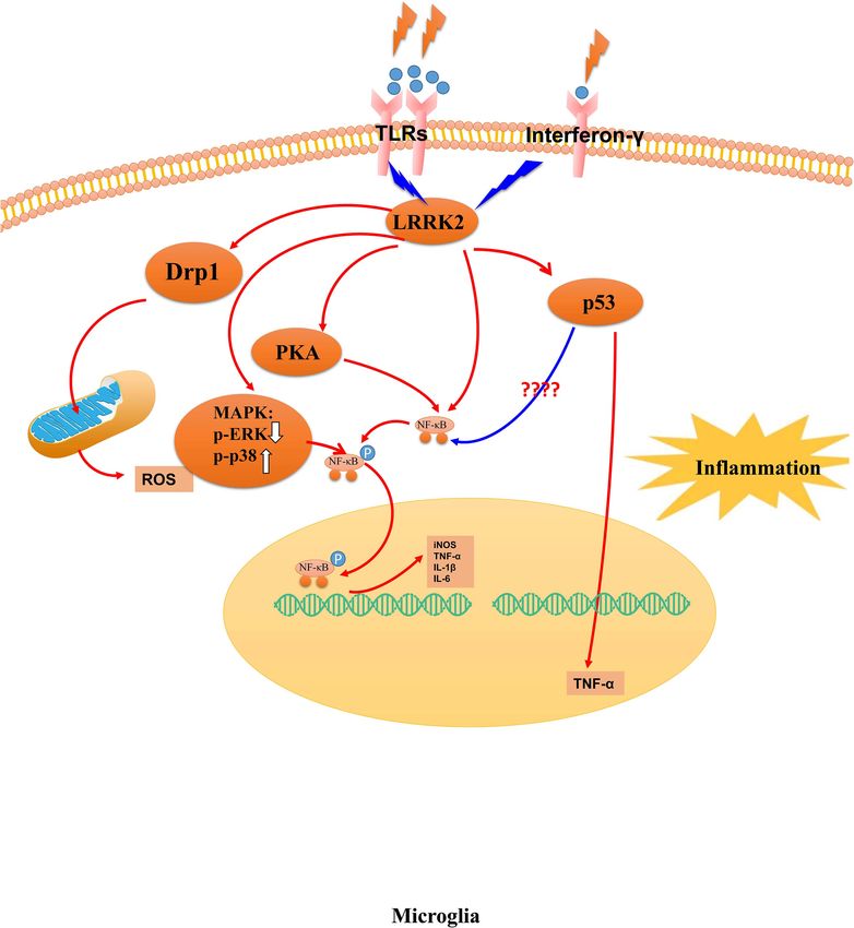
Yao et al.                                                                                                          Neuroinflammation in Parkinson’s Disease

a PD model (Gan et al., 2018). Polydatin treatment protects               TABLE 1 | The genes associated with the pathogenesis of PD.
DA neurons and ameliorates motor dysfunction by inhibiting
                                                                          Gene        Full name                        Locus     Location
microglial activation and the release of pro-inflammatory
mediators via regulation of the AKT/GSK3β-Nrf2/NF-κB                      SNCA        Synuclein alpha                  PARK1     4q22.1 Azizi and Azizi
signaling axis (Huang et al., 2018).                                                                                             (2018)
   Meanwhile, several NSAIDs, such as sodium salicylate,                  Parkin      Parkin RBR E3 ubiquitin          PARK2     6q26 Morato Torres et al.
                                                                                      protein ligase                             (2020)
celecoxib, aspirin, and diclofenac, have been found to exert
                                                                          PARK3       Parkinson disease 3              PARK3     2p13 Kachidian and
a neuroprotective role by decreasing NF-kB expression in
                                                                                                                                 Gubellini (2021)
an MPTP-induced model of PD (Bassani et al., 2015).                       SNCA        Synuclein alpha                  PARK4     4q22 Forero et al. (2020)
The peroxisome proliferator-activated receptor γ (PPAR-γ)                 UCHL1       Ubiquitin C-terminal             PARK5     4p13 Kachidian and
agonist pioglitazone mediates microglial activation and NF-                           hydrolase L1                               Gubellini (2021)
κB expression in the 6-hydroxydopamine model of PD (Goes                  PINK1       PTEN induced putative            PARK6     1p36 Li D. et al. (2020)
et al., 2018). A20 enzyme, which inhibits NF-κB by restricting                        kinase 1
the duration and intensity of its action, has been found to               PARK7       Parkinsonism associated          PARK7     1p36.23 Li Y. et al. (2020)
significantly decrease in a blood sample of patients with PD                          deglycase
(Mazo et al., 2017). The topics which are discussed next are widely       LRRK2       Leucine rich repeat kinase 2     PARK8     12q12 Li D. et al. (2020)
associated with NF-κB.                                                    ATPase      ATPase 13A2                      PARK9     1p36.13 Gago et al. (2020)
                                                                          13A2
                                                                          PARK10      Parkinson disease 10             PARK10    1p32 Kachidian and
                                                                                                                                 Gubellini (2021)
THE ROLE OF GENETICS IN THE
                                                                          GIGYF2      GRB10 interacting GYF            PARK11    2q37.1 Calatayud et al.
NEUROINFLAMMATION OF PD                                                               protein                                    (2017)
                                                                          PARK12      Parkinson disease 12             PARK12    Xq21-q25 Zanon (2020)
Since discovering the first mutation in the α-synuclein gene              HTRA2       HtrA serine peptidase 2          PARK13    2p13.1 Zanon (2020)
(SNCA) can cause disease-causing, PD has involved many genes              PLA2G6      Phospholipase A2 group VI        PARK14    22q13.1 Sudira et al. (2018)
and loci. For example, the deficiencies of genes such as LRRK2,           FBXO7       F-box protein 7                  PARK15    22q12.3 Yuan et al. (2017)
Parkin, SNCA, and PINK1 are risk factors for PD (including                PARK16      Parkinson disease 16             PARK16    1q32 Han et al. (2019)
family and sporadic PD). The genetic discoveries clearly illustrate       VPS35       VPS35, retromer complex          PARK17    16q11.2 Cutillo et al. (2020)
the cellular pathways and functions that are involved in the                          component
development of PD. To date, at least 23 loci and 19 genes (Table 1)       EIF4G1      Eukaryotic translation           PARK18    3q27.1 Li D. et al. (2020)
have been identified and designated as both 10 autosomal                              initiation factor 4 gamma 1
dominant and 9 autosomal recessive PD.                                    DNAJC6      DnaJ heat shock protein          PARK19    1p31.3 Velez-Pardo and
    Recent epidemiological and genetic studies have indicated                         family (Hsp40) member C6                   Jimenez-Del-Rio (2020)
that some PD-associated genes are involved in regulating                  SYNJ1       Synaptojanin 1                   PARK20    21q22.1 Zanon (2020)
neuroinflammation in the CNS. The discoveries of genetic factors          TMEM230     Transmembrane protein            PARK21    20p13 Deng et al. (2018)
                                                                                      230
highlight the biological mechanism of PD. According to the
                                                                          CHCHD2      Coiled-coil-helix-coiled-coil-   PARK22    7p11.2 Velez-Pardo and
literature, we summarized whether the 19 genes associated
                                                                                      helix domain                               Jimenez-Del-Rio (2020)
with PD are also associated with neuroinflammation (Table 1).                         containing 2
Understanding how genetic factors influence the inflammatory              VPS13C      Vacuolar protein sorting 13      PARK23    15q22.2
pathogenesis of PD can help decipher the disease’s etiology.              RIC3        homolog C                                  11p15.4 Zanon (2020)
                                                                                      acetylcholine receptor
                                                                                      chaperone RIC3
Leucine-Rich Repeat Kinase 2 (LRRK2)
Missense mutations in the LRRK2 gene are the most common
cause of autosomal-dominant inherited PD (Chan and Tan,                   non-manifesting LRRK2 mutation (Brockmann et al., 2016).
2017); the standard variants of the LRRK2 gene have also                  A recent study showed that the kinase activity of LRRK2
been associated with sporadic PD (Kluss et al., 2019). LRRK2              increased in microglia cells in sporadic PD postmortem tissue
has been a therapeutic target for family and sporadic PD                  (Di Maio et al., 2018). Besides, the accumulation of α-syn results
(Tufekci et al., 2012). The penetrance of LRRK2 mutations                 in increased ROS expression via inducing mitophagy in neurons,
is incomplete in PD because the lifetime risk is estimated to             a process linked to LRRK2 activity (Choubey et al., 2011; Saez-
be 22–32% in clinical populations, suggesting strong modifiers            Atienzar et al., 2014). LRRK2 can modulatesmokine (C–X3–C)
of LRRK2 disease (Goldwurm et al., 2007). Recently, genome-               receptor 1–mediated signaling pathways to modulate microglial
wide association studies show that LRRK2 is also involved                 activity (Ma et al., 2016). LRRK2 kinase activity contributes
in modifying immunogenic responses in PD (Moehle et al.,                  to neuroinflammation via phosphorylating p53 in PD, and
2012). Injecting LPS can strongly induce LRRK2 expression                 the phosphorylation of p53 induces the expression of TNF-α
in SNpc in mice. Idiopathic PD patients and those with an                 (Muda et al., 2014).
LRRK2 mutation have increased levels of pro-inflammatory                      LRRK2 is upstream of protein kinase A (PKA) and can
serum markers (Brockmann et al., 2016). However, no activated             negatively control PKA activity, thus modulating neuronal
inflammatory profiles are observed in PD patients with a                  functions (Greggio et al., 2017). Meanwhile, it has been found

Frontiers in Cell and Developmental Biology | www.frontiersin.org     5                                                 July 2021 | Volume 9 | Article 655819
Yao et al. Neuroinflammation in Parkinson’s Disease
that LRRK2 can control microglial inflammation by regulating in mice, accompanied by exacerbated neuroinflammation in
PKA-mediated NF-κB p50 phosphorylation in microglia cells the brain (Kozina et al., 2018). Overall, LRRK2 is associated
(Russo et al., 2015). The mutant variants of LRRK2 can vastly with the cellular pathways in microglia (Figure 2), and
enhance the transcriptional activity of NF-κB in microglia LRRK2 mutations with increased kinase activity might be
(Kim et al., 2012). LRRK2 knockdown increases the levels of one of the possible mechanisms for microglia- exacerbated
phosphorylated NF-κB p50 in primary microglial cells (Russo neuroinflammation in PD.
et al., 2015). Further, phosphorylated NF-κB translocates into the
nucleus, competes with, and displaces DNA-bound p50:p50 to Alpha-Synuclein
initiate mRNAs transcription (Zhong et al., 2002). Thus, LRRK2 Alpha-synuclein (α-syn), a 140-amino-acid protein, is
may control its activation to affect the consequent transcription abundantly expressed at a high level in the brain. Under
of pro-inflammatory mediators via NF-κB in microglia. A recent physiological conditions, the functions of α-syn include the
study demonstrated that LRRK2 acts as a negative regulator regulation of the dopamine transporter. The expression level of
of the nuclear factor of activated T cell (NFAT) transcription α-syn is regarded as a significant determinant of its neurotoxic
factors which are associated with the inflammatory response potential. In contrast, secreted extracellular α-syn has emerged
in a large set of immune cells (Liu et al., 2011). Moreover, as an additional important factor in PD’s pathological process
the abnormal activity of LRRK2 modulates the activation and (Chen Y. et al., 2018). However, dysfunctional modifications of
phagocytosis of microglia cells by the hyperpolymerization of α-syn ultimately cause the pathogenesis of neurodegeneration
cytoskeleton components such as actin and β-tubulin (Russo in PD. In this pathological process, it has been shown that
et al., 2014). Moreover, LRRK2 suppresses focal adhesion kinase α-syn could trigger inflammation and oxidative stress through
(FAK) Y397 phosphorylation through the phosphorylation of the activation of microglia (Harms et al., 2018). When the
Thr–X–Arg/Lys (TXR) motif(s) in FAK in microglial cells pathological deposition of α-syn occurs, microglia will migrate
(Choi et al., 2015). LRRK2 has a negative regulatory role in to the extracellular α-syn through endocytosis to prevent
αSYN clearance through down-regulation of the endocytosis α-syn accumulation in neurons; however, the extensive uptake
pathway in microglia (Maekawa et al., 2016). Besides, LRRK2 of α-syn with microglia could generate glial inclusions and
promotes mitochondrial alteration in microglia via Drp1 in induce inflammation (Vekrellis et al., 2011). Some in vitro and
a kinase-dependent manner, contributing to pro-inflammatory in vivo studies show that the release of α-syn from neurons can
responses, which is regarded as a potential therapeutic target activate microglia through TLRs, followed by the initiation of
in PD (Ho et al., 2018). LRRK2 has been found to modulate neuroinflammation and progressive neuronal damage in PD
neuroinflammation and neurotoxicity in models of human (Zhang et al., 2005; Kim et al., 2013) (Figure 3). Besides, the
immunodeficiency virus 1-associated neurocognitive disorders deposition of α-syn in glial cells induces neuroinflammation,
(Puccini et al., 2015). which promotes the degeneration of neurons and aggravates
The inhibition of LRRK2 kinase activity attenuates the the pathogenesis of PD, and the deposition of α-syn also could
expression of pro-inflammatory microglial signaling to modulate further propagate to other glial cells and neurons (Chistiakov and
neuroinflammation. In this context, several studies have Chistiakov, 2017). Specifically, the over-expression of α-syn could
identified that LRRK2 inhibitors show good physicochemical and drive microglia into having a reactive phenotype characterized
pharmacokinetic properties and good selectivity and blood-brain by enhanced levels of cytokine secretion, such as TNF-α and
barrier permeability against both kinases (Koshibu et al., 2015). IL-6, as well as nitric oxide (NO), arachidonic acid metabolizing
Disruption of LRRK2 activity prevents a complete inflammatory enzymes, and reactive nitrogen species, all superimposed
response and microglial morphological remodeling (Moehle upon impaired phagocytic potential (Rojanathammanee et al.,
et al., 2012). As of now, some inhibitors of the LRRK2 gene have 2011). Fibrillar α-syn, a potent inducer of pro-inflammatory
been found, showing a potential new neuroprotective role in PD immune, responds to microglia cells and highlights the level
(Lee et al., 2010). Manganese could induce neuroinflammation of fibrillization of α-syn as a significant feature for its efficient
and the up-regulation of LRRK2 in vitro and in vivo, and internalization and the activation process of microglia mainly
the know down of LRRK2 can attenuate manganese-induced depend on their aggregation state (Hoffmann et al., 2016). In the
autophagy dysfunction and inflammation in microglia (Chen J. cerebrospinal fluid and blood of PD patients, researchers have
et al., 2018). Wave2, an actin-cytoskeletal regulator which can found the aggregated and non-aggregated forms of α-syn. The
directly couple to LRRK2, mediates Lrrk2–G2019S-induced DA type of secretion of α-syn into the medium has implied that this
neuronal death in both macrophage-midbrain cocultures and form of release from neurons may activate the inflammatory
in vivo in PD (Kim K. S. et al., 2018). response in a microglial cell line (Alvarez-Erviti et al., 2011).
Meanwhile, LRRK2 can phosphorylate Wave2 at the spot Furthermore, α-syn deficiency promotes neuroinflammation by
of Thr470, stabilize, and prevent its proteasomal degradation increasing Th1 cell-mediated immune responses. Endogenous
in a murine microglia-like cell line (Kim K. S. et al., 2018). α-syn plays a functional role in immunological processes during
The computer-aided drug design can prevent LPS-induced early experimental autoimmune encephalomyelitis (EAE)
LRRK2 upregulation and microglia activation in a mouse as a new regulator of Th1 responses in neuroinflammation
model of neuroinflammation induced by LPS (Li et al., 2014). (Ettle et al., 2016).
The overexpression of human pathogenic LRRK2 mutations Dysfunctional modifications of α-syn affecting the activation
exhibits long-term lipopolysaccharide-induced DA neuronal loss of microglia are involved in many pathways in PD, and
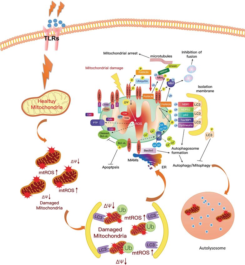
Frontiers in Cell and Developmental Biology | www.frontiersin.org 6 July 2021 | Volume 9 | Article 655819Yao et al. Neuroinflammation in Parkinson’s Disease FIGURE 2 | The mechanism of LRRK2 leading to microglia activation. Abnormal LRRK2 activity could regulate microglia cells’ activation through hyperphosphorylation of PKA, p53, MAPK family proteins, and Drp1. Thus, LRRK2 drives microglia toward a reactive phenotype with enhanced cell activity and inflammation in response to inflammatory stimuli, including LPS, environmental insults, and neuronal susceptibility. blocking these pathways can effectively control or attenuate neuroinflammation in the MPTP-probenecid-induced PD neuroinflammation in PD. For instance, suppressing the mouse model via targeting α-syn abnormalities in the Janus kinase/signal transducers and activators of transcription substantia nigra (Heng et al., 2016). FK506, as a well-known (JAK/STAT) pathway can prevent neuroinflammation and immunosuppressive drug, could decrease neuroinflammation neurodegeneration by inhibiting microglial activation, and DA neurodegeneration, pointing to a causal role of macrophage, and CD4(+) T-cell infiltration, and the neuroinflammation in an α-syn-based rat model of PD (Van production of pro-inflammatory cytokines/chemokines der Perren et al., 2015). The administration of hypoestoxide induced by α-syn (Qin et al., 2016). Furthermore, six could reduce neuroinflammation, neurodegeneration, and monosaccharides (6-mer), a specific inhibitor of the α-syn α-syn accumulation in a mouse model of PD via modulating output, could efficiently modulate neuroinflammation and the activity of NF-κB, suggesting that hypoestoxide may be a α-syn expression in neuron-like SH-SY5Y cells by blocking potent anti-PD drug (Kim et al., 2015). Curcumin has been NF-κB activation (Scuruchi et al., 2016). Meanwhile, α-M found to afford its neuroprotective effect and inhibit α-syn inhibits α-syn-induced microglial neuroinflammation and aggregation through the inhibition of oxidative stress generation, neurotoxicity by targeting nicotinamide adenine dinucleotide replenishing glutathione levels, and preventing glial-associated phosphate (NADPH) oxidase as a therapeutic possibility in inflammatory response in an LPS-induced PD model (Sharma preventing PD progression (Hu et al., 2016). Furthermore, et al., 2017; Sharma and Nehru, 2018). Endogenous high-mobility ginsenoside Rg1 could attenuate motor impairment and group protein B1, which has been demonstrated to mediate Frontiers in Cell and Developmental Biology | www.frontiersin.org 7 July 2021 | Volume 9 | Article 655819
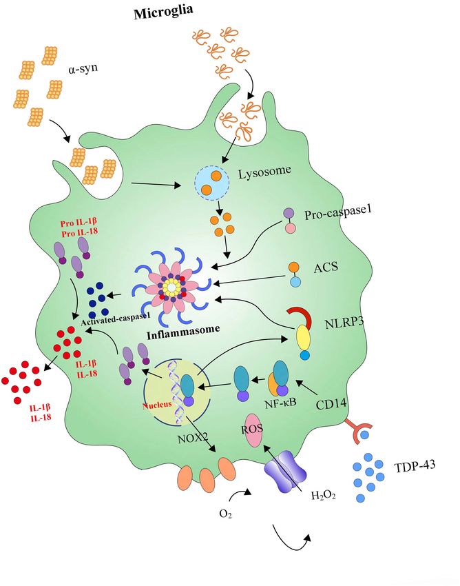
Yao et al.                                                                                                                  Neuroinflammation in Parkinson’s Disease

  FIGURE 3 | The signaling pathways and molecular factors involved in neuroinflammation. α-syn together with inflammasome form a network to regulate the
  activation of microglia. Blocking these signaling pathways and molecular factors can effectively improve apoptosis or the death of dopamine neurons caused by
  neuroinflammation.

persistent neuroinflammation and consequent progressive                               immune response, given the central role for microglial MHCII in
neurodegeneration by promoting multiple inflammatory and                              the activation of both innate and adaptive immune responses to
neurotoxic factors, could promote the autophagic degradation                          α-syn in PD (Harms et al., 2013).
α-syn via the Atg 5-dependent autophagy-initiation pathway
in PD (Guan et al., 2018). Immunotherapy targeting TLR2
alleviates α-syn accumulation in neuronal and astroglial                              The PINK1–Parkin Axis
cells and attenuates neuroinflammation, neurodegeneration,                            Parkin
and behavioral deficits in PD models of synucleinopathy by                            Parkin is predominantly expressed in the brain. It has been
modulating α-syn transmission and neuroinflammation (Kim C.                           implicated in many biological processes, such as synaptic
et al., 2018). Indeed, neuroinflammation might be mediated by                         excitability, inflammation, and immunity. The protein comprises
the microglial expression of MHC II, a vital regulator of the                         a C-terminal R1-in-between-ring-RING2 motif, an N-terminal

Frontiers in Cell and Developmental Biology | www.frontiersin.org                 8                                              July 2021 | Volume 9 | Article 655819
Yao et al.                                                                                                  Neuroinflammation in Parkinson’s Disease

ubiquitin-like (UBL) domain, RING0, RING1 IBR. In them,                    (TrxR) 2, a novel mediator of the inflammatory response, could
the Ubl and RING0 domains are unique to Parkin (Arkinson                   efficiently alleviate inflammation-mediated neuronal death by
and Walden, 2018). The UBL domain interacts with the R1                    activating the Akt–Parkin pathway and decreasing oxidative
environment, which negatively adjusts the activity of E3 ligase            stress (Gao et al., 2019). TNF-α-mediated neuronal inflammation
and parkin translocation to the mitochondria and parkin-                   could be attenuated by mitochonic acid-5 via augmenting the
dependent mitophagy. As an E3 ubiquitin-ligating enzyme,                   AMPK-Sirt3 pathways and activating Parkin-related mitophagy
Parkin plays a critical role in the cell, which works together with        (Huang et al., 2019).
E1 ubiquitin-activating enzymes and E2 ubiquitin-conjugating
enzymes in a ubiquubiquitin-proteasomeem to ubiquitinate                   PTEN-Induced Putative Kinase1 (PINK1)-Parkin Axis
the misfolded or aggregated proteins (van der Merwe et al.,                Recent studies have highlighted that mitochondrial dysfunction
2015). Thus, Parkin can act as an activator to disrupt the                 and DNA abnormalities complicatedly associate with the
autoinhibitory mechanisms and bridge the distance between                  pathogenesis of PD. PINK1 is a mitochondrial surveillance kinase
catalytic sites. Parkin shows conformational changes after point           that contributes to the processes involved in ridding the cell
mutation, disrupting the unique autoinhibitory features that               of damaged mitochondria; PINK1 mutations are a common
release both REP and Ubl domain activity (Tang et al., 2017).              genetic cause of familial PD. The mutations include truncating
Furthermore, the phosphorylation of Parkin leads to decreases              mutations, point mutations, missense, and deletions. PINK1-
in its E3 ubiquitin ligase activity (Aguirre et al., 2018). Besides,       associated PD has an earlier age of onset and slower progression
Parkin maintains mitochondrial quality control and turnover                (Kawajiri et al., 2011). The examination of humans’ brains with
(McWilliams and Muqit, 2017).                                              PINK1-linked PD shows the pathology of Lewy bodies and
    The knockout of Parkin is a crucial way to study the role of           neuronal loss in the substantia nigra, which are accompanied by
Parkin in the pathogenesis of PD (Matheoud et al., 2019). The              microgliosis and astrocytic gliosis (Steele et al., 2015).
decrease in Parkin’s solubility and stability is associated with the           The PINK1 protein sequence contains a predicted C-terminal
degeneration of substantia nigra neurons in PD (Lonskaya et al.,           kinase domain and a mitochondrial targeting sequence of
2013). The MPTP treatment on mice increased the expression                 the N-terminus (Pickrell and Youle, 2015). The protein is
of Parkin and neuroinflammation (Mendes et al., 2019). Earlier             imported into the mitochondria via the translocase of outer
studies reported the mutant Parkin in drosophila showed age-               and inner membrane complexes. PINK1 controls mitochondrial
dependent degeneration of dorsomedial dopaminergic neurons                 quality control by removing the dysfunctional mitochondria.
(Cha et al., 2005; Whitworth et al., 2005). Recent studies have            Mitochondrial damage is a significant cause of DA death in
shown that Parkin’s S-nitrosylation could diminish its protective          PD patients. PINK1 recruits the Parkin protein at the outer
effects against α-synuclein-mediated neurotoxicity and destroy             mitochondrial membrane while the damage of mitochondria
its ubiquitin ligase activity (Wahabi et al., 2018). It has been           occurred (van der Merwe et al., 2017). The activation of
reported patients with mutated Parkin have clinical symptoms               Parkin catalyzes the ubiquitination of outer mitochondrial
that are identical to patients with PD (Pickrell and Youle,                membrane proteins with ubiquitin which is then degraded
2015). Parkin mutation carriers are clinically characterized by            by the ubiquitin-proteasome system (Cornelissen et al., 2018).
slow disease progression and by having an excellent response to            Autophagosomes engulf the dysfunctional organelles, and then
levodopa treatment. To date, more than 100 different mutations             lysosomal enzymes typically digest both of them via the process
of Parkin have been reported, and the mutations are largely                of mitophagy. PINK1/parkin-mediated mitophagy has been
associated with the pathogenesis of PD (Abbas et al., 1999;                found in various neuronal and non-neuronal cells, especially
Ferreira and Massano, 2017). The overexpression of Parkin could            exposing to mitochondrial depolarizing agents (Akabane et al.,
protect against manganese-induced cell death and dopaminergic              2016; Newman and Shadel, 2018). The process prevents the
toxicity (Higashi et al., 2004; Jiang et al., 2004).                       accumulation of products from dysfunctional mitochondria, such
    Parkin shows a potential role in preventing                            as increased ROS and mtDNA damage (Truban et al., 2017).
neuroinflammation from progressing in PD. Mice with Parkin                     In a poor state of PINK1, the prevention of accumulated
mutations appear to have selective DA neuron degeneration and              products from dysfunctional mitochondria was obstructed, and
some motor deficits with intraperitoneal LPS (Vivekanantham                mtDNA mutational stress resulted in an inflammatory response
et al., 2015). Parkin levels and phenocopy Parkin loss-of-function         and activated the DNA-sensing cGAS–STING pathway, which
mutations were found to be decreased in chronic inflammatory               connected mitoflammation with PD pathology (Sliter et al.,
conditions, and the expression of TNF, IL-1β, and iNOS was                 2018). In this study, the researchers also found that the expression
increased in Parkin-null mice (Tran et al., 2011). In BV2                  of multiple cytokines such as IL-6, -12, and -13; IFNβ; CXCL1;
and primary microglia cell lines, the knockdown of Parkin                  and CCL2 and 4 increased efficiently in Pink1−/− and Parkin−/−
was found to increase LPS-induced microglial activation by                 mice. Meanwhile, it could also promote the expression of pro-
elevating the activity of NF-κB and JNK, which protected                   inflammatory type-I IFN and inflammatory cytokine production
neurons from zVAD-mediated necroptosis (Mouton-Liger                       and activate NF-κB signaling (West et al., 2015; West and Shadel,
et al., 2018; Dionisio et al., 2019). Parkin deficiency could              2017). Furthermore, the mtRNA and their double-stranded
enhance NLRP3 inflammasome signaling by attenuating an                     mitochondrial RNA activated the inflammasome and the RNA-
A20-dependent negative feedback loop in mice (Mouton-Liger                 sensing immune receptor MDA5 (Dhir et al., 2018; Zhong et al.,
et al., 2018). The overexpression of thioredoxin reductase                 2018). Moreover, one recent study by Yao et al. (2019a) reported

Frontiers in Cell and Developmental Biology | www.frontiersin.org      9                                        July 2021 | Volume 9 | Article 655819
Yao et al. Neuroinflammation in Parkinson’s Disease
that hydrogen-rich saline alleviated the inflammatory response consisted of a central protein, an adaptor protein ASC and
and apoptosis via PINK1/Parkin-mediated mitophagy (Figure 4). a caspase-1 protein, forming in response to a variety of
physiological and pathogenic stimuli. Inflammasome activation,
Inflammasome accrued in both health and disease in the CNS, is an
Inflammasomes, the central protein (varies with the type essential component of the innate immune. However, excessive
of inflammasome), which on activation recruits the adaptor activation of the inflammasome is also a significant driver of
apoptosis speck-like protein (ASC), is multimeric complexes autoimmune and metabolic disorders, underlying the importance
FIGURE 4 | PINK1 could phosphorylate Parkin on the mitochondrial surface, resulting in the activation of Parkin. The activated Parkin proteins form
phospho-polyubiquitin chains on damaged mitochondria. Finally, the dysfunctional mitochondria are cleared via autophagy. In this way, mitophagy inhibits
neuroinflammation in PD and increases microglial phagocytosis. However, the mutation of PINK1 or Parkin would alter the balance of fission to fusion by preventing
cells from responding to mitochondrial damage. The expression of ROS and pro-inflammatory factors are increased, which aggravates the development of PD. PD,
Parkinson’s disease; ROS, reactive oxygen species.
Frontiers in Cell and Developmental Biology | www.frontiersin.org 10 July 2021 | Volume 9 | Article 655819Yao et al. Neuroinflammation in Parkinson’s Disease of understanding these physiological and pathological contexts (AIM2) (Mariathasan et al., 2004; Boyden and Dietrich, 2006; (Sharma and Kanneganti, 2016; Mamik and Power, 2017). Recent Mariathasan et al., 2006; Rathinam et al., 2010; de Rivero work has mainly focused on the existence of inflammasome- Vaccari et al., 2014). Pattern recognition receptors have three mediated inflammatory pathways in CNS disorders. Pattern distinguishing features: universal expression, fast response, and recognition receptors (PRRs) which are primarily expressed recognizing many microbes (Zhu et al., 2018). Based on these by glial cells, play an integral role in the innate immune above features, PRRs efficiently initiate the signaling pathways response through the recognition of pathogen-specific proteins culminating in the activation of MAPK, NF-κB, and interferon (PAMPs) and damage-associated proteins (DAMPs) (Singhal regulatory factors (IRFs), which control the transcription of et al., 2014). So far, researchers have characterized four genes encoding pro-inflammatory factors (Zhu et al., 2018). different inflammasomes and their activators, including NLRP1, In neuroinflammation, inflammasomes can regulate microglial NLRP2, NLRP3, nod-like receptor family CARD domain- activation and subsequent neuroinflammatory processes in brain containing protein 4 (NLRC4), and absent in melanoma 2 pathology (Scholz and Eder, 2017). Otherwise, α-syn enters into FIGURE 5 | NLRP3 inflammasome activates neuroinflammation. Microglia are equipped with intracellular multi-molecule NLRP3 complexes, which α-syn can activate. NLRP3 inflammasomes could trigger the maturation of IL-1β and IL-18. High levels of IL-1β and IL-18 secretion enhances neuronal loss. Frontiers in Cell and Developmental Biology | www.frontiersin.org 11 July 2021 | Volume 9 | Article 655819
Yao et al.                                                                                                                             Neuroinflammation in Parkinson’s Disease

BV2 cells in an endocytosis-dependent manner and subsequently                                Currently, the signaling pathways and molecular factors involved
triggers NLRP3 inflammasome activation via inducing lysosomal                                in neuroinflammation have become an important research
swelling and increasing cathepsin B release (Zhou et al., 2016).                             method to identify PD’s pathogenesis. The anti-inflammatory
    Meanwhile, it also has been found that inflammasomes cause                               treatment has been found to exert a robust neuroprotective
caspase-1 activation following the stimulation of microglia                                  effect in a mouse model of PD. Studies on animal and
with lysophosphatidylcholine (LPC), depending on LPS                                         cell models of PD have shown that dietary supplements
prestimulation, NLRP3, and adaptor ASC, and knockdown                                        containing polyphenolic compounds have beneficial effects and
of inflammasome NLRC4 inhibits LPC-stimulated caspase-                                       are recommended for treating and preventing inflammation-
1 activity in microglia (Figure 5) (Scholz and Eder, 2017).                                  mediated neurodegeneration of DA neurons (Singh et al., 2020).
Further, inflammasomes are also involved in the inflammatory                                     Studies have shown that blocking these signaling pathways
pathogenesis of PD. For example, β-hydroxy butyrate (BHB),                                   and molecular factors can effectively improve apoptosis or the
an effective inhibitor of the NLRP3 inflammasome in response                                 death of dopamine neurons caused by neuroinflammation. This
to multiple activation stimuli including adenosine triphosphate                              paradigm is being shifted from theory to reality as a potential
(ATP), silica, and monosodium urate (MSU) crystals, almost                                   target for developing new drugs for PD. Going forward, focusing
completely blocks all aspects of inflammasome activation and                                 on these signaling pathways and molecular factors involved in
pyroptosis induced by ATP and MSU crystals in PD (Youm                                       neuroinflammation would provide a better understanding of
et al., 2015; Deora et al., 2017). Crucially, modern studies                                 the occurrence and development of PD. Ongoing research in
suggest that the NLRP3 inflammasome could be a major                                         this field may open a new door for developing pharmacological
disease-modifying therapeutic target in PD’s inflammatory                                    strategies toward the prevention and modification of the
pathogenesis. For instance, miR-7 directly targets NLRP3                                     pathogenesis of PD.
expression (besides α-syn) and modulates NLRP3 inflammasome
activation to attenuate DA neuronal degeneration accompanied
by the amelioration of microglial activation in an MPTP-                                     AUTHOR CONTRIBUTIONS
induced mouse model of PD (Zhou et al., 2016). In the
heightened microglial activation response, an exaggerated                                    LY and JW performed the majority of the literature search and
ROS/c-Abelson murine leukemia viral oncogene homolog                                         predominantly contributed to the writing of the article. SK and
(c-Abl)/NLRP3 signaling axis evaluates in LPS-primed rotenone                                GL assisted with the literature search. All authors read and
(ROT)-stimulated microglial cells and suggests that targeting                                approved the final manuscript.
c-Abl-regulated NLRP3 inflammasome signaling offers a novel
therapeutic strategy for PD treatment (Lawana et al., 2017).
Parkin deficiency modulates NLRP3 inflammasome activation                                    FUNDING
by attenuating an A20-dependent negative feedback loop in
Parkin’s pathogenesis (PARK2)-linked PD, paving the way for                                  This study was supported by the National Natural Science
the exploration of its potential as a biomarker and treatment                                Foundation of China (82060249), Key project of National Natural
target (Mouton-Liger et al., 2018).                                                          Science Foundation of Jiangxi Province (20202ACBL206005),
                                                                                             General project of Natural Science Foundation of Jiangxi
                                                                                             Province (20192BAB205042), General project of Natural Science
                                                                                             Foundation of Jiangxi Province (20202BABL206098), and the
CONCLUSION                                                                                   major academic and technical leaders training plan of Jiangxi
                                                                                             Province-Youth Training Program (20204BCJ23019).
Chronic inflammation of the CNS is mediated by neuroimmune
microglial cells and has been implicated as a pathological
contributor to PD. The activation of microglia and DA                                        ACKNOWLEDGMENTS
neuronal damage form a self-propelled degeneration cycle in
PD; thus, microglia are more likely to play critical roles in                                We would like to thank Yuqing Wang for helping us to draw
establishing and maintaining inflammatory responses in PD.                                   the illustrations.

REFERENCES                                                                                   Akabane, S., Uno, M., Tani, N., Shimazaki, S., Ebara, N., Kato, H., et al. (2016). PKA
                                                                                                regulates PINK1 stability and parkin recruitment to damaged mitochondria
Abbas, N., Lucking, C. B., Ricard, S., Durr, A., Bonifati, V., De Michele, G.,                  through phosphorylation of MIC60. Mol. Cell 62, 371–384. doi: 10.1016/j.
  et al. (1999). A wide variety of mutations in the parkin gene are responsible                 molcel.2016.03.037
  for autosomal recessive parkinsonism in Europe. French Parkinson’s disease                 Alvarez-Erviti, L., Couch, Y., Richardson, J., Cooper, J. M., and Wood, M. J. (2011).
  genetics study group and the European consortium on genetic susceptibility                    Alpha-synuclein release by neurons activates the inflammatory response in
  in Parkinson’s disease. Hum. Mol. Genet. 8, 567–574. doi: 10.1093/hmg/8.                      a microglial cell line. Neurosci. Res. 69, 337–342. doi: 10.1016/j.neures.2010.
  4.567                                                                                         12.020
Aguirre, J. D., Dunkerley, K. M., Lam, R., Rusal, M., and Shaw, G. S. (2018).                Anitua, E., Pascual, C., érez-Gonzalez, R. P., Orive, G., and Carro, E. (2015).
  Impact of altered phosphorylation on loss of function of juvenile Parkinsonism-               Intranasal PRGF-Endoret enhances neuronal survival and attenuates NF-κB-
  associated genetic variants of the E3 ligase parkin. J. Biol. Chem. 293, 6337–6348.           dependent inflammation process in a mouse model of Parkinson’s disease.
  doi: 10.1074/jbc.RA117.000605                                                                 J. Control. Release 203, 170–180.

Frontiers in Cell and Developmental Biology | www.frontiersin.org                       12                                                 July 2021 | Volume 9 | Article 655819
Yao et al. Neuroinflammation in Parkinson’s Disease Aoki, E., Yano, R., Yokoyama, H., Kato, H., and Araki, T. (2009). Role of nuclear neuroprotective, antioxidant and anti-inflammatory effects in a Parkinson’s transcription factor kappa B (NF-kappaB) for MPTP (1-methyl-4-phenyl- disease model in rats. J. Pharm. Pharmacol. 70, 787–796. doi: 10.1111/jphp. 1,2,3,6-tetrahyropyridine)-induced apoptosis in nigral neurons of mice. Exp. 12866 Mol. Pathol. 86, 57–64. doi: 10.1016/j.yexmp.2008.10.004 de Rivero Vaccari, J. P., Dietrich, W. D., and Keane, R. W. (2014). Activation Arkinson, C., and Walden, H. (2018). Parkin function in Parkinson’s disease. and regulation of cellular inflammasomes: gaps in our knowledge for central Science 360, 267–268. doi: 10.1126/science.aar6606 nervous system injury. J. Cereb. Blood Flow Metab. 34, 369–375. doi: 10.1038/ Azizi, S. A., and Azizi, S.-A. (2018). Synucleinopathies in neurodegenerative jcbfm.2013.227 diseases: accomplices, an inside job and selective vulnerability. Neurosci. Lett. Dehmer, T., Heneka, M. T., Sastre, M., Dichgans, J., and Schulz, J. B. (2004). 672, 150–152. Protection by pioglitazone in the MPTP model of Parkinson’s disease correlates Bassani, T. B., Vital, M. A., and Rauh, L. K. (2015). Neuroinflammation in with IκBα induction and block of NFκB and iNOS activation. J. Neurochem. 88, the pathophysiology of Parkinson’s disease and therapeutic evidence of anti- 494–501. inflammatory drugs. Arq. Neuropsiquiatr. 73, 616–623. doi: 10.1590/0004- Deng, H., Fan, K., and Jankovic, J. (2018). The role of TMEM230 gene in 282X20150057 Parkinson’s disease. J. Parkinsons Dis. 8, 469–477. Birla, H., Rai, S. N., Singh, S. S., Zahra, W., Rawat, A., Tiwari, N., et al. (2019). Deora, V., Albornoz, E. A., Zhu, K., Woodruff, T. M., and Gordon, R. (2017). Tinospora cordifolia Suppresses neuroinflammation in Parkinsonian mouse The ketone body beta-hydroxybutyrate does not inhibit synuclein mediated model. Neuromol. Med. 21, 42–53. doi: 10.1007/s12017-018-08521-7 inflammasome activation in microglia. J. Neuroimmune Pharmacol. 12, 568– Boyden, E. D., and Dietrich, W. F. (2006). Nalp1b controls mouse macrophage 574. doi: 10.1007/s11481-017-9754-5 susceptibility to anthrax lethal toxin. Nat. Genet. 38, 240–244. doi: 10.1038/ Dhir, A., Dhir, S., Borowski, L. S., Jimenez, L., Teitell, M., Rotig, A., et al. (2018). ng1724 Mitochondrial double-stranded RNA triggers antiviral signalling in humans. Brockmann, K., Apel, A., Schulte, C., Schneiderhan-Marra, N., Pont-Sunyer, C., Nature 560, 238–242. doi: 10.1038/s41586-018-0363-0 Vilas, D., et al. (2016). Inflammatory profile in LRRK2-associated prodromal Di Maio, R., Hoffman, E. K., Rocha, E. M., Keeney, M. T., Sanders, L. H., De and clinical PD. J. Neuroinflammation 13:122. doi: 10.1186/s12974-016-0588-5 Miranda, B. R., et al. (2018). LRRK2 activation in idiopathic Parkinson’s disease. Brundin, L., Bergkvist, L., and Brundin, P. (2018). Fire prevention in the Sci. Transl. Med. 10:eaar5429. doi: 10.1126/scitranslmed.aar5429 Parkinson’s disease brain. Nat. Med. 24, 900–902. doi: 10.1038/s41591-018- Dionisio, P. E. A., Oliveira, S. R., Amaral, J., and Rodrigues, C. M. P. (2019). Loss 0109-4 of microglial parkin inhibits necroptosis and contributes to neuroinflammation. Calatayud, C., Carola, G., Consiglio, A., and Raya, A. (2017). Modeling the genetic Mol. Neurobiol. 56, 2990–3004. doi: 10.1007/s12035-018-1264-9 complexity of Parkinson’s disease by targeted genome edition in iPS cells. Curr. Ehgoetz Martens, K. A., and Shine, J. M. (2018). The interactions between non- Opin. Genet. Dev. 46, 123–131. motor symptoms of Parkinson’s disease. Expert Rev. Neurother. 18, 457–460. Cha, G. H., Kim, S., Park, J., Lee, E., Kim, M., Lee, S. B., et al. (2005). Parkin doi: 10.1080/14737175.2018.1472578 negatively regulates JNK pathway in the dopaminergic neurons of Drosophila. Esposito, E., Di Matteo, V., Benigno, A., Pierucci, M., Crescimanno, G., Proc. Natl. Acad. Sci. U.S.A. 102, 10345–10350. doi: 10.1073/pnas.0500346102 and Di Giovanni, G. (2007). Non-steroidal anti-inflammatory drugs in Chan, S. L., and Tan, E. K. (2017). Targeting LRRK2 in Parkinson’s disease: an Parkinson’s disease. Exp. Neurol. 205, 295–312. doi: 10.1016/j.expneurol.2007. update on recent developments. Expert Opin. Ther. Targets 21, 601–610. 02.008 Charvin, D., Medori, R., Hauser, R. A., and Rascol, O. (2018). Therapeutic Ettle, B., Kuhbandner, K., Jorg, S., Hoffmann, A., Winkler, J., and Linker, R. A. strategies for Parkinson disease: beyond dopaminergic drugs. Nat. Rev. Drug (2016). alpha-Synuclein deficiency promotes neuroinflammation by increasing Discov. 17, 804–822. doi: 10.1038/nrd.2018.136 Th1 cell-mediated immune responses. J. Neuroinflammation 13:201. doi: 10. Chen, H., Zhang, S. M., Hernan, M. A., Schwarzschild, M. A., Willett, W. C., 1186/s12974-016-0694-4 Colditz, G. A., et al. (2003). Nonsteroidal anti-inflammatory drugs and the risk Fan, F. S. (2020). Ractopamine residue in meat might protect people from of Parkinson disease. Arch. Neurol. 60, 1059–1064. doi: 10.1001/archneur.60.8. Parkinson disease. Med. Hypotheses 145:110397. 1059 Ferreira, M., and Massano, J. (2017). An updated review of Parkinson’s disease Chen, J., Su, P., Luo, W., and Chen, J. (2018). Role of LRRK2 in manganese-induced genetics and clinicopathological correlations. Acta Neurol. Scand. 135, 273–284. neuroinflammation and microglial autophagy. Biochem. Biophys. Res. Commun. doi: 10.1111/ane.12616 498, 171–177. doi: 10.1016/j.bbrc.2018.02.007 Forero, D. A., Trujillo, M. L., and Lopez-Leon, S. (2020). “Genome plasticity and Chen, Y., Zhang, N., Ji, D., Hou, Y., Chen, C., Fu, Y., et al. (2018). Dysregulation neuropsychiatric disorders,” in Genome Plasticity in Health and Disease, eds of bcl-2 enhanced rotenone-induced alpha-synuclein aggregation associated D. A. Forero and G. P. Patrinos (Amsterdam: Elsevier). with autophagic pathways. Neuroreport 29, 1201–1208. doi: 10.1097/WNR. Fu, S. P., Wang, J. F., Xue, W. J., Liu, H. M., Liu, B. R., Zeng, Y. L., et al. 0000000000001097 (2015). Anti-inflammatory effects of BHBA in both in vivo and in vitro Cheng, C., and Zhu, X. (2019). Cordycepin mitigates MPTP-induced Parkinson’s Parkinson’s disease models are mediated by GPR109A-dependent mechanisms. disease through inhibiting TLR/NF-kappaB signaling pathway. Life Sci. 223, J. Neuroinflammation 12:9. doi: 10.1186/s12974-014-0230-3 120–127. doi: 10.1016/j.lfs.2019.02.037 Gago, M., Machado, A., and Rocha, S. (2020). “Current clinical approaches in Chistiakov, D. A., and Chistiakov, A. A. (2017). alpha-Synuclein-carrying neurodegenerative diseases,” in Handbook of Innovations in Central Nervous extracellular vesicles in Parkinson’s disease: deadly transmitters. Acta Neurol. System Regenerative Medicine, ed. A. J. Salgado (Amsterdam: Elsevier), 79. Belg. 117, 43–51. doi: 10.1007/s13760-016-0679-1 Gan, P., Xia, Q., Hang, G., Zhou, Y., Qian, X., Wang, X., et al. (2018). Knockdown Choi, I., Kim, B., Byun, J. W., Baik, S. H., Huh, Y. H., Kim, J. H., et al. (2015). of cathepsin D protects dopaminergic neurons against neuroinflammation- LRRK2 G2019S mutation attenuates microglial motility by inhibiting focal mediated neurotoxicity through inhibition of NF-kappaB signalling pathway adhesion kinase. Nat. Commun. 6:8255. doi: 10.1038/ncomms9255 in Parkinson’s disease model. Clin. Exp. Pharmacol. Physiol. 46, 337–349. doi: Choubey, V., Safiulina, D., Vaarmann, A., Cagalinec, M., Wareski, P., Kuum, 10.1111/1440-1681.13052 M., et al. (2011). Mutant A53T alpha-synuclein induces neuronal death by Gao, J., Li, Y., Li, W., and Wang, H. (2019). TrxR2 overexpression alleviates increasing mitochondrial autophagy. J. Biol. Chem. 286, 10814–10824. doi: inflammation-mediated neuronal death via reducing the oxidative stress and 10.1074/jbc.M110.132514 activating the Akt-Parkin pathway. Toxicol. Res. (Camb.) 8, 641–653. doi: 10. Cornelissen, T., Vilain, S., Vints, K., Gounko, N., Verstreken, P., and 1039/c9tx00076c Vandenberghe, W. (2018). Deficiency of parkin and PINK1 impairs age- Giacoppo, S., Rajan, T. S., De Nicola, G. R., Iori, R., Rollin, P., Bramanti, P., et al. dependent mitophagy in Drosophila. Elife 7:e35878. doi: 10.7554/eLife.35878 (2017). The isothiocyanate isolated from Moringa oleifera shows potent anti- Cutillo, G., Simon, D. K., and Eleuteri, S. (2020). VPS35 and the mitochondria: inflammatory activity in the treatment of murine subacute Parkinson’s disease. connecting the dots in Parkinson’s disease pathophysiology. Neurobiol. Dis. Rejuvenation Res. 20, 50–63. doi: 10.1089/rej.2016.1828 145:105056. Goes, A. T. R., Jesse, C. R., Antunes, M. S., Lobo Ladd, F. V., Lobo Ladd, A. A. B., de Araujo, D. P., Nogueira, P. C. N., Santos, A. D. C., Costa, R. O., de Lucena, J. D., Luchese, C., et al. (2018). Protective role of chrysin on 6-hydroxydopamine- Jataí Gadelha-Filho, C. V., et al. (2018). Viana: Aspidosperma pyrifolium Mart: induced neurodegeneration a mouse model of Parkinson’s disease: involvement Frontiers in Cell and Developmental Biology | www.frontiersin.org 13 July 2021 | Volume 9 | Article 655819
You can also read