Scientific Background Discovery of cancer therapy by inhibition of negative immune regulation - Nobel Prize
←
→
Page content transcription
If your browser does not render page correctly, please read the page content below
                                     Scientific Background

      Discovery of cancer therapy by inhibition of negative immune
                               regulation

The 2018 Nobel Prize in Physiology or Medicine         prognosis of the disease vary depending on the
is awarded to James P. Allison and Tasuku              tissue origin. The risk of cancer increases with
Honjo for their discovery of cancer therapy by         age and the remarkably longer life span of the
inhibition of negative immune regulation. Already      world population contributes to cancer as an
150 years ago attempts were made to use                increasing problem. Life style and environmental
infectious agents to stimulate immune responses        factors add to this development, with smoking as
to cancer. Since then many seminal discoveries         the most important factor.
in the fields of basic immunology and tumor
biology, including some awarded with the Nobel         According to WHO, more than 18 million persons
Prize, enabled a deeper understanding of such          in the world are estimated to be diagnosed with
processes. In 1996, James P. Allison and               cancer in 2018 (Global Cancer Observatory,
coworkers used this accumulated knowledge to           2018). Today, one in three will develop cancer in
demonstrate that antibodies directed against a         the economically more developed countries and it
cell surface molecule on T cells, CTLA-4, is           is estimated to be one in two in two decades. The
capable of unleashing an immune response,              number has increased steadily during the last 50
which cured mice from tumors. Prior to this, in the    years. Within the same period the proportion of
laboratory of Tasuku Honjo, a new molecule             cured patients, as estimated by five-year survival,
named PD-1 had been identified. Similar to             has increased from less than a third to more than
CTLA-4, PD-1 also serves as a brake, which can         two thirds. That said, these are average figures,
prevent T cells from killing cancer cells. With the    which conceal major differences – between
aid of biomedical companies Allison succeeded          cancers in different organs, between earlier and
in developing the new concept with anti-CTLA-4         more advanced forms of the disease in the same
into clinical therapy for patients with advanced       organ, and also between different countries. The
forms of melanoma. The use of antibodies               occurrence of metastasis often implies that it is
directed against PD-1 and its ligand PD-L1 has         impossible to cure the disease, even if life can be
now been approved for several cancer forms and         prolonged by different treatments. In countries
this treatment is even more efficacious. The best      with less developed economy, a lower proportion
clinical results have so far been obtained by          of patients are diagnosed and treated, resulting in
combining treatment with anti-CTLA-4 and anti-         fewer cures and reduced average survival.
PD-1. These antibodies are known as immune
checkpoint inhibitors and the seminal discoveries      Scientific discoveries have paved the way for
by the two laureates have added a new pillar for       better understanding of the disease, as well as
the treatment of cancer.                               for novel treatments. Etiology and pathogenesis
                                                       is complex, but all cancers display certain
Cancer                                                 hallmarks (Hanahan & Weinberg, 2011). Some of
                                                       these concern intrinsic cellular processes that
Cancer is a common term for a group of diseases        regulate division, death, metabolism and
caused by uncontrolled cell proliferation and          migration; others influence interactions in the
migration. This results in abnormal growth of a        tumor microenvironment with cells forming blood
tumor mass, first within an organ, then infiltrating   vessels or stroma, and cells of the immune
adjacent tissues. Eventually, cancer cells can         system. Each of the hallmarks is influenced by
also colonize distant organs via blood or              multiple pathways, which have been rendered
lymphatic vessels, so called metastases, causing       dysfunctional by alterations in the genes of
morbidity and death. The symptoms, course and          cancer cells. Some of these genetic changes can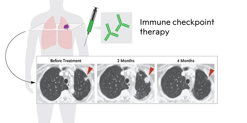
be inherited via the germ line DNA, while the             normal and cancer cells, with the aim of acquiring
majority is acquired as mutations in somatic cells        basic knowledge as a platform for new
in processes influenced by a variety of external          therapeutic concepts.
factors.

Several discoveries leading to today’s knowledge          Immunotherapy for tumors until 1995
about cancer represent breakthroughs that have
been awarded with The Nobel Prize in                      Throughout history there are many accounts on
Physiology or Medicine. Those discoveries                 tumors disappearing after infectious episodes,
awarded have included infection as an etiological         whereas the investigation of experimental
factor (e g Rous 1966, for tumor-inducing viruses;        infections as therapy for cancer patients dates
                                                                                th
zur Hauzen 2008, for Human Papilloma virus as             back to the late 19 century. The basic concept
a cause of cervical cancer); and the relation             behind such treatments has been the possibility
between cellular and viral genes in pathogenesis          that the infections stimulated the immune system,
(e g Baltimore, Dulbecco and Temin 1975, for              leading to an immune rejection of the tumor.
integration of retroviral genetic information into        Most well-known for studies in this field is the
DNA; Bishop and Varmus 1989, for the cellular             American surgeon William B. Coley, who
origin of viral oncogenes). In addition, novel            reported on the treatment of malignant tumors by
therapies have also been awarded the Nobel                repeated “inoculation of erysipelas”, i.e. of live,
Prize in Physiology or Medicine, e g Huggins              cultured Streptococci, in the early 1890s (Coley
1966, for hormone treatment of prostate cancer;           1891). However, the first attempts in this field
Elion and Hitchins 1988, for novel principles             were by German clinicians 150 years ago
leading to cytostatic drugs affecting the                 (Busch, 1868, Fehleisen, 1882). While infectious
metabolism of nucleic acids, and Thomas 1990,             therapy was practiced in numerous tumor
for discoveries concerning bone marrow                    patients, the clinical outcome varied, causing
transplantation, used to treat certain blood              disbelief within the medical community. Today,
cancers. These therapies were introduced in the           the concept is manifested in the form of
latter half of the previous century to complement         alternating     intradermal     and   intracavitary
the traditional methods of surgery and                    administration of Bacillus Calmette-Guerin to
radiotherapy. Additional Nobel Prizes, including          patients with bladder tumors (Morales et al 1976;
in Chemistry, have awarded groundbreaking                 Alexandroff et all 1999).
discoveries regarding basic cellular functions with
                                                                             th
relevance to cancer regarding DNA replication,            In the early 20        century, the mechanism
cell cycle mechanisms, apoptosis, and genome              underlying the effect of infectious agents on
integrity.                                                tumors was not understood, but the concept that
                                                          immunity could influence tumor development was
Despite this remarkable progress, the incidence           around. In the beginning of the last century Leo
of cancer is rising in most countries of the world,       Loeb mentioned the possible role of immunity for
partly due to an increasing life span and                 the growth of experimentally transplanted tumors
improved diagnosis; one out of three will develop         (Loeb, 1902). It was apparent that a basic
the disease and one of 6 will die of cancer               understanding of the immune system was
(Global Cancer Observatory, 2018). In 2018, the           needed. Inbred mouse strains were established.
global mortality from cancer is estimated to              Using this new possibility it was discovered that
surpass 9 million people. The scientific                  alleles within at the histocompatibility-2 (H-2)
knowledge has led to programs for prevention              locus in the mouse were key determinants for
and early diagnosis. However, even dramatically           tumor transplantation (Snell & Higgins, 1951) and
improved prevention programs will not solve the           George D. Snell became a Nobel laureate in
problem, since it is estimated that at least 50% of       1980 for his work in this field. These
cases may be caused by accumulation of                    groundbreaking discoveries paved the way for
spontaneous DNA mutations occurring during                the understanding of how lymphocytes use
normal ageing (Tomasetti et al., 2013). Novel             molecules of the Major Histocompatibility
types of treatment are strongly needed, and there         Complex (MHC) to discriminate ‘self’ from ‘non-
has been a huge investment in basic research on


                                                    2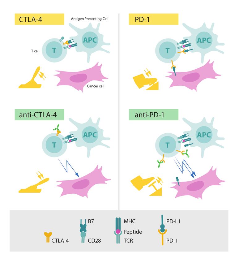
self’ and through this process can identify and                 one based on tumor-infiltrating lymphocytes
eliminate infectious intruders. Related to this, the            (Rosenberg et al., 1990) and another on a gene
Nobel Prize in 1996 was awarded to Peter C.                     therapy concept, namely the generation of
Doherty and Rolf M. Zinkernagel “for their                      chimeric antigen-receptor      (CAR) T cells
discoveries concerning the specificity of the cell              (reviewed in June & Sadelain, 2018). In 2017, the
mediated immune defence”.                                       US Food and Drug Administration, (FDA) and this
                                                                year, the European Medicines Agency (EMA)
                                             th
Another example from the early 20 century                       approved autologous CAR-T cell transfer for the
relates to the Nobel laureate Paul Ehrlich, who                 treatment of leukemia.
shared the 1908 Prize with Ilya Ilyich Mechnikov
"in recognition of their work on immunity". He                  T cell activation and the concept of
discussed in depth the potential effects of natural             costimulation
and acquired immunity for cancer (Ehrlich, 1909).
Sixty years later Sir Frank Macfarlane Burnet,                  T cells have been at the center stage in
who shared the 1960 Nobel Prize "for discovery                  immunology, but up until the 1980s their antigen-
of acquired immunological tolerance", proposed                  recognizing receptor remained elusive, whereas
that the immune system serves as a surveillance                 the antigen-specific receptor on B cells was
system for cancer (Burnet, 1970).                               already well characterized. Thus, during their
                                                                differentiation B cells were known to rearrange
A sign of the belief in immunotherapy for tumors                their immunoglobulin genes (Nobel Prize to
is that a very large part of the funding in the field           Susumu Tonegawa in 1987), express the
of immunology has been directed towards cancer                  resulting immunoglobulin protein on their surface
research. This led to a number of fundamental                   and secrete large amounts of it into body fluids.
discoveries in tumor immunology, e g the                        In 1975 the development of the monoclonal
demonstration of tumor-specific antigens (Klein &               antibody technology by George J F Köhler and
Klein, 1962) and their molecular nature (Lurquin                César Milstein, for which they were awarded the
et al., 1989), cytotoxic T cell killing of tumor cells          Nobel Prize in 1984, paved the way for the
(Brunner     et    al.,  1968),       tumor-infiltrating        identification of novel cell surface markers. This
lymphocytes (Klein et al., 1977) and their role in              technology became crucial not only for identifying
immunotherapy (Rosenberg et al., 1986) and                      key molecules on T cells, but also for the new
immunoselection and immunoediting during                        treatment that this year’s Nobel Prize awards.
tumor progression (Dunn et al., 2002). Numerous
reports    on     experimental       animal    studies          Major developments for the understanding of
demonstrated profound, beneficial effects of the                cellular adaptive immune responses took place in
immune system on tumor growth. However,                         the 1980s. During this decade the T cell receptor
translating these findings into clinical therapy                (TCR) was identified and its interaction with
failed almost without exception. Moreover,                      MHC-associated peptides on antigen-presenting
numerous case reports have described treatment                  cells was revealed. The gene for interleukin-2, an
effects    on    cancer     patients     related       to       important regulator of T cells, was cloned and the
immunotherapy, but also here, when such novel                   mechanism underlying the signaling of the TCR
approaches were tested in controlled clinical                   was beginning to be understood. It became clear
studies, there were typically failures or logistical            that the interaction between the TCR and MHC
problems in translating the complicated protocols.              antigen was insufficient for activation of T cells,
There are a few examples of success, one being                  leading to the concept of costimulation. The
that allogeneic bone marrow transplants, apart                  deciphering of the underlying mechanisms was
from serving as a replacement of hematopoietic                  an incremental process that took place during the
tissue, also cause a graft-versus-leukemia effect               1980s and 1990s involving many laboratories all
(Weiden et al., 1979).                                          over the world.

Moreover, in the 1980s two different approaches                 The first cell surface molecule identified in TCR
using complex protocols involving culture of                    costimulation was CD28, which was originally
patients’ lymphocytes ex vivo were developed,                   discovered by a monoclonal antibody recognizing


                                                          3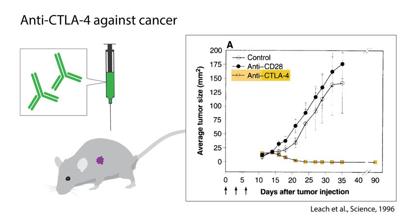
this protein on T cells and thymocytes (Hansen et          Sharpe and Tak Mak, confirmed and further
al., 1980). CD28 was found to synergize with the           substantiated its negative regulatory role, since
TCR complex in the activation of T cells (Martin           these mice developed very severe autoimmune
et al., 1986). Four years later a ligand for CD28          disease associated with proliferating T cells (Tivol
was identified as the B7 molecule (now known as            et al., 1995; Waterhouse et al., 1995). Soluble
CD80), which is expressed on antigen-presenting            CTLA4-Ig was later used by Linsley and
cells (Linsley et al., 1990). Lieping Chen, Peter          colleagues as a decoy to block costimulation,
Linsley and coworkers (Linsley et al., 1992)               developing it into a drug, abatacept, for treatment
suggested a role for costimulation in tumor                of rheumatoid arthritis (Linsley et al., 1992).
immunology by demonstrating that the transfer of
the B7 gene to tumor cells caused rejection, as            Inhibition of tumor growth by antibodies
shown also by James P. Allison’s group                     against CTLA-4 in animals
(Townsend & Allison, 1993).
                                                           While some investigators used this new
Identification of CTLA-4 as a negative                     knowledge       to    develop     treatments      for
regulator                                                  autoimmunity (Lenschow et al., 1992), Allison
                                                           followed another path. In spite of all the previous
In 1987, incidentally the same year as the CD28            failures, he intended to find a cure for cancer. He
gene was isolated, the cDNA for a T cell-                  attempted to block the negative effects that
expressed, CD28-related molecule, CTLA-4                   CTLA-4 induced, thereby unleashing an immune
(cytotoxic T lymphocyte antigen 4), also named             response. In contrast to most other concepts in
CD152, was cloned in the laboratory of Pierre              tumor immunology, there was no need to
Golstein (Brunet et al., 1987). The function of            understand which antigens the T cells would
CTLA-4 was unknown, but structurally both CD28             recognize, and the strategy was not selective for
and CTLA-4 belong to the immunoglobulin                    a particular type of tumor, but was in essence
superfamily. It is now known that CTLA-4 protein           universal. The first experiment was set up in his
resides intracellularly in resting T cells, but            laboratory at the University of California, Berkeley
translocates rapidly to the membrane after                 in the end of 1994 with an immediate blinded
activation (Lindsten et al., 1993; Linsley et al.,         repeat over the Christmas period. Mice that had
1996). In contrast to other T cells, it is                 been transplanted with tumors were treated with
constitutively expressed as a membrane protein             monoclonal antibodies against CTLA-4. The
in regulatory T cells (Takahashi et al., 2000).            result was spectacular (Fig. 1 and Fig. 4), and
Linsley et al. (1991) used soluble CTLA-4-Ig to            eventually led to the seminal discovery that this
demonstrate that CTLA-4, similar to CD28, binds            treatment directed towards the immune system,
to B7, but with higher affinity. In the following          had curative potential in tumor-bearing mice
years the generally held view was that CD28 and            (Leach et al 1996). Thus, (1) the authors
CTLA-4 acted in a similar, costimulatory way.              confirmed the hypothesis that blocking of CTLA-4
However, in 1994, Jeffrey Bluestone, Craig                 would strengthen the T cell anti-tumor response,
Thompson and colleagues (Walunas et al.1994),              (2) even pre-established tumors were found to be
and subsequently, Krummel & Allison (1995)                 sensitive, (3) rejection was followed by durable
came to the conclusion that CTLA-4 instead                 tumor immunity and (4) two types of tumors were
served as a negative regulator of T cell                   tested and both responded. One of the tumor cell
activation. This was initially interpreted as an           lines studied was a colon carcinoma, used with
intrinsic effect on the T cell, where CTLA-4 would         as well as without transfection with the murine
induce negative signalling interfering with positive       B7.1 (CD80) gene. Both cells responded to
signals elicited by TCR and costimulatory                  therapy with antibodies directed against CTLA-4.
receptors. Several alternative or additional
explanations are discussed today. Both the                 This work represents the birth of a new concept
above studies were possible through the                    for immunotherapy. Monoclonal antibodies with
generation of monoclonal antibodies directed               similar capacity to unleash responses against
against CTLA-4. Inactivation of the Ctla4 gene in          tumors are nowadays often referred to as
mice, performed in the laboratories of Arlene              immune checkpoint inhibitors (ICIs).


                                                     4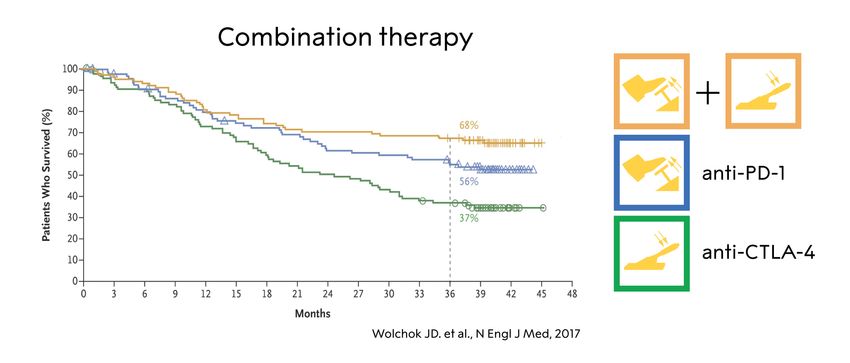
company, Medarex, in which an acquired
The Allison laboratory initially carried out a             technology based on transgenic mice made it
number of studies in other animal tumor models             possible to generate human monoclonal
including, prostate cancer (Kwon et al., 1997),            antibodies. An anti-CTLA-4 IgG1 monoclonal
mammary tumor (Hurwitz et al., 1998) and                   antibody named MDX-010 was developed in
melanoma (van Elsas et al., 1999). Some of                 1999, later named ipilimumab (Wolchock et al.,
these investigations included combined therapies           2013). The company Bristol-Myers Squibb
such as the use of anti-CTLA-4 and the                     subsequently acquired Medarex and continued
immunostimulatory           cytokine,       GM-CSF         the clinical development. With these studies,
(granulocyte-macrophage colony stimulating                 Jedd Wolchok and Stephen Hodi together with
factor). In a short period, Allison had thus proved        scientists at Bristol-Myers Squibb Company
that several different tumor types responded to            advanced the clinical program for the study of the
the same treatment strategy. Given the many                new ICI.
previous      failures   of     immune     therapies
demonstrated in mice, the challenge remained to            In the first-in-man study MDX-010 was given to 9
develop the concept into clinical practice – could         patients in a phase I clinical trial at a single dose
this be achieved?                                          of 3 mg/kg and a response was observed in
                                                           some melanoma patients (Hodi et al., 2003). The
                                                           same year complete regression was reported in
                                                           another trial in some treated melanoma patients,
                                                           while severe autoimmune side effects were also
                                                           observed (Phan et al., 2003). The responses
                                                           were frequently recorded later than for
                                                           chemotherapy. A special feature was the
                                                           observation that treatment initially could even
                                                           increase the tumor volume, “pseudoprogression”,
                                                           owing to the infiltration of immune cells, rather
                                                           than reducing it immediately, as usually seen with
                                                           chemo- or radiotherapy.

                                                           Additional clinical studies were carried out and
Figure 1. The discovery of James P. Allison and
                                                           the major breakthrough came from a phase III
coworkers, utilizing the role of CTLA-4 as an
                                                           trial for the treatment of unresectable, metastatic
inhibitor of activation and developing antibodies
                                                           melanoma showing a significantly increased
to release the brake. The graph shows the effect
                                                           overall survival (Hodi et al., 2010). This trial was
of anti-CTLA-4 treatment in tumor-bearing mice
                                                           also the basis for the subsequent approval of
compared to controls.
                                                           anti-CTLA-4 by the FDA and the EMA in 2011.
The development of clinical               immune
                                                           Discovery of the PD-1 receptor and its role in
checkpoint inhibitor therapy
                                                           immune responses
Translating this research into the clinics became
                                                           The second discovery awarded by this year’s
a long-term goal of Allison. At the time there was
                                                           Nobel Prize also originated in basic, curiosity-
very limited interest from the pharmaceutical
                                                           driven research, not primarily oriented towards
industry for a treatment based on the removal of
                                                           cancer. PD-1 (CD279) was identified and cloned
the brakes of immune responses, without
                                                           by Tasuku Honjo’s group at Kyoto University in
knowledge on which antigens were recognized
                                                           Japan already in the early 1990s (Ishida et al.,
on the tumor cells. Moreover, the severe
                                                           1992), during the explosive progress in the field
autoimmune phenotype of mice lacking CTLA-4
                                                           of costimulation, but before Allison’s discovery of
posed a major risk for severe side effects.
                                                           CTLA-4 inhibition to treat cancer. At the time,
However,      through     perseverance      Allison
                                                           Honjo and colleagues assumed that PD-1,
eventually managed to establish collaboration
                                                           identified by subtractive hybridization to isolate
with Alan Korman from the small biotech


                                                     5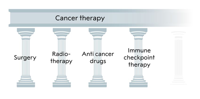
mRNAs overexpressed in dying mouse cells, was also the first to discuss the possibility that
would be involved in pathways regulating some tumors may use PD-L1 to inhibit an
apoptosis. Hence, they used the acronym PD (for antitumor immune response, based on the
Programmed Cell Death). The open reading observation that the molecule was expressed, not
frame predicted a protein with a transmembrane only by macrophages, dendritic cells and
region, distantly related to the immunoglobulin additional immune cells, but also by certain
gene superfamily. Follow-up studies reported the cancer cells. A second ligand for PD-1, PD-L2
structure and chromosomal location of the gene (CD273), was identified soon thereafter, in further
(Shinohara et al., 1994), as well as the collaboration between the groups of Freeman,
generation of a monoclonal antibody for PD-1 Honjo and Sharpe (Latchman et al., 2001).
used to demonstrate expression on activated T-
and B-cells as well as on immature thymocytes PD-1 blockade as a treatment of cancer
(Agata et al., 1996). The general concept that the PD-1/PD-L1
pathway might be involved in immune responses
The function of PD-1 remained elusive for many to tumors was first tested in two studies
years. Honjo launched an ambitious program to published in 2002, one from Chen’s laboratory
fully understand its function, in which the (Dong et al 2002) and another from the Kyoto
generation of mice deficient for this molecule was groups of Minato and Honjo, in collaboration (Iwai
central. The first “knock-out” mice on C57Bl et al 2002). Both reports focused on PD-L1
genetic background only showed a mild, molecules expressed on tumor cells,
intriguing phenotype with some splenomegaly demonstrating that expression of this ligand could
and augmented B cell proliferation (Nishimura et protect transformed cells from an immune attack
al., 1996; 1998). Late in life, the mice developed in vivo, and that this could be reversed by
a lupus like disease Nishimura et al., 1999). It antibodies to PD-Ll. The paper by Iwai et al was
took almost 10 years from the first discovery the first to discuss possible synergistic effects in
before a distinct picture emerged. By then, combination therapy based on blockade of the
additional knock-out strains on multiple genetic PD-1 and CTLA-4 pathway. The concept of a role
backgrounds had revealed different types of T of the PD-1/PD-L1 pathway in tumor immunology
cell driven autoimmune syndromes (Nishimura was now established (Carrera & Collins, 2002;
and Honjo, 2001; Nishimura et al, 2001), i.e. a Greenwald et al., 2002; Dong and Chen, 2003).
pattern resembling the CTLA-4 knock-out mice
described above. Honjo concluded that PD-1,
similar to CTLA-4, acts by negatively controlling
immune responses. By then it had already
emerged that PD-1 belongs to the extended
CD28/CTLA-4 family of receptors, and that the
cytoplasmic tail contains an essential
“immunoreceptor tyrosine based switch motif
(ITSM)” (Okazaki et al., 2001).
In parallel, Honjo and colleagues had engaged in
a hunt for the ligand of PD-1. They identified it
together with the groups of Gordon Freeman and
Clive Wood (Freeman et al, 2000) and it was
Figure 2. The discovery by Tasuku Honjo and
named PD-1 ligand (PD-L1). A human equivalent
coworkers, the identification of the PD-1 surface
of the molecule had been described already the
protein, recognizing its role as an inhibitor of
year before in the laboratory of Lieping Chen
activation and developing antibodies to release
(Dong et al., 1999), in search for genes belonging
the brake. The graph shows the effect of anti-PD-
to the B7 family of ligands. The identified
1 treatment in mice with metastasizing melanoma
molecule, B7-H1 was not a ligand for CD28 or
compared to untreated controls.
CTLA-4 and binding to PD-1 was not investigated
in this study. The Freeman et al. report (2000)
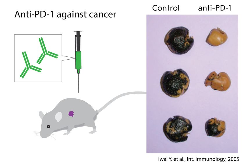
6The first studies where an antibody to the 3). Objective responses were seen in 20-25% of
receptor PD-1 was used to treat experimental patients with advanced non-small-cell-lung
cancer in mice were published in 2005 (Fig. 2 cancer (NSCLC), melanoma, or renal cancer,
and Fig. 4). The paper from Honjo’s group most of which were durable and some resulting in
presented several key conclusions that correctly complete tumor regression (Topalian et al.,
anticipated the clinical pattern emerging today: 2012). In 14% of patients, severe adverse
anti-PD-1 treatment could a) induce immune events, mainly pneumonitis, occurred. A phase I
responses, also to tumors that did not express trial of another PD-1 antibody, pembrolizumab,
detectable PD-L1 or PD-L2 b) be at least as (or developed by Merck (at the time called
even more) efficient as CTLA-4 therapy to treat lambrolizumab), in 135 melanoma patients also
tumors; c) lead to less severe autoimmune side showed very positive results, with clinical
effects than CTLA-4 (Iwai et al, 2005). In parallel, responses in 38% of patients, most of them
Chen’s group used B7-H1/PD-L1 transfected durable and some of them observed in patients
cancer cells to demonstrate efficacy of antibody who had not responded (or ceased to respond) to
inhibition of PD-1 or PD-L1 in the treatment of anti-CTLA-4 treatment (Hamid et al., 2013). In
tumors that expressed high levels of ligands parallel, several randomized phase III trials,
(Hirano et al, 2005). where treatment with the PD-1 antibodies was
compared to arms in which patients received
This development led to clinical studies based on standard chemotherapy, confirmed their efficacy
PD-1 inhibition and Honjo and coworkers had and safety (Brahmer et al., 2015; Borghaei et al.,
filed a patent based on this concept. This was 2015; Motzer et al., 2017). The first marketing
advanced by the company Ono Pharmaceuticals, and manufacturing approval was granted 2014 in
which at an early stage joined forces with Bristol- Japan. This was followed by FDA approval
Myers Squibb for the development of clinical through accelerated and breakthrough filing
grade antibodies directed against PD-1. The pathways, for two different anti PD-1 antibodies
clinical trials of the PD-1 antibody nivolumab later the same year, pembrolizumab and
were led by Suzanne Topalian. The first phase I nivolumab, for the treatment of unresectable or
study, initiated in 2006, showed that the drug was metastatic melanoma. The next year the approval
well tolerated (Brahmer et al., 2010). Two years was expanded to patients with metastatic
later, Topalian et al. reported the use of anti-PD-1 squamous non-small cell lung cancer in March
antibodies in 296 patients in a phase I study, 2015 by FDA and EMA approved the use of PD-1
indicating remarkable efficacy in treatment of blockade in Europe for treatment of melanoma
advanced disease of different tumor types (Fig. the same year.
7Figure 3. Immune checkpoint therapy in non-small cell lung cancer treated with anti-PD-1 (adopted from
Topalian et al., 2012). Please note the pseudoprogression at 2 months due to infiltrating immune cells
and the reduced tumor size after 4 months.
Cancer therapy using checkpoint inhibition and the curves that follow tumor-free survival
today show a plateau. In resected stage III and IV
melanoma anti-PD-1 yielded significantly
More than fifteen years has passed since the first improved results as compared to anti-CTLA-4
dose of anti-CTLA4 antibodies was given to a treatment with ipililumab (Weber et al., 2017).
patient, while the first treatment with anti-PD-1 Recurrence-free survival after 18 months of
antibodies was administered twelve years ago. treatment with pembrolizumab in resected stage
Today, there is a solid clinical experience, based III melanoma was 71% (Eggermont, 2018). This
not only on mono-treatment with each antibody, is a spectacular development when considering
but also on combination treatment. The metastatic disease of a solid tumor, which
introduction of ICIs has dramatically changed the previously meant death within two years for most
situation for patients with advanced, metastatic patients.
melanoma. Most of the responses are durable,
Figure 4. The role of CTLA-4 and PD-1 as inhibitors of activation (upper panels) and the effect of
releasing the corresponding brakes using antibodies (lower panels).
While the outcome of multiple studies suggest CTLA-4, combined therapy with blockade of both
that ICIs against PD-1 or its ligand, PD-L1, yield CTLA-4 and PD-1 seems to induce an even
better treatment effects as compared to anti- stronger anti-tumor effect in both melanoma
8(Wolchock et al., 2013; Wolchock et al., 2017), patients treated with anti-CTLA-4 or anti-PD-
depicted in Fig. 5, and in renal cell carcinoma 1/PD-L1. Adrenal insufficiency whether caused
(Motzer et al., 2018), possibly because of by inflammation of the pituitary or destruction of
different mechanisms of action. As expected, the adrenal cortex can be life-threatening due to
there are severe side effects in the form of inability to produce endogenous cortisol, but can
autoimmunity, in particular during anti-CTLA-4 be easily treated with hydrocortisone
treatment (Eggermont et al., 2018; Postow et al., supplementation, whereas e.g. pneumonitis may
2018). Regardless of which therapy that is require aggressive immunomodulatory treatment
employed, immune-related adverse events occur with e.g. synthetic steroid or monoclonal antibody
at a high rate, as the immune system is no longer therapy directed against tumor necrosis factor
fully controlled, although with different clinical (anti-TNF). As with many cancer treatment
presentations. With inhibition of CTLA-4, severe modalities, the adverse reactions associated with
colitis or endocrine dysfunction affecting the immune checkpoint therapy can in some
pituitary-adrenal axis are not uncommon, instances be fatal.
whereas thyroiditis shows a similar frequency in
Figure 5. The effect of anti-CTLA-4, anti-PD-1 and combination therapy in a subgroup of patients with
melanoma (adopted from Wolchock et al., 2017).
The ICIs have revolutionized the treatment of on cure and long-term survival. There are so far
tumors, where there previously was precious little six anti-PD-1 or anti–PD-L1 antibodies approved
to offer, and while still many patients do not by the FDA. The tumors that show the highest
respond, there are numerous long-term survivors. frequency of responses (50-90%) are Hodgkin´s
Using other forms of treatment, durable lymphoma especially in patients with
recurrence-free survival is extremely rare among overexpression of PD-L1 and PD-L2 caused by
patients with certain tumors, such as advanced gene amplification (Ansell et al., 2015), Merkel
melanoma and NSCLC. cell carcinoma of the skin, being of viral origin
(Nghiem et al., 2016), in microsatellite-instability
Currently very large numbers of studies for cancers of any origin having high mutational load
almost all cancer forms are being carried out from mismatch-repair deficiency (Le et al., 2017)
worldwide and the development initiated by the and in desmoplasmic melanoma carrying
study of CTLA-4 and PD-1 and its ligands is likely numerous, UV-induced mutations (Eroglu et al.,
to continue. Promising results have been 2018). Incidentally, the anti PD-1 antibodies were
reported for anti-PD-1 treatment in several types the first class of agents granted approval by the
of cancer, even if there is still limited information FDA based on a genetic characteristic of the
9cancer rather on the organ or histological origin depending on their genetic constitution as well as
of the disease in 2016. on their previous immune responses.
The development is likely to continue, with There are several interesting observations on
combination treatments – using different agents how processes upstream of the initial T cell
to release the negative regulation of immune activation can correlate with the response of the
responses, but also by combining them with patient. These processes include mutational load
strategies to activate or improve immune in the tumor, and, as a consequence, its
responses (e g vaccination, adoptive cell therapy) expression of antigens, previous infections or
or simply combining them with existing modalities vaccination, and the microenvironment and the
(surgery, chemotherapy, radiotherapy, hormone microbiome in niches such as the colon or the
treatment and drugs targeting specific pathways) skin (Snyder et al., 2014; Rizvi et al., 2015; Van
in adjuvant or neo-adjuvant protocols. Novel ICIs Allen et al., 2015; Le et al, 2017; Mariathasan,
will undoubtedly add additional fuel into this 2018; Tauriello et al., 2018; Gopalakrishnan et
development with numerous candidates in the al., 2018; Routy et al., 2018).
pipeline of the biotech and pharmaceutical
industry. The actual mechanisms whereby the antibodies
interfere with negative regulation are also
Mechanisms in the clinical response and how complex. It has been suggested that temporal
to predict it and spatial differences between the CTLA-4 and
PD-1 pathways, due to receptor expression
It is so far clear that the common and general kinetics and ligand expression pattern, lead to
mechanism by which checkpoint therapy different and complementary roles in the immune
functions is via unleashing of the negative control response, and consequently, synergistic effects
of T cells, leading to stronger and more sustained in combination therapy. It was initially assumed
immune response to the tumor. Immuno- that anti-CTLA-4 antibodies worked by interfering
histological and transcriptomic studies of biopsies with intrinsic T cell signaling and mainly acting in
indicate that a proportion of tumor infiltrating T lymph nodes to prevent initial T cell activation.
cells, and signs of immune activation in the tumor However, the importance of intrinsic signaling is
prior to or during treatment, are associated with a debated, and later studies by several groups,
favorable outcome (reviewed in Ribas & including Allison’s, have provided evidence for at
Wolchock, 2018). An analysis of 1535 advanced least four different mechanisms 1) Blockade of a
cancer patients treated with immune checkpoint direct inhibitory intracellular signaling pathway in
inhibitors demonstrated that maximal effector T cells 2) Competition for, or
heterozygosity at HLA class I loci improved sequestration of, activating ligands on antigen
overall survival, suggesting that antigen presenting cells 3) Interference with the function
presentation to T cells plays a key role (Chowell of regulatory T cells 4) Elimination of regulatory T
et al., 2018). The molecular mechanisms of cells. Note that the last mechanism may be
primary as well as acquired resistance of tumor exerted in the local tumor environment via
cells to anti PD-1 therapy converge in alterations antibody dependent cell mediated cytotoxicity.
in the antigen presentation machinery (e.g. loss
of β2-microglobulin, Zaretsky et al., 2016) and in Regarding PD-1, there is strong evidence that the
the IFN-γ receptor pathway controlling PD-L1 antibodies act predominantly via T cell intrinsic
expression (Ribas & Wolchock, 2018). signaling, mediated by the ITSM in the
cytoplasmic tail of the molecule (Okazaki et al.,
The responses can include and be influenced by 2001). Furthermore, a major part of the effect
several downstream mechanisms, mediated also occurs intratumorally to counteract exhaustion of
by other cells of the immune system. These cells infiltrating T cells. Indeed, cancer cells frequently
and various molecules are intensively studied as express PD-L1, and PD-L1 staining of tumor
biomarkers, and it should be emphasized that the biopsies has therefore been examined
critical effector mechanisms may vary between extensively as a predictive marker. While there is
tumors, stage of disease and among patients a correlation between tumor cell expression of
10the ligand PD-L1 by immunohistochemistry and identification of CTLA-4 and PD1/PD-L1 is only
the likelihood of response to anti-PD-1, some the beginning and additional molecules with
patients (and mice) with undetectable PD-Ll on similar functions are already being investigated.
their tumors also respond (Brahmer et al., 2010; So far there is no single or combination of
Topalian et al., 2012), and effects in the lymph immune biomarkers that can predict the clinical
nodes are not excluded, nor are effects exerted response with certainty before or early in the
via PD-1 expressed by regulatory T cells. treatment. There is intensive research going on,
and the identification of key mechanisms and
A crucial aspect in the future development is to biomarkers is likely to improve clinical efficacy
improve understanding of these mechanisms, as and safety as well as patient selection.
well as the ones leading to adverse events. The
Figure 6. The three pillars of cancer treatment, all directed against the cancer cell, and the fourth,
immune checkpoint inhibitor, based on unleashing an immune response against the tumor, added by
Allison and Honjo.
Concluding remarks the perhaps unprecedented research activities in
the immune checkpoint field, it is likely that there
This year’s Nobel Prize in Physiology or Medicine will be major developments regarding this
awards the discovery of a novel principle for therapy at all levels. This demonstrates how
tumor therapy based on removal of the brakes in influential the discoveries of Allison and Honjo
T cells. This new form of immunotherapy have been. Their findings have conferred great
unleashes a vigorous, and often durable, immune benefit on mankind; they add a new pillar to the
response directed against essentially any tumor existing cancer treatments (Fig. 6).
already recognized by the immune system. Given
References
Agata Y, Kawasaki A, Nishimura H, Ishida Y, Tsubata T, Yagita H, Honjo T. Expression of the PD-1
antigen on the surface of stimulated mouse T and B lymphocytes. Int Immunol. 1996, 8(5):765-72.
Ansell SM, Lesokhin AM, Borrello I, Halwani A, Scott EC, Gutierrez M, Schuster SJ, Millenson MM, Cattry
D, Freeman GJ, Rodig SJ, Chapuy B, Ligon AH, Zhu L, Grosso JF, Kim SY, Timmerman JM, Shipp MA,
11Armand P. PD-1 blockade with nivolumab in relapsed or refractory Hodgkin's lymphoma. N Engl J Med. 2015, 372(4):311-9. Bailey MH, Tokheim C, Porta-Pardo E, Sengupta S, Bertrand D, Weerasinghe A, Colaprico A, Wendl MC, Kim J, Reardon B, Ng PK, Jeong KJ, Cao S, Wang Z, Gao J, Gao Q, Wang F, Liu EM, Mularoni L, Rubio- Perez C, Nagarajan N, Cortés-Ciriano I, Zhou DC, Liang WW, Hess JM, Yellapantula VD, Tamborero D, Gonzalez-Perez A, Suphavilai C, Ko JY, Khurana E, Park PJ, Van Allen EM, Liang H; MC3 Working Group; Cancer Genome Atlas Research Network, Lawrence MS, Godzik A, Lopez-Bigas N, Stuart J, Wheeler D, Getz G, Chen K, Lazar AJ, Mills GB, Karchin R, Ding L. Comprehensive Characterization of Cancer Driver Genes and Mutations. Cell. 2018, 173(2):371-385.e18. Borghaei H, Paz-Ares L, Horn L, Spigel DR, Steins M, Ready NE, Chow LQ, Vokes EE, Felip E, Holgado E, Barlesi F, Kohlhäufl M, Arrieta O, Burgio MA, Fayette J, Lena H, Poddubskaya E, Gerber DE, Gettinger SN, Rudin CM, Rizvi N, Crinò L, Blumenschein GR Jr, Antonia SJ, Dorange C, Harbison CT, Graf Finckenstein F, Brahmer JR. Nivolumab versus Docetaxel in Advanced Nonsquamous Non-Small- Cell Lung Cancer. N Engl J Med. 2015, 373(17):1627-39. Brahmer JR, Drake CG, Wollner I, Powderly JD, Picus J, Sharfman WH, Stankevich E, Pons A, Salay TM, McMiller TL, Gilson MM, Wang C, Selby M, Taube JM, Anders R, Chen L, Korman AJ, Pardoll DM, Lowy I, Topalian SL.Phase I study of single-agent anti-programmed death-1 (MDX-1106) in refractory solid tumors: safety, clinical activity, pharmacodynamics, and immunologic correlates. J Clin Oncol. 2010, 28(19):3167-75. Brahmer JR, Rodríguez-Abreu D, Robinson AG, Hui R, Csőszi T, Fülöp A, Gottfried M, Peled N, Tafreshi A, Cuffe S, O'Brien M, Rao S, Hotta K, Zhang J, Lubiniecki GM, Deitz AC, Rangwala R, Reck M. Health- related quality-of-life results for pembrolizumab versus chemotherapy in advanced, PD-L1-positive NSCLC (KEYNOTE-024): a multicentre, international, randomised, open-label phase 3 trial. Lancet Oncol. 2017, 18(12):1600-1609. Brunet JF, Denizot F, Luciani MF, Roux-Dosseto M, Suzan M, Mattei MG, Golstein P. A new member of the immunoglobulin superfamily--CTLA-4. Nature. 1987, 328(6127):267-70. Brunner KT, Mauel J, Cerottini JC, Chapus B. Quantitative assay of the lytic action of immune lymphoid cells on 5"Cr-labelled allogeneic target cells in vitro; inhibition by isoantibody and by drugs Immunology. 1968, 14:181-96. Burnett FM. The concept of immunological surveillance. Prog Exp Tumor Res. 1970, 13:1-27. Busch W. Aus der Sitzung der medicinischen Section vom 18. November 1867. Berliner klinische Wochenscr. 1868, 5:137-8. Carreno BM, Collins M. The B7 family of ligands and its receptors: new pathways for costimulation and inhibition of immune responses. Annu Rev Immunol. 2002, 20:29-53. Chowell D, Morris LGT, Grigg CM, Weber JK, Samstein RM, Makarov V, Kuo F, Kendall SM, Requena D, Riaz N, Greenbaum B, Carroll J, Garon E, Hyman DM, Zehir A, Solit D, Berger M, Zhou R, Rizvi NA, Chan TA. Patient HLA class I genotype influences cancer response to checkpoint blockade immunotherapy. Science. 2018, 359(6375):582-587. Coley WB. Contribution to the knowledge of sarcoma. Ann Surg. 1891, 14(3):199-220. Dong H, Chen L. B7-H1 pathway and its role in the evasion of tumor immunity. J Mol Med 2003, 81(5):281-7. Dong H, Strome SE, Salomao DR, Tamura H, Hirano F, Flies DB, Roche PC, Lu J, Zhu G, Tamada K, Lennon VA, Celis E, Chen L. Tumor-associated B7-H1 promotes T-cell apoptosis: a potential mechanism of immune evasion. Nat Med. 2002, 8(8):793-800. Dong H, Zhu G, Tamada K, Chen L. B7-H1, a third member of the B7 family, co-stimulates T-cell proliferation and interleukin-10 secretion. Nat Med. 1999, 5(12):1365-9. Dunn GP, Bruce AT, Ikeda H, Old LJ, Schreiber RD. Cancer immunoediting: from immunosurveillance to tumor escape. Nat Immunol. 2002, 3:991-8. Eggermont AMM, Robert C, Ribas A. The new era of adjuvant therapies for melanoma. Nat Rev Clin Oncol. 2018, 15(9):535-536. 12
Ehrlich P. Über den jetzigen Stand der Karzinomforschung. Nederlandsch Tijdschrift voor Geneeskunde
1909, 5:273-90.
Eroglu Z, Zaretsky JM, Hu-Lieskovan S, Kim DW, Algazi A, Johnson DB, Liniker E, Ben Kong, Munhoz R,
Rapisuwon S, Gherardini PF, Chmielowski B, Wang X, Shintaku IP, Wei C, Sosman JA, Joseph RW,
Postow MA, Carlino MS, Hwu WJ, Scolyer RA, Messina J, Cochran AJ, Long GV, Ribas A. High response
rate to PD-1 blockade in desmoplastic melanomas. Nature. 2018, 553(7688):347-350
Fehleisen F. Ueber die Zuchtung der Erysipelkokken auf künstlichem Nährboden und ihre
Uebertragsbarkeit auf den Menschen. Deutsche Med. Wochenschr. 1882, 8:553-4.
Forde PM, Chaft JE, Smith KN, Anagnostou V, Cottrell TR, Hellmann MD, Zahurak M, Yang SC, Jones
DR, Broderick S, Battafarano RJ, Velez MJ, Rekhtman N, Olah Z, Naidoo J, Marrone KA, Verde F, Guo
H, Zhang J, Caushi JX, Chan HY, Sidhom JW, Scharpf RB, White J, Gabrielson E, Wang H, Rosner GL,
Rusch V, Wolchok JD, Merghoub T, Taube JM, Velculescu VE, Topalian SL, Brahmer JR, Pardoll DM.
Neoadjuvant PD-1 Blockade in Resectable Lung Cancer. N Engl J Med. 2018, 378(21):1976-1986.
Freeman GJ, Long AJ, Iwai Y, Bourque K, Chernova T, Nishimura H, Fitz LJ, Malenkovich N, Okazaki T,
Byrne MC, Horton HF, Fouser L, Carter L, Ling V, Bowman MR, Carreno BM, Collins M, Wood CR, Honjo
T. Engagement of the PD-1 immunoinhibitory receptor by a novel B7 family member leads to negative
regulation of lymphocyte activation. J Exp Med. 2000, 192(7):1027-34.
Gettinger S, Horn L, Jackman D, Spigel D, Antonia S, Hellmann M, Powderly J, Heist R, Sequist LV,
Smith DC, Leming P, Geese WJ, Yoon D, Li A, Brahmer J. Five-Year Follow-Up of Nivolumab in
Previously Treated Advanced Non-Small-Cell Lung Cancer: Results From the CA209-003 Study. J Clin
Oncol. 2018, 36(17):1675-1684.
Global Cancer Observatory, 2018. http://gco.iarc.fr/
Greenwald RJ, Latchman YE, Sharpe AH. Negative co-receptors on lymphocytes. Curr Opin Immunol.
2002, 14(3):391-6.
Gopalakrishnan V, Spencer CN, Nezi L, Reuben A, Andrews MC, Karpinets TV, Prieto PA, Vicente D,
Hoffman K, Wei SC, Cogdill AP, Zhao L, Hudgens CW, Hutchinson DS, Manzo T, Petaccia de Macedo M,
Cotechini T, Kumar T, Chen WS, Reddy SM, Szczepaniak Sloane R, Galloway-Pena J, Jiang H, Chen
PL, Shpall EJ, Rezvani K, Alousi AM, Chemaly RF, Shelburne S, Vence LM, Okhuysen PC, Jensen VB,
Swennes AG, McAllister F, Marcelo Riquelme Sanchez E, Zhang Y, Le Chatelier E, Zitvogel L, Pons N,
Austin-Breneman JL, Haydu LE, Burton EM, Gardner JM, Sirmans E, Hu J, Lazar AJ, Tsujikawa T, Diab
A, Tawbi H, Glitza IC, Hwu WJ, Patel SP, Woodman SE, Amaria RN, Davies MA, Gershenwald JE, Hwu
P, Lee JE, Zhang J, Coussens LM, Cooper ZA, Futreal PA, Daniel CR, Ajami NJ, Petrosino JF, Tetzlaff
MT, Sharma P, Allison JP, Jenq RR, Wargo JA. Gut microbiome modulates response to anti-PD-1
immunotherapy in melanoma patients. Science. 2018, 359(6371):97-103.
Hanahan D, Weinberg RA. Hallmarks of cancer: the next generation. Cell. 2011, 144:646-74.
Hansen JA, Martin PJ, Nowinski RC. Monoclonal antibodies identifying a novel T-cell antigen and Ia
antigens on human lymphocytes. Immunogenetics. 1980, 10:247-60.
Hellmann MD, Rizvi NA, Goldman JW, Gettinger SN, Borghaei H, Brahmer JR, Ready NE, Gerber DE,
Chow LQ, Juergens RA, Shepherd FA, Laurie SA, Geese WJ, Agrawal S, Young TC, Li X, Antonia SJ.
Nivolumab plus ipilimumab as first-line treatment for advanced non-small-cell lung cancer (CheckMate
012): results of an open-label, phase 1, multicohort study. Lancet Oncol. 2017, 8(1):31-41.
Herbst RS, Soria JC, Kowanetz M, Fine GD, Hamid O, Gordon MS, Sosman JA, McDermott DF,
Powderly JD, Gettinger SN, Kohrt HE, Horn L, Lawrence DP, Rost S, Leabman M, Xiao Y, Mokatrin A,
Koeppen H, Hegde PS, Mellman I, Chen DS, Hodi FS. Predictive correlates of response to the anti-PD-L1
antibody MPDL3280A in cancer patients. Nature. 2014, 515(7528):563-7.
Hirano F, Kaneko K, Tamura H, Dong H, Wang S, Ichikawa M, Rietz C, Flies DB, Lau JS, Zhu G, Tamada
K, Chen L. Blockade of B7-H1 and PD-1 by monoclonal antibodies potentiates cancer therapeutic
immunity. Cancer Res. 2005, 65(3):1089-96.
Hodi FS, Mihm MC, Soiffer RJ, Haluska FG, Butler M, Seiden MV, Davis T, Henry-Spires R, MacRae S,
Willman A, Padera R, Jaklitsch MT, Shankar S, Chen TC, Korman A, Allison JP, Dranoff G. Biologic


                                                     13
activity of cytotoxic T lymphocyte-associated antigen 4 antibody blockade in previously vaccinated
metastatic melanoma and ovarian carcinoma patients. Proc Natl Acad Sci U S A. 2003, 100(8):4712-7.
Hodi FS, O'Day SJ, McDermott DF, Weber RW, Sosman JA, Haanen JB, Gonzalez R, Robert C,
Schadendorf D, Hassel JC, Akerley W, van den Eertwegh AJ, Lutzky J, Lorigan P, Vaubel JM, Linette
GP, Hogg D, Ottensmeier CH, Lebbé C, Peschel C, Quirt I, Clark JI, Wolchok JD, Weber JS, Tian J,
Yellin MJ, Nichol GM, Hoos A, Urba WJ. . Improved survival with ipilimumab in patients with metastatic
melanoma. N Engl J Med. 2010, 363(8):711-23. Erratum in: N Engl J Med. 2010, 363(13):1290.
Hurwitz AA, Yu TF, Leach DR, Allison JP. CTLA-4 blockade synergizes with tumor-derived granulocyte-
macrophage colony-stimulating factor for treatment of an experimental mammary carcinoma. Proc Natl
Acad Sci U S A. 1998, 95(17):10067-71.
Ishida Y, Agata Y, Shibahara K, Honjo T. Induced expression of PD-1, a novel member of the
immunoglobulin gene superfamily, upon programmed cell death. EMBO J. 1992, 11(11):3887-95.
Iwai Y, Ishida M, Tanaka Y, Okazaki T, Honjo T, Minato N. Involvement of PD-L1 on tumor cells in the
escape from host immune system and tumor immunotherapy by PD-L1 blockade. Proc Natl Acad Sci U
S A. 2002, 99(19):12293-7.
Iwai Y, Terawaki S, Honjo T. PD-1 blockade inhibits hematogenous spread of poorly immunogenic tumor
cells by enhanced recruitment of effector T cells. Int Immunol. 2005, 17(2):133-44.
June CH, Sadelain M. Chimeric antigen receptor therapy. N Engl J Med. 2018, 379(1):64-73.
Klein G, Klein E. Antigenic properties of other experimental tumors. Cold Spring Harb Symp Quant Biol
1962, 27: 463-70.
Klein E, Svedmyr E, Jondal M, Vanky F. Functional studies on tumor-infiltrating lymphocytes in man. Isr J
Med Sci. 13:747-52, 1977
Krummel MF, Allison JP. CD28 and CTLA-4 have opposing effects on the response of T cells to
stimulation. J Exp Med. 1995, 182(2):459-65.
Kwon ED, Hurwitz AA, Foster BA, Madias C, Feldhaus AL, Greenberg NM, Burg MB, Allison JP.
Manipulation of T cell costimulatory and inhibitory signals for immunotherapy of prostate cancer. Proc
Natl Acad Sci U S A. 1997, 94(15):8099-103.
Lamm DL, Thor DE, Harris SC, Reyna JA, Stogdill VD, Radwin HM.Bacillus Calmette-Guerin
immunotherapy of superficial bladder cancer. J Urol. 1980, 124(1):38-40.
Latchman Y, Wood CR, Chernova T, Chaudhary D, Borde M, Chernova I, Iwai Y, Long AJ, Brown JA,
Nunes R, Greenfield EA, Bourque K, Boussiotis VA, Carter LL, Carreno BM, Malenkovich N, Nishimura H,
Okazaki T, Honjo T, Sharpe AH, Freeman GJ. PD-L2 is a second ligand for PD-1 and inhibits T cell
activation. Nat Immunol. 2001, 2(3):261-8.
Le DT, Durham JN, Smith KN, Wang H, Bartlett BR, Aulakh LK, Lu S, Kemberling H, Wilt C, Luber BS,
Wong F, Azad NS, Rucki AA, Laheru D, Donehower R, Zaheer A, Fisher GA, Crocenzi TS, Lee JJ,
Greten TF, Duffy AG, Ciombor KK, Eyring AD, Lam BH, Joe A, Kang SP, Holdhoff M, Danilova L, Cope L,
Meyer C, Zhou S, Goldberg RM, Armstrong DK, Bever KM, Fader AN, Taube J, Housseau F, Spetzler D,
Xiao N, Pardoll DM, Papadopoulos N, Kinzler KW, Eshleman JR, Vogelstein B, Anders RA, Diaz LA Jr.
Mismatch repair deficiency predicts response of solid tumors to PD-1 blockade. Science. 2017,
357(6349):409-413.
Leach DR, Krummel MF, Allison JP. Enhancement of antitumor immunity by CTLA-4 blockade. Science.
1996, 271(5256):1734-6.
Leibson PJ. The regulation of lymphocyte activation by inhibitory receptors. Curr Opin Immunol. 2004,
16(3):328-36.
Lindsten T, Lee KP, Harris ES, Petryniak B, Craighead N, Reynolds PJ, Lombard DB, Freeman GJ,
Nadler LM, Gray GS,Thompson CB, June CH. Characterization of CTLA-4 structure and expression on
human T cells. J Immunol. 1993, 151(7):3489-99.


                                                 14
Linsley PS, Bradshaw J, Greene J, Peach R, Bennett KL, Mittler RS. Intracellular trafficking of CTLA-4
and focal localization towards sites of TCR engagement. Immunity. 1996, 4(6):535-43.
Linsley PS, Brady W, Urnes M, Grosmaire LS, Damle NK, Ledbetter JA. CTLA-4 is a second receptor for
the B cell activation antigen B7. J Exp Med. 1991, 174(3):561-9
Linsley PS, Clark EA, Ledbetter JA. T-cell antigen CD28 mediates adhesion with B cells by interacting
with activation antigen B7/BB-1. Proc Natl Acad Sci U S A. 1990, 87(13):5031-5.
Linsley PS, Wallace PM, Johnson J, Gibson MG, Greene JL, Ledbetter JA, Singh C, Tepper MA.
Immunosuppression in vivo by a soluble form of the CTLA-4 T cell activation molecule. Science. 1992,
257:792-5.
Loeb L. Further Investigations in Transplantation of tumors. J Med Res. 1902, 8(1):44-73.
Lurquin C, Van Pel A, Mariamé B, De Plaen E, Szikora JP, Janssens C, Reddehase MJ, Lejeune J, Boon
                               -
T. Structure of the gene of tum transplantation antigen P91A: the mutated exon encodes a peptide
recognized with Ld by cytolytic T cells. Cell. 1989, 58:293-303.
Mariathasan S, Turley SJ, Nickles D, Castiglioni A, Yuen K, Wang Y, Kadel EE III, Koeppen H, Astarita
JL, Cubas R, Jhunjhunwala S, Banchereau R, Yang Y, Guan Y, Chalouni C, Ziai J, Şenbabaoğlu Y,
Santoro S, Sheinson D, Hung J, Giltnane JM, Pierce AA, Mesh K, Lianoglou S, Riegler J, Carano RAD,
Eriksson P, Höglund M, Somarriba L, Halligan DL, van der Heijden MS, Loriot Y, Rosenberg JE, Fong L,
Mellman I, Chen DS, Green M, Derleth C, Fine GD, Hegde PS, Bourgon R, Powles T. TGFβ attenuates
tumour response to PD-L1 blockade by contributing to exclusion of T cells. Nature. 2018, 554(7693):544-
548.
Martin PJ, Ledbetter JA, Morishita Y, June CH, Beatty PG, Hansen JA. A 44 kilodalton cell surface
homodimer regulates interleukin 2 production by activated human T lymphocytes. J Immunol. 1986,
136(9):3282-7.
Morales A, Eidinger D, Bruce AW. Intracavitary Bacillus Calmette-Guerin in the treatment of superficial
bladder tumors. J Urol. 1976, 116(2):180-3.
Motzer RJ, Escudier B, McDermott DF, George S, Hammers HJ, Srinivas S, Tykodi SS, Sosman JA,
Procopio G, Plimack ER, Castellano D, Choueiri TK, Gurney H, Donskov F, Bono P, Wagstaff J, Gauler
TC, Ueda T, Tomita Y, Schutz FA, Kollmannsberger C, Larkin J, Ravaud A, Simon JS, Xu LA, Waxman
IM, Sharma P; CheckMate 025 Investigators.Nivolumab versus Everolimus in Advanced Renal-Cell
Carcinoma. N Engl J Med. 2015, 373(19):1803-13.
Nghiem PT, Bhatia S, Lipson EJ, Kudchadkar RR, Miller NJ, Annamalai L, Berry S, Chartash EK, Daud A,
Fling SP, Friedlander PA, Kluger HM, Kohrt HE, Lundgren L, Margolin K, Mitchell A, Olencki T, Pardoll
DM, Reddy SA, Shantha EM, Sharfman WH, Sharon E, Shemanski LR, Shinohara MM, Sunshine JC,
Taube JM, Thompson JA, Townson SM, Yearley JH, Topalian SL, Cheever MA. PD-1 Blockade with
Pembrolizumab in Advanced Merkel-Cell Carcinoma. N Engl J Med. 2016, 374(26):2542-52
Nishimura H, Agata Y, Kawasaki A, Sato M, Imamura S, Minato N, Yagita H, Nakano T, Honjo T.
Developmentally regulated expression of the PD-1 protein on the surface of double-negative (CD4-CD8-)
thymocytes. Int Immunol. 1996, 8(5):773-80.
Nishimura H, Honjo T. PD-1: an inhibitory immunoreceptor involved in peripheral tolerance. Trends
Immunol. 2001, 22(5):265-8.
Nishimura H, Minato N, Nakano T, Honjo T. Immunological studies on PD-1 deficient mice: implication of
PD-1 as a negative regulator for B cell responses. Int Immunol. 1998, 10(10):1563-72.
Nishimura H, Nose M, Hiai H, Minato N, Honjo T. Development of lupus-like autoimmune diseases by
disruption of the PD-1 gene encoding an ITIM motif-carrying immunoreceptor. Immunity. 1999, 11(2):141-
51.


                                                 15
Nishimura H, Okazaki T, Tanaka Y, Nakatani K, Hara M, Matsumori A, Sasayama S, Mizoguchi A, Hiai H, Minato N, Honjo T. Autoimmune dilated cardiomyopathy in PD-1 receptor-deficient mice. Science. 2001, 291(5502):319-22. Okazaki T, Maeda A, Nishimura H, Kurosaki T, Honjo T. PD-1 immunoreceptor inhibits B cell receptor- mediated signaling by recruiting src homology 2-domain-containing tyrosine phosphatase 2 to phosphotyrosine. Proc Natl Acad Sci U S A. 2001, 98(24):13866-71. Phan GQ, Yang JC, Sherry RM, Hwu P, Topalian SL, Schwartzentruber DJ, Restifo NP, Haworth LR, Seipp CA, Freezer LJ, Morton KE, Mavroukakis SA, Duray PH, Steinberg SM, Allison JP, Davis TA, Rosenberg SA. Cancer regression and autoimmunity induced by cytotoxic T lymphocyte-associated antigen 4 blockade in patients with metastatic melanoma. Proc Natl Acad Sci U S A. 2003, 100(14):8372- 7. Postow MA, Sidlow R, Hellmann MD. Immune-Related Adverse Events Associated with Immune Checkpoint Blockade. N Engl J Med. 2018, 378(2):158-168. Rizvi NA, Hellmann MD, Snyder A, Kvistborg P, Makarov V, Havel JJ, Lee W, Yuan J, Wong P, Ho TS, Miller ML, Rekhtman N, Moreira AL, Ibrahim F, Bruggeman C, Gasmi B, Zappasodi R, Maeda Y, Sander C, Garon EB, Merghoub T, Wolchok JD, Schumacher TN, Chan TA. Cancer immunology. Mutational landscape determines sensitivity to PD-1 blockade in non-small cell lung cancer. Science. 2015, 348(6230):124-8. Robert C, Ribas A, Wolchok JD, Hodi FS, Hamid O, Kefford R, Weber JS, Joshua AM, Hwu WJ, Gangadhar TC, Patnaik A, Dronca R, Zarour H, Joseph RW, Boasberg P, Chmielowski B, Mateus C, Postow MA, Gergich K, Elassaiss-Schaap J, Li XN, Iannone R, Ebbinghaus SW, Kang SP, Daud A. Anti- programmed-death-receptor-1 treatment with pembrolizumab in ipilimumab-refractory advanced melanoma: a randomised dose-comparison cohort of a phase 1 trial. Lancet. 2014, 384(9948):1109-17. Rodig SJ, Gusenleitner D, Jackson DG, Gjini E, Giobbie-Hurder A, Jin C, Chang H, Lovitch SB, Horak C, Weber JS, Weirather JL, Wolchok JD, Postow MA, Pavlick AC, Chesney J, Hodi FS. MHC proteins confer differential sensitivity to CTLA-4 and PD-1 blockade in untreated metastatic melanoma. Sci Transl Med. 2018, 18;10(450). Rosenberg SA, Aebersold P, Cornetta K, Kasid A, Morgan RA, Moen R, Karson EM, Lotze MT, Yang JC, Topalian SL, Merino MJ, Culver K, Miller D, Blaese RM, Anderson WF. Gene transfer into humans-- immunotherapy of patients with advanced melanoma, using tumor-infiltrating lymphocytes modified by retroviral gene transduction. N Engl J Med. 1990, 323(9):570-8. Rosenberg SA, Spiess P, Lafreniere R A new approach to the adoptive immunotherapy of cancer with tumor-infiltrating lymphocytes. Science. 1986, 233:1318-21. Routy B, Le Chatelier E, Derosa L, Duong CPM, Alou MT, Daillère R, Fluckiger A, Messaoudene M, Rauber C, Roberti MP, Fidelle M, Flament C, Poirier-Colame V, Opolon P, Klein C, Iribarren K, Mondragón L, Jacquelot N, Qu B, Ferrere G, Clémenson C, Mezquita L, Masip JR, Naltet C, Brosseau S, Kaderbhai C, Richard C, Rizvi H, Levenez F, Galleron N, Quinquis B, Pons N, Ryffel B, Minard-Colin V, Gonin P, Soria JC, Deutsch E, Loriot Y, Ghiringhelli F, Zalcman G, Goldwasser F, Escudier B, Hellmann MD, Eggermont A, Raoult D, Albiges L, Kroemer G, Zitvogel L. Gut microbiome influences efficacy of PD- 1-based immunotherapy against epithelial tumors. Science. 2018, 359(6371):91-97 Schachter J, Ribas A, Long GV, Arance A, Grob JJ, Mortier L, Daud A, Carlino MS, McNeil C, Lotem M, Larkin J, Lorigan P, Neyns B, Blank C, Petrella TM, Hamid O, Zhou H, Ebbinghaus S, Ibrahim N, Robert C. Pembrolizumab versus ipilimumab for advanced melanoma: final overall survival results of a multicentre, randomised, open-label phase 3 study (KEYNOTE-006). Lancet. 2017, 390(10105):1853- 1862. Shinohara T, Taniwaki M, Ishida Y, Kawaichi M, Honjo T. Structure and chromosomal localization of the human PD-1 gene (PDCD1). Genomics. 1994, 23(3):704-6. Snyder A, Makarov V, Merghoub T, Yuan J, Zaretsky JM, Desrichard A, Walsh LA, Postow MA, Wong P, Ho TS, Hollmann TJ, Bruggeman C, Kannan K, Li Y, Elipenahli C, Liu C, Harbison CT, Wang L, Ribas A, 16
You can also read