A Neuroprotective Dose of Isatin Causes Multilevel Changes Involving the Brain Proteome: Prospects for Further Research - MDPI
←
→
Page content transcription
If your browser does not render page correctly, please read the page content below
International Journal of
            Molecular Sciences

Article
A Neuroprotective Dose of Isatin Causes Multilevel
Changes Involving the Brain Proteome:
Prospects for Further Research
Alexei Medvedev *, Arthur Kopylov , Olga Buneeva, Leonid Kurbatov , Olga Tikhonova,
Alexis Ivanov and Victor Zgoda
 Department of Proteomic Research and Mass Spectrometry, Institute of Biomedical Chemistry,
 10 Pogodinskaya Street, 119121 Moscow, Russia; a.t.kopylov@gmail.com (A.K.); olbuneeva@gmail.com (O.B.);
 kurbatovl@mail.ru (L.K.); tiolika@gmail.com (O.T.); professor-ivanov@yandex.ru (A.I.);
 victor.zgoda@gmail.com (V.Z.)
 * Correspondence: professor57@yandex.ru; Tel.: +7-495-245-0509

 Received: 29 April 2020; Accepted: 9 June 2020; Published: 11 June 2020

 Abstract: Isatin (indole-2,3-dione) is an endogenous regulator, exhibiting a wide range of biological
 and pharmacological activities. At doses of 100 mg/kg and above, isatin is neuroprotective in different
 experimental models of neurodegeneration. Good evidence exists that its effects are realized via
 interaction with numerous isatin-binding proteins identified in the brain and peripheral tissues studied.
 In this study, we investigated the effect of a single dose administration of isatin to mice (100 mg/kg, 24 h)
 on differentially expressed proteins and a profile of the isatin-binding proteins in brain hemispheres.
 Isatin administration to mice caused downregulation of 31 proteins. However, these changes
 cannot be attributed to altered expression of corresponding genes. Although at this time point isatin
 influenced the expression of more than 850 genes in brain hemispheres (including 433 upregulated and
 418 downregulated genes), none of them could account for the changes in the differentially expressed
 proteins. Comparative proteomic analysis of brain isatin-binding proteins of control and isatin-treated
 mice revealed representative groups of proteins sensitive to isatin administration. Control-specific
 proteins (n = 55) represent specific targets that interact directly with isatin. Appearance of brain
 isatin-binding proteins specific to isatin-treated mice (n = 94) may be attributed to the formation of
 new clusters of protein–protein interactions and/or novel binding sites induced by a high concentration
 of this regulator (ligand-induced binding sites). Thus, isatin administration produces multiple effects
 in the brain, which include changes in gene expression and also profiles of isatin-binding proteins
 and their interactomes. Further studies are needed for deeper insight into the mechanisms of the
 multilevel changes in the brain proteome induced by isatin. In the context of the neuroprotective
 action, these changes may be aimed at interruption of pathological links that begin to form after
 initiation of pathological processes.

 Keywords: neuroprotector isatin; molecular targets; differentially expressed proteins; proteome analysis;
 isatin-binding proteins; interactome

1. Introduction
     Isatin (indole-2,3-dione) is an endogenous oxidized indole found in the brain, peripheral tissues,
and biological body fluids of humans and animals [1–5]. The interest of researchers in this “talented
molecule” [6] is determined by various regulatory effects described in the literature (see [1,7] for review)
and the role of isatin as a core structure in numerous synthetic isatin-based compounds exhibiting
different pharmacological activities [7–9]. Numerous isatin analogues have been synthesized and tested

Int. J. Mol. Sci. 2020, 21, 4187; doi:10.3390/ijms21114187                          www.mdpi.com/journal/ijms
Int. J. Mol. Sci. 2020, 21, 4187                                                                       2 of 21

as anticonvulsants [7,10], antibacterial [7,11], antiviral [7,12–14], anticancer agents [8], and inhibitors
of apoptosis [15,16]. Sunitinib (marketed by Pfizer as Sutent), a 5-fluoro-3-substituted isatin derivative,
was approved by the FDA (U.S. Food and Drug Administration) for treatment of advanced renal
cell carcinoma [17].
      Isatin has also been found in mammalian brain, peripheral tissues, and body fluids [18–23],
and there is experimental evidence for its endogenous origin [24]. Various types of stress have
a significant impact on isatin levels in the brain, serum, urine, and examined tissues. In rats exposed to
immobilization/audiogenic stress, the isatin levels in the brain, heart, and serum were 2–4-fold higher
than in control animals [21]. Cold-stressed rats (for 2 h at 4◦ C) exhibited a significantly higher (2–3-fold)
isatin content in the daily (24 h) urine [22]. Food deprivation for three days (with free access to water)
caused even a more pronounced (~5-fold) increase in isatin in the daily urine [22]. Administration of
the proconvulsant, pentylenetetrazole, increased (~1.5-fold) the brain isatin content [23].
      Exogenous isatin readily crosses the blood brain barrier; for example, isatin injection to rats
at a dose of 50–100 mg/kg increased the level of brain isatin up to 9 µg/g [23]. Isatin administered
in vivo produced various (dose-dependent) physiological/pharmacological effects. Low doses of isatin
(15–20 mg/kg) were anxiogenic in open field and elevated plus maze tests in albino mice [25,26] and
in social interaction test in rats, while the locomotor activity of these rats remained unchanged [25].
In contrast to mice, rats were insensitive to the anxiogenic doses of isatin in the open field and forced
swim tests [27], and higher doses of isatin (80–160 mg/kg) caused sedation manifested as the reduced
distance in the open field and increased immobility in the forced swim test [27]. The higher doses of
isatin (from 60 mg/kg to 200 mg/kg) produced an anticonvulsive effect evaluated in different models
including audiogenic seizures in rats [28,29] and pentylenetetrazole administration [30].
      In rats with the 6-hydroxydopamine model of Parkinsonism, isatin (100 mg/kg) inhibited rotations
induced by apomorphine [31]. In the context of other experimental models of Parkinson’s disease,
administration of isatin (100 mg/kg) also decreased locomotor impairments in rats with Parkinsonism
induced by Japanese encephalitis virus [32,33] and in mice with Parkinsonism induced by the neurotoxin
MPTP (1-methyl-4-phenyl-1 2 3 6-tetrahydropyridine) [34,35]. Isatin administration (100 mg/kg) also
influenced the profile of ubiquitinated mitochondrial proteins in the mouse brain. These changes were
observed 2 h after isatin administration [35].
      Biological activities of isatin involve isatin-binding proteins, identified during proteomic
profiling of brain preparations of mice and rats [1,36–38]. Interestingly, many of these proteins
are actively investigated in the context of neurodegenerative diseases such as Parkinson’s disease
and Alzheimer’s’ disease [37,39,40]. In vitro physiological concentrations of isatin (1–10 µM)
inhibit monoamine oxidase B (MAO B) and natriuretic peptide receptor coupled guanylate
cyclases [1,5]; higher (neuroprotective) concentrations (50–400 µM) inhibit non-glycolytic activity of
glyceraldehyde-3-phopshate dehydrogenase [39], induce apoptosis of various (including malignant)
cells, and affect gene expression associated with apoptosis [1,41–46]. Some authors even believe that
due to its cytotoxic and antiproliferative activities, isatin should be considered a good candidate as
a pharmacologically attractive chemotherapeutic substance [47].
      It should be noted that after administration of the neuroprotective doses of isatin (50–100 mg/kg),
its concentration in the brain may exceed 70 µM [1,4]. Our recent study has shown that treatment
of mice with a neuroprotective dose of isatin significantly influenced the proteomic profile of brain
isatin-binding proteins [48]. The detected changes in the profile of isatin-binding proteins are
consistent with the accumulation of administered isatin in the brain and its binding to target proteins,
thus preventing subsequent protein binding to the affinity sorbent containing an isatin analogue as the
affinity ligand [48]. Earlier, we also demonstrated that within a slightly different scheme of the animal
experiment, pretreatment of rats with a neuroprotector dose of isatin prevented brain MAO B against
irreversible inhibition by the mechanism-based inhibitor phenelzine [49]. Cell culture studies revealed
that incubation with high concentrations of isatin (100 µM and above) influenced the expression of
some genes [43–45,50,51].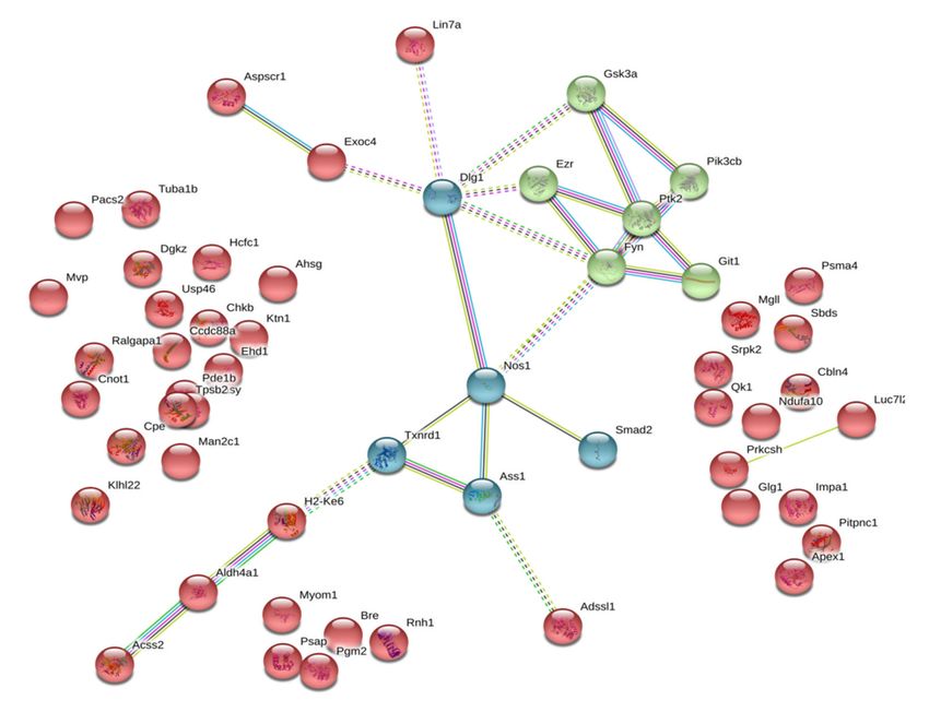
Int. J. Mol. Sci. 2020, 21, 4187                                                                                                                 3 of 21

       Thus, multiple biological/pharmacological effects of isatin may be attributed to both isatin
interaction with particular protein targets (isatin-binding proteins) and also regulation of
isatin-responsive genes. However, proteomic profiling of differentially expressed proteins and
their possible association with altered expression of corresponding genes have never been investigated
after isatin administration in vivo. Moreover, possibility of the regulation of gene expression in
the brain of animals by administration of a single dose of isatin has not been investigated at all;
certain evidence only exists that chronic treatment of animals with a low dose of isatin influenced the
expression of some selectively studied genes in the brain [52].
       Thus, the aim of this work was to investigate the effect of administration of isatin (100 mg/kg) to
mice one day before analysis on the following: (i) differently expressed proteins in brain hemispheres;
(ii) their possible association with altered gene expression; and (iii) profile of isatin-binding proteins in
the brain hemispheres. Previous studies have shown that this dose of isatin attenuated manifestations
of MPTP-induced Parkinsonism in mice [34,35]. The time interval of 24 h was chosen on the basis of
our analysis of literature data on altered expression of genes in cell cultures treated with isatin [1].
Such a time interval is often used for the drug-induced preconditioning of the brain [53].

2. Results

2.1. The Effect of Isatin Administration to Mice on Differently Expressed Proteins in Brain Hemispheres
     Isatin administration to mice caused downregulation of 31 proteins (Table 1). The most pronounced
changes were found in the case of calcium/calmodulin-dependent protein kinase type IV (about 11-fold),
fructose-1,6-bisphosphatase 1 (more than 8-fold), serine protease inhibitor A3K (more than 4-fold),
nucleolar protein 3 (almost 4-fold), and neurobeachin (more than 3-fold). These and other proteins
listed in Table 1 participate in processes involved in cell signaling, regulation of cell death and
proliferation, and also in protein synthesis. In this context, it is particularly interesting to note that
fructose-1,6-bisphosphatase 1 (FBP1), a classical glycolytic enzyme, may block the transcriptional
activity of the hypoxia-inducible factor (HIF-1α) and prevent activation of the RAS/RAF/MEK/ERK
pathway (see [54] for review).

      Table 1. Differentially expressed proteins detected in brain hemispheres 24 h after administration of
      a neuroprotective dose of isatin (100 mg/kg, s.c.).
               Entry     Entry Name   ANOVA (p)        Fold Change             Description                              Function
                                                                                                               E3 ubiquitin ligase of the
      1       Q9JLV5        CUL3           0             −2.681                  Cullin-3
                                                                                                             ubiquitin-proteasome system
      2       O35143        ATIF1     1.44 × 10⁻¹⁵       −2.655      ATPase inhibitor, mitochondrial          Regulation of ATP synthesis
                                                                     Ran-specific GTPase-activating         Regulation of nucleocytoplasmic
      3        P34022       RANG      2.00 ×   10−15     −2.736
                                                                                 protein                               transport
                                                                                                         Binding of iron ions and stimulation of
      4       Q921I1        TRFE      1.11 × 10⁻¹⁴       −2.062              Serotransferrin
                                                                                                                    cell proliferation
      5       Q8C8N2         SCAI     9.71 × 10⁻¹⁴       −2.521               Protein SCAI                         Tumor suppressor
                                                                                                           BAG family molecular chaperone
      6       Q9Z1R2        BAG6      1.32 × 10⁻¹³       −2.275      Large proline-rich protein BAG6        regulator 6 is involved in DNA
                                                                                                              damage-induced apoptosis
                                                                                                            E-selectin ligand 1, cysteine-rich
      7       Q61543        GSLG1     9.94 × 10⁻¹²       −2.164         Golgi apparatus protein 1
                                                                                                            fibroblast growth factor receptor
                                                                     Ubiquitin-conjugating enzyme               The component of the
      8       Q9D2M8        UB2V2     1.38 × 10⁻¹¹       −2.224
                                                                              E2 variant 2                   ubiquitin-proteasome system
                                                                     Nascent polypeptide-associated
      9        P70670      NACAM      4.60 × 10⁻¹¹       −2.293         complex subunit alpha,                   Molecular chaperone
                                                                          muscle-specific form
                                                                     Eukaryotic translation initiation
      10      Q8BGD9         IF4B     1.88 × 10⁻¹⁰       −2.453                                                    Protein translation
                                                                                factor 4B
Int. J. Mol. Sci. 2020, 21, 4187 4 of 21
Table 1. Cont.
Entry Entry Name ANOVA (p) Fold Change Description Function
In CNS, participates in reverse
11 Q00623 APOA1 4.70 × 10⁻¹⁰ −2.363 Apolipoprotein A-I
cholesterol transport
12 P84309 ADCY5 6.87 × 10⁻¹⁰ −2.72 Adenylate cyclase type 5 Cell signaling
Propionyl-CoA carboxylase beta
13 Q99MN9 PCCB 7.83 × 10⁻¹⁰ −2.249 Fatty acid beta-oxidation
chain, mitochondrial
A member of serine protease inhibitors,
required for normal synaptic plasticity
14 P07759 SPA3K 4.90 × 10⁻⁹ −4.156 Serine protease inhibitor A3K
and regulation of serine protease
mediated cell death
A protein required for central synapses
15 Q9EPN1 NBEA 5.62 × 10⁻⁹ −3.393 Neurobeachin
formation and functioning
A member of serine protease inhibitors,
required for normal synaptic plasticity
16 P22599 A1AT2 5.78 × 10⁻⁹ −2.595 Alpha-1-antitrypsin 1-2
and regulation of serine
protease-mediated cell death
A component of the extracellular matrix
17 Q62059 CSPG2 2.58 × 10⁻⁸ −2.126 Versican core protein
component of the brain
Binds to specific sequences of DNA
18 D3YXK2 SAFB1 3.06 × 10⁻⁸ −2.383 Scaffold attachment factor B1 needed for genome organization into
functional units in the cell nucleus
An anti-apoptotic protein implicated in
19 Q9D1X0 NOL3 3.43 × 10⁻⁷ −3.614 Nucleolar protein 3 down-regulation of activities of
caspases-2 and -8 and tumor protein p53
Calcium/calmodulin-dependent
20 P08414 KCC4 7.85 × 10⁻⁷ –10.97 Cell signaling
protein kinase type IV
Required for stability of synapses and
21 Q9D2N4 DTNA 1.05 × 10⁻⁶ −2.133 Dystrobrevin alpha clustering of nicotinic
acetylcholine receptors
Endoribonuclease causing translation
22 P52760 UK114 3.24 × 10⁻⁶ −2.077 Ribonuclease UK114
inhibition by cleaving mRNA
C-1-tetrahydrofolate synthase,
23 Q922D8 C1TC 4.95 × 10⁻⁶ −2.019 Crucial for de novo synthesis of purines
cytoplasmic
Vacuolar protein A component of the retromer
24 Q8C0E2 VP26B 9.11 × 10⁻⁶ −2.027
sorting-associated protein 26B cargo-selective complex (CSC)
A member of the SOGA (suppressor of
25 Q6NZL0 SOGA3 1.70 × 10⁻⁵ −2.171 Protein SOGA3 glucose by autophagy) family involved
in regulation of autophagy
Lamina-associated polypeptide 2, Involved in maintenance of the
26 Q61029 LAP2B 2.69 × 10⁻⁵ −2.127 isoforms structural organization of the nuclear
beta/delta/epsilon/gamma envelope and DNA binding
Translational regulator that helps to
27 O55091 IMPCT 2.85 × 10⁻⁵ −2.608 Protein IMPACT maintain constant high levels of
translation under stress conditions
Tyrosine-protein phosphatase Participates in the signal transduction
28 P35235 PTN11 2.89 × 10⁻⁵ −2,467
non-receptor type 11 from the cell surface to the nucleus
The enzyme involved in
29 P23492 PNPH 4.66 × 10⁻⁵ −2.104 Purine nucleoside phosphorylase
purine metabolism
Glycolysis and non-canonical functions
including blockade of the transcriptional
30 Q9QXD6 F16P1 1.31 × 10⁻⁴ −8.295 Fructose-1,6-bisphosphatase 1
activity of HIF-1α and activation of the
RAS/RAF/MEK/ERK cascade
The regulatory subunit of the protease
calpain, responsible for limited
31 O88456 CPNS1 6.14 × 10⁻³ −2.523 Calpain small subunit 1 proteolysis of substrates involved in
cytoskeleton remodeling and
signal transduction
However, the changes in differentially expressed proteins cannot be attributed to altered expression
of corresponding genes. Although at this time point isatin influenced the expression of more than
850 genes in brain hemispheres (including 433 upregulated and 418 downregulated genes), none of
them could account for the changes in the differentially expressed proteins listed in Table 1. In total,
transcripts of 19,903 genes (including 19,052 non-changed transcripts) were detected. Table 2 shows
the top 20 upregulated and downregulated genes. Most genes listed in this table are involved in
processes related to cell proliferation and signaling. This trend is also supported by Gene Ontology
classification terms for all differentially expressed genes where several groups for different biological
process regulations and gene expressions were found (Figure 1).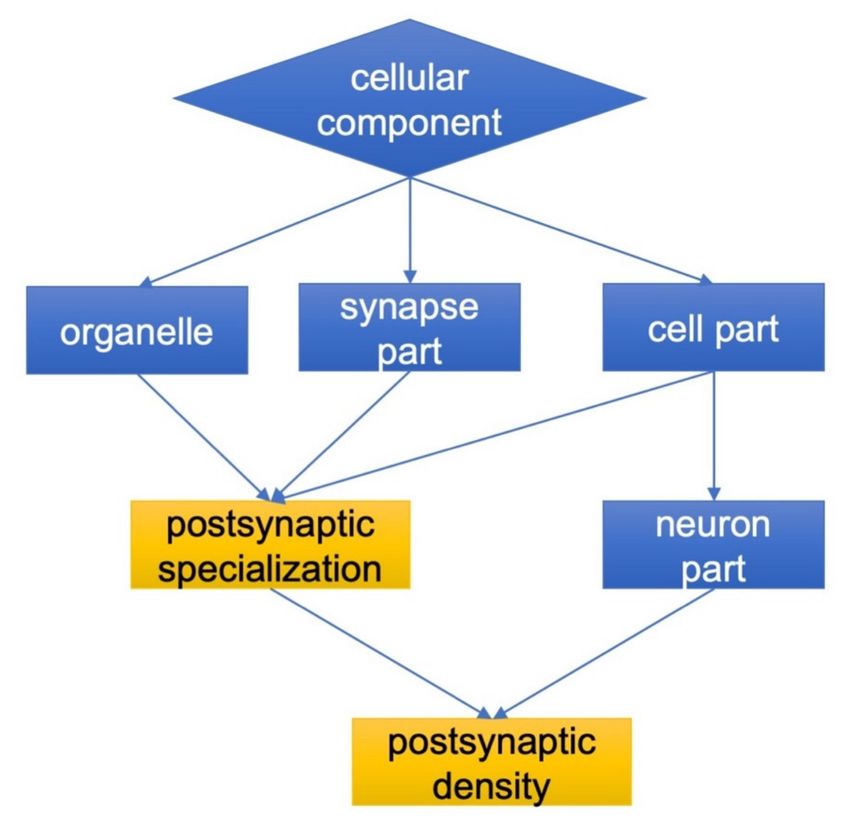
Int. J. Mol. Sci. 2020, 21, 4187                                                                                                        5 of 21

      Figure 1. Functional annotation of transcriptome data in terms of Gene Ontology (GO) biological
      processes. Color intensity reflects the significance of enrichment: darker colors indicate higher statistical
      significance. The biological process in terms of GO supported the prevalence of downregulated
      transcripts related to the regulation and development of neurons including those guiding gliogenesis
      and myelination (A). The upregulated transcripts are related to processes involved in the response
      to DNA damage and consequent regulation of cell death as well as negative regulation of common
      cellular processes in the population of upregulated transcripts (B). Since each gene may be involved in
      several biological processes, the number of up- and downregulated transcripts does not match the sum
      of biological processes shown in the Figure. Other explanations are given in the text.

      Proteomic analysis revealed 1058 proteins corresponding to detected transcripts. However, correlation
between the expression of all identified proteins and their transcripts was very low (R = 0.14) and did
not reach the level of statistical significance. Combined transcriptomic and proteomic analyses revealed
only two matched transcript/protein coding genes. However, in both cases these included an increase
in the transcript level and a decrease in the protein level: (i) Q9JLV5, cullin 3: a 3.2-fold increase in the
transcript level and a 2.7-fold decrease in the protein level; (ii) Q61029, thymopoietin: a 5.9-fold increase in
the transcript level and a 2.1-fold decrease in the protein level (Table 1).

      Table 2. Top 20 upregulated genes detected in brain hemispheres 24 h after the administration of the
      neuroprotective dose of isatin (100 mg/kg, s.c.) to mice.
                                                                     −Log
      Gene Symbol                  Gene Description                                LogFold Change               Role/Effect
                                                                   (p value)
                                                               Upregulated genes
                        cytotoxic T lymphocyte-associated
         Ctla2a                                                      4.154             3.132         Induces apoptosis in some cells
                                  protein 2 alpha
                        alpha, nuclear factor of kappa light
                                                                                                        Negative regulator of cell
         Nfkbia        polypeptide gene enhancer in B cells          4.191             2.876
                                                                                                             proliferation
                                     inhibitor
                        cytotoxic T lymphocyte-associated
         Ctla2b                                                      4.376             2.735         Induces apoptosis in some cells
                                   protein 2 beta
                       cyclin-dependent kinase inhibitor 1A
         Cdkn1a                                                      3.165             2.730           Delays or stops the cell cycle
                                     (P21)
                                                                                                    Negative regulator of several EGFR
          Errfi1        ERBB receptor feedback inhibitor 1           4.676             2.236
                                                                                                             family members
                              serum/glucocorticoid                                                      Regulation of vascular cell
          Sgk1                                                       3.642             1.940
                                regulated kinase 1                                                            proliferation
                                                                                                       One of the key elements in
                         transformation related protein 53
        Trp53inp1                                                    3.301             1.887        p53-mediated cell cycle arrest and
                            inducible nuclear protein 1
                                                                                                     apoptosis in different cell types
Int. J. Mol. Sci. 2020, 21, 4187 6 of 21
Table 2. Cont.
−Log
Gene Symbol Gene Description LogFold Change Role/Effect
(p value)
Involved in the regulation of cell
mechanistic target of rapamycin
Mtor 4.091 1.787 growth, proliferation, autophagy,
(serine/threonine kinase)
and protein synthesis
chromatin licensing and DNA
Cdt1 4.554 1.747 DNA replication
replication factor 1
A member of the exportin family,
Xpo7 exportin 7 4.626 1.722 responsible for nucleocytoplasmic
trafficking of regulatory proteins
Enzyme involved in
tetrahydrobiopterin (BH4)
Gch1 GTP cyclohydrolase 1 4.055 1.716
biosynthesis; its expression is
increased in pathological proliferation
enhancer of polycomb homolog 2 Epc plays an important role in cellular
Epc2 3.578 1.691
(Drosophila) differentiation and development
A member of the exportin family,
Xpo4 exportin 4 4.535 1.608 responsible for nucleocytoplasmic
trafficking of regulatory proteins
Acts as an E3 ubiquitin ligase; is
Rbbp6 retinoblastoma binding protein 6 4.721 1.606 a negative regulator of p53 and
promotes cell proliferation
Involved in regulation of the protein
Ubqln1 ubiquilin 1 3.187 1.596
quality control system
DNA helicase that belongs to the
Recql4 RecQ protein-like 4 3.030 1.575
RecQ helicase family
Involved in the functional recruitment
Sap30 sin3 associated polypeptide 30 kDa 3.040 1.575 of the Sin3-histone
deacetylase complex
Involved in neuron specific signal
Stac3 SH3 and cysteine rich domain 3 4.708 1.573
transduction
A member of the conserved family of
RNA-binding proteins, which play
Srsf6 serine/arginine-rich splicing factor 6 3.077 1.457
an important role in the regulation of
gene expression
Interacts with various nuclear
proline-rich nuclear receptor
Pnrc1 3.679 1.453 receptors and functions as
coactivator 1
a tumor suppressor
Downregulated genes
Has a significant impact on
Cx3cr1 chemokine (C-X3-C) receptor 1 −3.709 −2.796
cell proliferation
Plays an important role in neuron
T cell lymphoma invasion and
Tiam2 −5.154 −2.105 development and malignant
metastasis 2
proliferation in humans
Members of this superfamily of solute
carriers are crucial for brain
Slc38a5 member 5, solute carrier family 38 −3.087 −2.103
physiology and are implicated in
various brain disorders
Regulates expression of CREB1
Lyl1 lymphoblastomic leukemia 1 −4.446 −1.956
target genes
Regulates the cellular response to
Ier5l immediate early response 5-like −3.599 −1.910
mitogenic signals
This guanine nucleotide-exchange
Cyth4 cytohesin 4 −5.331 −1.882 protein is an important regulator of
signal transduction
Is necessary for growth, development,
Limd2 LIM domain containing 2 −3.070 −1.867
and adaptive responses
Gpr17 G protein-coupled receptor 17 −3.345 −1.853 Cell signaling
A Ras-related protein that belongs to
Rasl10a RAS-like, family 10, member A −3.816 −1.760 the superfamily of small GTPases and
exhibits tumor suppressor potential.
B cell CLL/lymphoma 11A
Bcl11a −3.983 −1.759 DNA binding protein
(zinc finger protein)
A molecular adapter involved in
transforming growth factor beta 1
Tgfb1i1 −5.006 −1.748 coordination of multiple
induced transcript 1
protein–protein interactions
A ligand for CXC chemokine receptor
Cxcl12 chemokine (C-X-C motif) ligand 12 −3.073 −1.685 4 involved in neuronal regeneration
in the adult brain
In the brain it is crucial for cortical
Cckbr cholecystokinin B receptor −3.012 −1.683
development
transmembrane 4 superfamily A cell surface glycoprotein involved
Tm4sf1 −3.198 −1.681
member 1 in cell–cell interactionsInt. J. Mol. Sci. 2020, 21, 4187 7 of 21
Table 2. Cont.
Involved in structural rearrangements
Tbcc tubulin-specific chaperone C −5.848 −1.629 of tubulin and actin during the
cell cycle
A calcium-binding adapter molecule
Aif1 allograft inflammatory factor 1 −3.263 −1.627
specifically expressed in microglia.
Functions as a ubiquitination ligase;
Trim59 tripartite motif-containing 59 −4.371 −1.602 its overexpression stimulates cell
proliferation and migration
A transcription factor required for
Pbx2 pre B cell leukemia homeobox 2 −5.499 −1.579
normal brain development
An ATP-dependent
microtubule-based motor protein
Kif21b kinesin family member 21B −4.166 −1.497
involved in the intracellular
transport processes
Rap guanine nucleotide exchange Involved in brain development and
Rapgef3 −3.312 −1.488
factor (GEF) 3 brain diseases
Using the PROTEOME™ database for functional classification, we found downregulated
transcripts important for nervous system development (n = 8, GO:0048709 oligodendrocyte
differentiation, adjusted p value = 0.022; n = 10, GO:0042552 myelination, adjusted p value = 0.027;
n = 11, GO:0042063 gliogenesis, adjusted p value = 0.043) and function (n = 12, GO:0001508 regulation
of action potential in neuron, adjusted p value = 0.026; Figure 1A). These included the following
transcripts: Fa2h, Gjb1, Gpr17, Qk, Rcor2, Sox10, Traf4, Yy1, Dok4, Gal3st1, Gmfb, Kcnj9, and Notch2.
The mean log-fold decrease for 418 downregulated transcripts (Figure 1A) was FC= –0.87 and ranged
between –2.796 and –0.501 with a p value < 0.001).
The most upregulated transcripts (Figure 1B) were related to biological processes involved in
the cell metabolic process (n = 166, GO:0044260, adjusted p value = 1.44 e-12) and its regulation
(n = 99, GO:0060255, adjusted p value = 5.35e-06), gene expression (n = 111, GO:0010467, adjusted
p value = 1.68e-06), cellular macromolecule biosynthetic process (n = 88, GO:0034645, adjusted
p value = 2.98 e-06), cellular response to stress (n = 36, GO:0033554, adjusted p value = 1.04e-05),
regulation of cell cycle (n = 31, GO:0010941, adjusted p value = 8.14e-05), and RNA processing (n = 23,
GO:0006396, adjusted p value = 2.98e-06). The mean log-fold increase for all upregulated transcripts
(n = 433) was FC = 0.83 and ranged between 0.504 and 3.132 at p < 0.001. Remarkably, some GO
groups related to metabolic and biosynthetic processes (e.g., response to DNA damage stimulus (n = 45,
GO:0006974, adjusted p value = 1.84e-06), regulation of cell death (n = 82, GO:0010941, adjusted
p value = 7.24e-06), and the group responsible for nucleic acid metabolic process (n =193, GO:0090304,
adjusted p value = 2.59e-12) were similar for both groups.
Expectedly, both up- and downregulated transcripts were localized in the nucleus (n = 296,
GO:0005634, adjusted p value = 2.19e-15) or were components of the nucleoplasm (n = 79, GO:0005654,
adjusted p value = 2.70e-10). The prevalence of downregulated transcripts in the cytosol (n = 209,
GO:0005737, adjusted p value = 1.10e-07) or in macromolecular complexes (n = 271, GO:0032991,
p = 3.22e-05) was higher compared to the upregulated elements (n = 154, adjusted p value = 1.63e-07,
and n = 169, adjusted p value = 3.19e-05, respectively) (see Supplementary Materials S1).
Results of our study suggest that at 24 h after administration, the neuroprotective dose of
isatin downregulated some brain-specific processes (and corresponding genes) and upregulated the
expression of genes involved in the stress response.
2.2. Isatin-Binding Proteins
Besides isatin-binding proteins common in brain hemispheres of control and isatin-treated mice
(Supplementary Materials S2), there were isatin-binding proteins specific to each group of animals
(Supplementary Materials S3). Control-specific isatin-binding proteins (n = 55) represent direct targets
of isatin, as their interaction with the immobilized isatin analogue was influenced (inhibited) by
isatin administration to animals. In this context, the accumulation of exogenously administered
isatin in the brain and its interaction with target proteins in vivo (i.e., before brain tissue processingInt. J. Mol. Sci. 2020, 21, 4187 8 of 21
for subsequent analysis) had a significant impact on the proteomic profiling. Such homologous
competition for particular targets in the brain was used earlier for evaluation of the specific binding of
[3 H]isatin in various brain structures [36]. In addition, according to our previous calculations made on
the basis of the brain isatin content and the brain tissue water content [1,4], after administration of
50–100 mg/kg isatin to animals, its concentrations in the brain could be as high as 70 µM (provided
that all the exogenously administered isatin was evenly distributed in the brain) or even higher (in
the case of uneven distribution in the brain tissue). Earlier, it was also found that pretreatment of
rats with a dose of isatin 80 mg/kg significantly attenuated irreversible inactivation of brain MAO B
by the mechanism-based inhibitor phenelzine [49]. All this supports the mechanism of competitive
interactions between isatin accumulated in the brain (and bound to its targets) in vivo and the affinity
ligand, 5-aminoisatin-Sepharose, added to concentrated brain lysates in vitro (see Materials and
Methods Section).
In the case of brain isatin-binding proteomes of isatin-treated mice, there were 94 proteins that
were absent in the fraction of brain isatin-binding proteins of control mice. Their appearance may
be associated with several possible mechanisms including isatin-induced changes in protein–protein
interactions as well as isatin-induced formation of new binding sites (see the Discussion section).
However, regardless of the particular mechanism(s), it should be noted that these isatin-binding
proteins have particular cellular localization and functional links as evidenced by GO terms (Figure 2,
see also Supplementary Materials 3). In contrast to control-specific isatin-binding proteins which were
not linked (assigned) to the particular cell compartments, the brain isatin-binding proteins specific to
the isatin-treated mice could be well characterized by targeted localization related to neuronal synapses
(see Figure 2).
Figure 2. Cellular localization and functional links of brain isatin-binding proteins specific to
isatin-treated mice. Details are given in the Materials and Methods Section.
3. Discussion
Administration of the neuroprotective dose of isatin (100 mg/kg, s.c.) to mice had a significant
impact on both the expression of genes and the profile of isatin-binding proteins.
Results of transcriptome and proteome analyses of differentially expressed genes suggest the
lack of concerted changes 24 h after administration of the neuroprotective dose of isatin. In other
words, changes in the transcripts of genes have not been translated into corresponding changes of their
protein products. In addition, unidirectional changes occurred in genes/proteins involved in opposite
processes such as cell proliferation and cell death. This may be attributed to the known phenomenon
of uncoupling of transcription and translation when some delay between transcription and translationInt. J. Mol. Sci. 2020, 21, 4187 9 of 21
occurs in the course of dynamic adaptation processes [55]. Since the time interval of 24 h after isatin
administration was selected as the earliest period for the detection of altered gene expression, it is
possible that concerted changes in the gene expression stages may appear later, after reaching a new
steady state of the dependence of the protein level on the respective mRNA concentration [55]. On the
other hand, the protein level in the cell is not strictly dependent on the expression of particular genes as
it involves other factors, including posttranslational modifications, needed for both biological activity
of particular proteins and also for their (targeted) elimination/degradation. These discrepancies clearly
need further studies including several time points (i.e., longer intervals after isatin administration) and
validation by independent approaches.
Administration of the neuroprotective dose of isatin to mice influenced the profile of brain
isatin-binding proteins. Although profiles of brain isatin-binding proteins of control and isatin-treated
mice shared significant similarity, pools of specific isatin-binding proteins were recognized in both
groups of animals. Figure 3 shows that functionally control-specific isatin-binding proteins mainly
exist as independent molecules unrelated to each other: the majority of these isatin-binding proteins
(37 of 55) demonstrate rather poor functional interactions, thus suggesting their involvement in distinct,
unrelated processes. This is the only axis that links documented interactions between neuronal nitric
oxide synthase (NOS1), generating nitric oxide from arginine, argininosuccinate synthetase, the enzyme
crucial for arginine synthesis, and the antioxidant enzyme thioredoxin reductase 1 (Txnrd1), as well as
components of the regulatory pathways, which share Disks large homolog 1 (Dlg1) as a hub.
Figure 3. Functional links between control-specific isatin-binding proteins of the mouse brain
hemispheres. Line thickness and type (solid or dashed) reflect combined score and evidence suggesting
a functional link. Bold lines indicate direct interactions between the identified proteins, annotated
in String (v. 11.0 of January 19, 2019). Bold dashed lines show experimentally proven interactions
between the identified proteins. Colors indicate different functional clusterization: red color shows
lack of functional interactions, green color designates interaction between structural and regulatory
proteins, blue color indicates interaction of proteins involved redox-dependent regulation.Int. J. Mol. Sci. 2020, 21, 4187 10 of 21
DLG1 is a member of the membrane-associated guanylate kinase (MAGUK) family of proteins,
which function as molecular scaffolds. Dlg1, also known as synapse-associated protein 97 or SAP97,
is a scaffolding protein that belongs to the MAGUK family [56]. Being involved in the formation
of specific multiprotein complexes, including receptors, ion channels, and signaling proteins, it is
implicated in the control of cell polarity. Dlg1 promotes the arrangement of various assemblies of
cell adhesion molecules, receptors and/or ion channels, and other signaling pathway components
with cytoskeleton proteins at particular plasma membrane regions [56]. Recently, it was demonstrated
that Dlg1 (SAP97) co-precipitated with neuronal NOS (NOS1), and this interaction is important for
regulatory thiol modification of GluA1 AMPA receptors [57]. Implication in local redox modifications
explains the direct interaction between NOS1 and Thioredoxin reductase 1 (Txnrd1), while NOS1 and
Smad2 are both involved in TGF-β signaling [58,59].
The other functional cluster containing well-documented interacting proteins includes proteins
GSK3a, Pik3cb, Ptk2, Fyn (nonreceptor tyrpsine kinase), Git1 (ARF GTPase-activating protein), and Ezr,
which interacts with Dlg1 via its FERM domain [60]. Being one of the ERM proteins (ezrin, radixin,
and moesin) forming a highly conserved branch of the FERM domain (four-point-one, ezrin, radixin,
moesin) [61], ezrin functions as a membrane-cytoskeleton linker involved in the connections of major
cytoskeleton structures to the plasma membrane [62]. It adopts various conformational states [60],
which promote ezrin interactions with various partners including different kinases, which can
phosphorylate ezrin and other members of this family (radixin and moesin) at the FERM domain [61].
Ezrin forms a functional triagle, including protein tyrosine kinase 2 (PTK2), also known as focal
adhesion kinase (FAK), and Git1. Protein tyrosine kinase 2 (PTK2) is a non-receptor tyrosine-protein
kinase [63]; it directly interacts with Ezrin in connections of major cytoskeleton structures to the plasma
membrane [62]. FAK (PTK2) can directly interact with Fyn, a member of the Src family of protein
tyrosine kinases (SFKs) in lipid rafts [64]. GIT1, GTPase-activating protein for the ADP ribosylation
factor family, acts as a scaffold crucial for signaling modules with various kinases, including PTK2 [65].
Interestingly, GIT1 may serve as an endothelial nitric-oxide synthase (eNOS) interactor, contributing to
phosphorylation and activation of this enzyme [65], and as the adaptor for actin regulators and local
modulator of Rac activity at synapses [66].
Glycogen synthase kinase-3 alpha (GSK3A) is a constitutively active protein kinase involved in
the hormonal regulation of glucose homeostasis, Wnt signaling and also in the regulation of various
transcription factors and microtubules [67]. This enzyme is an important target of the PI3K pathway [68].
Good evidence also exists that PI3K signaling via GSK3 may regulate DNA methylation [69].
In the case of brain isatin-binding proteomes of isatin-treated mice, there were 94 proteins that
were absent in the fraction of brain isatin-binding proteins of control mice. Most of them form
a deeply integrated protein network in which another member of the membrane-associated guanylate
kinase (MAGUK) family of proteins, DLG4 (known as synapse-associated protein 90, SAP90, and the
PSD-95 protein), serves as a hub linking together several clusters of proteins involved in chromatin
modification, cytoskeleton formation/rearrangement and intracellular trafficking, metabolic processes,
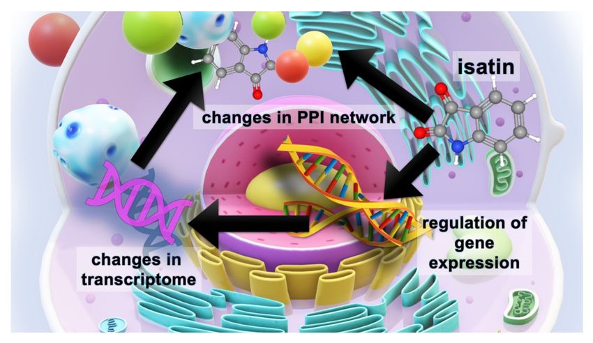
posttranslational modification of proteins, and signaling (Figure 4).Int. J. Mol. Sci. 2020, 21, 4187 11 of 21
Figure 4. Functional links between isatin-binding proteins specific to the brain hemispheres of
isatin-treated mice. Line thickness and type (solid or dashed) reflect combined score and evidence
suggesting a functional link. Bold lines indicate direct interactions between the identified proteins,
annotated in String (v. 11.0 of January 19, 2019). Bold dashed lines show experimentally proven
interactions between the identified proteins. Colors indicate different functional clusterization: red
color shows intracellular molecular scaffolds with various intracellular proteins, aquamarine designates
proteins involved in regulations of RNA splicing, green color shows enzymes, involved in metabolic
processes, blue color shows proteins involved in regulation of neuron-specific proliferation.
Appearance of these rather large groups of proteins may be attributed to changes in the
protein–protein interaction (PPI) networks induced by high isatin concentrations in the brain. In some
cases, the appearance of such PPIs may be accompanied by formations of new binding sites.
For example, in Escherichia coli cells, temperature- and nucleotide-dependent modulation of the
interaction between the central component of translocase (SecA) and the integral SecYEG membrane
protein complex (so called SecA–SecYEG interaction) is involved in the formation of new binding
sites [70]. The other relevant example includes human serum albumin which is able to adopt numerous
conformations in dependence on the nature of bound ligand [71].
Platelet glycoprotein IIb/IIIa receptor antagonists can induce conformational changes in the
receptor, known as a ligand-induced binding site [72]. In addition, some of brain isatin-binding proteinsInt. J. Mol. Sci. 2020, 21, 4187 12 of 21
specific to the group of isatin-treated mice were already identified among the proteins specifically
bound to the other isatin analogue (e.g., glial fibrillary acidic protein, GFAP) [37]. Although it remains
unclear whether these new PPIs result in the formation of tight complexes that bind to the affinity
ligand or whether they represent secondary interactions facilitating the isolation of these proteins
in the group of isatin-binding proteins [73], there is increasing evidence that isatin may act as a PPI
regulator [1]. It should be noted that isatin promoted protein complex formation at least in some model
systems, in which individual components demonstrated low isatin-binding activity [74].
The appearance of new functional protein clusters related to the regulation of gene expression-
transcription and splicing as well as protein synthesis can account for uncoupling between transcription
and translation observed after administration of the neuroprotector dose of isatin: isatin-induced
changes in the expressions of genes did not result in corresponding changes of their protein products
(Tables 1 and 2).
In this context, the Tra2b protein (the transformer 2 beta homolog) is particularly interesting
as it is considered one of the most important splicing factors involved in RNA processing needed
for both normal (e.g., brain development) and pathological (e.g., cancer) cell proliferation [75,76].
The RNA binding protein Rbmx, also known as heterogenous nuclear ribonucleoprotein G (hnRNPG),
also functions as an important protein, being involved in the regulation of gene expression
and alternative splicing. Using its so-called low-complexity region rather than the canonical
RNA-binding domain, hnRNPG recognizes and binds a motif exposed by RNA m6A modification
(N6-methyladenosine) [77]. Another small nuclear ribonucleoprotein-associated protein B (SNRPB)
has a significant impact on the expression and processing of spliceosome components [78].
Gars (glycyl-tRNA synthetase) catalyzes the reaction of glycine ligation to its cognate tRNA [79,80]
needed for global protein synthesis. In humans, impaired functioning of Gars results in the development
of hereditary axonal neuropathy known as Charcot–Marie–Tooth disease (CMT) [79,80]. In addition,
it is suggested that GARS may exhibit axon-specific functions unrelated to tRNA charging [80].
These selected examples suggest that brain isatin-binding proteins specific to isatin-treated
animals may directly affect both transcription and translation processes. Taking into consideration
that all known effects of isatin on catalytically active protein targets result in the decrease of their
activities [1], it is reasonable to suggest that the appearance of new clusters of interactomes in the
brain of isatin-treated mice causes a coordinated decrease of several groups of biological processes.
All these considerations suggest that the presence of functionally related proteins in the same fraction
was not accidental.
Our previous proteomic studies have shown that in mice exposed to a shorter period of time after
administration of isatin (100 mg/kg; 90–120 min), there was a significant decrease in the repertoire of
brain proteins specifically bound to the Rpn-10 subunit of proteasomes [24] and a number of proteins
associated with the brain mitochondrial fraction [25].
This study has shown that 24 h after the administration of the neuroprotective dose of isatin,
the brain hemispheres responded by downregulation of 31 proteins, altered expression of genes,
uncoupling between transcription and translation processes, as well as by changes in the profiles
of isatin-binding proteins. The changes in the profiles of isatin-binding proteins included the
disappearance of control-specific individual proteins from the isatin-binding pattern due to loading
with administered isatin (and homologous competition with an isatin analogue as the affinity sorbent)
and the formation of new clusters of interacting proteins specific to the isatin-treated brain.
These effects are aimed at interruption of pathological links that begin to form after initiation of
pathological processes such as MPTP-induced parkinsonism [34,35]. It is possible that some of the
recognized proteins and genes sensitive to the neuroprotective dose of isatin may be used as potential
targets for numerous analogues synthesized in many laboratories worldwide [81–83].Int. J. Mol. Sci. 2020, 21, 4187 13 of 21
4. Materials and Methods
4.1. Reagents
Sucrose, Triton X-100, triethylammonium bicarbonate, potassium phosphates, deoxycholic acid
sodium salt, urea, 4-vinylpyridine, CNBr-activated Sepharose 4B, and isatin were purchased from
Sigma (St. Louis, MO, USA); formic acid was obtained from Merck (Darmstadt, Germany); acetonitrile
was obtained from Fisher Chemical (Leicestershire, UK); and tris-(2-carboxyethyl) phosphine was
obtained from Pierce-Thermo Scientific (Rockford, IL, USA). Trypsin (modified sequencing grade)
was obtained from Promega (Madison, WI, USA), and 5-aminoisatin was synthesized using standard
methods [84].
4.2. Animals and Isatin Administration
Sixteen male C57BL/6 mice (20–25 g; n = 8 in each group), obtained from the Stolbovaya nursery
(Moscow region), were used in this study. Experiments were performed one week after their arrival
from the nursery. Animals were kept under natural illumination and received standard laboratory
chow and water ad libitum. Isatin was injected subcutaneously (s.c., 100 mg/kg); control mice were
treated with the same volume of saline (0.1 mL/kgl s.c.). All procedures conformed to the Russian
version of the Guide for the Care and Use of Laboratory Animals (Washington, 1996) were approved
by the Animal Care and Use Committee of the Institute of Biomedical Chemistry (approval certificated
issued from November 12, 2019, local code IBMC-RFBR-00042).
4.3. Sample Preparation for Transcriptome Analysis and Proteomic Profiling of Isatin-Binding Proteins
The animals were decapitated 24 h after injection under light ether anesthesia. The extracted brains
were quickly washed in ice-cold saline, and brain hemispheres were immediately used in subsequent
experiments. Four samples per group of mice were used for transcriptomic and proteomic analyses.
4.3.1. Transcriptome Analysis
Total RNA from brain hemispheres samples (~15–20 mg) was isolated using the Absolutely RNA
Miniprep Kit (Agilent, cat. no. 5190-0444). The quality of obtained RNA samples was assessed using
an Agilent Bioanalyser 2100 (Agilent, Santa Clara, CA, USA). RNA samples with an RIN of at least 8
were used for subsequent analysis. Sample preparation was carried out in accordance with the Two
Color Microarray-Based Gene Expression Analysis Version 6.6 protocol for two-color hybridization
using the two-color Low input Quick-Amp Labeling Kit (cat. no. 5190-2331; Agilent, Santa Clara,
CA, USA). The experimental samples were labeled with Cy5 –CTP, and the reference RNA (Universal
Mouse Reference RNA (cat. no. 740100; Agilent, Santa Clara, CA, USA) was labeled with Cy3-CTP.
According to the standard protocol recommended by the manufacturer, the corresponding amounts of
RNA from the Agilent RNA Spike-In Kit (cat. no. 5188-5279; Agilent, Santa Clara, CA, USA) were
added to the samples. Evaluation of gene expression was carried out using microarrays for mouse
whole-genome microarray analysis (cat. no. G2519F-026655; 4x44K V2 format; Agilent, Santa Clara,
CA, USA).
After fragmentation of cRNA and hybridization performed in accordance with the standard
protocol, data acquisition was performed by using a DNA Microarray Scanner G2505C (Agilent,
Santa Clara, CA, USA).
Primary transcriptome data were treated using Feature Extraction software (version 10.1.3.1;
Agilent Technologies).
Further data processing was performed using the GeneXplain platform [85]. Non-logarithmic
normalization of the raw data for Control and Isatin treatment conditions was carried out pairwise
using module “Normalize Agilent experiment and control” with methods in the Bioconductor LIMMA
package. The up- and down-regulated transcripts were identified, and the log-fold change values were
calculated by the workflow “Compute differentially expressed genes (Agilent probes)”. This up andInt. J. Mol. Sci. 2020, 21, 4187 14 of 21
down identification analysis applies Student’s t-test and calculates p values. The following criteria
were used: LogFoldChange > 0.5 and –log_p_value > 3 for upregulated probes; LogFoldChange < –0.5
and –log_p_value < –3 for downregulated probes.
4.3.2. Functional Analysis of Differentially Expressed Genes
Functional analysis of up- and down-regulated genes was performed individually using the
GeneXplain platform with default settings of the “Mapping to ontologies (PROTEOME™)” workflow.
The “Functional classification” module was used for the annotation of differentially expressed genes in
terms of Gene Ontology. The algorithm of the functional analysis is intended to detect statistically
significant representation of certain functional groups of genes among all genes of the input sets.
Statistical significance of the classification was estimated with the adjusted p value, and only statistically
significant GO groups with adjusted p value < 0.05 were considered.
4.3.3. Affinity Chromatography and Sample Preparation for Mass Spectrometric Analysis
The brain hemispheres were immediately dissected and homogenized using a SilentCrusher S
homogenizer (Heidolph, Germany) at 50,000 rpm in 0.05 M potassium phosphate buffer, pH 7.4 (buffer
A) (1:3, w/v). Homogenates were then lysed with 3% Triton X-100 (final concentration) at 4 ◦ C for
1 h, diluted three-fold with buffer A, and centrifuged at 16,000 rpm (Eppendorf 5415 R centrifuge;
Eppendorf, Germany) for 20 min at 4 ◦ C.
5-aminoisatin-Sepharose was prepared as in [38]. Lysates of brain hemisphere homogenates
(protein concentration of about 10 mg/mL) were added to 5-aminoisatin-Sepharose and incubated in
a suspension (1:1) at 4 ◦ C overnight under gentle stirring.
The affinity sorbent was washed with 100 volumes of buffer A to remove non-specifically bound
proteins, and then the remaining proteins were eluted using a column (1 × 2 cm) at room temperature:
initially, with 30 mL of 1 mM isatin in buffer A, then with the same volume of 1 M NaCl in buffer A.
The eluate (30 mL) was concentrated to 0.25 mL using an Amicon Ultra centrifuge device (Millipore,
Bedford, MA, USA). Proteins were extracted with a mixture of chloroform–methanol [35]; trypsinolysis
was performed directly on a Vivaspin 500 centrifuge membrane filter (Sartorius Stedim Biotech,
Germany) with a 10,000 Da membrane, as described in [35].
Samples were evaporated using a 5301 vacuum concentrator (Eppendorf, Germany), dissolved in
0.1% formic acid, and analyzed using LC-MS/MS.
In order to determine proteins non-specifically bound to the affinity sorbent, control CNBr-activated
Sepharose was used; it was subjected to the same procedures as 5-aminoisatin-Sepharose, but without
the addition of the affinity ligand.
4.3.4. Mass Spectrometric Analysis
Chromatographic separation was performed using an Ultimate 3000 RSLC Nano system (Thermo
Scientific, Rockford, IL, USA). A sample (2 µL) was loaded for 4 min in mobile phase C (water, 2.5%
acetonitrile, 0.1% formic acid, 0.03% acetic acid) onto enrichment column. Peptides were separated in
a gradient of mobile phase A (aqueous solution of 0.01% formic acid, 0.03% acetic acid), and mobile
phase B (90% acetonitrile, 10% methanol, 0.1% formic acid, 0.03% acetic acid) on an Acclaim Pepmap®
analytical column (geometry 75 µm × 150 mm, 1.8 µm; Thermo Scientific, Rockford, IL, USA). Initial
conditions for the elution gradient were 98% A: 2% B, flow rate of 0.3 µL/min, and the loading rate
on the enrichment column was 15 µL/min. To increase the washing efficiency of the column during
chromatographic separation (removal of bound hydrophobic components), the dynamic change in the
flow rate to 0.45 mL/min was used at a high relative content of the mobile phase B.
Mass spectrometric analysis was performed on an Orbitrap Fusion high-resolution hybrid mass
spectrometer (Thermo Scientific, Rockford, IL, USA) with an NSI ionization source in the mode of
positive electrostatic ionization. Precursor ions were acquired at a resolution of 60K in the m/z range of
425–1250 with the maximum integration time of 15 ms or a minimum. Precursor ions with charge stateInt. J. Mol. Sci. 2020, 21, 4187 15 of 21
z = 2+ . . . 4+ were isolated with an isolation window of ± 0.75 m/z and offset of 0.25 m/z and triggered
for fragmentation in HCD (higher-energy collision dissociation) mode with the normalized activation
energy at 27%. Fragment ions were acquired in an orbital mass analyzer with a resolution of 15K and
with a maximum accumulation time of 85 ms.
Data files obtained after LC-MS/MS analysis were converted in peak list format and used for
protein identification. Peak lists obtained from spectra were identified using X!Tandem, and the
search was conducted using Search GUI version 2.3.17 (Compomics, Belgium) [86]. Proteins were
identified against a concatenated target/decoy database of human proteins (Uniprot database revision
April 2019). The decoy sequences were created by reversing the target sequences in Search GUI.
The identification settings were as follows: trypsin as a specific protease, with a maximum of 1
missed cleavage, tolerance of ±5 ppm as the MS1 level, and tolerance of ±0.005 Da as the MS2 level
tolerances; variable modifications: oxidation of M (+15.99), deamination of N (+0.98 u), deamination
of Q (+0.98 u); cysteine pyridilethylation C (+105 u) was set as a fixed modification. Peptide Spectrum
Matches (PSMs), peptides, and proteins were validated at a 1.0% false discovery rate (FDR) estimated
using the decoy hit distribution [87]. Proteins were inferred from the spectrum identification results
and quantified using normalized spectrum abundance factor (NSAF) in Peptide Shaker version 1.16.15
(Compomics, Belgium) [88,89].
In each proteomic experiment, the total preparations obtained during the affinity enrichment of
lysates of brain homogenates of at least two mice were used. Each of the proteins presented in the
tables was identified in at least three independent experiments.
5. Conclusions
Isatin (indole-2,3-dione) is an endogenous indole found in mammalian tissues and body fluids.
It was originally identified as an endogenous MAO (B) inhibitor [90]. Now good evidence exists
that it exhibits various biological functions which are unrelated to MAO inhibition. Previous studies
performed in vitro and in cell cultures revealed a large group of isatin-binding proteins, including
nucleoproteins, and also some isatin-responsive genes [1,36–38,41–46]. This suggests that isatin
could be potentially involved in the multilevel regulatory hierarchy from genes to proteins (and
their posttranslational modifications) [1]. This study was the first research aimed at investigation
of the effect of the neuroprotective dose of isatin to mice (100 mg/kg; 24 h) on gene expression and
also on the profile of isatin-binding proteins in the brain hemispheres. Results of transcriptome and
proteome analyses of differentially expressed genes suggest the lack of concerted changes 24 h after
administration of the neuroprotective dose of isatin. This may be attributed to the known phenomenon
of uncoupling of transcription and translation, when some delay between transcription and translation
occurs in the course of dynamic adaptation processes [55]. Whether this delay is associated with
isatin binding to nucleoproteins [1] and altered ribosome functioning remains to be experimentally
investigated. Little correspondence between proteomic and transcriptomic datasets requires the use of
both independent validation approaches (e.g., Western blot and qPCR, respectively) and a significant
extension of time intervals after isatin administration. Figure 5 summarizes the main results of this
study and further research hotspots.Int. J. Mol. Sci. 2020, 21, 4187 16 of 21
Figure 5. The scheme illustrating the multilevel effects of the neuroprotective dose of isatin (100 mg/kg,
24 h). Isatin has a significant impact on the expression of responsive genes and thus influences the cell
transcriptome. Acting on isatin-binding proteins, isatin changes protein–protein interactions (PPI),
which are also influenced by the altered proteome, thus causing global changes in the PPI network.
The results of this study have shown that isatin administration produces multilevel changes at
least in the brain hemispheres, which include altered gene expression and changes in the profiles of
isatin-binding proteins and their interactomes. Appearance of isatin-binding proteins that were not
found in the hemispheres of control mice could be associated with changes in PPI networks induced
by the accumulation of a high isatin concentration in the brain.
Since isatin-responsive genes (Table 2) and isatin-binding proteins are involved in the control of
various important cell functions, it is reasonable to suggest that some isatin analogues may selectively
act on particular targets in various brain regions and produce a certain therapeutic effect. The latter
is especially important because many isatin-binding proteins have also been implicated in various
neurodegenerative processes.
Supplementary Materials: Supplementary materials can be found at http://www.mdpi.com/1422-0067/21/11/4187/s1.
Author Contributions: A.M. conceived and designed the experiments. O.B. and A.M. performed animal
experiments, isolation of brain hemispheres, and collection of brain samples. L.K. and O.T. performed the
transcriptome and gene expression analyses. O.B. performed the isolation of isatin-binding proteins and sample
preparation for mass spectrometry (MS). A.K. and V.Z. performed MS analysis. A.I. helped to perform interactome
analysis and data interpretation. A.M., A.K., O.B. prepared the original draft and wrote the paper. All authors
have read and agreed to the published version of the manuscript.
Funding: This work performed within the framework of the Program for Basic Research of State Academies
of Sciences for 2013–2020 was partially supported by the Russian Foundation for Basic Research (project
no.18-15-00042).
Acknowledgments: The mass spectrometry identification of mouse brain mitochondrial proteins was performed
using the “Human Proteome” Core Facility (Institute of Biomedical Chemistry, Moscow).
Conflicts of Interest: The authors declare no conflict of interest.You can also read