Interplay between Autophagy and the Ubiquitin-Proteasome System and Its Role in the Pathogenesis of Age-Related Macular Degeneration - MDPI
←
→
Page content transcription
If your browser does not render page correctly, please read the page content below
International Journal of
           Molecular Sciences

Review
Interplay between Autophagy and the
Ubiquitin-Proteasome System and Its Role in the
Pathogenesis of Age-Related Macular Degeneration
Janusz Blasiak 1 , Elzbieta Pawlowska 2 , Joanna Szczepanska 3 and Kai Kaarniranta 4,5, *
 1    Department of Molecular Genetics, Faculty of Biology and Environmental Protection, University of Lodz,
      90-236 Lodz, Poland; janusz.blasiak@biol.uni.lodz.pl
 2    Department of Orthodontics, Medical University of Lodz, 92-216 Lodz, Poland;
      elzbieta.pawlowska@umed.lodz.pl
 3    Department of Pediatric Dentistry, Medical University of Lodz, 92-216 Lodz, Poland;
      joanna.szczepanska@umed.lodz.pl
 4    Department of Ophthalmology, University of Eastern Finland, Kuopio 70211, Finland
 5    Department of Ophthalmology, Kuopio University Hospital, 70029 Kuopio, Finland
 *    Correspondence: kai.kaarniranta@kuh.fi; Tel.: +358-440-593290

 Received: 10 December 2018; Accepted: 2 January 2019; Published: 8 January 2019

 Abstract: Age-related macular degeneration (AMD) is a complex eye disease with many pathogenesis
 factors, including defective cellular waste management in retinal pigment epithelium (RPE). Main
 cellular waste in AMD are: all-trans retinal, drusen and lipofuscin, containing unfolded, damaged
 and unneeded proteins, which are degraded and recycled in RPE cells by two main machineries—the
 ubiquitin-proteasome system (UPS) and autophagy. Recent findings show that these systems can act
 together with a significant role of the EI24 (etoposide-induced protein 2.4 homolog) ubiquitin ligase
 in their action. On the other hand, E3 ligases are essential in both systems, but E3 is degraded by
 autophagy. The interplay between UPS and autophagy was targeted in several diseases, including
 Alzheimer disease. Therefore, cellular waste clearing in AMD should be considered in the context
 of such interplay rather than either of these systems singly. Aging and oxidative stress, two major
 AMD risk factors, reduce both UPS and autophagy. In conclusion, molecular mechanisms of UPS and
 autophagy can be considered as a target in AMD prevention and therapeutic perspective. Further
 work is needed to identify molecules and effects important for the coordination of action of these two
 cellular waste management systems.

 Keywords: age-related macular degeneration; autophagy; mitophagy; ubiquitin-proteasome system;
 cellular waste elimination; proteostasis

Int. J. Mol. Sci. 2019, 20, 210; doi:10.3390/ijms20010210                          www.mdpi.com/journal/ijms
Int. J. Mol. Sci. 2019, 20, 210                                                                                            2 of 23

1. Introduction
     Int. J. Mol. Sci. 2019, 20, x FOR PEER REVIEW                                                                   2 of 23

          Figure
     Figure       1. Schematic
             1. Schematic        representationofofthe
                             representation          thepathogenesis
                                                         pathogenesis ofofage-related
                                                                           age-related  macular
                                                                                           macular degeneration  (AMD)
                                                                                                       degeneration   (AMD)
          with  an  important   role of cellular waste   (yellow  highlight). Oxidative    stress
     with an important role of cellular waste (yellow highlight). Oxidative stress (red thunder)   (red thunder)  can be
                                                                                                                       can be
          generated by many environmental/life style risk factors as well as yet unidentified sources. Visual
     generated by many environmental/life style risk factors as well as yet unidentified sources. Visual cycle
          cycle (VA) by-products can contribute to cellular waste. A complex interplay between oxidative stress,
     (VA) by-products can contribute to cellular waste. A complex interplay between oxidative stress, chronic
          chronic inflammation, variants of genes encoding the complement and cellular waste clearing may
     inflammation,     variants of genes encoding the complement and cellular waste clearing may lead to
          lead to degeneration of retinal cells and clinically detectable AMD, which in its advanced stage may
     degeneration
          acquire theretinal
                     of   form cells  and clinically
                                  of geographic        detectable
                                                    atrophy    (GA)AMD,   which
                                                                     or wet       in its characterized
                                                                               AMD,      advanced stage   bymay   acquire the
                                                                                                               choroidal
     form neovascularization
           of geographic atrophy       (GA)  or  wet  AMD,    characterized   by  choroidal     neovascularization
                                (CNV). AMD symptoms include loss of central vision. Sharp black arrows indicate        (CNV).
     AMDstimulation/consequences,
            symptoms include loss of      centralblunt
                                         whereas    vision.  Sharp
                                                           black    black–arrows
                                                                 arrows             indicate
                                                                            inhibition.         stimulation/consequences,
                                                                                          PR—photoreceptors,      RPE—
     whereas   blunt
          retinal       black
                  pigment      arrows—inhibition.
                            epithelium,                   PR—photoreceptors,
                                         BM—Bruch’s membrane,                       RPE—retinal pigment epithelium,
                                                                     Ch—choriocapillaris.
     BM—Bruch’s membrane, Ch—choriocapillaris.
          Age-related diseases are frequently associated with oxidative stress and free radicals and
     mitochondrialdiseases
     Age-related        theories ofareaging  link aging
                                        frequently          with increased
                                                        associated     with production
                                                                               oxidative of       reactive
                                                                                               stress   andoxygen     species and
                                                                                                              free radicals
     (ROS) [1,2].theories
mitochondrial      These theories
                              of aginghavelink
                                            beenaging
                                                   criticized
                                                           with and now it is production
                                                                  increased     rather assumed    of that  errorsoxygen
                                                                                                      reactive     associated
                                                                                                                           species
     with   every  biological    process  increase    with   aging  resulting   in an   increased
(ROS) [1,2]. These theories have been criticized and now it is rather assumed that errors associated  amount    of  damaged
     biological molecules [3]. Proteins seem to be at the first line of the attack of the products of decreased
with every biological process increase with aging resulting in an increased amount of damaged
     accuracy of vital processes with age as they are the most abundant biomolecules. Mature proteins are
biological molecules [3]. Proteins seem to be at the first line of the attack of the products of decreased
     precisely folded to be stable and functional. ROS can unfold them or contribute to their misfolding
accuracy  of vital
     making   themprocesses        with age
                      prone to damage      andasaggregation.
                                                  they are theTherefore,
                                                                   most abundant       biomolecules.
                                                                              age-related     diseases canMature      proteins
                                                                                                             be featured   by are
precisely  folded    to  be stable   and  functional.
     an increased number of unfolded/damaged proteins.     ROS    can unfold     them    or   contribute    to  their misfolding
making them prone to damage and aggregation. Therefore, age-related diseases can be featured by an
increased number of unfolded/damaged proteins.
Int. J. Mol. Sci. 2019, 20, 210                                                                    3 of 23

     Age-related macular degeneration (AMD) is a progressive and degenerative eye disease affecting
the macula in the central region of the retina and leading to sight distortion. Many genetic and
environmental/lifestyle factors may play a role in AMD pathogenesis, but accumulation of cellular
waste and impairment in its clearing seem to be of a special significance, which was discussed by
Kaarniranta et al. in 2010 [4]. This review updates and extends information and conclusions contained
in that work.
     AMD is a primary cause of vision loss in the elderly in developed countries. Estimated number of
individuals affected by AMD in 2020 is 196 million and 288 million in 2040 [5]. Such high numbers
imply high personal and public costs and an urgent need to develop efficient treatment.
     AMD can occur in dry (atrophic) or wet (exudative, neovascular) form. Dry AMD is manifested
in 80–85% of all cases, but treatment options are available only to patients with wet AMD and do not
address disease causes, but rely on the inhibition of vascular endothelial growth factor (VEGF) by
antiangiogenic agents [6]. Aging is the most serious risk factor for AMD and the number of individuals
affected increases significantly after the age of 50 years [7]. Aging is associated with intracellular
accumulation of lipid and protein deposits [8]. As we will present further, such deposits are observed
in AMD.
     AMD is a multifactorial disease and its etiology is not completely known (Figure 1). It is
suggested that oxidative stress can play a major role in the pathogenesis of AMD. The retina is
susceptible to oxidative stress caused by its constant exposure to visible light and high consumption of
oxygen [9]. Several life-style factors, including smoking and fat-rich diet can contribute to increased
ROS production in the retina, but in general all sources of oxidative stress in the retina are unknown.
Retinal pigment epithelium (RPE) is a major site of pathological alterations in AMD. In normal
conditions, RPE cells regulate ion balance, secrete growth factors and maintain the blood-retina barrier.
Altered bioenergetics in RPE cells manifested by reduced glycolysis and oxidative phosphorylation
can contribute to AMD pathology [10]. RPE cells derived from AMD donors show an increased
susceptibility to oxidative stress and produce more ROS [11].
     Homeostasis of proteins depends on their folding, translocation and degradation (reviewed
in [12]). Increased oxidative stress can cause protein misfolding and accumulation of lipid/protein
aggregates observed in AMD (reviewed in [13]). Consequently, there is a need for an efficient
removal of cellular waste in retinal cells to prevent AMD or slowing down its progression. Waste
clearing in RPE cells includes proteasomal degradation, heterophagy, autophagy and mitophagy
(reviewed in [14]). Exosomes can also be involved in waste removal in RPE cells [15]. The ubiquitin
proteasome system (UPS) is mainly responsible for degradation of damaged or no longer needed
proteins. Autophagy can degrade damaged organelle and may also take a part in degradation
proteins when other clearance processes are failed (reviewed in [16]). RPE cells phagocytose used
photoreceptors outer segments (POS) with their subsequent autophagy-lysosomal degradation [17].
The removal of POS by heterophagy occurs at the apical side of RPE cells that is linked with the
photoreceptor layer [18]. Disturbances in waste clearing leads to accumulation of harmful lipid and
protein aggregates that can act as a physical barrier to intracellular transport and disturb proper
functioning of RPE cells.

2. Major Cellular Waste in Retinal Pathophysiology

2.1. All-Trans-Retinal
      All-trans-retinal (atRAL) is the product of isomerization of 11-cis-retinal, which is an essential
reaction of the visual cycle, a process occurring through a series of reactions catalyzed by
membrane-bound enzymes located in photoreceptors and RPE cells [19] (Figure 2). 11-cis-retinal
is a chromophore of rhodopsin and cone pigments. atRAL is a reactive aldehyde, whose accumulation
causes toxic conjugates with proteins inducing degeneration of the mouse retina [20]. Many AMD risk
factors, including aging, smoking, ultraviolet (UV) and blue light exposure, chronic inflammation and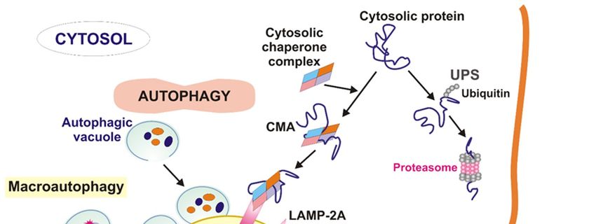
Int. J. Mol. Sci. 2019, 20, 210 4 of 23
improper diet can be related to oxidative stress, but it is not known, whether oxidative stress associated
with AMD belongs to the reasons or consequences of the disease or both. In any case, reduction of the
stress can be important in both prevention and therapy of AMD.
Int. J. Mol. Sci. 2019, 20, x FOR PEER REVIEW 4 of 23
Figure 2. The visual cycle produces all-trans-retinal (atRAL), which is a major cellular waste in retinal
Figure 2. The visual cycle produces all-trans-retinal (atRAL), which is a major cellular waste in
cells. Light is absorbed by photoreceptors (PR) and causes isomerization of 11-cis-retinal to atRAL,
retinal cells. Light is absorbed by photoreceptors (PR) and causes isomerization of 11-cis-retinal to
which is transported and reduced to all-trans-retinol by ATP-binding transporter (ABCA4) and all
atRAL, which is transported and reduced to all-trans-retinol by ATP-binding transporter (ABCA4)
trans retinal dehydrogenases RDH8/12, respectively. atRAL moves into retinal pigment epithelium
and all trans
(RPE), retinal
where dehydrogenases
it is converted RDH8/12,
to all-trans-retinyl respectively.
esters atRAL
by lecithin retinol moves into(LRAT).
acyltransferase retinal RPE-
pigment
epithelium
specific(RPE),
proteinwhere
(RPE65) it isomerized
is converted toesters
these all-trans-retinyl esters
to 11-cis-retinol, by lecithin
which retinol by
is then oxidized acyltransferase
RDH5 to
(LRAT). RPE-specific
11-cis-retinal. Black protein
arrows(RPE65)
indicate aisomerized
way from a these esterstotoits11-cis-retinol,
compound derivative. which is then oxidized
by RDH5 to 11-cis-retinal. Black arrows indicate a way from a compound to its derivative.
To maintain vision, atRAL released from light-activated visual pigments must be isomerized to
To
its maintain
11-cis isomer vision,
[21]. atRAL
The Rdh8 released from light-activated
gene encodes an enzyme that visual reducespigments
atRAL in rod mustandbecone
isomerized
outer to
segments
its 11-cis isomer and theThe
[21]. Abca4Rdh8 gene
gene encodes
encodes theanATP-binding
enzyme thattransporter
reduces atRAL of atRAL,
in rodcatalyzing
and coneitsouter
movement
segments and the from the gene
Abca4 insideencodes
to the outside of disc membranes
the ATP-binding transporter of rods and cones.
of atRAL, Mice carrying
catalyzing a
its movement
double knock-out in the Rdh8 and Abca4 genes accumulated atRAL
from the inside to the outside of disc membranes of rods and cones. Mice carrying a double knock-out condensation products and
showed altered phenotype of photoreceptors and RPE cells [20]. This phenotype is characterized by
in the Rdh8 and Abca4 genes accumulated atRAL condensation products and showed altered phenotype
the presence of yellowish deposits called drusen, intracellular lysosomal lipofuscin, basal laminar
of photoreceptors and RPE cells [20]. This phenotype is characterized by the presence of yellowish
deposits and thickening of Bruch’s membrane and is escalated by light. Intense light exposure of
deposits
thesecalled drusen, intracellular
mice increased atRAL levels inlysosomal lipofuscin,
the retina leading basal (the
to NADPH laminar
reduceddeposits
form ofand thickening of
nicotinamide
Bruch’s membrane
adenine and is escalated
dinucleotide phosphate) by light. Intense lightoverproduction
oxidase-mediated exposure of these of mice increased
intracellular atRAL
ROS [22].levels
in theTherefore,
retina leading
aberrant to NADPH
release of (the reduced
byproducts of form of nicotinamide
the visual cycle could lead adenine dinucleotide
to retinal phosphate)
degeneration. N-
oxidase-mediated overproduction of intracellular
retinylidene-N-retinylethanolamine ROS [22].ofTherefore,
(A2E) is a derivative vitamin A,aberrant
which isrelease
producedof byproducts
in the
of thevisual
visualcycle
cycle[23].
could A2Elead is to also a major
retinal lipofuscin N-retinylidene-N-retinylethanolamine
degeneration. component inducing damage to RPE cells. (A2E) is
Photosensitization of A2E leads to
a derivative of vitamin A, which is produced in the visualtelomere dysfunction and DNA
cycle damage
[23]. A2E in RPE cells
is also triggering
a major lipofuscin
cellular inducing
component senescence, a process
damage tocontributing to retinal degeneration
RPE cells. Photosensitization of[24,25].
A2E leads to telomere dysfunction
and DNA damage in RPE cells triggering cellular senescence, a process contributing to retinal
2.2. Lipofuscin
degeneration [24,25].
Dysfunction in POS degradation leads to accumulation of lipid-protein aggregates resulting
from oxidation of unsaturated fatty acids.
These aggregates are called lipofuscin and are composed from covalently cross-linked proteins,
lipids and small amount of saccharides [26] (Figure 3). Lipofuscin accumulation reflects a weakened
ability to degrade protein debris and is a hallmark of RPE cells aging [27]. One of the cytotoxic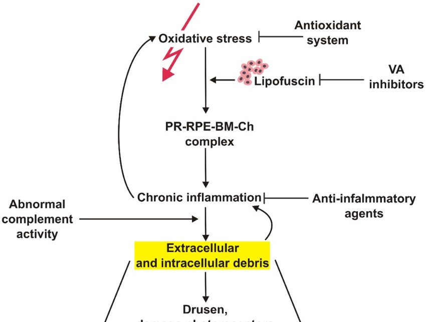
Int. J. Mol. Sci. 2019, 20, 210 5 of 23
2.2. Lipofuscin
Dysfunction in POS degradation leads to accumulation of lipid-protein aggregates resulting from
oxidation of unsaturated fatty acids.
These aggregates are called lipofuscin and are composed from covalently cross-linked proteins,
lipids and small amount of saccharides [26] (Figure 3). Lipofuscin accumulation reflects a weakened
ability to degrade protein debris and is a hallmark of RPE cells aging [27]. One of the cytotoxic
lipofuscin activity is the inhibition of degradation of oxidized proteins by binding to proteasome and
lysosomalInt.proteases. The
J. Mol. Sci. 2019, 20, x binding of lipofuscin to the proteasome may result in the inhibition
FOR PEER REVIEW 5 of 23 of its
activity [28]. It was shown that lipofuscin-bound iron is a major intracellular source of oxidants in
lipofuscin activity is the inhibition of degradation of oxidized proteins by binding to proteasome and
senescent lysosomal
fibroblasts so it has the ability to incorporate iron and promote the Fenton reaction [29]. We
proteases. The binding of lipofuscin to the proteasome may result in the inhibition of its
and othersactivity
showed that
[28]. It was disturbed
shown thatiron metabolism
lipofuscin-bound might
iron playintracellular
is a major a role in AMD source pathogenesis
of oxidants in [30–32].
Studies revealed that the inhibition of mitochondrial fission led to an increased formation
senescent fibroblasts so it has the ability to incorporate iron and promote the Fenton reaction of lipofuscin.
[29]. We
and others showed that disturbed iron metabolism might play
Higher lipofuscinogenesis is also associated with downregulation of the Lon protease thata role in AMD pathogenesis [30–32].
is responsible
Studies revealed that the inhibition of mitochondrial fission led to an increased formation of
for selective degradation of abnormal proteins in mitochondria [33]. The A2E fluorophore is the main
lipofuscin. Higher lipofuscinogenesis is also associated with downregulation of the Lon protease that
hydrophobic component
is responsible of lipofuscin
for selective and itof isabnormal
degradation the product of inthemitochondria
proteins interaction[33].between
The A2E atRAL and
ethanolamine. A2E iswas
fluorophore reported
the main to accumulate
hydrophobic component of inlipofuscin
aging RPE and itcells
is theand increase
product the expression of
of the interaction
VEGF andbetween atRAL and ethanolamine.
some interleukins as well asA2E otherwasinflammatory
reported to accumulate
moleculesin aging RPE cells and increase
[23,34].
the expression of VEGF and some interleukins as well as other inflammatory molecules [23,34].
Figure 3. Fundus autofluorescence image from a degenerated macula indicating increased lipofuscin
Figure 3. Fundus autofluorescence image from a degenerated macula indicating increased lipofuscin
accumulation with increased autofluorescence signal.
accumulation with increased autofluorescence signal.
2.3. Drusen
2.3. Drusen
Drusen are extracellular products located between the basal lamina of the RPE cells and collagen
Drusenlayer of extracellular
are Brunch’s membrane (Figurelocated
products 4). They between
are composed
theofbasal
neutral lipids and
lamina protein
of the RPE derivatives,
cells and collagen
substantial cellular waste products. Large areas with small drusen are associated with the incidence
layer of Brunch’s membrane (Figure 4). They are composed of neutral lipids and protein derivatives,
of AMD [35]. Deposits accumulate when the balance between production and clearance of cellular
substantialcomponents
cellular waste products.
is disturbed. MoreLarge
than 40%areas
of with
drusensmall
volume drusen
is made areup
associated with the incidence
of lipid components
of AMD [35]. Deposits
dominated accumulate
by esterified when
cholesterol and the balance between
phosphatidylcholine [36]. production
They also contain and clearance of cellular
apolipoprotein
components E, amyloid β, vitronectin,
is disturbed. More thancollagens
40% ofanddrusen
complement
volumeproteins
is made[37].upTheoflatter
lipidsuggests that thedominated
components
formation of drusen is associated witch local inflammatory events, such as activation of the
by esterified cholesterol and phosphatidylcholine [36]. They also contain apolipoprotein E, amyloid
complement cascade. The impairment of the phagocytosis of the Aβ42 peptide leads to the formation
β, vitronectin, collagensand
of its aggregates and complement
contributes proteins
to drusen [37].
formation. OneThe latter
of the suggests
components that the
involved formation of
in Aβ42
drusen is associated
clearing is thewitch local receptor
triggering inflammatory
expressedevents, such cells-2
on myeloid as activation
(TREM2).ofItthe wascomplement
shown that cascade.
expression
The impairment ofoftheTREM2 was decreased
phagocytosis in human
of the Aβ42AMD retinas
peptide compared
leads to the to formation
control samples [38].
of its aggregates
miRNA-34a downregulates TREM2 expression in retinas obtained from AMD donors. This
and contributes to drusen formation. One of the components involved in Aβ42 clearing is the
downregulation is triggered by ROS and inflammatory cytokines. The level of miRNA-34a is also
triggeringincreased
receptorinexpressed
AMD retinas onleading
myeloid cells-2 (TREM2).
to dysfunctional It was
phagocytosis of shown that expression
Aβ42 peptides and depositsof TREM2
was decreased in human
formation [38]. AMD retinas compared to control samples [38]. miRNA-34a downregulates
TREM2 expression in retinas obtained from AMD donors. This downregulation is triggered by ROS
and inflammatory cytokines. The level of miRNA-34a is also increased in AMD retinas leading to
dysfunctional phagocytosis of Aβ42 peptides and deposits formation [38].Int. J. Mol.
Int. J.Sci.
Mol.2019, 20, 210
Sci. 2019, 20, x FOR PEER REVIEW 6 of 236 of 23
Int. J. Mol. Sci. 2019, 20, x FOR PEER REVIEW 6 of 23
Figure 4. Drusen are extracellular waste located between retinal pigment epithelium (RPE) cells and
Figure
Figure 4. Drusen
4. Drusen areextracellular
are extracellular waste
wastelocated between
located betweenretinal pigment
retinal epithelium
pigment (RPE) cells
epithelium and cells
(RPE)
Bruch’s membrane (BM), which can disturb forward vision. They are clearly visible in fundus
Bruch’s
and Bruch’s membrane
membrane (BM), which
(BM), light
which can
can disturb forward vision. They are clearly visible in fundus
disturb forward vision. They are clearly visible in fundus
fluorescence as scattered stains. PR—photoreceptors.
fluorescence
fluorescence as scattered
as scattered light
light stains.
stains. PR—photoreceptors.
PR—photoreceptors.
3. Waste Clearing in RPE Cells
3. Waste
3. Waste Clearing
Clearing in RPE
in RPE Cells
Cells
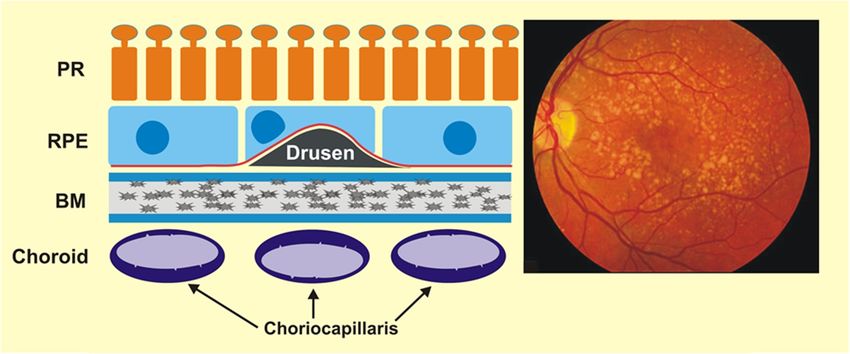
Figure 5. Cellular and extracellular waste clearing. Cellular waste, including misfolded, aggregated
and damaged proteins as well as damaged organelles (presented as small ovals or squares of different
colors) is subjected by two main machineries: ubiquitin-proteasome system (UPS) and autophagy,
which can be in the form of macroautophagy, including mitophagy, microautophagy and chaperone-
Figure 5. Cellular and
5. Cellular extracellular waste clearing. Cellular waste,including
includingmisfolded,
misfolded, aggregated
aggregated and
Figure mediated and extracellular
autophagy waste
(CMA). Unfolded clearing. Cellular
proteins are waste,
a substrate for unfolded protein response (UPR,
and damaged
damaged proteins proteins
as well
not represented
as well
as damaged
here),
as damaged
organelles
which directs
organelles (presented
(presented
them to degradation
as
as small
either
small ovals
ovals or
by autophagy
or squares
or squares
of different
of different
UPS. Heterophagy, colors)
colors) is
which subjected
degrades by two main machineries: ubiquitin-proteasome system (UPS) and autophagy,
is subjected by two mainextracellular
machineries: debrisubiquitin-proteasome
inside the cell, is of a particular
system importance
(UPS) and in retinal pigmentwhich can
autophagy,
whichepithelium
can be in cells
the form
and isof macroautophagy,
usually including mitophagy,can
carried out by endocytosis. microautophagy and chaperone-
be in the form of macroautophagy, including mitophagy,Exosomes microautophagytransport waste material out
and chaperone-mediated
mediated
of theautophagy (CMA). Unfolded
cell. LAMP-2A—lysosomal proteins
associated are a substrate
membrane protein for
2A. unfolded
The black protein response
arrows indicate the(UPR,
autophagysequence
(CMA). Unfolded proteins are a substrate for unfolded protein response (UPR, not
of here),
events.which directs them to degradation either by autophagy or UPS. Heterophagy,
not represented
represented here), which directs them to degradation either by autophagy or UPS. Heterophagy,
which degrades extracellular debris inside the cell, is of a particular importance in retinal pigment
The proteasome
whichepithelium
degrades is the main
extracellular machinery
debris inside of eukaryotic
the cell, is ofcells that degrades
a particular misfoldedinand damaged
cells and is usually carried out by endocytosis. Exosomes can importance
transport waste retinal
material pigment
out
proteins.
epithelium Ubiquitin
cells and is proteasome
usually systemout
carried (UPS)
by targets soluble proteins
endocytosis. Exosomes thatcan
are ubiquitinated
transport priormaterial
waste to
of the cell. LAMP-2A—lysosomal associated membrane protein 2A. The black arrows indicate the
their degradation [39] (Figure 5).
out ofsequence
the cell.ofLAMP-2A—lysosomal
events. associated membrane protein 2A. The black arrows indicate the
Substrate proteins are delivered to lysosomes from the extracellular media (heterophagy) or
sequencefromofinside
events.
the cell (autophagy). The best described heterophagic pathway is endocytosis. Three
The proteasome
different types ofis the main machinery
autophagy have beenof eukaryotic
described cells that degrades
in mammalian misfolded
cells: and damaged
macroautophagy,
proteins. Ubiquitin proteasome system (UPS) targets soluble proteins
The proteasome is the main machinery of eukaryotic cells that degrades misfolded and that are ubiquitinated prior to
damaged
their degradation [39] (Figure 5).
proteins. Ubiquitin proteasome system (UPS) targets soluble proteins that are ubiquitinated prior to
Substrate [39]
their degradation proteins are 5).
(Figure delivered to lysosomes from the extracellular media (heterophagy) or
from inside the cell (autophagy). The best described heterophagic pathway is endocytosis. Three
Substrate proteins are delivered to lysosomes from the extracellular media (heterophagy)
different types of autophagy have been described in mammalian cells: macroautophagy,
or from inside the cell (autophagy). The best described heterophagic pathway is endocytosis.
Three different types of autophagy have been described in mammalian cells: macroautophagy,
microautophagy, and chaperone-mediated autophagy (CMA). In macroautophagy intracellular
components are sequestered by a limiting membrane to form an autophagic vacuole that then fuses
with lysosomes. In microautophagy, substrates are directly internalised through invaginations ofInt. J. Mol. Sci. 2019, 20, x FOR PEER REVIEW 7 of 23
microautophagy, and chaperone-mediated autophagy (CMA). In macroautophagy intracellular
components are sequestered by a limiting membrane to form an autophagic vacuole that then fuses
with lysosomes. In microautophagy, substrates are directly internalised through invaginations of
Int. J. Mol. Sci. 2019, 20, 210
the
7 of 23
lysosomal membrane. In contrast to this “in-bulk” degradation, in CMA, selective substrate proteins
are translocated into the lysosomes one by one after binding to a lysosomal receptor LAMP-2A
the lysosomal
(lysosomal membrane.
associated In contrast
membrane to this
protein “in-bulk”
2). The degradation, in system
ubiquitin-proteasome CMA, selective
(UPS) is substrate
the other
proteins are translocated into the lysosomes one by one after binding to
major pathway for degradation of intracellular proteins inside cells. Substrates are tagged with a lysosomal receptor LAMP-2A
(lysosomal
ubiquitin that associated membrane
is recognised by theprotein 2). The the
proteasome, ubiquitin-proteasome
protease of this pathway. system Damaged
(UPS) is the other major
organelle and
pathway for degradation
protein aggregates of intracellular
that cannot be degradedproteinsby inside cells. Substrates
proteasome are subjected are tagged
by systemwithdegrading
ubiquitin
that
marked is recognised
targets inby the proteasome,
specific the protease of this pathway.
organelles—autophagosomes [40]. It is Damaged organelle
called autophagy inand
the protein
case of
aggregates
degradation thatof cannot be degraded
intracellular waste, by proteasomewhen
heterophagy are subjected by system
extracellular degrading
compounds aremarked
degraded targets
and
in specific organelles—autophagosomes
mitophagy in the case of degradation of damaged [40]. It ismitochondria.
called autophagy Damagedin theproteins
case of or
degradation
organelles of in
intracellular
both systemswaste, heterophagy
are detected whenchaperone
by specific extracellular andcompounds
co-chaperone areproteins,
degradedand andthis
mitophagy in the
action leads to
case of degradation
the selection of damaged
of degradation mitochondria.
pathway Damaged and
[41]. Chaperone proteins or organelles
co-chaperone alsoinparticipate
both systems in are
the
detected
ubiquitinationby specific chaperone
process. Proteinsanddirected
co-chaperoneto theproteins,
autophagy and this
pathwayactionundergo
leads to the selection of
engulfment in
degradation
autophagosomes pathwaywhere [41].they
Chaperone and co-chaperone
are subject to degradation. alsoUbiquitin
participateininboththe ubiquitination
of these systems process.
is a
Proteins
degradation directed to theexcluding
marker, autophagyubiquitin-independent
pathway undergo engulfment autophagy in autophagosomes
pathways [42]. where they
Membrane
are subject
proteins of to degradation.
damaged Ubiquitin
organelle in both ofare
and aggregates these systems
marked is aattachment
by the degradation marker, excluding
of ubiquitin particles.
ubiquitin-independent
Then autophagosomal degradation autophagy pathways
system detects[42]. Membrane proteins proteins
those ubiquitinated of damaged and organelle
engulfs wholeand
aggregates
organelle and areaggregates.
marked by Some the attachment of ubiquitin
of the signalling particles.
proteins, ThenMfn1
including autophagosomal
and 2 (mitofusindegradation
1 and 2),
system detectsinthose
participating ubiquitinated
mitophagy proteins and
before formation engulfs whole organelle
of autophagosome are degraded and aggregates.
by proteasome Some of
[43].
the signalling
Moreover, UPS proteins,
plays anincluding
important Mfn1
roleand
in the2 (mitofusin
regulation1ofand 2), participating
intracellular level ofinROS.
mitophagy before
Proteasome is
formation
responsible of autophagosome
for degradation areof
degraded
damaged by proteasome
mitochondrial [43]. Moreover,
membraneUPS plays ancomponent
proteins, important role of
in the regulationelectron
photosynthetic of intracellular
transport level of ROS.
chain. Damage Proteasome is responsible
in these proteins leads for degradation
to abnormal of damaged
increase in ROS
mitochondrial membrane degradation
production. Proteasomal proteins, component
of these of photosynthetic
proteins results inelectron transport
restoration chain. oxidative
of normal Damage
in these proteins leads
phosphorylation (OXPHOS)to abnormal
process increase in ROS production.
and reduction Proteasomal
of ROS. Proteasome degradation
activation can of these
mediate
proteins
mitophagy results
[43].inExosomes,
restoration of normal oxidative
extracellular phosphorylation
vehicles released by almost (OXPHOS) process
all if not all and reduction
eukaryotic cells, are
of ROS. Proteasome
important element of activation cancommunication,
cell-to-cell mediate mitophagy [43].can
but they Exosomes,
also play extracellular vehicles
a role in waste released
clearing [44].
by almost all ifunfolded
Accumulated not all eukaryotic
and damaged cells,proteins
are important element
are detected byofunfolded
cell-to-cell communication,
protein response (UPR)but they
and
can also play
targeted a role in waste
for degradation by clearing [44]. Accumulated unfolded and damaged proteins are detected
UPS or autophagy.
by unfolded protein response (UPR) and targeted for degradation by UPS or autophagy.
3.1. Unfolded Protein Response and Endoplasmic Reticulum-Associated Degradation
3.1. Unfolded Protein Response and Endoplasmic Reticulum-Associated Degradation
Figure 6.
Figure 6. Unfolded
Unfoldedprotein
proteinresponse.
response.When
Whenunfolded,
unfolded,misfolded
misfolded and
and damaged
damaged proteins
proteins accumulate
accumulate in
in endoplasmic reticulum (ER), they can induce unfolded protein response (UPR), a signaling
endoplasmic reticulum (ER), they can induce unfolded protein response (UPR), a signaling cascade cascade
with the
with the involvement
involvement ofof protein
protein kinase-like
kinase-like endoplasmic
endoplasmic reticulum
reticulum kinase
kinase (PERK),
(PERK), inositol
inositol requiring
requiring
enzyme11(IRE1),
enzyme (IRE1),
andand activating
activating transcription
transcription factorfactor 6 (ATF6).
6 (ATF6). This cascade
This cascade leads to aleads totranslation
stop in a stop in
translation
of of faulty
faulty proteins, proteins, degradation
degradation of misfoldedofproteins
misfoldedandproteins and
increased increased
synthesis of synthesis
chaperonsofinvolved
chaperons
in
protein folding. If these mechanisms fall, UPR switch to pro-apoptotic response. XBP1s—X-box binding
protein 1 specificity protein, eIF2—translation initiation factor 2, ERAD—ER-associated degradation,
ATF6f—the transcriptional activator domain of ATF6, P—phosphate residue.Int. J. Mol. Sci. 2019, 20, 210 8 of 23
Unfolded protein response is a signaling cascade activated in response to endoplasmic reticulum
(ER) stress manifested by the accumulation of unfolded and damaged proteins. UPR may also be
activated by damaged mitochondria. Expression of UPR genes increases in oxidative stress and it
was shown that the stress caused by cigarette smoke extracts induced UPR in RPE cells [42,43]. In ER
UPR is activated by pathways initiated by three different sensors: protein kinase-like endoplasmic
reticulum kinase (PERK), inositol requiring enzyme 1 (IRE1), and activating transcription factor
6 (ATF6) (Figure 6) [45–49]. Oxidative stress enhances the transcription of these proteins in RPE
cells [43]. The UPR signalling activates transcription factors and protein kinases leading to an adaptive
response involving the activation of proteasomal degradation and autophagy, chaperone induction
and enhancement of antioxidant defence [50,51]. Inefficient attempts to restore homeostasis cause UPR
to induce cell death by apoptosis. UPR increases apoptosis in RPE cells treated with cigarette smoke
extract [52–54]. Therefore, UPR can play a role in AMD pathogenesis as it is involved in detecting of
improper proteins and their degradation in RPE.
Endoplasmic reticulum-associated degradation (ERAD) has been considered as an integral part of
UPR as many ERAD genes are controlled by UPR. However, recent studies suggest that ERAD plays a
direct role in protein clearance, which is mainly underlined by IRE1α activation [55,56].
3.2. Ubiquitin Proteasome System
The process of UPS degradation is initiated by specific enzymes, E1–E3, that attach ubiquitin
to the substrate in an ATP-dependent manner. Substrates with polyubiquitin tail are transported to
proteasome where they undergo degradation.
Typical 26S proteosomal complex contains three domains: a core particle (20S) with two regulatory
particles (19S, caps, lids) [57]. Regulatory particles are involved in the recognition and binding of
polyubiquitinated proteins and their transport to the catalytic core located at the inner surface of
the 20S subunit. The core particle is composed of four heptameric annular complexes—two outer α
subunits, which play a structural function and two inner β subunits with catalytic activity. The β
subunits contain proteolytic active sites located on proteasomal interior surface. Energy from ATP
hydrolysis is used to open lid, unfold polyubiquitinated proteins and their transport inside 20S subunit.
Oxidative stress, a major factor of AMD pathogenesis, is associated with an increased production
of cellular waste, but on the other hand it may damage components of cellular waste clearing systems,
which can contribute to AMD progression. A mild oxidative stress can regulate UPS activity through
the stimulation of E1 and E2 to bind ubiquitin as well as the 26S proteasome [58,59]. However, strong
oxidative stress can damage E1 and E2, blocking ubiquitin binding. This effect can be associated with
a high concentration of oxidized glutathione, competing with E1 and E2 on their binding sites in
ubiquitin [60]. Oxidative stress could also directly inactivate 26S proteasome through detachment of
the 19S subunit from 20S core particle [52,61].
Reduction of proteasome activity during lifetime is associated with aging, another critical factor
in AMD pathogenesis. Aging results in reduced expression of UPS genes and may lead to the
collapse of proteasome complex, resulting in an accumulation of cellular debris [62–66]. These effects
are correlated with weakening of the sustaining activity of the proteins quality control system and
especially chaperone proteins, which play a significant role in UPS [67–69]. Several other effects can
underline lowering of the efficacy of UPS with age, including alternations in the composition of the
proteasomal subunits, reduced stability of proteasomes or their inactivation [14,65–67]. It has been
shown that during replicative senescence the level of the β subunits decreases [70]. Proteasome activity
is associated with the rate of cellular aging and the entry of cells into the senescence pathway [71].
Moreover, decreasing activity of proteasome results in increase of damaged proteins of the respiratory
chain, resulting in mitochondrial dysfunction and an increase of cellular ROS level [72,73].
Insufficient activity of UPS leads to the escalation of protein deposits followed by an increase
in the level of ROS and induction of chronic inflammation. Inflammation combined with constantlyInt. J. Mol. Sci. 2019, 20, 210 9 of 23
weakening waste cleaning system in RPE cells can induce their senescence, resulting in the
development and progression of AMD [24].
Liu et al. showed that photooxidative stress decreased the activity of UPS in RPE. This
interaction increased the expression of genes encoding proinflammatory interleukins 6 and 8
(IL-6 and -8) and downregulated the anti-inflammatory genes MCP-1 (monocyte chemoattractant
protein-1) and CFH (complement factor H) [74]. Similar results obtained Qin et al. who showed
that a decrease in proteasome activity in RPE led to dysregulation of the NF-κB (nuclear factor
kappa-light-chain-enhancer of activated B cells) signalling pathway [75]. It was demonstrated in a
mouse model that dysregulation of UPS led to retinal degeneration through photoreceptor cell death
by apoptosis in a caspase-independent pathway [76]. De Carvalho. et al. suggested that proteasomal
regulation may play a significant role in the control of neovascularization process important in
wet AMD [77]. They showed an efficient action of UPS counteracting degenerative changes in
ARPE-19 (human retinal pigment epithelial) cells through the control of the TGFβ (transforming
growth factor-β) signalling.
3.3. Autophagy
Autophagy degrades damaged or unneeded proteins in lysosomes. Many proteins are involved
in this process, including autophagy related proteins (ATGs), mechanistic target of rapamycin (mTOR),
the serine/threonine uncoordinated-51-like kinases 1 and 2 (ULK1 and ULK2), FIP-200, p62/SQSTM1,
microtubule-associated protein light chain 3 (LC3) and others. Several modes of autophagy
can function, including macroautophagy (usually referred as to autophagy), chaperone-mediated
autophagy and microautophagy.
Autophagy is initiated by the formation of a double-membrane vesicle, autophagosome, enclosing
material to be degraded (cargo) that is delivered to the lysosome, where degradation and recycling
occur [78].
Autophagy impairment, caused by the depletion of the core autophagy genes ATG5 and ATG7,
was associated with an AMD-like phenotype in mouse RPE cells. This phenotype was manifested by
RPE thickening, hypertrophy or hypotrophy, pigmentary abnormalities and accumulation of oxidized
proteins [79]. A2E, the main hydrophobic constituent of lipofuscin can induce damage to RPE cells
through the inhibition of autophagy [80]. These findings suggest that autophagy prevents detrimental
effects of A2E and inhibits the production of inflammatory factors in RPE.
Many reports imply that oxidative stress induces autophagy in RPE cells [81–84]. Studies on
RPE cells from AMD donors and mice with AMD-like phenotype suggest that autophagy increases
during aging and AMD [82]. However, the autophagosomes formation in late AMD was reported
to occur at lower rate than in early stages. This study also revealed that chronic oxidative stress
decreased autophagic flux. Autophagy can prevent retinal cells from the damaging effects of oxidative
stress [81,82]. Rapamycin induced autophagy in RPE cells and led to reduced accumulation of
lipofuscin, whereas leupeptin, a blocker of autophagy, caused an increase in lipofuscin formation [82].
Autophagy can protect RPE cells from cell death induced by oxidative stress. RPE cells treated
with paraquat, an inducer of oxidative stress and cultured with autophagy inhibitor 3-methyladenine
(3-MA) showed an increase in the number of apoptotic cells compared to cells with undisturbed
autophagy [81]. RPE cells under oxidative stress increased the expression of p62/SQSTM1 and
autophagy [83]. Rotenone, an agent inducing mitotic catastrophe, increased autophagy and mitophagy
in RPE cells protecting these cells from death [84].
The FIP200 protein is important in autophagy induction as it is involved in the formation of
autophagosomes [85]. The conditional knockout of gene encoding FIP200 (FIP200 cKO) in mice
resulted in a reduction of autophagy. These animals also displayed changes in the phenotype of RPE
cells, including lipid accumulation, increasing with age. The reduction of autophagy in FIP200 cKO
mice led to photoreceptors loss and retinal dysfunction [86].Int. J. Mol. Sci. 2019, 20, 210 10 of 23
Mice with knockout of ATP-binding cassette subfamily A member 4 (Abca4) and retinol
dehydrogenase 8 (Rdh8) genes are characterized by impaired clearing of atRAL and they were a model
of light-induced retinal degeneration [20]. Retinas of these mice were characterized by a delay in atRAL
removal after light exposure [87]. Additionally, light illumination led to an increased expression of
the LC3B-II and PARKIN proteins, a marker of autophagosome formation and a mitophagy regulator,
respectively. These results suggest that autophagy plays an important role in protecting the retina
from damage caused by light.
Injection of amyloid-β, which is a main component of drusen, to murine vitreous resulted in
a upregulation of autophagy markers LC3, ATG5 and BECLIN-1. Human RPE cells treated with
amyloid-β also showed autophagy induction and upregulated expression of cytokines [88].
3.4. Mitophagy
Damaged mitochondria are removed in a highly specific and selective pathway called mitophagy.
All mechanistic aspects of this process are not exactly known and several models of it have been
presented [89].
In a model proposed by Ding and Yin mitophagy is a two-steps process involving induction of
canonical autophagy with ATG proteins and priming of mitochondria [90]. Canonical autophagy
is underlined by several mechanisms, including AMPK activation induced by ATP depletion and
suppression of mTOR mediated by mitochondrial damage resulting in ROS overproduction. These
ROS induce further mitochondrial damage, which amplifies the inducing signal. Mitochondria
priming could be PARKIN-dependent or independent. In the former, depolarization of mitochondrial
membrane results in compromised cleavage of the PINK1 (PTEN (phosphatase and tensin
homolog) induced kinase 1) protein mediated by the mitochondrial rhomboid protease PARL
(presenilins-associated rhomboid-like protein, mitochondrial). Stabile PINK1 recruits PARKIN to
mitochondria resulting in subsequent ubiquitination of proteins localized on the outer mitochondrial
membrane. These proteins can be degraded by UPS or bound by p62/SQSTM1, which directly interacts
with LC3 to bind autophagosome to faulty mitochondria. Selective mitophagy can be supported by
the PI3K (phosphoinositide 3-kinase) complex activated by Ambra1. An enhanced expression of the
FUNDC1 (FUN14 domain containing 1) and BNIP3L (BCL2 (B-cell lymphoma 2) interacting protein 3
like) proteins in impaired mitochondria may occur in the PARKIN-independent pathway of mitophagy.
These proteins induce autophagosome to target mitochondria by a direct interaction with LC3. In this
pathway, damaged mitochondria can be also targeted by Smurf1 (SMAD specific E3 ubiquitin protein
ligase 1) to ubiquitinate mitochondrial proteins and induce mitophagy. ULK1 can phosphorylate
ATG13 upon activation by Hsp90 (heat shock protein 90 kDa) to promote mitophagy. PINK1 is cleaved
by mitochondrial proteases and degraded in the proteasome [91]. PINK1 activates PARKIN by its
phosphorylation at S65 [92]. PINK1 targets the same residue in phosphorylation of ubiquitin in the
S65 position [93]. Several other mechanisms of mitophagy, both PARKIN-dependent and independent
could be considered.
Aging reduces the efficacy of mitophagy, which leads to the accumulation of damaged
mitochondria (reviewed in [91]). Aged RPE cells have more mitochondrial DNA (mtDNA) damage and
display a decreased ability to repair it as compared to young RPE [94–97]. The number of mitochondria
decreased with age in rhesus RPE and aging mitochondria had an increased length and formed
clusters [94]. Studies comparing RPE cells from healthy elderly with AMD patients showed that the
latter had a decreased number of mitochondria [98]. RPE cells from elderly donors were more sensitive
to oxidative stress than RPE from young individuals [99,100]. These findings suggest that mitophagy
can be important in AMD pathogenesis.
3.5. Exosomal Degradation
Exosomes are extracellular vehicles released by various cells, including epithelial cells [101]. They
are an important element of the cell-to-cell communication (reviewed in [102]). They can carry outInt. J. Mol. Sci. 2019, 20, 210 11 of 23
of the cells various molecules, including peptides, proteins, lipids, RNA and DNA, so they can be
also considered as an important element of cellular waste clearing. In fact, waste elimination function
was attributed to exosomes earlier than their communicative potential. However, some of molecules
exported from cells by exosome can be only carriers of biological information. Exosome is a 9–11
protein complex having, similarly to proteasome, ring-like core structure that in humans contains nine
subunits [103]. Core proteins of eukaryotic exosomes display RNase activity and belong to the RNase
PH class [41]. Exosomes are present and display activity in the cytoplasm, nucleus and nucleolus.
RNA degradation by exosomes is their best known and likely the main function. To perform it, the core
exosomes display both exo- and endoribonuclease activities. Many aspects of exosome functioning,
both as a cellular garbage bin and as an important element of the cell-to-cell communication, need
further research. It is worth noting that exosomes can cooperate with autophagy in cellular waste
clearing [104].
Some proteins which can be found in drusen, including annexin, enolase, CD63 are features of
exosomes [15,105,106].
Blue light is an environmental AMD risk factor and it induces detrimental changes in the retina,
which are associated with oxidative stress and overproduction of cellular waste. An increase in
the proinflammatory molecules in the content of exosomes released by RPE cells after photoactive
blue-light stimulation was observed [107]. Moreover, a higher level of the NLRP3 (NACHT (neuronal
apoptosis inhibitor protein, class 2 transcription activator of the MHC, heterokaryon incompatibility
and telomerase-associated protein 1), NLR (nucleotide-binding domain, leucine-rich repeat-containing
family), and PYD (pyrin domain)-containing protein 3) inflammasome was observed in that study.
Emerging evidence suggests the role of exosomes in the activation of the complement in the
immediate vicinity of RPE cells [108]. Exosomes were suggested to play a role in the occurrence and
development of choroidal neovascularization, so they can be important in mechanisms of wet AMD
pathogenesis and developing therapeutic strategies in this disease [109]. Exosomal proteins found in
aqueous humor were postulated to be an independent molecular marker in wet AMD [110]. Exosomal
miRNA, which can be important to stimulate target cells, was recently discovered in AMD [111].
Therefore, exosomes may play a multiple role in AMD pathogenesis, but uncovering the precise
mechanism of this role requires further studies.
3.6. Heterophagy
Heterophagy, a digestion of extracellular material inside the cell, is intensively carried out in RPE
cells as they constantly degrade POS to maintain the function of photoreceptors. Each RPE cell is
challenged by digestion of POS from 30 to 40 photoreceptors [112].
Heterophagy in RPE cells involves the recognition and attachment of a POS discs, its digestion,
the formation of phagosome and its fusion with lysosome and the final degradation [18]. Integrins,
including ITGAV (integrin alpha V)-ITGB5 (integrin subunit beta 5) are necessary to bind POS, which
ingestion requires the MERTK (c-mer proto-oncogene tyrosine kinase) protein [113]. After ingestion
of extracellular cargo into vesicles they are transported to the basal end of the cell and fuse with
lysosomes to degrade the cargo as it does in autophagy [114]. Heterophagy impairment can lead to
accumulation of photoreceptor cell waste resulting in chronic inflammation. POS recognition by RPE
may be a critical step in heterophagy as defects in this process results in death of photoreceptors [115].
4. Interplay of Autophagy and UPS in AMD
Autophagy and UPS are two main pathways to eliminate damaged and misfolded proteins from
the cell. Despite the inhibition of UPS activates autophagy, these two systems were considered as
independent for a long time. Some proteins, e.g. α-synuclein can be degraded at the same time by UPS
and autophagy [116]. These systems share more common substrates (reviewed in [117]). However,
some of them are too large to fit the proteasome. Both systems use ubiquitin as a signal moleculeInt. J. Mol. Sci. 2019, 20, 210 12 of 23
to label protein to degrade. This suggests that these two pathways interplay to maintain cellular
proteostasis and many proteins can regulate this interplay.
Pandey et al. showed that histone deacetylase 6 (HDAC6) can be essential in the regulation of
both UPS and autophagy [118]. Moreover, this regulation was shown to play an important role in
the pathogenesis of various neurodegenerative diseases—HDAC6 repressed degeneration resulting
from proteasome mutations in an autophagy-dependent fashion. This suggests, that HDAC1 can be
important for a compensatory mechanism between UPS and autophagy.
ARPE-19 cells treated with proteasome inhibitor MG132 and chloroquine, an inhibitor of
autophagy, showed an increase in ubiquitinated protein aggregates and enhanced levels of LC3-I,
LC-3II and LAMP1 [119]. However, an increased level of γ-tubulin and p62/SQSTM1 was also
observed, suggesting that autophagy was upregulated in that study. Chloroquine increased the levels
of ubiquitinated aggregates and LC3-II and p62/SQSTM1. Prolonged inhibition of autophagy resulted
in compromising of proteasome activity. These results confirm the interplay between autophagy
and UPS in the retina, so waste clearing in AMD should be rather considered in the context of such
interplay than in either system separately.
Ubiquitin tagging is performed with the involvement of several proteins, mainly ubiquitin ligases
E1-E3 and the interaction between the RING (really interesting new gene) domain of E3 with E2 results
in the final stage of attachment of ubiquitin to the protein to be degraded. It was reported that the
protein EI24 (etoposide-induced protein 2.4 homolog) promoted degradation of RING E3 ligases in
autophagy, which can be important in cancer transformation [120]. The gene encoding EI24 was shown
to be essential for autophagy [121]. Therefore, EI24 can be the main connection between UPS and
autophagy underlined by its ability to degrade RING E3 (Figure 7). It was also shown that E3 ligases,
major functional proteins in UPS, can be degraded by autophagy. Although the biological relevance of
EI24 has been evidenced only in cancer, it seems reasonable to consider its action as a mechanism of
pathogenesis of any disorder associated with impaired clearing of cellular debris.
p62/SQSTM1 is multifunctional protein that interacts non-covalently with ubiquitin and mediates
delivery of damaged proteins for degradation in both the UPS and autophagic pathways [122,123].
Phosphorylation of p62/SQSTM1 at serine 403 leads to the attachment of ubiquitinated proteins and
their targeting to degradation in autophagosomes [124]. We showed that treatment of ARPE-19 cells
with the proteasome inhibitor MG-132 led to accumulation of perinuclear aggregates which rapidly
colocalized with p62/SQSTM1 [125]. Based on studies in which autophagy was inhibited, it was
found that p62/SQSTM1 is degraded mainly by autophagy [121]. We showed that p62/SQSTM1 was
accumulated in AMD donors in macular area with a large number of drusen, supporting important
role of autophagy in AMD pathogenesis [126]. p62/SQSTM1 can be also phosphorylated by ULK1 at
serines 409 and 405 [127]. This phosphorylation occurs when UPS does not work properly leading to
proteotoxic stress. Additionally, phosphorylation executed by ULK1 does not occur under nutrient
deficiency. Proteosomal stress induced by MG-132 leads to phosphorylation of p62/SQSTM1 at serine
28 by the short form of protein kinase PINK1 (PINK1-s). This phosphorylation occurs only in cells
with inhibited proteasome. Phosphorylation of p62/SQSTM1 on serine 28 is required for aggregosome
formation in cells under proteasomal stress. These results suggest that PINK1-s can act as a sensor of
UPS activity that can stimulate the formation of aggregosome [128].Int. J. Mol. Sci. 2019, 20, 210 13 of 23
Int. J. Mol. Sci. 2019, 20, x FOR PEER REVIEW 13 of 23
Figure 7. EI24 (etoposide-induced protein 2.4 homolog) is the main connection between
Figure 7. EI24 (etoposide-induced
ubiquitin-mediated protein(UPS)
proteasomal system 2.4 homolog) is the main
and autophagy. The connection between
concerted action of ubiquitin-
ubiquitin
mediated proteasomal system (UPS) and autophagy. The concerted action of ubiquitin ligases
ligases E1-E3 results in ubiquitination of target proteins to label for UPS-mediated degradation. E1-E3
results in ubiquitination of target proteins to label for UPS-mediated degradation. Ubiquitin
Ubiquitin chain transfer to target proteins is catalyzed by the RING-domain E3 ligases. EI24, chain
an
transfer to target proteins is catalyzed by the RING-domain E3 ligases. EI24, an autophagy-inducing
autophagy-inducing protein, can cause autophagy-mediated degradation of RING-domain E3 ligases.
protein,
Thick can represent
arrows cause autophagy-mediated
main pathways, thindegradation
arrows—side of pathways.
RING-domain E3 ligases. Thick arrows
represent main pathways, thin arrows – side pathways.
Oxidative stress induced by H2 O2 leads to inhibition of proteasome activity in RPE cells and an
Oxidative
increase stress induced
in p62/SQSTM1 by H2O[83].
expression 2 leads to inhibition
Oxidative stressofinduced
proteasome activity smoke
by cigarette in RPEalso
cellsinhibits
and an
increase in p62/SQSTM1 expression [83]. Oxidative stress induced by cigarette
the UPS pathway in RPE cells [129]. It was shown that cigarette smoke upregulated the expression smoke also inhibits
thep62/SQSTM1
of UPS pathwaymRNA in RPEin cells [129]. Itcells.
ARPE-19 was shown
Silencingthatofcigarette
p62/SQSTM1smokeincreased
upregulated the the expression of
accumulation of
p62/SQSTM1 mRNA in ARPE-19 cells. Silencing of p62/SQSTM1 increased
protein aggregates caused by cigarette smoke in RPE cells that showed decreased autophagy and the accumulation of
protein aggregates caused by cigarette smoke in RPE cells that showed decreased
Nrf2 (nuclear factor (erythroid-derived 2)-like 2)-mediated antioxidant response [36]. This finding autophagy and
Nrf2 (nuclear
suggests factor (erythroid-derived
that p62/SQSTM1 plays a major2)-like
role in2)-mediated
the protection antioxidant
of RPE cells response
against [36].
stressThis finding
induced by
suggests that
protein damage. p62/SQSTM1 plays a major role in the protection of RPE cells against stress induced by
protein damage.
Proteins of the Hu family are RNA-binding proteins. In vertebrates there are four members of
Proteins
the Hu family:ofHuR,
the Hu family
HuB, HuC areand
RNA-binding
HuD. HuR proteins.
plays a roleIn vertebrates there are
in cellular stress four members
response and in the of
the Hu family: HuR, HuB, HuC and HuD. HuR plays a role in cellular stress
regulation of cell cycle (reviewed in [130]). Treatment of ARPE-19 cell with proteasome inhibitors response and in the
regulation
MG132 andofAICAR
cell cycle (reviewed in [130]). Treatment ribonucleotide)
(5-aminoimidazole-4-carboxamide of ARPE-19 cellled withto proteasome
activation ofinhibitors
the HuR
MG132 and AICAR (5-aminoimidazole-4-carboxamide
protein that increased p62/SQSTM1 expression [131]. ribonucleotide) led to activation of the HuR
protein that increased p62/SQSTM1 expression [131].
Hsp70 is involved in proteostasis maintaining by preventing protein aggregation, refolding of
Hsp70 isand
denaturated involved in proteostasis
aggregated proteins and maintaining
acting asby preventingfor
a chaperone protein aggregation,
degradation refolding or
in proteasome of
denaturated and aggregated proteins and acting as a chaperone for degradation
lysosome (reviewed in [132]). Hsp70 can also trigger the permeability of lysosomal membrane [133]. in proteasome or
lysosome (reviewed in [132]). Hsp70 can also trigger the permeability of lysosomal membrane [133].
Hsp70 protects ARPE-19 cells against oxidative stress. ARPE-19 cells treated with recombinantYou can also read