New Insights into the Multifaceted Role of Myeloid-Derived Suppressor Cells (MDSCs) in High-Grade Gliomas: From Metabolic Reprograming ...
←
→
Page content transcription
If your browser does not render page correctly, please read the page content below
cells
Review
New Insights into the Multifaceted Role of Myeloid-Derived
Suppressor Cells (MDSCs) in High-Grade Gliomas: From
Metabolic Reprograming, Immunosuppression, and
Therapeutic Resistance to Current Strategies for
Targeting MDSCs
Senthilnath Lakshmanachetty                   , Joselyn Cruz-Cruz † , Eric Hoffmeyer † , Allison P. Cole †
and Siddhartha S. Mitra *

                                          Morgan Adams Brain Tumor Research Program, Department of Pediatrics, Division of Hematology/Oncology/
                                          Bone Marrow Transplant, University of Colorado Anschutz Medical Campus, Aurora, CO 80045, USA;
                                          senthilnath.lakshmanachetty@cuanschutz.edu (S.L.); Joselyn.cruzcruz@cuanschutz.edu (J.C.-C.);
                                          eric.hoffmeyer@cuanschutz.edu (E.H.); ALLISON.P.COLE@CUANSCHUTZ.EDU (A.P.C.)
                                          * Correspondence: siddhartha.mitra@cuanschutz.edu; Tel.: +1-303-724-3886
                                          † These authors contributed equally to this work.

                                Abstract: Cancer cells “hijack” host immune cells to promote growth, survival, and metastasis. The

                                          immune microenvironment of high-grade gliomas (HGG) is a complex and heterogeneous system,
Citation: Lakshmanachetty, S.;
                                          consisting of diverse cell types such as microglia, bone marrow-derived macrophages (BMDMs),
Cruz-Cruz, J.; Hoffmeyer, E.; Cole,
                                          myeloid-derived suppressor cells (MDSCs), dendritic cells, natural killer (NK) cells, and T-cells.
A.P.; Mitra, S.S. New Insights into the
                                          Of these, MDSCs are one of the major tumor-infiltrating immune cells and are correlated not only
Multifaceted Role of Myeloid-Derived
Suppressor Cells (MDSCs) in High-
                                          with overall worse prognosis but also poor clinical outcomes. Upon entry from the bone marrow
Grade Gliomas: From Metabolic             into the peripheral blood, spleen, as well as in tumor microenvironment (TME) in HGG patients,
Reprograming, Immunosuppression,          MDSCs deploy an array of mechanisms to perform their immune and non-immune suppressive
and Therapeutic Resistance to             functions. Here, we highlight the origin, function, and characterization of MDSCs and how they are
Current Strategies for Targeting          recruited and metabolically reprogrammed in HGG. Furthermore, we discuss the mechanisms by
MDSCs. Cells 2021, 10, 893. https://      which MDSCs contribute to immunosuppression and resistance to current therapies. Finally, we
doi.org/10.3390/cells10040893             conclude by summarizing the emerging approaches for targeting MDSCs alone as a monotherapy or
                                          in combination with other standard-of-care therapies to improve the current treatment of high-grade
Academic Editors: Justin Lathia
                                          glioma patients.
and Defne Bayik

                                          Keywords: MDSCs; glioma; glioblastoma; high-grade glioma; brain tumors; metabolic reprogram-
Received: 31 March 2021
                                          ming; immune suppression; therapeutic resistance; therapeutic targeting; immunotherapy; tumor mi-
Accepted: 10 April 2021
Published: 14 April 2021
                                          croenvironment

Publisher’s Note: MDPI stays neutral
with regard to jurisdictional claims in
                                          1. Introduction
published maps and institutional affil-
iations.                                        Malignant gliomas that originate from glial, neural stem cells and astrocytes are the
                                          most aggressive tumors of the central nervous system (CNS) and spinal cord with a median
                                          survival of less than 12–15 months [1]. The current standard of care therapies such as
                                          surgery, radiotherapy, and chemotherapy have had only limited success in increasing the
Copyright: © 2021 by the authors.
                                          lifespan of glioma patients [2]. Although recent advances in immune checkpoint blockade
Licensee MDPI, Basel, Switzerland.
                                          (ICD) therapies such as anti-PD-1/PD-L1 and anti-CTLA4 have yielded promising results
This article is an open access article
                                          in melanoma and non-small lung cancer [3], glioma patients not only failed to respond in
distributed under the terms and           clinical trials but also developed resistance to ICB in a multitude of ways [4]. One such way
conditions of the Creative Commons        is the development and maintenance of an immune-suppressive tumor microenvironment
Attribution (CC BY) license (https://     (TME) that thwarts the efficacy of existing therapies and host anti-tumor immune responses.
creativecommons.org/licenses/by/                Extensive analysis of the immune microenvironment in high-grade glioma (HGG) us-
4.0/).                                    ing single-cell RNA-seq, mass cytometry (CyTOF), immunohistochemistry, flow cytometry,

Cells 2021, 10, 893. https://doi.org/10.3390/cells10040893                                                 https://www.mdpi.com/journal/cells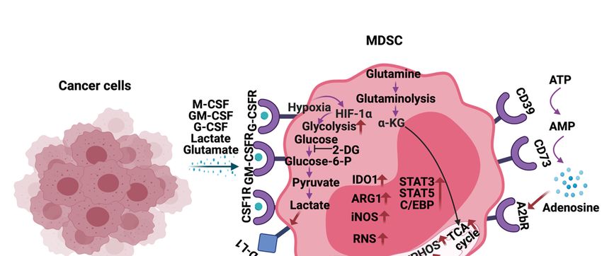
Cells 2021, 10, 893                                                                                              2 of 21

                      and other “omics” technologies indicate the presence of higher numbers of immune-
                      suppressive macrophages, microglia dendritic cells, regulatory T-cells, and myeloid-
                      derived suppressor cells (MDSCs) [5]. Together, these cells interact with the neoplastic
                      cells to promote tumor growth, progression, metastasis, angiogenesis and contribute to the
                      extreme immunosuppression observed in HGG.
                            In healthy humans and mice, MDSCs are present at very low frequencies and con-
                      stitute only ~0.5–2% of peripheral blood mononuclear cells [6]. Nevertheless, 30–50% of
                      the tumor mass in HGGs are found to be MDSCs [7,8]. Originally, derived from the bone
                      marrow, MDSCs are a very heterogeneous population of immature myeloid cells (IMCs)
                      present at various stages of myelopoiesis. Under normal conditions, IMCs can be differ-
                      entiated into macrophages, granulocytes, and dendritic cells. However, in pathological
                      conditions such as HGG, the differentiation of IMCs is subverted, resulting in the genera-
                      tion, recruitment, expansion, and activation of MDSCs [9] not only in the tumor bed but
                      also in the peripheral blood [10,11].
                            Recently, there is a great deal of interest to identify, quantify, characterize, and target
                      the different MDSC populations in brain tumors. In this review, we aim to provide an
                      overview regarding the origin, characterization, and metabolic reprogramming of MDSCs.
                      Moreover, we illustrate the mechanisms by which MDSCs contribute to immunosuppres-
                      sion and resistance to existing therapies. Finally, we conclude by discussing the current
                      strategies and clinical trials that are being pursued to effectively target MDSCs in the setting
                      of high-grade glioma.

                      2. History, Origin, and Characterization of MDSCs in Mice and Humans
                            Under non-pathological conditions, myelopoiesis is a tightly controlled process by
                      which the body can effectively protect itself from the insult. Conversely, under chronic
                      inflammatory conditions or neoplasia, the immune system cannot keep up with the demand
                      for neutralization as a result leading to deregulated myelopoiesis. One subpopulation of
                      cells which expands prodigiously under such conditions is myeloid-derived suppressor
                      cells (MDSCs).
                            In the late 1970s, the presence of an immune-suppressive subpopulation of myeloid
                      cells was first reported in mice following myeloablative radiation therapy [12]. Initially,
                      these cells were referred to as natural suppressor (NS) cells since they did not express
                      any markers related to macrophages, T-cells or B-cells, however, they shared similar
                      characteristics as natural killer (NK) cells [13]. Nearly 20 years later, this population of
                      “suppressor cells” was reported ex vivo in the peripheral blood of patients following
                      cytokine mobilization and apheresis [14]. Around this time, the first characteristic surface
                      antigens of “suppressor cells” derived from the spleen of mice were identified to be Mac-1
                      and Gr-1 [15]. In 2007, it was proposed that this population be referred to as MDSCs
                      to reflect their shared origin and function, and to lessen confusion in this growing field
                      of interest [16].
                            In HGGs, MDSCs are derived from the immature myeloid progenitors present in the
                      bone marrow (Figure 1). More recently, single-cell RNA-sequencing (scRNA-seq) analysis
                      on mouse breast tumors suggests that abnormal differentiation of monocyte and neutrophil-
                      like cells in the spleen can lead to the generation of MDSCs [17]. Reprogramming or
                      activation of monocytes and granulocytes by exposure to Toll-like receptors, IL-10, WNT5A,
                      LPS, and INFγ can also give rise to MDSCs [18]. Lastly, MDSCs can be generated and
                      activated ex vivo by the addition of GM-CSF, G-CSF, IL-6, and IL-10 to bone marrow
                      precursors obtained from healthy individuals and detailed functional and phenotypic
                      characterization revealed these bone marrow-derived MDSCs (BM-MDSCs) to be similar
                      to the MDSCs present in different cancer patients [19,20].
                            MDSCs in mice are usually identified by the surface expression of CD11b and Gr1.
                      Further, based on the surface expression, density, and morphology, two subsets of MDSCs
                      are identified: Monocytic (M-MDSC) and polymorphonuclear (PMN-MDSC) or granu-
                      locytic MDSC (G-MDSC) [21]. As the name suggests, M-MDSCs are mononuclear cells,
Cells 2021, 10, 893                                                                                                            3 of 21

                                 whereas PMN-MDSCs are polymorphonuclear cells. Moreover, M-MDSCs are very similar
                                 to inflammatory monocytes, and PMN-MDSCs shared some similar characteristics with
                                 neutrophils [22]. Irrespective of these similarities, MDSC subsets are a very distinct popu-
                                 lation and are phenotypically distinguished by the expression of CD11b+ Ly6G− Ly6Chigh
                                 (M-MDSC) or CD11b+ Ly6G+ Ly6Clow (PMN-MDSCs) (Figure 1) [23].

      Figure 1. Origin and subsets of myeloid-derived suppressor cells (MDSCs) in mice and humans. Under normal conditions,
      immature myeloid cells (IMCs) derived from the bone marrow differentiate into Monocytes, Neutrophils, Macrophages,
      and Dendritic cells. However, pathological conditions such as high-grade gliomas prevent IMC differentiation, resulting in
      the generation and accumulation of different MDSC subsets. Phenotypic markers that are commonly used to identify these
      subsets in mice and humans are described.

                                       MDSCs in humans are generally defined as CD33+ major histocompatibility class
                                 II− or HLA-DR− cells. Further, expression of CD115, CD124, CD80, programmed death
                                 ligand-1 (PD-L1), ARG1, and iNOS has been proposed to identify MDSCs [24]. In hu-
                                 mans, three subsets of MDSCs exist: Early-stage MDSC (e-MDSC), monocytic MDSC
                                 (M-MDSC), and polymorphonuclear (PMN-MDSC) or granulocytic MDSCs (gMDSCs)
                                 (Figure 1) [25]. Phenotypic markers used to identify them are lin− (including CD3, CD14,
                                 CD15, CD19, and CD56) HLA-DR− CD33+ (e-MDSCs), CD11b+ CD14+ CD15− (M-MDSC),
                                 and CD11b+ CD14+ CD66b+ (PMN-MDSC) (Figure 1) [22]. Furthermore, expression of
                                 Lectin-type oxidized LDL receptor 1 (LOX-1), which is associated with ER stress and lipid
                                 metabolism, is specifically seen in PMN-MDSC [26]. Aside from the phenotypic mark-
                                 ers, suppression of T-cells and reactive oxygen species (ROS) production are also used
                                 to functionally characterize MDSCs [20]. M-MDSCs rely on increased nitric oxide (NO)
                                 and inducible NO synthase, whereas PMN-MDSCs are dependent on reactive oxygen
                                 species (ROS). NO reacts with superoxide in the TME to create peroxynitrite (PNT) which
                                 then adds nitrate to T-cell receptors thereby reducing T-cell affinity. On the other hand,
                                 ROS production is required for inducing antigen-specific T cell tolerance [27]. Despite
                                 these established phenotypic and functional characterization, the accurate identification of
                                 different MDSC subsets in mouse and human tumors remains a challenge to date.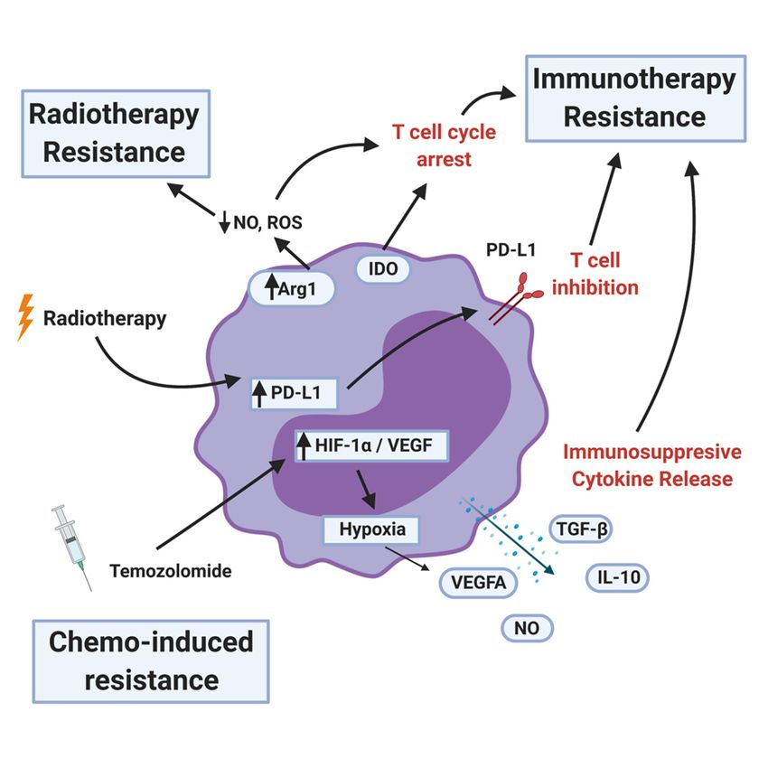
Cells 2021, 10, 893                                                                                        4 of 21

                      3. MDSCs in High-Grade Gliomas
                            Gliomas are classified into four different histopathologic grades: Low-grade gliomas
                      (LGG) (WHO grade I and II) and high-grade gliomas (HGG) (WHO grade III and IV) [28,29].
                      LGG are rare, occur mostly in children, and commonly have mutations in BRAF, FGR1,
                      ATRX, IDH1, and IDH2 [30]. On the contrary, HGGs are brain tumors that primarily affect
                      adults and have mutations in H3F3A G34R, TP53, EGFR, PTEN, IDH1, and IDH2. The
                      most common and aggressive grade IV, glioblastoma (GBM) typically does not have IDH
                      mutations, nonetheless, frequent intratumoral MGMT promoter methylation is found in
                      these patients [31]. In general, LGG have a lower number of tumor-infiltrating immune
                      suppressive cells compared to HGG [32].
                            In rat glioma models, an increase in the percentage of MDSCs in the peripheral
                      blood has been reported [33]. Furthermore, it can also serve as a biomarker for tumor
                      recurrence [33]. An increase in MDSCs is also reported in syngeneic and xenograft mouse
                      models of glioma [34]. Corroborating these findings, in humans, elevated levels of MDSCs
                      in the peripheral blood of glioblastoma (GBM) patients have been observed [35], however,
                      this increase is yet to be correlated with increased MDSCs at the glioma site. One possible
                      explanation for this is attributed to the existence of different subsets of MDSCs. In fact,
                      Gielen et al. reported that although both M-MDSCs and PMN-MDSCs are increased in
                      the peripheral blood of GBM patients, PMN-MDSCs and not M-MDSCs are increased
                      in the GBM TME [36]. Conversely, in mouse GBMs, M-MDSCs and not PMN-MDSCs
                      are enriched at the tumor site [37]. Thus, the predominant type of MDSCs present in the
                      TME of HGG is still not clear. Adding to this complexity, Bayik et al. reported that sexual
                      dimorphism can determine which type of MDSC subsets expand and contribute to immune
                      suppression in GBM patients. For instance, higher frequencies of M-MDSCs found in male
                      GBM patients promote tumor progression, whereas increased PMN-MDSCs in female
                      patients prevents anti-tumor immunity and is associated with poor prognosis [38]. In
                      agreement with these findings, in mouse GBM models, depletion of PMN-MDSCs with
                      anti-LY6G antibody only prolongs the survival of female and not male mice. Similarly,
                      reduction of M-MDSC frequencies with fludarabine increases the survival of male and not
                      female mice [38].
                            The precise mechanisms by which MDSCs traffic from bone marrow to the gliomas are
                      not known. To recruit MDSCs from the bone marrow, glioma cells overexpress CD200 [39],
                      indoleamine 2,3-dioxygenase (IDO1) [40] and secrete CCL20 [41], macrophage migration
                      inhibitory factor (MIF) [37], in addition to other growth factors. Along with glioma cells,
                      immune cells such as glioma-associated microglia and macrophages (GAMs) secrete CCL2
                      to recruit regulatory T-cells (Tregs) and MDSCs (Figure 2). Furthermore, the hypoxic tumor
                      environment upregulates the expression of vascular endothelial growth factor (VEGF) and
                      hypoxia-inducible factor-1 alpha (HIF1α) in glioma cells, which then induces ectonucleo-
                      side triphosphate diphosphohydrolase 2 (ENTPD2) and aids in the accumulation of MDSCs
                      by converting extracellular ATP to 50 -AMP [42]. In addition, hypoxia recruits CX3CR1
                      expressing MDSCs by increasing the expression of CCL26 on tumor cells [43]. Finally,
                      in vitro exposure of MDSCs from the spleen to hypoxia results in their differentiation into
                      immunosuppressive macrophages [44].
                            Once MDSCs are recruited to the HGG site their expansion and activation are tightly
                      controlled by cytokines (IL-6, IL-10, TGF-β, M-CSF, GM-CSF, INFγ), chemokines (CCR2,
                      and other factors (VEGF) secreted by tumor cells, T cells, microglia, and macrophages
                      (Figure 2) [9,33,45]. Glioma-derived exosomes also induce the expansion of MDSCs by
                      transferring miR-29a and miR-92a [46]. Briefly, the above-mentioned factors and exo-
                      somes activate Janus tyrosine kinase (JAK), signal transducer and activator of transcrip-
                      tion 3 (STAT3), STAT1, STAT6, CCAAT/enhancer-binding protein (C/EBPs), S100A8,
                      S100A9, prostaglandin E2 (PGE2) in MDSCs to promote their survival, expansion, and
                      function [32,33]. Furthermore, activated C/EBPs promote the expression of immune sup-
                      pressive arginase 1 (ARG1), reactive oxygen species (ROS), and inducible nitric oxide
                      synthase (iNOS) in MDSCs [47]. Higher levels of arginase activity and G-CSF levels in the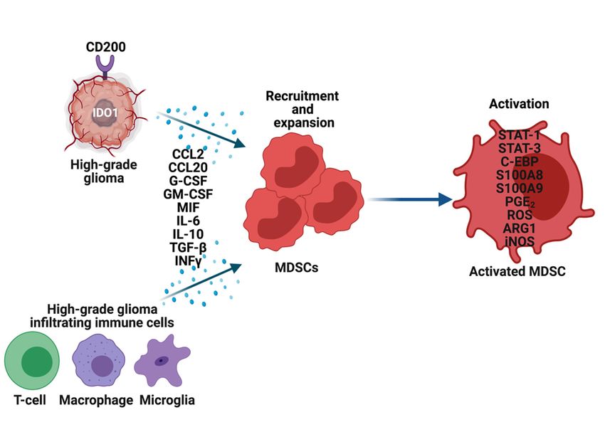
ells 2021, 10, x FOR PEER REVIEW 5 of
Cells 2021, 10, 893 5 of 21
peripheral blood of GBM patients have been used as an indicator for the activation an
immune suppressive functions of MDSCs (Figure 2) [48].
In summary, glioma and immune cells develop and maintain immunosuppressi
peripheral blood of GBM patients have been used as an indicator for the activation and
TME by recruiting, expanding, and activating MDSCs from the bone marrow and sple
immune suppressive functions of MDSCs (Figure 2) [48].
into the peripheral
In summary, blood,
glioma as
andwell as atcells
immune the develop
tumor site.
and maintain immunosuppressive
TME by recruiting, expanding, and activating MDSCs from the bone marrow and spleen
into the peripheral blood, as well as at the tumor site.
Figure 2. Recruitment, expansion, and activation of MDSCs in high-grade gliomas (HGG). Glioma cells, T-cells,
Figure 2.Macrophages,
Recruitment,and Microglia in
expansion, andtheactivation
tumor microenvironment
of MDSCs inoverexpress
high-grademultiple
gliomas genes and secrete
(HGG). Glioma ancells,
array T-cells,
of cy- Macro-
tokines, chemokines, and other factors to recruit and expand MDSCs. These factors also activate MDSCs through
phages, and Microglia in the tumor microenvironment overexpress multiple genes and secrete an array of cytokines, various
mechanisms, which then perform their immune-suppressive functions in HGG.
chemokines, and other factors to recruit and expand MDSCs. These factors also activate MDSCs through various mecha-
nisms, which then perform their immune-suppressive functions in HGG.
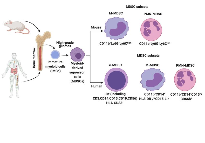
4. Metabolic Reprogramming of MDSCs
Cancer cells use glycolysis to generate ATP even under aerobic conditions (Warburg ef-
4. Metabolic Reprogramming of MDSCs
fect). This altered metabolism compared to normal cells was first reported by Otto Warburg
Cancer
in cells
1930 [49]. usetheglycolysis
Over to generate
last few decades, ATP
metabolic even under aerobic
reprogramming conditions
has emerged as a new(Warbu
hallmark that cancer cells use to evade growth suppression and
effect). This altered metabolism compared to normal cells was first reported cell death/apoptosis [50].
byToOtto Wa
this end, they preferentially utilize glycolysis, amino acid metabolism, pentose-phosphate
burg in 1930 [49]. Over the last few decades, metabolic reprogramming has emerged a
pathway (PPP), and tricarboxylic acid (TCA) cycle to thrive under the harsh environmental
newconditions
hallmarkpresented
that cancerby thecells
TME use to evade growth suppression and cell death/apopto
[51].
[50]. To Recently,
this end,thethey preferentially
critical utilize
role of metabolism glycolysis,
in controlling theamino acid
functions metabolism,
of immune cells is pentos
phosphate pathway (PPP), and tricarboxylic acid (TCA) cycle to thrive under
becoming greatly appreciated [52,53]. Along those lines, increased glycolysis, ROS, fattythe har
acid oxidationconditions
environmental (FAO), glutamine metabolism,
presented by theoxidative
TME [51]. phosphorylation (OXPHOS), lipid
uptake, and extracellular adenosine are observed in MDSCs infiltrating into the tumors,
Recently, the critical role of metabolism in controlling the functions of immune ce
including HGG (Figure 3) [54]. MDSCs exhibit these metabolic changes to support their
is becoming greatly
development, appreciated
survival, [52,53].
differentiation, Along and
activation, those lines, increased activity.
immunosuppressive glycolysis,
Thus,ROS, fat
acidinhibitors
oxidation (FAO),the
targeting glutamine metabolism,
above-mentioned oxidative
metabolic pathways phosphorylation (OXPHOS), lip
are being actively explored
to block the MDSC activity and improve anti-cancer immunotherapies.
uptake, and extracellular adenosine are observed in MDSCs infiltrating into the tumo
including HGG (Figure 3) [54]. MDSCs exhibit these metabolic changes to support th
development, survival, differentiation, activation, and immunosuppressive activi
Thus, inhibitors targeting the above-mentioned metabolic pathways are being actively e
plored to block the MDSC activity and improve anti-cancer immunotherapies.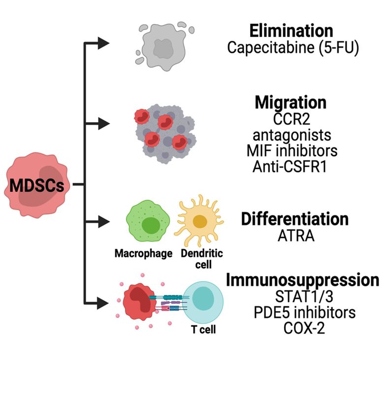
Cells 2021, 10, x FOR PEER REVIEW 6 of 21
Cells 2021, 10, 893 6 of 21
Figure 3. Metabolic reprogramming of MDSCs in the tumor microenvironment. In response to the cytokines and metabolites
Figure 3. Metabolic reprogramming of MDSCs in the tumor microenvironment. In response to the cytokines and metabo-
secreted by the cancer cells, MDSCs alter their metabolism by increasing glycolysis, glutamine/glutaminolysis, fatty acid
lites secreted by the cancer cells, MDSCs alter their metabolism by increasing glycolysis, glutamine/glutaminolysis, fatty
oxidation (FAO), oxidative phosphorylation (OXPHOS), tricarboxylic acid (TCA) cycle, reactive oxygen species (ROS), and
acid oxidation (FAO), oxidative phosphorylation (OXPHOS), tricarboxylic acid (TCA) cycle, reactive oxygen species
expression of ectonucleotidases CD39/CD73. Additionally, metabolites such as lactate, lipids glutamate, and adenosine in
(ROS), and expression of ectonucleotidases CD39/CD73. Additionally, metabolites such as lactate, lipids glutamate, and
the tumor
adenosine microenvironment
in the (TME) also(TME)
tumor microenvironment play aalso
vitalplay
role ainvital
regulating
role in the immune-suppressive
regulating functions of functions
the immune-suppressive MDSCs inof
HGG. The arrows in red indicate activation/upregulation.
MDSCs in HGG. The arrows in red indicate activation/upregulation.
Increased glycolysis in tumor cells makes them secrete cytokines such as M-CSF and
Increased glycolysis in tumor cells makes them secrete cytokines such as M-CSF and
GM-CSF into the TME [55]. These cytokines bind to CSF1R and GM-CSFR on MDSCs
GM-CSF into the TME
and upregulate [55]. These
glycolysis, cytokines
glucose, amino acidbinduptake,
to CSF1Randand
ATPGM-CSFR
generationon[56–58].
MDSCs and
Fur-
upregulate glycolysis, glucose, amino acid uptake, and ATP generation
thermore, glycolysis in tumor cells indirectly supports MDSC development and function [56–58]. Further-
more, glycolysisthe
by increasing in expression
tumor cellsofindirectly supports MDSC protein
CCAAT/enhancer-binding development
(CEBP),and function by
liver-enriched
increasing
activatorthe expression
protein (LAP),of CCAAT/enhancer-binding
AMP-activated protein kinaseprotein (CEBP), liver-enriched
(AMPK)-ULK1, and autophagy ac-
tivator protein (LAP), AMP-activated protein kinase (AMPK)-ULK1,
pathways [56]. Importantly, inhibiting glycolysis using 2-deoxy-D-glucose (2-DG) de- and autophagy path-
ways [56].the
creased Importantly,
levels of IDO inhibiting
in MDSCs glycolysis
and enhancedusingtheir
2-deoxy-D-glucose
apoptosis [55]. In (2-DG)
additiondecreased
to the
thetumor-derived
levels of IDOfactors, in MDSCs and TME
hypoxic enhanced
inducestheir apoptosis
HIF-1α [55]. glycolysis
dependent In addition in to the tumor-
MDSCs and
promotes
derived theirhypoxic
factors, differentiation into immune
TME induces HIF−1α suppressive
dependenttumor-associated
glycolysis in MDSCs macrophages
and pro-
(TAMs)
motes their[59]. Moreover, hypoxia
differentiation into also
immune directssuppressive
the oxidationtumor-associated
of pyruvate to lactate in tumor
macrophages
(TAMs) [59]. Moreover, hypoxia also directs the oxidation of pyruvate to lactate MDSCs
cells. The lactate produced and secreted by tumor cells increases the frequencies of in tumor
andThe
cells. causes the differentiation
lactate produced and of tumor-infiltrating
secreted by tumor neutrophils into MDSCs.
cells increases Lactate gen-of
the frequencies
erated in MDSCs via glycolysis increases their programmed cell death-ligand 1 (PD-L1)
MDSCs and causes the differentiation of tumor-infiltrating neutrophils into MDSCs. Lac-
expression, which then suppresses the T-cell activity through interaction with programmed
tate generated in MDSCs via glycolysis increases their programmed cell death-ligand 1
cell death protein (PD-1) and cytotoxic T lymphocyte-antigen associated protein 4 (CTLA-
(PD-L1) expression, which then suppresses the T-cell activity through interaction with
4) [60]. In line with these findings, inhibition of Lactate dehydrogenase-A (LDH-A) reduces
programmed
the number cell death arising
of MDSCs proteinfrom
(PD−1) and in
spleens cytotoxic T lymphocyte-antigen
tumor-bearing mice. Similar results associated
were
protein 4 (CTLA−4) [60]. In line with
also observed in human cancer patients [61]. these findings, inhibition of Lactate dehydrogenase-
A (LDH-A)Uponreduces theinto
infiltrating number of MDSCs
the tumor, arising from
both M-MDSCs spleens in tumor-bearing
and PMN-MDSCs mice.
prefer lipid uptake
Similar results were also observed in human cancer patients [61].
and fatty acid oxidation (FAO) and partake in tricarboxylic acid (TCA) cycle and oxidative
Upon infiltrating
phosphorylation into the in
(OXPHOS) tumor, both M-MDSCs
the mitochondria and PMN-MDSCs
to generate ATP [62]. MDSCsprefer achieve
lipid up-
take and
this fatty acid
metabolic oxidation
switch (FAO) CD36/FAT
by increasing and partake in tricarboxylic
(fatty acidscavenging
acid translocase) (TCA) cycle and ox-
receptor,
idative phosphorylation (OXPHOS) in the mitochondria to generate ATP [62]. MDSCs
achieve this metabolic switch by increasing CD36/FAT (fatty acid translocase) scavenging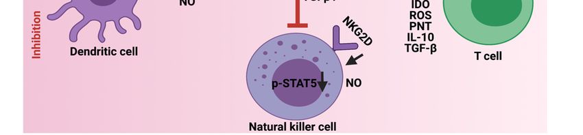
Cells 2021, 10, 893                                                                                           7 of 21

                      fatty-acid transport proteins (FATP), FAO enzymes (carnitine palmitoyltransferase (CPT1)
                      and 3-hydroxy acyl-COA dehydrogenase (HADHA)), mitochondrial and oxygen consump-
                      tion, and JAK-STAT (STAT3 and STAT5) signaling [54,63]. Furthermore, continuous ER
                      stress and ROS production as a result of FAO induce the immune suppressive function of
                      MDSCs by increasing the expression of iNOS, ARG1, reactive nitrogen species (RNS), and
                      NADPH oxidase 2 (NOX2). In agreement with these results, pharmacologic inhibition of
                      FATP and FAO blocked immune-suppressive functions of MDSCs and enhanced immune
                      checkpoint therapies [62,64].
                            Glioma stem cells metabolize glutamine and secrete high levels of glutamate into
                      the TME to evade immune surveillance. A higher concentration of glutamate enables
                      the maturation and infiltration of MDSCs to the glioma site [65]. Glutamine-derived α-
                      ketoglutarate (α-KG) aids in the expansion of MDSCs via the NDMA receptors [66]. The
                      mechanisms by which α-KG regulates MDSCs in HGG are not known. Recent studies
                      suggest that blocking glutamine/glutaminolysis metabolism prevents the recruitment,
                      generation, and metabolic reprogramming of MDSCs by inhibiting tumor-derived G-CSF
                      secretion and promotes the generation of anti-tumor inflammatory macrophages [67].
                            MDSCs express high levels of the ectonucleotidases CD39 and CD73 and release
                      immunosuppressive adenosine into the TME. TGF-β-mTOR-HIF1-α signaling axis induces
                      the expression of CD39/CD73 on MDSCs in the peripheral blood and tumor tissues [68].
                      CD39/CD73 expression on MDSCs performs their suppressive activity by inhibiting NK
                      and T-cells via paracrine signaling. Consistent with these observations, pharmacological
                      activation of AMP-activated protein kinase α using metformin caused the inhibition of HIF-
                      1α induced CD39/CD73 expression on MDSCs [69]. Extracellular adenosine contributes to
                      tumor angiogenesis by activating the A2B adenosine receptor on MDSCs. This effect and ac-
                      cumulation of MDSCs are blocked by the addition of A2B receptor antagonist PSB1115 [70].
                            Overall, the mechanisms by which the upregulation of the above-mentioned metabolic
                      pathways regulate MDSC maturation, infiltration, accumulation, and function is currently
                      unknown in HGG and warrants further investigation. However, the promising results
                      obtained by inhibiting these pathways in other cancer types provide the necessary impetus
                      to try those in the context of high-grade gliomas.

                      5. Immunosuppression Mediated by MDSCs
                            In steady-state or homeostatic conditions MDSCs lack any immunosuppressive activ-
                      ity. This is due to the shared characteristics of MDSCs with neutrophils and monocytes.
                      However, in pathological diseases, inflammation, obesity, sepsis, and cancer, immuno-
                      suppression is one of the major functions orchestrated by MDSCs. To this end, MDSCs
                      employ a variety of mechanisms to inhibit the function of T-cells, NK, dendritic cells
                      (DC), and macrophages and promote the recruitment of Tregs and immune-suppressive
                      B-cells to support the development and progression of HGG (Figure 4). Furthermore,
                      MDSCs generate and maintain an immunosuppressive TME and limit the efficacy of
                      cancer immunotherapies.
                            MDSCs suppress T-cell functions using different mechanisms. Specifically, in rat
                      glioma models, MDSCs produce NO, iNOS, ARG1, and IDO to suppress T-cell proliferation
                      and induce T-cell apoptosis [71]. Furthermore, ARG1 and IDO catabolize extracellular
                      arginine and tryptophan, the essential amino acids required for T-cell activation and
                      function [72]. Consistent with this, MDSCs express xc-transporter and sequester cysteine,
                      another essential amino acid that is vital for T-cell proliferation and activation [73]. MDSCs
                      also produce ROS, peroxynitrite (PNT), and secrete anti-inflammatory cytokines such
                      as IL-10 and TGF-β to interfere with T-cell cytotoxic function. Briefly, ROS and PNT
                      cause the nitration of the T-cell receptor, which then prevents its binding to peptide-major
                      histocompatibility complex [74,75]. IL-10 downregulates the expression levels of L-selectin
                      (CD62L) on naïve T-cells to prevent their migration to the tumor site [76], whereas TGF-β
                      promotes the differentiation of TH17 cells (a subset of CD4 T-cells) into Tregs [77]. Finally,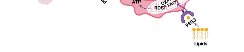
Cells 2021, 10, 893                                                                                                        8 of 21

2021, 10, x FOR PEER REVIEW
                                        MDSCs upregulate the expression of immune checkpoint molecules such as PD-L1 that
                                        interacts with PD-1 on T-cells and causes their exhaustion [78].

Figure 4. Immunosuppressive        role ofrole
           Figure 4. Immunosuppressive      MDSCs
                                               of MDSCsin HGG.
                                                           in HGG. MDSCs
                                                                      MDSCs employ      different
                                                                                employ different      mechanisms
                                                                                                  mechanisms         to inhibit
                                                                                                               to inhibit         the cytotoxic an
                                                                                                                          the cytotoxic
antigen presentation    functions of
           and antigen presentation    T-cells,
                                    functions    Natural
                                              of T-cells,    killer
                                                          Natural    cells,
                                                                  killer     and
                                                                         cells, andDendritic    cells.
                                                                                    Dendritic cells.   Additionally,
                                                                                                     Additionally, MDSCsMDSCs
                                                                                                                           promote promote
                                                                                                                                    the      the re
           recruitment and activation of immune-suppressive    Tregs,  Bregs,   and M2 macrophages.    The downward
cruitment and activation of immune-suppressive Tregs, Bregs, and M2 macrophages. The downward arrows in blac           arrows  in black
           indicate downregulation.
indicate downregulation.
                                             Similar to T-cells, NK cells have the tremendous potential to eliminate glioma cells [79].
                                      MDSCs
                                       However,suppress
                                                    the presenceT-cell  functions
                                                                   of MDSCs   in HGG  using    different
                                                                                         decreases           mechanisms.
                                                                                                      the cytotoxicity   of NKSpecifically,
                                                                                                                                cells. For   in r
                                       instance,   NO   produced   by MDSCs    inhibits  the  Fc receptor-mediated
                                 oma models, MDSCs produce NO, iNOS, ARG1, and IDO to suppress T-cell prolife           NK   function  in a
                                       contact-independent fashion [80]. Further, membrane-bound TGFβ1 on MDSCs reduces
                                 and induce     T-cell apoptosis [71]. Furthermore, ARG1 and IDO catabolize extracellu
                                       the cytotoxic function of NK cells by downregulating the expression of INFγ and acti-
                                 ginine  and receptor
                                       vating   tryptophan,NKG2D  the  essential
                                                                    [81].          amino
                                                                          Importantly,   NK acids      required
                                                                                              cells that            for T-cell
                                                                                                           are deficient         activation
                                                                                                                         for TGF-β    were   and
                                 tion [72].
                                       immuneConsistent      with this, MDSCs
                                                  to the MDSC-mediated      suppressiveexpress
                                                                                           effectsxc-transporter       andGr1+
                                                                                                    [82]. Lastly, CD11b+      sequester
                                                                                                                                  MDSCs cystein
                                       were found to accumulate in the spleens of tumor-bearing mice and there it suppresses
                                 other essential amino acid that is vital for T-cell proliferation and activation [73]. M
                                       the perforin production by NK cells through the modulation of JAK-STAT (decreasing
                                 also produce       ROS, peroxynitrite
                                       p-STAT5) signaling     pathway [83]. (PNT), and secrete anti-inflammatory cytokines su
                                 IL−10 andThe  TGF-β      to interfere
                                                  frequencies   of maturedwith  T-cellcells
                                                                            dendritic    cytotoxic      function.
                                                                                              (DCs) decrease          Briefly,
                                                                                                                 as the  MDSC ROSnumbersand PNT
                                       increase  in  tumors  via MyD88-NF-κB     signaling  [84].  Furthermore,    MDSCs
                                 the nitration of the T-cell receptor, which then prevents its binding to peptide-majo      secreted IL-10
                                       in the TME inhibit the IL-12 induced T-cell stimulatory function of DCs. In line with
                                 tocompatibility       complex [74,75]. IL−10 downregulates the expression levels of L-se
                                       these findings, the Il-12 treatment induces the expansion of CD11c (a marker for DCs) in
                                 (CD62L)    on naïve
                                       splenocytes         T-cells to prevent
                                                      of tumor-bearing  mice and their    migration
                                                                                   differentiation        to the tumor
                                                                                                     of M-MDSC              siteMoreover,
                                                                                                                   to DCs [85].  [76], whereas T
                                 promotes the differentiation of TH17 cells (a subset of CD4 T-cells) into Tregs [77]. F
                                 MDSCs upregulate the expression of immune checkpoint molecules such as PD-L
                                 interacts with PD−1 on T-cells and causes their exhaustion [78].
                                      Similar to T-cells, NK cells have the tremendous potential to eliminate gliom
Cells 2021, 10, 893 9 of 21
NO produced by MDSCs in the TME hampers the antigen presentation from DCs to CD4+ T-
cells and this effect was reversed by the addition of iNOS inhibitors [86]. Moreover, MDSCs
deliver lipid bodies (LBs) enclosed with oxidatively truncated lipids to DCs. These LBs then
interact with heat shock protein 70 (HSP70) and prevent the expression of peptide-MHC
class 1 expression on the surface of DCs, thereby impairing the antigen cross-presentation
function of DCs to T-cells [87].
Along with inhibiting the function of NK, DC, and T-cells as described above, MDSCs
contribute to the immunosuppressive TME in glioma by recruiting alternatively activated or
M2 macrophages, Bregs, and Tregs. The cross-talk between MDSCs and tumor-associated
macrophages is well-documented in glioma [88]. MDSCs reduce the antigen presentation
function of macrophages by reducing the MHC-class II expression on them. Further-
more, the presence of MDSCs in tumors reduces the anti-inflammatory M1 macrophages
and expands pro-tumor M2 macrophages [89]. Moreover, MDSCs secrete IL-10 to pre-
vent macrophages from generating an inflammatory cytokine, IL-12 that activates CD4+
T-cells, DCs, and NK cells [90]. In addition, hypoxic conditions in the TME environ-
ment recruit MDSCs into the tumor site, which then differentiates into tumor-associated
macrophages (TAMs) [91].
MDSCs secrete IL-10, INFβ, and TGF-β to promote the differentiation of naïve CD4+
T-cells into Tregs and provide a suppressive TME that is conducive for the expansion of
Tregs [92]. The cytokines, chemokines (CCL3, CCl4, CCL5) secreted by MDSCs directly
recruit Tregs via CCR5 into the TME [93]. Recent studies suggest that direct interaction
between MDSCs and Tregs in mice and human tumors suppressed CD8+ T-cell effector
responses and promoted their apoptosis [94]. Depletion of Tregs using anti-CD25 mAb
results in the accumulation of non-immune suppressive macrophages and granulocytes in
mouse HGG glioma models [95].
MDSCs use the recruited regulatory B-cells (Bregs) in the HGG microenvironment to
perform their immune-suppressive functions. Bregs account for about 40% of the immune-
infiltrating cells in GBMs. In the TME, Bregs express elevated levels of PD-L1 and CD155
as well as secrete immunosuppressive cytokines TGF-β and IL-10 to prevent the cytotoxic
activity of CD8+ T-cells. One of the mechanisms by which Bregs upregulate immune
checkpoint molecules and cytokines is through the uptake of MDSC-derived microvesicles
containing PD-L1. Consistent with these findings, depletion of B-cells using the anti-CD20
monoclonal antibody increased the survival. Therefore, a dialog between MDSCs and
B-cells plays an important role in GBM progression [96]. On the other hand, B-cells are
shown to induce T-cell mediated anti-tumor immunity by acting as antigen-presenting
cells (APCs) [97]. Moreover, the presence of B-cells correlates with reduced tumor growth,
invasion, and increased survival in GBM patients [98]. Thus, the exact role of B-cells in
HGG is still not clear and remains to be determined.
Taken together, MDSCs are one of the key players in avoiding immune surveillance in
HGG. To achieve this, MDSCs deploy different strategies to prevent the cytotoxic functions
and maturation of T-cells, NK cells, and DCs. Additionally, they provide a favorable TME
to support the recruitment and inhibitory functions of M2 macrophages, Tregs, and Bregs.
Whether some of these outlined mechanisms contribute to the immunosuppression by
MDSCs in HGGs remain to be investigated.
6. MDSCs Induced Therapeutic Resistance
MDSCs levels in the peripheral blood as well as at the tumor site are used as a
biomarker to predict if the existing standard-of-care therapies would work in cancer pa-
tients. High levels of MDSCs are often correlated with chemo, radiation, and immunother-
apy resistance (Figure 5).
MDSCs contribute to chemotherapy in two ways: Initial chemotherapy resistance
and induced immunosuppression following chemotherapy. Limited information on which
subsets of MDSCs contribute to chemoresistance is available until now. Furthermore,Cells 2021, 10, 893 10 of 21
1, 10, x FOR PEER REVIEW
the underlying mechanisms of how MDSCs contribute both directly and indirectly to
chemotherapy resistance are not well understood.
MDSCs increase the recruitment of suppressive T-regulatory cells to the tumor site,
which results in both radiotherapy and chemotherapy resistance [99]. Several subpop-
MDSCs
ulationslevels
of MDSCsin the
haveperipheral
been shown to blood as well roles
play important as atinthe
theirtumor sitecancer
respective are used a
omarker to predict
models while alsoif serving
the existing standard-of-care
as a proxy therapies would
for response to chemotherapy. work in can
Glioma patients
have shown elevated levels of MDSCs expressing S1008/9 along with
tients. High levels of MDSCs are often correlated with chemo, radiation, and im increased arginase
production [100]. Similarly, S100A9(+) inflammatory monocytes in patients with non-small
therapycell
resistance
lung cancer(Figure
(NSCLC)5).are shown to suppress T-cells by production of arginase, iNOS,
and the IL-13/IL-4Rα axis. The amount of these inflammatory monocytes is associated
with poor response to chemotherapy [101].
Figure 5. MDSC mechanisms of therapy resistance. MDSCs contribute to radiotherapy resistance by depleting nitric oxide
(NO) and reactive oxygen species (ROS) and upregulating programmed death ligand-1 (PD-L1) after the treatment. MDSCs
inhibit immunotherapy by arresting or inhibiting T-cell function, recruiting T-regulatory cells, and secreting immunosup-
gure 5. MDSC mechanisms of therapy resistance. MDSCs contribute to radiotherapy resistance by depleting nitric oxi
pressive cytokines and other factors. Less is known about chemotherapy resistance, but certain MDSC populations can be a
O) and reactive oxygen
proxy for species MDSCs
therapy response. (ROS)that and upregulating
survive programmed
the initial temozolomide death
treatment ligand-1
upregulates (PD-L1) after
hypoxia-inducible factor-1the treatme
DSCs inhibitalpha
immunotherapy by arresting
(HIF-1α) and vascular endothelial or inhibiting
growth T-cell
factor (VEGF), function,
resulting recruiting
in pro-tumor T-regulatory cells, and secreting i
angiogenesis.
unosuppressive cytokines and other factors. Less is known about chemotherapy resistance, but certain MDSC popu
In many
ns can be a proxy for therapy response. clinical
MDSCs trials,
that increased
survive theimmunosuppression
initial temozolomide is, unfortunately,
treatment aupregulates
side effect of hypox
chemotherapy
ducible factor−1 alpha (HIF−1α) and treatments. Temozolomide
vascular endothelial is one
growth factor of the common
(VEGF), resultingchemotherapeutic agents
in pro-tumor angiogenesis
used for treating glioblastoma. While it does have anti-tumor effects and increased patient
MDSCs contribute to chemotherapy in two ways: Initial chemotherapy res
and induced immunosuppression following chemotherapy. Limited informat
which subsets of MDSCs contribute to chemoresistance is available until now. FCells 2021, 10, 893 11 of 21
survival, it can also induce increased expression of hypoxia-related genes HIF-1α and
VEGF [102]. These factors promote a cascade of pro-tumor pathways including angiogene-
sis and cell repair (Figure 5). Systemic temozolomide administration also results in immun-
odepletion, which can be an advantage when used in combination with immunotherapies
but presents another hurdle for establishing anti-tumor activity in lymphocytes. In lung
cancer mouse models, carboplatin potentiates an MDSC dependent pathway that triggers
resistance through upregulated VCAM/RANTES and activating of IL13/33 pathways [103].
These chemo-induced MDSCs perform their immune suppressive role by promoting the
accumulation of IL-10 producing CD4+/Foxp3+ Tregs (regulatory T-cells) [103].
The application of ionizing radiation, known as radiotherapy (RT), is largely based on
its cytocidal activity combined with the ability to selectively target tumors. Radiotherapy
facilitates anti-tumor immunity both locally and at distant, non-radiated sites (abscopal
effect) [104]. RT can reduce MDSC levels in the TME, but MDSCs are relatively short-lived
and frequently replenished by circulating MDSCs, which are often unaffected by RT and
elevated in glioma patients [48]. High Arginase-1 (Arg1) expression by subsets of MDSCs
deplete local levels of nitric oxide and other reactive oxidative species which confer RT
sensitivity under hypoxic conditions (Figure 5) [105,106].
RT can also promote the tumor expression of known checkpoint inhibitors such as
PD-L1 [106]. As a result of this increased immunosuppression, many ongoing studies
seek to combine the anti-tumor effects of RT while using checkpoint inhibitors to mitigate
any potential immunosuppressive side effects. An alternative approach is to modify the
intensity and schedule of RT. Recent studies have utilized ablative hypofractionated RT
(AHFRT) rather than the conventional fractionated RT (CFRT). Compared with CFRT,
AHFRT significantly inhibits tumor growth and reduces recruitment of MDSCs, most likely
through downregulation of VEGF and decreasing tumor hypoxia [107]. However, in the
context of brain cancers, high doses of radiation can have detrimental side effects. Utilizing
AHFRT plus temozolomide in glioblastoma found no increase in the overall survival [108].
Currently, AHFRT is an accepted standard of care only in patients with poor performance
status or advanced age [109].
MDSCs represent a large obstacle to the immune checkpoint blockade due to their
direct suppression and indirect induction of immunosuppressive phenotypes in other
immune cells in the tumor microenvironment. In glioblastoma, MDSCs are influenced
by tumor cells, including cancer stem cells, towards their immunosuppressive phenotype
both locally and systemically [48,100].
As discussed earlier (see immunosuppression mediated by MDSCs), MDSCs secrete
immunosuppressive cytokines and other factors such as TGF-β, IL-10, NO, and ROS. They
also upregulate proteins that induce T-cell dysfunction: Arg1, IDO1, and PD-L1 (Figure 5).
A further consequence of MDSCs is the induction of immunosuppressive phenotypes in
other immune cell types in the TME. MDSCs promote the differentiation of Treg cells [110].
Tregs are potent suppressors of both the adaptive and innate immune system [111]. MDSCs
contribute to increased NK cell dysfunction [81] and T-cell exhaustion by inducing T-cell
expression of TIGIT, CTLA-4, TIM3, LAG3, and CD160 [112,113]. Whether these effects
seen in other tumor types translate to HGG remains to be investigated.
The result of MDSCs in the TME is a self-reinforcing network of immune suppression
that supports tumor proliferation. As a result of these known suppressive mechanisms,
it is no surprise that the presence of MDSCs can affect the efficacy of IC therapy. In a
model of metastatic melanoma, MDSC levels could be used as a predictor of ipilimumab
(anti-CTLA4 monoclonal antibody) treatment efficacy [114]. Future treatment models may
need to include an anti-MDSC component. Promising research in mouse models has
shown rejection of tumor and immunologic memory when combining immune checkpoint
inhibitors (nivolumab, avelumab, and ipilimumab) with MDSC targeted therapies as
discussed below [115].rejection of tumor and immunologic memory when combining immune chec
itors (nivolumab, avelumab, and ipilimumab) with MDSC targeted therapies
below [115].
Cells 2021, 10, 893 12 of 21
7. Strategies for Therapeutic Targeting of MDSCs
Current therapies for HGG have failed until now, in part due to the i
pressive
7. Strategies for functions
Therapeuticof MDSCsofasMDSCs
Targeting described above. Henceforth, several appro
ing pursued
Current tofor
therapies therapeutically
HGG have failed target
until MDSCs. Standard
now, in part ofimmune
due to the care and current i
sup-
apies
pressive for brain
functions tumors
of MDSCs have failed,
as described above.inHenceforth,
part due to the approaches
several immunosuppressive
are
being MDSCs.
pursued toSeveral
therapeutically target MDSCs. Standard of care and current immunother-
strategies are being developed to therapeutically target MD
apies cancers
for brain (Figure
tumors have
6). failed, in part due to the immunosuppressive function of
MDSCs. Several strategies are being developed to therapeutically target MDSCs in solid
cancers (Figure 6).
Figure 6. Strategies for targeting MDSCs in HGG. Chemotherapies are being used to deplete MDSCs
in solid tumors. The migration of these cells is inhibited using CCR2, macrophage inhibitory factor
(MIF) or CSFR1 inhibitors. Other therapies are used to decrease the immunosuppression pathways
and promote MDSCs differentiation.
The immunosuppressive activity of MDSCs can be disrupted by inhibiting stem cell
Figure 6. Strategies forfactors
targeting MDSCs
primarily in HGG.
produced Chemotherapies
by tumor are being used
cells. The JAK2-STAT3 pathwayto deplete MDSCs
is known in solid tum
to regulate
migration of these cellsimmune
is inhibited using
responses viaCCR2, macrophage
cytokines. inhibitory factor
JAK2 phosphorylation (MIF) or cause
and activation CSFR1 inhibitors. Other t
translocation
are used to decrease the immunosuppression pathways and promote MDSCs differentiation.
of STAT proteins into the nucleus regulating anti-apoptotic and pro-proliferative genes in
MDSCs. For instance, STAT1 controls arginase-1 (ARG1) and iNOS in M-MDSCs, while
STAT3 is involved in proliferation and reactive
The immunosuppressive activityoxygen speciescan
of MDSCs (ROS)
be formation
disruptedinby G- inhibit
MDSCs [116,117]. A STAT3 inhibitor has been identified from the herb Curcuma longa
factors primarily produced by tumor cells. The JAK2-STAT3 pathway is kn
Linn commonly known as curcumin. Studies have shown the benefits of curcumin in brain
tumorlate
cells,immune
decreasingresponses via cytokines.
their proliferative JAK2 phosphorylation
and antiapoptotic and activation
capacity [118,119]. However,
location
the effects of STAT
of STAT3 proteins
inhibition in MDSCsintointhe
the nucleus regulating
setting of brain tumors anti-apoptotic
remain unexploredand pro-
to thisgenes
date. in MDSCs. For instance, STAT1 controls arginase−1 (ARG1) and iNOS i
Phosphodiesterase
while STAT3 is5involved(PDE5) is another promising therapeutic
in proliferation and reactive targeting immunosup-
oxygen species (ROS)
pressive pathway in MDSCs. PDE5 is a hydrolase that regulates the NO/cyclic guanosine
G-MDSCs [116,117]. A STAT3 inhibitor has been identified from the herb Cu
monophosphate (cGMP) pathway [120]. Mice with carcinomas treated with sildenafil, a
PDE5 Linn commonly
inhibitor, known as of
had downregulation curcumin.
ARG1, NOS2, Studies have in
and IL4Rα shown
MDSCs the benefits
[121]. Preop-of curcu
erativetumor cells, decreasing
administration their
of sildenafil proliferative
has been and antiapoptotic
shown to reduce levels of ARG1capacity
and ROS[118,11
the effects of STAT3 inhibition in MDSCs in the setting of brain tumors remain
to this date.Cells 2021, 10, 893 13 of 21
without disturbing the accumulation of gMDSCs increasing antitumoral NK cell cytotoxic-
ity towards murine melanoma tumors [122]. In the phase 2 clinical trial, patients treated
with tadalafil, another PDE5 inhibitor, resulted in a reduction of MDSCs in the blood and
tumor possibly by decreasing pro-survival IL4Rα on MDSCs [123]. Inhibition of PDE5
by sildenafil has been shown to increase chemotherapeutic response in medulloblastoma
cells [124]. Preclinical studies using syngeneic mouse models and clinical trials should be
performed to assess the effects of PDE5 inhibitors in brain cancer patients.
Tumor cells producing PGE2 can promote MDSCs immunosuppression by activating
TGFβ through PGE2 receptors Ep2 and Ep4 found in monocytes [125]. The generation
of PGE2 by tumor cells is partially regulated by cyclooxygenase-2 (COX-2). The COX-2
inhibitors acetylsalicylic acid (ASA) and celecoxib decreased systemic levels of PGE2 and
MDSCs immunosuppression in the murine glioma model [126].
The release of colony-stimulating factor 1 (CSF-1) from tumors allows CSF-1R+
myeloid cells to proliferate and differentiate into MDSCs. In proneural gliomas, CSF-
1R inhibition initially increased the survival of mice and decreased M2-like TAMs [127].
However, gliomas developed resistance to CSF-1R through the PI3K mediated release of
insulin-like growth factor 1 (IGF-1) [128]. In a concluded clinical trial, the oral admin-
istration of CSF-1R inhibitor PLX3397 was well tolerated but not efficient to treat GBM
patients [129]. These studies have shown partial efficacy of CSF-1R inhibition and the
potential benefit of combining it with other therapies to overcome resistance.
The migration and accumulation of MDSCs into glioma TME has been reported to
be mediated by CCL2. Glioma tumor cells release CCL20 and induce CCL2 production
in TAMs and GAMs attracting MDSCs into TME [41]. Accumulation of MDSCs in glioma
was abrogated in CCR2 deficient mice [41]. The use of CCR2 antagonist CCX872 reduced
intratumoral MDSCs and enhanced the anti-PD-1 treatment in GBM-bearing mice [130].
In addition to CCL2, the macrophage inhibitory factor (MIF) is secreted by glioma cells
and regulates MDSCs recruitment into TME [131]. Sulforaphane, a MIF inhibitor derived
from broccoli sprouts, decreased levels of MDSCs in monocytes cultured with hypoxic
glioma conditioned media [132]. In vivo, sulforaphane induces apoptosis of glioma flank
tumors [133]. Ibudilast is another MIF inhibitor shown to target M-MDSCs through the
CD74 receptor decreasing the release of MCP-1 [37].
Low levels of chemotherapies can be used to deplete MDSCs. The use of 5-FU
on mice bearing GBM was able to decrease tumor-derived MIF, resulting in decreased
arginase-1 expression in MDSCs [131]. Similarly, a phase 0/1 clinical trial was conducted
showing that a combination of capecitabine (an orally 5-FU drug) and bevacizumab (anti-
angiogenic antibody) were able to decrease circulating MDSCs in patients with recurrent
GBM [134]. These studies suggest the benefit of combining chemotherapies with other
targeted therapies.
Other therapeutic approaches are being developed to promote MDSCs differenti-
ation dampening their pro-tumor activity. MDSCs isolated from cancer patients were
differentiated into dendritic cells (DC) using ATRA and GM-CSF (Al). In MDSCs, ATRA
causes upregulation of glutathione synthase resulting in the accumulation of glutathione
and reduction of ROS [135]. In brain cancer cells (glioma and medulloblastoma), ATRA
treatments cause cell growth arrest [136–138]. However, the combination of ATRA with
epigenetic drugs SAHA and 5-AZA exacerbated tumor growth in a glioma xenograft mouse
model [139]. The effects of ATRA in MDSCs of brain tumors remain to be determined.
Most treatments targeting MDSCs directly or indirectly have been evaluated in other
solid tumors than in brain cancers (Table 1). Studies have shown the effects of some of
these MDSCs targeted therapies in brain tumor cells but not in MDSCs. Future work
should be done to fully understand the effects of MDSC targeted therapies in the immune
microenvironment of brain tumors.Cells 2021, 10, 893 14 of 21
Table 1. Clinical trials targeting MDSCs directly or indirectly in HGGs.
Tumor Type Drug Target Combination Strategy Status Clinical Trial Identifier Effects on Clinical Outcome References
Reduction in MDSC
frequencies and increase in
Glioblastoma Capecitabine Tumor cells Bevacizumab Active, recruiting NCT02669173 [134]
cytotoxic immune cells is
observed in GBM patients
Early results show reduction
of regulatory T-cells and
Glioblastoma Atezolizumab PDL1 Ipatasertib Active, recruiting NCT03673787 [140]
increase in effector T-cells in
solid tumor patients
Sustained CD8 and CD4 T-cell
IMA 950 (multi
responses leads to an increase
Glioblastoma tumor-associated Immune cells Vaccine adjuvant Poly ICLC Completed NCT01920191 [141,142]
in medial survival of GBM
peptides vaccine)
patients by 19 months
Decreases circulating
monocytes in solid tumor
Advanced solid
patients. Some adverse effects
tumors (including Cabiralizumab CSFR1 Nivolumab Completed NCT02526017 [143,144]
such as increase in creatine
malignant glioma)
kinase and serum liver
enzymes are reportedCells 2021, 10, 893 15 of 21
8. Conclusions, Open Questions, and Future Perspectives
MDSCs are one of the abundant immune suppressive cells that are present in pe-
ripheral blood as well as at the tumor site in HGG patients. Recent observations have
started to inform that similar to cancer cells, MDSCs alter their metabolic pathways to
adapt and thrive in extreme conditions offered by the glioma TME. Furthermore, a growing
body of evidence as outlined in this review highlight the profound effects of MDSCs in
inducing immunosuppression, therapeutic resistance, and how targeting them in mouse
and rat GBM models is beneficial for increasing the survival of these animals. However,
so far clinical approaches aimed at targeting MDSCs in human cancer patients have not
lived up to the expectations. The reasons for this are due to the incomplete understanding
of MDSC phenotypic and functional heterogeneity and the strategies utilized by MD-
SCs to resist chemo, radiation, and immunotherapies. Some of this can be addressed
in preclinical tumor models using state-of-the-art technologies such as single-cell RNA-
sequencing (scRNA-seq), nanostring, mass cytometry, multiplexed immunohistochemistry,
single-cell assay for transposase accessible chromatin using sequencing (scATAC-seq), and
whole-genome sequencing.
Several outstanding questions remain: First, what are the true phenotypic markers that
separate MDSC subsets from monocytes and neutrophils. Second, what are the molecular
mechanisms that govern the generation, recruitment, and accumulation of MDSCs in HGG?
Third, what metabolic programs are altered in MDSCs? Are these alterations common or
different in M-MDSCs compared to PMN-MDSCs? Finally, whether targeting MDSCs using
different approaches (as mentioned in Strategies for therapeutic targeting of MDSCs) alone
as monotherapy is sufficient for inducing significant and long-term durable responses in
the clinic or a combination with other treatments is needed? If combination therapy is
required then the details regarding the type, sequence, and timing of these treatments have
to be worked out. Finding the answer to these questions in the upcoming years will result
in novel and efficient treatment options that would improve the survival of high-grade
glioma patients by precisely targeting MDSCs.
Author Contributions: Conceptualization, S.L. and S.S.M.; review literature, S.L., J.C.-C., E.H., A.P.C.
and S.S.M.; writing—original draft preparation, S.L., J.C.-C., E.H., A.P.C. and S.S.M.; writing—review
and editing, S.L. and S.S.M.; prepared figures, S.L., J.C.-C., E.H., A.P.C. and S.S.M.; supervision, S.S.M.
All authors have read and agreed to the published version of the manuscript.
Funding: SSM is funded through the Morgan Adams Foundation, Michele Plachy-Rubin Foundation,
American Cancer Society Institutional Research Grant (ACS-IRG #16-184-5), Broncos Foundation,
Alex’s Lemonade Stand Crazy8 Pilot Fund, and The Andrew McDonough B+ (Be positive) Founda-
tion. SL is funded through Cancer League of Colorado.
Institutional Review Board Statement: Not applicable.
Informed Consent Statement: Not applicable.
Data Availability Statement: Not applicable.
Acknowledgments: In this section, you can acknowledge any support given which is not covered by
the author contribution or funding sections. This may include administrative and technical support,
or donations in kind (e.g., materials used for experiments). All the figures in this review were created
with BioRender.com.
Conflicts of Interest: The authors declare no conflict of interest.
References
1. Stylli, S.S. Novel Treatment Strategies for Glioblastoma. Cancers 2020, 12, 2883. [CrossRef]
2. Hegi, M.E.; Diserens, A.C.; Gorlia, T.; Hamou, M.F.; de Tribolet, N.; Weller, M.; Kros, J.M.; Hainfellner, J.A.; Mason, W.;
Mariani, L.; et al. MGMT gene silencing and benefit from temozolomide in glioblastoma. N. Engl. J. Med. 2005, 352, 997–1003.
[CrossRef]
3. Bonaventura, P.; Shekarian, T.; Alcazer, V.; Valladeau-Guilemond, J.; Valsesia-Wittmann, S.; Amigorena, S.; Caux, C.; Depil, S.
Cold Tumors: A Therapeutic Challenge for Immunotherapy. Front. Immunol. 2019, 10, 168. [CrossRef]You can also read