Novel Technological Advances in Functional Connectomics in C. elegans - MDPI
←
→
Page content transcription
If your browser does not render page correctly, please read the page content below
Journal of
           Developmental
           Biology

Review
Novel Technological Advances in Functional
Connectomics in C. elegans
Elizabeth M. DiLoreto, Christopher D. Chute, Samantha Bryce and Jagan Srinivasan *
 Biology and Biotechnology Department, Worcester Polytechnic Institute, Worcester, MA 01605, USA;
 emdiloreto@wpi.edu (E.M.D.); cdchute@wpi.edu (C.D.C.); sbryce@wpi.edu (S.B.)
 * Correspondence: jsrinivasan@wpi.edu; Tel.: +1-508-831-6564

 Received: 1 November 2018; Accepted: 13 February 2019; Published: 23 April 2019

 Abstract: The complete structure and connectivity of the Caenorhabditis elegans nervous system
 (“mind of a worm”) was first published in 1986, representing a critical milestone in the field of
 connectomics. The reconstruction of the nervous system (connectome) at the level of synapses
 provided a unique perspective of understanding how behavior can be coded within the nervous
 system. The following decades have seen the development of technologies that help understand how
 neural activity patterns are connected to behavior and modulated by sensory input. Investigations
 on the developmental origins of the connectome highlight the importance of role of neuronal cell
 lineages in the final connectivity matrix of the nervous system. Computational modeling of neuronal
 dynamics not only helps reconstruct the biophysical properties of individual neurons but also allows
 for subsequent reconstruction of whole-organism neuronal network models. Hence, combining
 experimental datasets with theoretical modeling of neurons generates a better understanding of
 organismal behavior. This review discusses some recent technological advances used to analyze and
 perturb whole-organism neuronal function along with developments in computational modeling,
 which allows for interrogation of both local and global neural circuits, leading to different behaviors.
 Combining these approaches will shed light into how neural networks process sensory information
 to generate the appropriate behavioral output, providing a complete understanding of the worm
 nervous system.

 Keywords: connectomics; synapses; calcium imaging; optogenetics; sonogenetics; multisensory
 integration; sensory-motor integration

1. Introduction
     The field of connectomics attempts to link brain function with behavior by comprehensively
mapping the anatomical links between all constituent neurons within different brain regions [1,2].
Caenorhabditis elegans, a microscopic roundworm, has served as a model for macroscopic research for
over six decades (Figure 1). To date C. elegans remains the only organism to be fully mapped at the level
of the nervous system [3–5]. The nervous system of both sexes; hermaphrodite (302 neurons) and male
(385 neurons) have been completely mapped at the level of electron microscopy [4,6]. This has served
as a prototype for analytical studies of larger scale connectome networks. However, can this complete
mapping of synaptic connections in the brain (connectome) shape the understanding of the mechanistic
basis of behavior? In other words, does structural connectivity define function within the nervous
system? One mindset is that wiring diagrams can serve as a starting point for generating mechanistic
hypotheses for the investigation of the neural basis of behavior. Therefore, knowledge of connectome
structure enables the generation of testable hypotheses about the specific as well as general roles of
individual neurons [7]. However, the physical connectivity patterns of the C. elegans nervous system,
are unable to predict how the nervous system functions as a whole in order to enact behaviors [8–10].

J. Dev. Biol. 2019, 7, 8; doi:10.3390/jdb7020008                                   www.mdpi.com/journal/jdb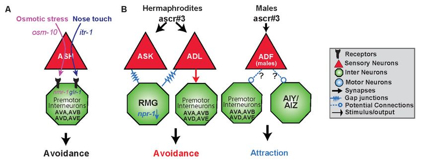
J. Dev. Biol. 2019, 7, 8 2 of 18
J. Dev. Biol. 2019, 7, 8 2 of 18
Along with synapses, most nervous systems also contain gap junctions, which mediate fast, potentially
to enact behaviors
bidirectional electrical[8–10]. Along
coupling with synapses,
between most In
cells [11,12]. nervous systems
addition, also containsignaling
extrasynaptic gap junctions,
between
which mediate fast, potentially bidirectional electrical coupling between cells
neurons within nervous system happens via monoamines and neuropeptides, occurring primarily [11,12]. In addition,
extrasynaptic signaling between neurons within nervous system happens via monoamines and
outside the synaptic connectome [13]. These signaling systems act over both short and long ranges and
neuropeptides, occurring primarily outside the synaptic connectome [13]. These signaling systems
are independent of synaptic connections, allowing them to shape behavioral responses to either the
act over both short and long ranges and are independent of synaptic connections, allowing them to
same or different stimuli [13,14]. Therefore, a complete characterization of a neuronal dynamics under
shape behavioral responses to either the same or different stimuli [13,14]. Therefore, a complete
different conditionsofis aessential
characterization neuronalfor generating
dynamics undera complete
different functional
conditions is understanding of the nervous
essential for generating a
system. Sincefunctional
complete behaviorunderstanding
is not a linearofsummation
the nervous of sensory
system. information,
Since the asame
behavior is not linearcircuit can lead
summation
to different behavioral
of sensory information,outputs. Thiscircuit
the same couldcan
be lead
a result of molecular
to different events,
behavioral stimuli
outputs. concentration
This could be a or
physiological states—all of which are non-linear in nature—generating different behaviors
result of molecular events, stimuli concentration or physiological states—all of which are non-linear via the same
connectome. Hence, we define
in nature—generating “functional
different behaviors connectomics” as the study
via the same connectome. of thewe
Hence, relationship between a
define “functional
connectomics”
neuron’s as the
function and itsstudy of the relationship
connections—both betweenand
anatomical a neuron’s function and its connections—
extrasynaptic.
both anatomical and extrasynaptic.
1986: Wiring diagram mapped
by electron microscopy (4)
2007:Microfluidic devices 2015: OpenWorm Project (132)
1964: Discovered by 1999:Automated behavioral for neuronal imaging (44)
Sydney Brenner (5) tracking (51) 2015-16: Whole brain
2012: Optofluidic
2003:Neural imaging Lens-Free Devices (130) imaging (59,60)
with GECIs (51)
1985
1960 2019
1965
2005: Optogenetics, 2012: Multi-sensory
1985: Laser Ablation 1998-99: Electrophysiology controlling neurons integration (81)
of neurons (16) of neurons & muscles by light (69) 2015: Sonogenetics,
(31,32) perturbing cells with
sound (134)
Figure 1. Timeline
Figure of of
1. Timeline neuroscience
neurosciencerelated discoveriesininC.C.
related discoveries elegans.
elegans. TheThe
pastpast 60 years
60 years have have resulted
resulted in
in development
development in technologies and breakthroughs in understanding the neural connections andand
in technologies and breakthroughs in understanding the neural connections the the
nervous system.
nervous system.
2. Functional Characterization
2. Functional CharacterizationofofNeural
NeuralCircuits: UsingConnectome
Circuits: Using Connectometoto Generate
Generate Hypotheses
Hypotheses
C. elegans neurons
C. elegans neurons can bebedivided
can dividedinto
intothree
three functional “classes”ofofneurons:
functional “classes” neurons: sensory
sensory neurons,
neurons,
motor neurons,
motor neurons, and andinterneurons
interneurons or premotor
premotorneurons
neurons [7,9].
[7,9]. The The sensory
sensory neuronsneurons have dendrites
have dendrites that
that extend
extendtotothe
thetiptipofof
the nose
the nose and terminate
and intointo
terminate diverse ciliated
diverse structures
ciliated to detect
structures stimulistimuli
to detect from thefrom
environment. These
the environment. These neurons
neurons account for afor
account third of theofneurons
a third with more
the neurons withconnections
more connectionsbeing pre-being
synaptic than post-synaptic. Conversely, motor neurons, another third
pre-synaptic than post-synaptic. Conversely, motor neurons, another third of all neurons, have of all neurons, have moremore
post-synaptic
post-synaptic connections.
connections. TheTheremaining
remainingneurons
neurons areare considered
consideredtotobebepremotor
premotor interneurons,
interneurons,withwith
large numbers of both pre- and post-synaptic connections [10]. Understanding the connections alone
large numbers of both pre- and post-synaptic connections [10]. Understanding the connections alone
between these neuron classes helps increase our understanding of how a signal is transduced,
between these neuron classes helps increase our understanding of how a signal is transduced, processed,
processed, to ultimately produce behavioral outputs, as seen in previous work on C. elegans’
to ultimately produce behavioral outputs, as seen in previous work on C. elegans’ navigation [15].
navigation [15].
OneOneof the first
of the major
first majorfindings
findingsofofthe
theworm
worm connectome
connectome from fromthethestructural
structural connectivity
connectivity datadata
was was
the the
characterization of the mechanosensory circuitry [16]. Using laser
characterization of the mechanosensory circuitry [16]. Using laser ablations, components of ablations, components
of a amechanosensory
mechanosensory circuit circuitwere
wereidentified,
identified, consisting
consisting of sensory
of sensory neurons,
neurons, premotor premotor interneurons,
interneurons, and
and ventral
ventralcord
cord motorneurons,
motorneurons, responsible
responsible for escape
for escape behavior behavior in response
in response to body
to body touch [16].touch [16]. By
By testing
eacheach
testing neuron’s function
neuron’s by laserbymicrosurgery,
function [16,17], a [16,17],
laser microsurgery, set of premotor
a set ofinterneurons were identified
premotor interneurons were
that control the direction of locomotion; six neurons that promoted forward
identified that control the direction of locomotion; six neurons that promoted forward locomotion and locomotion and four
fourneurons
neuronspromoting
promoting backward
backward locomotion
locomotion [16]. ThisThis
[16]. work worknot not
onlyonly
opened
openedup upthe the
genetic andand
genetic
molecular studies of the C. elegans touch circuit, but also implicated these
molecular studies of the C. elegans touch circuit, but also implicated these major premotor interneurons major premotor
interneurons in several other behaviors [18–21]. This study was a landmark in understanding how
in several other behaviors [18–21]. This study was a landmark in understanding how structural
structural changes within the connectome can impact function and signaling between neurons.
changes within the connectome can impact function and signaling between neurons.
The compact nervous system of C. elegans allows for a single neuronal class to be involved in the
The compact nervous system of C. elegans allows for a single neuronal class to be involved in the
sensation of diverse stimuli or elicitation of different behaviors [7,9,22]. From a functional
sensation of diverse
connectomic stimuli or
perspective, thiselicitation of different
suggests that behaviors
not all stimuli utilize[7,9,22].
the same From a functional
pathways connectomic
and connections.
perspective, this suggests that not all stimuli utilize the same pathways and connections.
This suggests a non-linearity and complexity in the information processing of stimuli, for example, This suggests
a non-linearity
the polymodal, and amphid,
complexity in the information
single-ciliated, processing
nociceptive neuron,of stimuli,
ASH, for example,
detects a myriad the polymodal,
of different
amphid, single-ciliated, nociceptive neuron, ASH, detects a myriad of different mechano, osmo, and
chemo stimuli that result in aversive behaviors [23–29]. The diversity in neuronal circuitries may alsoJ. Dev. Biol. 2019, 7, 8 3 of 18
be due to the intracellular machinery used within individual neurons. C. elegans are equipped with a
large set of G protein subunits that exhibit overlapping expression, rendering particular intracellular
pathways important in different behavioral circuits [30]. The nematodes genome codes for 21 Gα protein
subunits, and 2 subunits each of both Gβ and Gγ proteins [30]. Of the 21 Gα subunits, 16 are expressed
throughout the chemosensory neurons, and many overlap in their expression profiles [30]. For example,
on its own, ASH expresses ten different Gα subunits, while a different amphid, single-ciliated, ASE,
expresses onlyJ.three
Dev. Biol. 2019, 7, 8
[30]. 3 of 18
mechano, osmo, and chemo stimuli that result in aversive behaviors [23–29]. The diversity in
3. Technologies Employed
neuronal circuitriesto Unravel
may the
also be due Functional
to the intracellular Connectome
machinery used within individual neurons.
C. elegans are equipped with a large set of G protein subunits that exhibit overlapping expression,
One of therendering
earliestparticular
methodsintracellular
to monitor neuron
pathways function
important was patch
in different clamp
behavioral electrophysiology
circuits [30]. The [31–34].
This techniquenematodes
lends genome codes for 21 Gα protein subunits, and 2 subunits each of both Gβ and Gγ proteins
itself to understanding functional connectomics as it
[30]. Of the 21 Gα subunits, 16 are expressed throughout the chemosensory neurons, and many
monitors the flow of ions
across neuronaloverlap
membranes
in their expression C. elegans
[33]. Inprofiles [30]. Foritexample,
was usedon itstoown,
understand thetenrole
ASH expresses of graded
different Gα potentials,
subunits,
in opposition to all orwhile a different
nothing amphid,
action single-ciliated,
potentials ASE, expresses
observed only three [30].
in mammals [31–34]. This method is rather
invasive, requiring fixed samples
3. Technologies Employedof individual
to Unravel neurons
the Functional for testing [35].
Connectome
One of the earliest methods to monitor neuron function was patch clamp electrophysiology, [31–
3.1. Neuronal Imaging in C. elegans
34]. This technique lends itself to understanding functional connectomics as it monitors the flow of
ions across neuronal membranes [33]. In C. elegans it was used to understand the role of graded
Optical techniques
potentials, inare available
opposition to allinormany
nothingexperimental
action potentialssystems
observed but are highly
in mammals applied
[31–34]. This in C. elegans
research as themethod
nematodes
is rather are optically
invasive, requiring transparent
fixed samples ofand can be
individual imaged
neurons while
for testing fully intact. In addition,
[35].
a variety of Genetically
3.1. NeuronalEncoded Calcium
Imaging in C. elegans Indicators (GECIs) are available targeting individual neuron/s
of interest with specific
Optical techniques are [36,37].
promoters available inThese GECIs cansystems
many experimental specifically targetapplied
but are highly the neuronal
in C. cell body
and/or distribute throughout the entire neuron. Fluorescent imaging with GECIs has achieved rapid
elegans research as the nematodes are optically transparent and can be imaged while fully intact. In
addition, a variety
progress in visualizing Ca2+offluxGenetically Encoded Calcium Indicators (GECIs) are available targeting
at the levels of cell populations [38], single cells [39], or even
individual neuron/s of interest with specific promoters [36,37]. These GECIs can specifically target
subcellular compartments [40]. Among
the neuronal cell body and/or distribute available GECIs,
throughout Green
the entire fluorescent
neuron. Calmodulin
Fluorescent imaging with M13 fusion
Protein (GCaMP) is one of the most successful and popular, due to its ability to convey Ca2+ levels
GECIs has achieved rapid progress in visualizing Ca 2+ flux at the levels of cell populations [38],
single cells [39], or even subcellular compartments [40]. Among available GECIs, Green
with impressive signal-noise
fluorescent ratios
Calmodulin M13(Figure 2A) (GCaMP)
fusion Protein [41–43].is one of the most successful and popular,
due to its ability to convey Ca2+ levels with impressive signal-noise ratios (Figure 2A) [41–43].
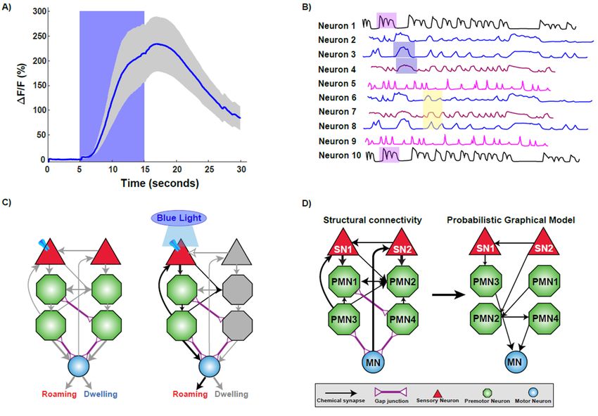
Figure 2. Technologies used to decipher
Figure 2. Technologies the functional
used to decipher connectome.
the functional connectome. A)(A) Imaging
Imaging calciumcalcium
changes changes using
using GCaMP sensor. The trace plot shows the activation of a sensory neuron upon stimulus
GCaMP sensor. The trace plot shows the activation of a sensory neuron upon stimulus presentation
presentation by increase in calcium influx as measured by increase in fluorescence intensity. Increase
by increase in calcium influx as measured by increase in fluorescence intensity. Increase in calcium is
sustained while the worm experiences the stimulus, as fluorescence decreases upon stimulus removal.
(B) A representative brain phase plot where neurons are activated in different phases (shaded regions)
during exposure to a stimulus. (C) Optogenetic interrogation of the connectome. Expressing and
activating Channelrhodopsin via blue light exposure in a particular sensory neuron activates a subset
of downstream neurons, resulting in the elicitation of roaming behavior. (D) Computational modeling
helps to unravel the functional connections within a structural framework of neurons.J. Dev. Biol. 2019, 7, 8 4 of 18
Calcium imaging of neurons is widely employed in C. elegans neuroscience as it allows for
the activity of a single or multiple neurons to be monitored over different time scales. Advances in
microfabrication technology have permitted the construction of well-controllable microenvironments for
monitoring neural function in C. elegans. One of the first microfluidic devices used to monitor neuronal
calcium dynamics was termed the ‘olfactory chip’. This device was used to examine stimulus-response
relationships in chemosensory neurons over a short temporal timescale [44]. Over the last decade, the
applications of microfabrication techniques have exponentially increased in neuroscience with chips
being designed for high-throughput and high resolution- based applications [44–48].
3.2. Whole-Brain Imaging in C. elegans
Measuring neural activity of a single neuron over a short time course is helpful in identifying
neuronal dynamics upon stimuli exposure and characterizing circuits of activity [31,33,44,45,47,49–51].
However, it offers little insight into the processes at play during long-term behaviors. To execute
motor commands during a particular behavior, more is occurring than cross-talk via the connection
between neurons. Global-brain or whole-brain imaging enables characterization of behaviors with
the integration of sensory neurons with motor neurons to be quantified [40,52]. While imaging a
single neuron, sensory or premotor neurons are often the focus [45,53], whole-brain imaging enables
elucidating on the role of multiple neurons within the connectome simultaneously. Studies in zebrafish
have been accomplished on a global-brain scale with single-neuron resolution with specific regard to
motor neurons [54,55].
The first whole-brain calcium imaging experiment in C. elegans was conducted in an immobilized
setting, imaged neural activity across 100 neurons in a small channel. This technique relied on
existing neural maps to match captured neural responses to individual neurons [4,38]. This study
achieved single-neuron resolution across most of the brain and, when combined with the extensive
existing knowledge of C. elegans neural anatomy, supported identification of most neurons. Using
this technology, it is possible to track the calcium changes through circuits, generating a brain state
phase plot (Figure 2B) [38]. This temporal experiment was key in identifying repeating patterns of
stimulation similar to models of central pattern or rhythmic motor generation [38,56–58]. Tracking the
whole-brain changes in calcium signals during this repetitive behavior allows for different classes of
neurons as well as other movement variables, such as speed, to be incorporated into the understanding
of this behavior [38]. While this information generated is sufficient to understand the culmination of a
behavior, the neural information can also be parsed out to understand the signaling occurring in each
part of the action, such as moving forward, slowing, reversing, or turning, on the scale of an individual
neuron or across the brain [38]. This study suggested that high-level organization of behavior is
encoded in the brain by globally distributed, continuous, and low-dimensional dynamics [38].
Recent developments have allowed researchers to develop two methods for imaging the neurons
of C. elegans while roaming. These techniques rely on simultaneously recording several neurons
expressing a calcium indicator using spinning disk confocal microscopy to ultimately produce
volumetric imaging [59,60]. Both methods allow for the simultaneous recording of approximately
80 neurons, gathering and correlating information on body posture and location in a moving worm.
One method expresses both the calcium indicator and another fluorescent protein, and also infers
body posture and position via head ganglia orientation [59]. In contrast, the other method utilizes
a custom software, which ensures that the head of the worm is always in the correct location of the
microscope stage despite the worm’s movement, and actively records the body’s position [60]. The
ability to generate multi-neuron data of freely roaming worms will be vital in moving towards a more
thorough understanding of the functional connectome [61].
3.3. Optogenetics and the Worm Connectome
Optogenetics offers temporal control of activity of individual neurons utilizing light-controlled
ion channels [62]. This unique optical control of neurons allows researchers to probe neural circuitsJ. Dev. Biol. 2019, 7, 8 5 of 18
and investigate neuronal function in a highly specific and controllable fashion [63–69]. C. elegans is
a popular platform for probing the nervous system at length, with scales spanning from synapse
to whole circuit [63,64,67]. The initial studies on optogenetic control of C. elegans neurons involved
using whole-field illumination together with specific genetic mutations, involving activation of
Channelrhodopsin (ChR2) in excitable motor neurons [68,69]. Specific neurons or muscles expressing
ChR2 can be quickly and reversibly activated by light in both live and behaving animals [68,69].
Expressing a light-sensitive protein in specific neurons using specific promoters highlighted that
functional neuronal circuits during behaviors can be elucidated in C. elegans (Figure 2C).
A drawback of whole-field illumination was lack of cellular specificity as the illumination
occurred over a region of the worm’s body. Newer technologies have been generated wherein targeted
illumination of multiple fluorophores expressing optically sensitive proteins, and extensively employed
to observe behavior [67,70–72]. Using these illumination systems, it is now possible to track a freely
moving C. elegans and spatiotemporally excite and/or inhibit specific nodes of neural networks to probe
for function across several types of locomotory behaviors. In addition, this technology enables the
use of combinations of optogenetic tools and fluorescent GECIs with high reproducibility and light
intensity control. In addition to development of illumination systems, progress has been made in terms
of development of variants of the optogenetic proteins that both depolarize and hyperpolarize neurons
over longer temporal scales [64]. These highly light-sensitive optogenetic tools, are highly effective
and display fast kinetics, allowing better investigation prolonged neuronal activity states in C. elegans.
3.4. Computational Strategies
C. elegans is a powerful biophysical system that can be understood at different levels such as sensory
stimulation and motor output. Computational models serve as an excellent platform to implicate how
dynamics of different neurons can affect network connectivity [73]. The development of neuroimaging
tools and technologies enable us to record neural dynamics over a longer timescale, which help generating
dynamic models of synaptic connectivity. Newer computational approaches help elucidate the dynamic
connectome by comparing longer timescale studies containing higher-dimensions of data [73].
An interesting application of computational strategies is the characterization of sensory-motor
integration [74]. Sensory-motor integration attempts to understand the neural pathways from stimuli
input to motor-neuron driven behavioral responses. Given the connectome data, physical circuits
can be identified, and then simulated to understand responses [74]. Modeling the dynamics of the
neural network is possible by combining the known structural connectome data of C. elegans with a
physiologically model of a neuron [74]. Models such as probabilistic graphical models (PGMs), use
known circuits of repetitive behavior, (Figure 2D), to predict responses to novel situations [75,76].
A recent study has developed the “dynome” of the worm nervous system [77]. This predictive
system relies on the simulation of neural dynamics, or temporal experiments on multiple neurons,
to allow application of stimuli to neurons to examine network properties. Such a model enables the
user to apply or modify stimuli to the network, observe the neural dynamics on various time and
population scales, and allow for network structural changes. Changing these parameters allows for
calculation of neural response patterns associated with different stimuli. The strength of this approach
lies in the ability of removing neurons from a network and studying the dynamics of the circuit [77,78].
This unique computational approach is a first step in making any nervous system’s architecture being
able to predict a behavior based on network properties.
4. Analysis of the Functional Connectome
The development of the technologies discussed above, allow us to investigate specific aspects of
the connectome, such as sensation or processing of diverse multisensory stimuli. Sensory systems
continuously receive complex types of environmental input, which are processed by the nervous
system to identify relevant facts about the surroundings and internally represent that information
to help the animal successfully navigate its environment. This complex neural function is further4. Analysis of the Functional Connectome
The development of the technologies discussed above, allow us to investigate specific aspects of
the connectome, such as sensation or processing of diverse multisensory stimuli. Sensory systems
J.continuously receive
Dev. Biol. 2019, 7, 8 complex types of environmental input, which are processed by the nervous 6 of 18
system to identify relevant facts about the surroundings and internally represent that information to
help the animal successfully navigate its environment. This complex neural function is further
complicated
complicated when when combined
combined withwith the
the internal
internal physiological
physiological state
state of
of the
the animal. In Drosophila,
animal. In Drosophila, recent
recent
work on the multilevel multimodal convergence circuit, which relies
work on the multilevel multimodal convergence circuit, which relies on multisensory on multisensory integration, was
integration,
limited
was limitedby the bylack
the of connectomic
lack data unveiling
of connectomic multisensory
data unveiling neuronal
multisensory convergence
neuronal [79]. [79].
convergence
Multisensory C. elegans
Multisensory integration in C. elegans falls into two broad categories: co-exposure to two
integration in falls into two broad categories: co-exposure to two distinct
distinct
stimuli, aversive and attractive, or exposure to one stimuli in conjunction
stimuli, aversive and attractive, or exposure to one stimuli in conjunction with an environmental with an environmental
indicator,
indicator, bothboth of of which
which can
can be
betested
testedvia
viaavoidance
avoidanceassays
assays[80–82].
[80–82]. Furthermore,
Furthermore, with
with only
only sixteen
sixteen
pairs
pairs of chemosensory neurons, neuronal ablation techniques such as the laser ablation method as
of chemosensory neurons, neuronal ablation techniques such as the laser ablation method as
well
well asas genetic
genetic ablations
ablations have
have become
become effective
effective tools
tools for
for understanding
understanding the the diverse
diverse roles
roles of
of individual
individual
sensory
sensory neurons
neurons within
within the
the realm
realmof ofthe
theconnectome
connectome[83,84].
[83,84].
4.1.
4.1. Divergent
Divergent Functions
Functions within
within aa Single
Single Neuronal
Neuronal Class
Class
Modifications
Modifications in in functional
functional connections
connections downstream
downstream of of sensory
sensory neurons
neurons can
can be
be achieved
achieved byby
differential
differential cellular signaling mechanisms both within sensory neurons and between sensory and
cellular signaling mechanisms both within sensory neurons and between sensory and
premotor
premotor interneurons.
interneurons. The The nociceptive
nociceptive sensory
sensory neuron
neuron ASH
ASH detects
detects various
various stimuli;
stimuli; chemical,
chemical,
mechanical,
mechanical, osmotic,
osmotic, all
all ofof which
which result
result in
in the
the same
same behavioral
behavioral outcome: avoidance [25,28,29,85–87].
outcome: avoidance [25,28,29,85–87].
Studies
Studies have
have characterized
characterized differential
differential use
use of
of both
both intra-
intra- and
and inter-signaling
inter-signaling molecules
molecules by by ASH
ASH
sensory neuron leading to the same behavior (Figure 3A) [85]. For example, nose
sensory neuron leading to the same behavior (Figure 3A) [85]. For example, nose touch avoidance, touch avoidance,
requires
requires expression
expression of of itr-1
itr-1 in
in ASH
ASH neurons
neurons and
and isis not
not required
required for
for osmotic
osmotic aversive responses [85].
aversive responses [85].
Conversely, specific genes within ASH (such as osm-10) are specific for detecting osmotic
Conversely, specific genes within ASH (such as osm-10) are specific for detecting osmotic stress, and stress, and not
required for tactile
not required response.
for tactile This implies
response. that inthat
This implies addition to expression
in addition of specific
to expression G protein-coupled
of specific G protein-
subunits,
coupled subunits, distinct downstream effectors within the signaling pathway are recruited
distinct downstream effectors within the signaling pathway are specifically to
specifically
initiate functional connections. [85].
recruited to initiate functional connections. [85].
Figure 3. Functional circuits are created by differential use of neurons and distinct intra- and intercellular
Figure 3. pathways.
signaling Functional(A) circuits are created
The sensory neuron byASH
differential
responds use of neurons
to two differentand distinct
stimuli intra-inand
resulting an
intercellular signaling pathways. A) The sensory neuron ASH responds to two
avoidance response via the premotor interneurons AVA, AVB, AVD and AVE. Osmotic stress is mediated different stimuli
resulting
via osm-10 in an avoidance
whereas nose touchresponse
utilizesvia
itr-1the premotor
within interneurons of
ASH. Downstream AVA,
ASH,AVB, AVD
osmotic andtargets
stress AVE.
Osmotic stress is mediated via osm-10 whereas nose touch utilizes itr-1 within ASH.
the N-methyl-D-aspartate (NMDA)-type receptor nmr-1, whereas nose touch activates the glutamate Downstream of
ASH, osmotic stress targets the N-methyl-D-aspartate (NMDA)-type receptor nmr-1,
receptor glr-1 in the premotor interneurons [86,88]. (B) Gender and type of synapse govern behavioral whereas nose
touch activates
output the stimulus.
to the same glutamateADL receptor
sensesglr-1
theinascaroside,
the premotor interneurons
ascr#3, [86,88].
and in solitary B) Gender and(high
hermaphrodites type
of synapse
npr-1) govern
results behavioral
in avoidance via output to the
electrical same stimulus.
synapses. However,ADL senses the ascaroside,
hermaphroditic ascr#3,
animals with lowand in
npr-1
activity dampen or even reverse the valence of response to ascr#3 by the hub-and-spoke gap junction
circuit, specifically ASK and RMG. Gender also shapes the response to ascr#3. Males are also able to
detect ascr#3 via the masculine mab-3 expressing state of ADF and are attracted to the compound. It
is likely that ADF is opposing the ADL promoted avoidance response either via input to command
interneurons or the first layer amphid interneurons it synapses with.
ASH has synaptic connections with AVA, AVB, and AVD, AVE, the forward and backward
premotor interneurons [4]. Differential release of glutamate from ASH neurons may activate differentJ. Dev. Biol. 2019, 7, 8 7 of 18
types of glutamate receptors on these premotor interneurons mediating the nociceptive escape response
(Figure 3A) [25,28,87]. The glutamate receptor, glr-1, is utilized primarily in nose touch avoidance,
in downstream, premotor neurons (Figure 3A) [29,86,87]. Weak activation of ASH, elicited by nose
touch, activates non-NMDA ionotropic glutamate receptor (iGluR) subunits GLR-1 (Figure 3A).
Hyperosmolarity evokes higher levels of Ca2+ release, activating the NMDA ionotropic glutamate
receptor NMR-1 along with GLR-1 [87]. Therefore, differential activation of inter/intra-signaling
pathways leads to specific downstream neurons establishing different functional connections within
the structural connectome.
While certain intracellular components and synaptic connections are vital in some behaviors, they
may be irrelevant in other behavioral circuits. One example of this is the amphid sensory neuron,
ADL, and its involvement in the response to ascaroside #3 (ascr#3) (Figure 3B). Ascarosides (ascr) are
small-molecule signals which serve diverse functions in inter-organismal chemical signaling [88–91].
Ascr#3 is a small-molecule pheromone that causes different behavioral responses in males and
hermaphrodites. Males are attracted by ascr#3, hermaphrodites are repelled by the cue [89,92,93].
Hermaphrodites avoid ascr#3 via ADL chemical synaptic transmission, presumably, to the backward
command interneurons AVA and AVD [4,92]. ADL neuron’s avoidance to ascr#3 is regulated via the
gap junction hub-and-spoke RMG circuit, whereas the interneuron RMG serves as a hub to modulate
sensory neuron responses [92,93]. RMG, through the activity level of the neuropeptide receptor npr-1,
and input from the sensory neuron ASK, can inhibit ADL triggered avoidance by altering gap junction
properties [92,93]. Therefore, chemical synapses are involved in the avoidance to ascr#3, whereas gap
junctions are necessary for modulating the response in an npr-1 dependent manner to elicit aggregation
or attraction (Figure 3B).
4.2. Developmental Connectomics: Rewiring of the Connectome during Larval Development
Throughout development, the nervous system undergoes drastic changes in neuron number and
neural connectivity. From sensory systems to neuromuscular junctions, new cellular components
expand neural circuits as they differentiate from progenitors [4,94,95]. These structural changes at
each larval stage update sensorimotor responses and adapt to changing body plans. Though the
adult wiring diagram has long been completed, no other developmental stage or other organism has
been mapped with such synaptic resolution [4,96]. However, emerging technologies has allowed
neurogenesis of the connectome to be followed throughout the development of C. elegans, utilizing the
well-established electron miscopy data of the adult worm and embryonic cellular information [97].
From the first cell division to hatching, the C. elegans connectivity matrix [6,96] can be followed
throughout the development of the C. elegans [97]. A fertilized C. elegans undergoes the first round
of divisions to form two cells called AB and P1, named for their anterior and posterior locations,
respectively [98]. The AB cell gives rise to the neurons [95]. The larval connectome is a dynamic
structure with connections changing very quickly over a given period of development. For instance, the
first neuronal cell RMEV, is born just shy of 5 hours post fertilization. This neuron has no connections
to the four other neurons present, ADF and AWB right and left. However, ten minutes later, RMEV
temporarily becomes hub of connection within the group of >30 neurons [97]. At the 5 hours mark,
connectivity is more complex with neurons forming feedback loops among other large scale networking
features [99]. After 10 hours, the embryo hatches, entering the L1 stage of development [100].
Neuronal cells migrate from their point of origin during development and rewiring occurs
throughout the development of the worm [97]. At the L1 larval stage, the worm has 222 neurons [94],
22 of which are motor neurons [101,102]. Of the 80 neurons added into adulthood, the majority of these
neurons belong to the motor class [94]. The 22 motor neurons of the L1 worm migrate to innervate
the dorsal region in adulthood with the addition of 36 ventral motor neurons throughout the larval
stages [102]. The point of genesis of a neuron in development does not have a great influence on its level
of connectivity, with no clear connection between cells having greater connections in adulthood with
neurons that have a large number of connections during the development of the worm [97]. However,J. Dev. Biol. 2019, 7, 8 8 of 18
two studies which investigated the origins of the C. elegans connectome in the embryo suggest that
there are some relationships between neurons that are born early versus those that are born later in
development [97,103]. Both studies used a complex network approach combining elements of the
published connectome with the birth times and spatial locations of neurons. Using this approach, the
researchers found that as the C. elegans embryo develops, a neural network emerges that is shaped by
their ancestral developmental cell lineages and proximal relationships between these cells [97]. The
growth of the network transitions from an accelerated to a constant increase in the number of synaptic
connections as a function of the neuronal number [103]. These investigations highlight the fact that a
full understanding of the interplay between anatomical, functional, and behavioral changes across
development, requires dynamic and structural models of complete neural circuits at different stages
of development.
4.3. Sex Differences in the Functional Connectome
Interestingly, the sex of the animal can establish the synaptic connection and function of a
neuron. One example of a sex-specific circuit change is in the sensation of ascr#3 (Figure 3B).
Ascr#3 is also sensed by ADF, but only in males, and hermaphrodites which have been masculinized
through expression of fem-3, a sex-determination protein which inhibits the sexual regulator gene,
tra-1 [104,105]. Neuronal activation of ADF by ascr#3 also requires mab-3, which is naturally inhibited
in hermaphroditic animals [104]. As ADL activation in males, results in attraction, masculinized ADF
in hermaphrodites inhibits the aversive response to ascr#3. This inhibition may be taking place via
extrasynaptic connections, or through serotonin signaling on downstream neuronal target of ADL
(Figure 3B). Sex can also result in different physical circuits, where synapses between certain neurons
are only present in males, and pruned in hermaphrodites [106].
There are 294 neurons that are present in both the hermaphrodite and male worm [104,107–112].
These common neurons constitute a significant portion of the 302 hermaphrodite neurons [4] but
the male has an additional 83 neurons primarily localized in the nose and tail [113]. In embryonic
development, only two sets of sex specific cells develop in both sexes, HSN and CEM; the other cells
develop throughout the larval stages [95]. In hermaphrodites, HSN cells are motor neurons involved
in egg laying. The male-specific CEMs are involved with detecting hermaphrodite pheromones and
help innervate cephalic sensilla [108,114]. Programed apoptosis eliminates the unnecessary category of
neuron in each sex [115,116]. Complete differentiation doesn’t occur embryonically but in larval stages
when it is important to complete sexually different neural circuits prior to their use in adulthood [94,113].
Sexually dimorphic neuronal connectivity comes about primarily in the L4 stage, when sexual maturity
is reached [4,6,117,118]. At this stage of development, pruning occurs in particular neurons which
later have an impact of sex specific behaviors especially those related to mate finding. Beyond physical
changes, pruning of cells impacts sensory circuits leading to sex-specific reception of chemosensory
information [104,119–122].
The importance in pruning of connections is prevalent in the PHB and AVA connection. In worms
with this connection, hermaphrodites and young worms, there is an avoidance to noxious chemicals,
e.g. SDS, [82,117] closely related to kairomone secreted their predator Pristionchus pacificus [123]. This
connection is pruned in L4 males and they do not avoid noxious chemicals [82,117,118]. This difference
in behavior is necessary to alter the way that males seek mates. Males must actively seek a mate
to reproduce and may perform more ecologically dangerous behaviors to pass genetic information.
Hermaphrodites do not need to do this and take action to preserve life. Etiological studies [124]
focusing on this valence have shown that which changes in food availability effects this connection.
When there is a lack of food as a juvenile L1, the male neuronal pruning is altered, affecting reproduction
efficacy as the male does not maintain contact with the hermaphrodite [125]. This behavior is rescued
with the addition of food prior to the L3 state [118].
Sex-specific circuits have been identified that govern the male response to sex pheromones,
demonstrating the importance of fully mapping neural circuits in both hermaphrodites and males [122].J. Dev. Biol. 2019, 7, 8 9 of 18
This highlights the necessity of investigating how specific connections underlying a behavioral circuit
is regulated by sex of the organism, not merely the requisite neuron, in order to generate a more
complete functional connectome.
4.4. Modulation of Neural Circuits
Behavioral circuits are dependent on the state of the animal. While receptor expression profiles
and the sex of the animal are set variables, more flexible states—such as the physiological state of
the animal—shape and modulate these circuits. Sensory networks are altered by neuromodulators
(neurotransmitters and neuropeptides) in a context specific manner; over varying distances and
timescales. The effect of these modulations varies based on site of release and local concentration as
governed by release, degradation, and reuptake of neuromodulators [7,10].
The neurotransmitter serotonin (5-HT) has been shown to have a large role in behaviors related to
foraging, egg laying, and locomotion, dependent on the presence or absence of food, as expression levels
are correlated with being either fed or starved [126]. Interestingly, it was found that the site of release
is important, able to generate opposing effects of 5-HT-mediated locomotion [127]. These findings
highlight how a single neurotransmitter, within the same circuit, can give rise to different synaptic
strengths and fine-tuned behavioral outputs. Moreover, the same stimulus does not necessarily utilize
the same circuit at different concentrations [126]. Furthermore, the duration of stimulus detection is
coded into neural circuits suggesting a role of temporal activity in shaping functional circuits. For
instance, avoidance to copper, is a short-term behavioral state mediated by a cross-talk ASI and ASH
inhibition circuit that fine tunes the behavioral response. ASH neurons respond quickly and robustly
in comparison to a slower, weaker response by ASI, which inhibits further ASH activation [128].
Worms also exhibit long-term behavioral states for example, roaming and dwelling states in the
presence of food alternate, and last for minutes at a time (Figure 4). This switch is achieved via two
opposing neuromodulators: serotonergic signaling promotes dwelling, whereas the neuropeptide
PDF-1, pigment dispersing factor, promotes the roaming state [129]. The neurons that produce and
respond to each neuromodulator form a distributed circuit independent to the classical wiring diagram,
with several essential neurons that express each molecule (Figure 4). Serotonergic signaling through
mod-1 initiates and extends dwelling states by inhibiting the neurons that promote roaming, whereas
PDF signaling through pdfr-1 initiates and extends roaming states (Figure 4). Despite the compact
size of the C. elegans nervous system, the serotonin and PDF that regulate roaming and dwelling each
have several important sources, and their receptors each act in several target neurons. Strikingly, this
functional circuit defies classical circuit logic of sensory to motor organization: motor and interneurons
modulate the activity of sensory neurons [129]. This largely extrasynaptic, long-term timescale circuit
has many potential inputs that can bias signaling of one state over the other.
Together, functional connectomes often take shape in drastically different ways than wiring
diagrams suggest, with particular synaptic importance being dictated by physiological states and
timescales. Additionally, functional circuits do not work in isolation, the final behavioral output is a
readout of the fine tuning of multiple functional circuits converging to create a functional connectome.
Just as the internal state modulates the response to a particular cue, the presence of multiple
stimuli is integrated into larger networks. How integration of cues allows for the modulation of circuits
can further be exemplified by looking at threat tolerance. Well-fed C. elegans do not cross a high osmotic
barrier to chemotax the food-odor attractant, diacetyl: the risk is not worth reward. However, animals
which are deprived of food will cross the same osmotic barrier, presumably weighing that the risk
no longer outweighs the reward [81]. This modulation requires slow accumulation of tyramine—the
expression of which increases during extended times of starvation, thereby desensitizing ASH to the
osmotic stressor—and requires a few hours of starvation to reach a level of tyramine which allows for
the switch to decide to cross the osmotic barrier [81].J. Dev. Biol. 2019, 7, 8 10 of 18
J. Dev. Biol. 2019, 7, 8 10 of 18
HSN/
NSM
Serotonin (5HT)
Ion channel
m
od
-1 mod-1
Increased Receptors
AIY RIF d-1 Dwelling Sensory Neurons
mo
Inter Neurons
ASI
Motor Neurons
pdfr-1
Synapses
RIM/ PVP Increased
RIA
AVB Roaming
pdfr-1 PDF
Figure 4. Functional circuits may be shaped by both neurotransmitters and neuropeptides and by
Figure 4. Functional circuits may be shaped by both neurotransmitters and neuropeptides and by
short and long timescales. C. elegans display long-term behavioral dwelling or roaming states which
short and long timescales. C. elegans display long-term behavioral dwelling or roaming states which
are triggered by serotonin and PDF, respectively. PDF prolongs roaming and shortens dwelling states,
are triggered by serotonin and PDF, respectively. PDF prolongs roaming and shortens dwelling states,
whereas serotonin has reciprocal effects. PVP has been hypothesized to secrete PDF. Roaming and
whereas serotonin has reciprocal effects. PVP has been hypothesized to secrete PDF. Roaming and
dwelling behavior seems to be modulated by a distributed circuit with the switch between dwelling
dwelling behavior
and roaming seems to be
is seemingly modulatedNSM
spontaneous. by a neurons
distributed circuit with
are implicated in the switch
feeding, andbetween dwelling
HSN neurons
and are
roaming is seemingly
implicated spontaneous.
in egg laying. NSMneurons
AIY and RIM neurons are implicated
regulate in feeding, ASI
reversal frequencies. andneurons
HSN neurons
are
are implicated
sensory neurons in egg laying. that
(triangles) AIY sense
and RIM
food,neurons regulate
pheromones, reversal
to regulate dauerfrequencies. ASI neurons
larva development. RIA are
sensory neurons regulate
interneurons (triangles)headthat sense during
curving food, pheromones,
locomotion. AVB to regulate dauerare
interneurons larva development.
forward commandRIA
neurons inregulate
interneurons the motor circuit.
head Interestingly,
curving functional connections
during locomotion. can be extrasynaptic
AVB interneurons are forwardand defy
command
sensory to motor circuit logic as HSN/NSM serotonin inhibits ASI in this behavior.
neurons in the motor circuit. Interestingly, functional connections can be extrasynaptic and defy Circuit adapted
fromto
sensory Flavell
motor et al. [129].logic as HSN/NSM serotonin inhibits ASI in this behavior. Circuit adapted
circuit
from Flavell et al. [129].
The aforementioned examples showcase the complexity underlying functional circuits, as there
seem to be multiple levels of neuronal processing acting in parallel to finely adjust how the animal
Together, functional connectomes often take shape in drastically different ways than wiring
responds, including specific intercellular machinery allows for rapid adjustment of neuronal responses,
diagrams
therebysuggest,
affectingwith particular
the output, and synaptic importance
these modulations can being dictated
also take place over by physiological states and
long time scales—not
timescales. Additionally,
merely minutes, functional
but instead hours.circuits do not
Therefore, work
to truly in isolation,
decipher the final
a functional behavioral
circuit, output is a
many different
readout of the fine tuning of multiple functional circuits converging
time points and stimulus concentrations must be investigated, as at one concentration and time to create a functional
scale
connectome.
there is likely hidden information acting at a deeper level.
Just as the internal state modulates the response to a particular cue, the presence of multiple
5. Discussion and Future Directions
stimuli is integrated into larger networks. How integration of cues allows for the modulation of
circuits can further
Nervous be exemplified
systems by looking
are comprised at threat
of structurally tolerance. Well-fed
interconnected neuronal C.networks
elegans do notbrain
and cross a
regions with complex connectivity patterns [78]. As mapping and recording techniques
high osmotic barrier to chemotax the food-odor attractant, diacetyl: the risk is not worth reward. become
increasingly
However, capable
animals of capturing
which neural of
are deprived structure
food willand activity
cross theacross widely
same distributed
osmotic barrier,circuits and
presumably
systems, there is a growing need for new analytical tools and modeling
weighing that the risk no longer outweighs the reward [81]. This modulation requires slowapproaches interpret these
rich sets of of
accumulation “big data”. C. elegans,
tyramine—the with its well
expression characterized
of which increasesphysical
duringconnections,
extended provides
times of astarvation,
strong
platform for functional connectomic analysis and elucidation of connectivity patterns. Future functional
thereby desensitizing ASH to the osmotic stressor—and requires a few hours of starvation to reach a
connectome studies will require a strong push for rapid whole-brain imaging techniques that maintain
level of tyramine which allows for the switch to decide to cross the osmotic barrier [81].
a resolution which allows for detailed analysis of individual neurons. Currently, these techniques
The aforementioned examples showcase the complexity underlying functional circuits, as there
are not optimized in many organisms. In the case of C. elegans, using a technique to image a single
seem to be requires
neuron multiplethe levels of neuronal
animal to be moved processing actingtesting
into a separate in parallel to finelyBy
environment. adjust how the animal
this methodology,
responds,
the worm including specifictointercellular
is often reduced machinery
a fixed space [44]. This idea allows for rapid
of imaging in vivoadjustment
yet removedof fromneuronal
the
responses,
natural environment rings to the same tune as MRI or other imaging techniques that require largetime
thereby affecting the output, and these modulations can also take place over long
scales—not
pieces ofmerely minutes,
equipment. butother
On the instead
endhours.
of this Therefore, to truly
spectrum, exists decipherthat
techniques a functional
do not evencircuit,
requiremany
different time points and stimulus concentrations must be investigated, as at one concentration and
time scale there is likely hidden information acting at a deeper level.J. Dev. Biol. 2019, 7, 8 11 of 18
a microscope. These lens-free methods include an optofluidic microscope: C. elegans are still able to
move around freely in solution while their behavior is monitored [130]. This technique goes further
than a behavioral assay, as it obtains real time results from internal structures.
Whole-brain imaging studies suggest that a population coding mechanism allows for the smooth
transitioning of network activity [38,59,60]. This allows the worm to switch between different
programs (forward to backward or vice versa) during locomotor behaviors. Novel computational
methods will need to be developed to verify in a quantitative manner that population-level features
indeed encode behaviors. Moreover, whole-brain imaging in freely moving worms should reveal
whether other possible population-level features have indeed behavioral correlates. Optogenetics is
especially well suited to uncovering compartment-specific processes. Developing the optogenetic
toolkit further to localize photosensitive proteins to specific subcellular locations with precise activation
is an area of future research. These technologies will help decipher subcellular dynamics of sensory
and interneurons.
In silico approaches that utilize the C. elegans connectome to model known behaviors and also
predict novel outcomes to a known stimulus are currently being developed. One such open-source
platform is the OpenWorm, with the aim of building a complete digital organism to simulate all features
of C. elegans’ behavior [131,132]. Computational modeling using novel algorithms and superimposition
of these models on the experimental data can provide insights into how network/s function after
stimulus exposure [73–75]. However, developing models that can predict network function based on
simulations is still an area that requires further study. Additionally, dynamics of multitudes of neurons
during a certain behavior, allow for new approaches in modeling incorporating both the structural
connectome data and layering it with the neurophysiological responses and interactions [77]. The
‘Dynome’ model depicts the dynamical systems overlaying the structural connectivity [77]. These
models are more akin to the realistic nervous system and have amazing potential for revealing novel
neural pathways and functionalities of the network [7,10,78].
There has also been a substantial amount of developments in non-invasive techniques for probing
neural mechanisms. One technique utilizes ultrasound waves to stimulate neural circuits in worms
and other excitatory cells [133]. This field of sonogenetics delivers ultrasound to manipulate the neural
circuit through a variety of mediums. One method uses repeated exposure of low-pressure ultrasound
with microbubbles, while C. elegans remain on agar plates [134]. Others turn to microfluidic chip
devices to deliver a single, short pulse of ultrasound [135]. As of 2016, the use of sonogenetics has been
approved by the FDA to treat essential tremors in humans. This high-intensity focused ultrasound uses
the mechanisms of MRI to map structures, before ablating damaged structures exacerbating tremors,
typically localized in the thalamus [136].
In conclusion, connectomics (both structural and functional) are likely to expand significantly in
coming years. Several large-scale national and international projects and consortia directed at brain
science are underway, including the Human Connectome Project and the BRAIN initiative in the U.S. as
well as the Human Brain Project in the E.U. [137–139]. Given the rate of data generation, an interesting
avenue is development of frameworks that can span different scales and neural systems, helping make
sense of “big brain data” [140].
Author Contributions: E.M.D. wrote the entire manuscript with inputs from C.D.C. and J.S. Figures were made
by E.M.D. with inputs from J.S.; S.B. provided inputs on the section pertaining to multi-sensory integration.
Funding: This work was supported in by grants from the NIH to J.S. (R01DC016058). We thank the Caenorhabditis
Genetics Center (CGC), which is funded by the NIH Office of Research Infrastructure Programs (P40 OD010440).
Acknowledgments: We thank Douglas K. Reilly and the Srinivasan lab for critical comments on the manuscript
and the Zhen lab for the EM picture of the worm connectome.
Conflicts of Interest: The authors declare no conflicts of interest. The funders had no role in the design of the
study; in the collection, analyses, or interpretation of data; in the writing of the manuscript, or in the decision to
publish the results.You can also read