Proteomic and Transcriptomic Techniques to Decipher the Molecular Evolution of Venoms - MDPI
←
→
Page content transcription
If your browser does not render page correctly, please read the page content below
toxins
Review
Proteomic and Transcriptomic Techniques to Decipher the
Molecular Evolution of Venoms
Stephanie Mouchbahani-Constance                       and Reza Sharif-Naeini *

                                          Department of Physiology and Cell Information Systems Group, Alan Edwards Center for Research on Pain,
                                          McGill University, Montreal, QC H3A 0G4, Canada; stephanie.mouchbahaniconstance@mail.mcgill.ca
                                          * Correspondence: reza.sharif@mcgill.ca

                                          Abstract: Nature’s library of venoms is a vast and untapped resource that has the potential of
                                          becoming the source of a wide variety of new drugs and therapeutics. The discovery of these
                                          valuable molecules, hidden in diverse collections of different venoms, requires highly specific genetic
                                          and proteomic sequencing techniques. These have been used to sequence a variety of venom glands
                                          from species ranging from snakes to scorpions, and some marine species. In addition to identifying
                                          toxin sequences, these techniques have paved the way for identifying various novel evolutionary
                                          links between species that were previously thought to be unrelated. Furthermore, proteomics-based
                                          techniques have allowed researchers to discover how specific toxins have evolved within related
                                          species, and in the context of environmental pressures. These techniques allow groups to discover
                                          novel proteins, identify mutations of interest, and discover new ways to modify toxins for biomimetic
                                          purposes and for the development of new therapeutics.

                                          Keywords: venom; toxin; predator; prey; high-performance liquid chromatography; mass spectrometry

                                Key Contribution: Herein, we provide a review of transcriptomic and proteomic tools and techniques

                                          to elucidation inter- and intra-species variations in venom toxins. These techniques allow for detailed
Citation: Mouchbahani-Constance,          examinations of venoms and their molecular evolution, which will lend insights into understanding
S.; Sharif-Naeini, R. Proteomic and       the co-evolution of toxins and toxin-resistance mechanisms.
Transcriptomic Techniques to
Decipher the Molecular Evolution of
Venoms. Toxins 2021, 13, 154.
https://doi.org/10.3390/toxins1302        1. Introduction
0154
                                               Our planet possesses libraries of molecules that have the potential to become the next
Received: 16 January 2021
                                          generation of therapeutics, and these libraries are located within venoms. Venoms are
Accepted: 10 February 2021
                                          secretions produced by animals and are composed of a cocktail of toxin molecules that
Published: 16 February 2021               work together to execute the venom’s final function. Venoms give species an adapted
                                          advantage for either defense, predation, or competition, but understanding exactly what
Publisher’s Note: MDPI stays neutral
                                          these adaptations are and how they came about mostly remains a mystery [1,2]. Venomics
with regard to jurisdictional claims in   describes the integrated study of venoms from a genomic, transcriptomic, and proteomic
published maps and institutional affil-   point of view, to uncover the molecular underpinnings of venoms and the glands that
iations.                                  produce them [3]. Furthermore, by studying evolutionary adaptations of venoms to
                                          different environments and other evolutionary pressures, we can understand how certain
                                          evolutionary toxin modifications may ascribe an advantage to a given molecule. These
                                          modifications can then be harnessed and applied to drug design processes, allowing us to
Copyright: © 2021 by the authors.
                                          develop more efficient, effective, and stable drugs [4].
Licensee MDPI, Basel, Switzerland.
                                               Both predator and prey species have evolved venoms—in the case of the predator
This article is an open access article    as an aid in catching dinner, and in the case of prey as a protective mechanism against
distributed under the terms and           becoming dinner. The evolution of these tools, and various resistance mechanisms against
conditions of the Creative Commons        them, has resulted in an evolutionary arms race. If a predator happens to lose the race,
Attribution (CC BY) license (https://     it can still hunt again, but if the prey loses, it will die. Thus, adaptation plays a central
creativecommons.org/licenses/by/          role in driving the phenotypic evolution of venoms. By unveiling the genetic variants
4.0/).

Toxins 2021, 13, 154. https://doi.org/10.3390/toxins13020154                                                https://www.mdpi.com/journal/toxins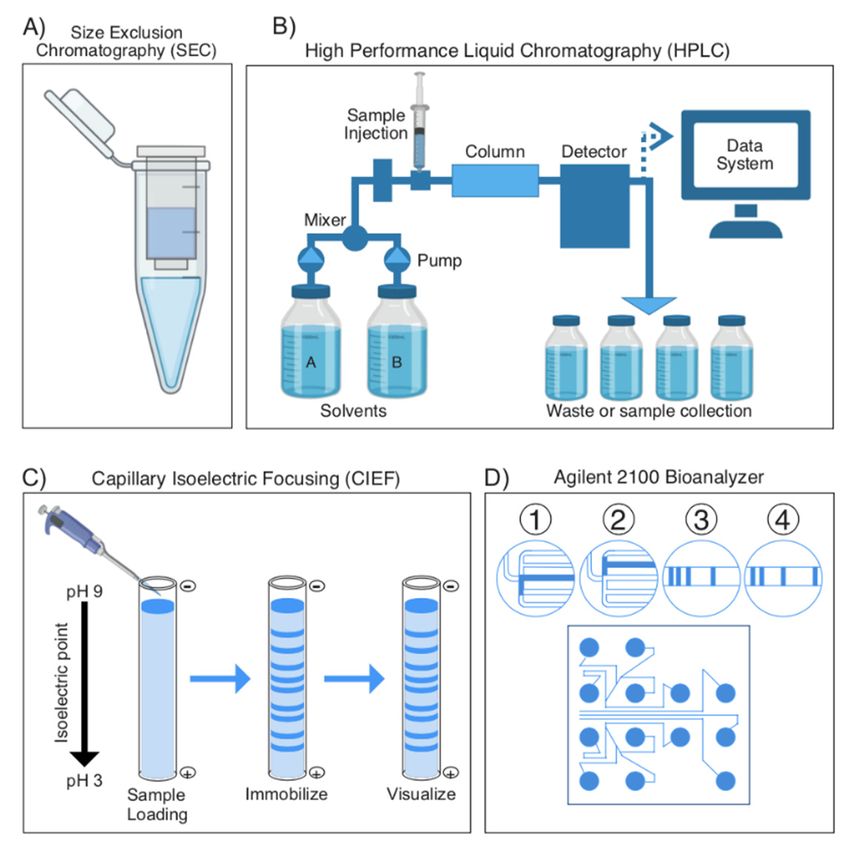
Toxins 2021, 13, 154                                                                                           2 of 15

                       that underlie these changes, one can begin to uncover the molecular basis of different
                       evolutionary processes as they relate to venoms [5–8].
                            From a predator’s point of view, venom is its most important weapon for capturing
                       prey. To maximize the chances of a meal, these venoms have evolved an impressive di-
                       versity of biochemical components. In addition to a powerful chemical weapon, many
                       species have also optimized their venom administration techniques (such as the miniature
                       harpoon-like teeth from cone snails, which serve to directly inject venom into victims [9])
                       to guarantee the incapacitation of their prey [10]. For many species, these venom deliv-
                       ery systems can be observed by the naked eye, but the complex composition of the key
                       weapon—the venom—remains to be uncovered. These venoms are almost analogous to a
                       molecular swiss-army knife, with each “arm” being a toxin that has been evolutionarily
                       specialized to execute its particular role.
                            This review will focus on overviewing the current techniques in venom proteomics,
                       as well as venom gland transcriptome sequencing. We will further describe how advances
                       in these techniques have allowed the field to slowly uncover the molecular composition
                       of nature’s vast microscopic arsenal of chemical weapons: venoms. Furthermore, we will
                       address how future developments in this field will provide insights into the molecular
                       evolution of venoms, which are key cheat codes that we can apply to any molecule in the
                       future to maximize its efficiency.

                       2. Techniques in Venomics
                            Historically, a generally similar workflow has been followed for elucidating the molec-
                       ular components of venoms (see Figure 1). Typically, following venom extraction and
                       purification, groups follow separation techniques to separate the venom into specific frac-
                       tions based on one, or a combination of different molecular properties ranging from size,
                       polarity, and charge to solubility and substrate affinity. One such example of a fractiona-
                       tion technique is high-performance liquid chromatography (HPLC). Groups would then
                       inject fractions into mice to observe which fraction elicited the final behavior of interest.
                       Finally, “active” fractions were purified to homogeneity with subsequent fractionations,
                       and then amino acid sequencing would be determined by reducing the toxins and carboxy-
                       methylating them. These could then be analyzed using Edman degradation with devices,
                       such as the Procise sequencing system or the Beckman890D spinning-cup sequencer [10,11].
                       However, these techniques were complicated to execute, and additional levels of complex-
                       ity were added by the fact that toxins are often multi-peptide proteins, which could not be
                       identified using such basic techniques.

                       2.1. Proteomics: Separation and Mass Spectrometry Techniques
                       2.1.1. General Overview of Separating Proteins, Bottom up vs. Top down
                             It is clear that the most efficient approach to increasing the resolution of current se-
                       quencing techniques lies in the proteomic steps that precede them. By using the modern
                       methods described herein, it is possible to achieve a high efficiency of toxin fractionation
                       and sequence identification to reliably characterize venoms. These methods improve the
                       fractionation and separation of toxin isoforms, allowing for sequencing techniques to
                       analyze a higher diversity of toxin isoforms, as opposed to older techniques that may not
                       separate highly similar toxin fragments as efficiently. This increased resolution at the pro-
                       tein level can provide greater insights into toxin variability among highly similar isoforms
                       and identifies different single nucleotide polymorphisms (SNPs) and post-translational
                       modifications (PTMs) that may occur within toxin families. Ultimately, these techniques
                       will provide the field with much deeper insights into toxin evolution [12].
Toxins 2021, 13, x FOR PEER REVIEW 3 of 16
Toxins 2021, 13, 154 3 of 15
Figure 1.Figure Classic workflow
1. workflow
Classic for studying
for studying toxin components
toxin components from venoms.
from venoms. In most In most studies,
studies, the se- the series
ries of experiments employed
of experiments employed to elucidate the molecular
to elucidate components
the molecular componentsof venoms involve
of venoms studying
involve studying the
the venom gland
venom and and
gland the venom
the venomitselfitself
separately. TheThe
separately. venom gland
venom is subject
gland to RNA
is subject to RNA extraction,
extraction, mRNA
mRNA enrichment,
enrichment,cDNA
cDNAlibrary
librarypreparation
preparationand andRNARNAsequencing
sequencingtotoobtain
obtainthe gland’s
the gland’stran-
transcriptome.
scriptome. In parallel, the whole venom is fractionated using any number of separation tech-
In parallel, the whole venom is fractionated using any number of separation techniques, high-
niques, high-performance liquid chromatography (HPLC) is pictured above. These fractions are
performance liquid chromatography (HPLC) is pictured above. These fractions are subject to mass
subject to mass spectrometry to identify peptides and obtain a proteome with the gland’s tran-
scriptomespectrometry to identify
(obtained from peptides and
RNA sequencing) obtain
being a proteome
utilized with thedatabase
as a reference gland’s transcriptome
for the mass (obtained
from
spectrometry. RNA sequencing) being utilized as a reference database for the mass spectrometry.
Historically, toxin elucidation has occurred mostly using bottom-up proteomic (BUP)
2.1. Proteomics: Separation and Mass Spectrometry Techniques
studies (see Figure 2), whereby toxin proteins are broken down into peptides using en-
2.1.1. General
zymatic Overview of Separating
or chemical reactions, Proteins, Bottom uptypically
then fractionated, vs. Top with
downliquid chromatography
It is clear that technique
(LC)-based the most efficient
and then approach to by
identified increasing
studying the resolution
their of current
intact mass se-
and fragmenta-
quencing tiontechniques lies comparing
patterns and in the proteomic
them to steps that precede
a protein sequence them. By using
database the modern
[12–14]. This approach
methods hasdescribed
been used herein, it isstudies,
in toxin possiblesince
to achieve a high efficiency
early gel-based proteomicsof toxin
untilfractionation
shotgun proteomic
and sequence identification
techniques to reliably
[13]. However, characterize
peptide-centric venoms. These
proteomics cannotmethods improve
necessarily give anthe
accurate
biological
fractionation interpretation
and separation of toxins,
of toxin sinceallowing
isoforms, different
forpeptides
sequencingmaytechniques
be presenttoinan-
different
alyze a combinations
higher diversity in multiple toxins, may
of toxin isoforms, as be presenttoinolder
opposed different proteoforms,
techniques or not
that may may have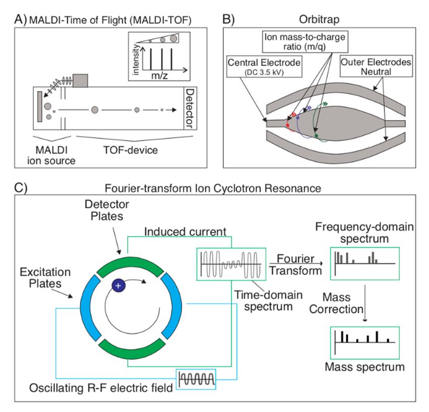
patterns of these whole proteins can bring to light information about toxins that would
                        otherwise be inaccessible using traditional bottom-up techniques, such as identification of
                        proteoforms, post-translational modifications (PTM), and alternative splicing [12,15–23].
                             Top-down proteomics allows the field to study how changes to toxins may ascribe
Toxins 2021, 13, 154    evolutionary advantages to venomous species, a possibility that was unlikely by simply   4 of 15
                        studying a venom gland’s transcriptome. This is especially advantageous for investigat-
                        ing the venoms of novel subspecies, whose close relatives have already been studied using
                        a bottom-up approach [19]. For these, TDP can not only save time in analyzing toxin com-
                       undergone   post-translational
                        ponents, but  also provide new modifications  with
                                                          insights into themajor functional
                                                                           specific         implications,
                                                                                    evolution             termed
                                                                                               of that species      the
                                                                                                               in com-
                       “protein inference  problem”   [14].
                        parison to its close relatives, uncovering the unique traits of this subspecies.

                       Figure 2. Top-down vs. Bottom-up proteomics approaches. The two main approaches in discerning
                       toxins from one another in venoms are top-down and bottom-up proteomics. Top-down proteomics
                       describes an approach whereby a venom’s whole proteins (notice: native, folded proteins in tubes on
                       top left) are holistically analyzed without the need for breaking proteins down into their constituent
                       fragments. On the other hand, bottom-up proteomics refers to techniques that involve the denaturing
                       of whole proteins (notice: unfolded proteins on top right) into fractions, and the study of these protein
                       fractions separately, before reassembling these fragments into proteins in silico to identify a venom’s
                       constituent toxin proteins. Typically, top-down proteomics is more accurate in discerning between
                       closely related toxins with minimal sequence variation or toxins with post-translational modifications.

                            With technological developments, new methods have been developed which allow
                       for top-down proteomic (TDP) studies. In these, rather than breaking proteins down
                       into their component peptides, intact proteins (or gas-phase fragmented proteins) can be
                       holistically analyzed using tandem mass spectrometry techniques without any requirement
                       for digestion, although some degree of denaturing can also be done to compare the resulting
                       proteomes of denatured vs. native samples (see Figure 2) [15–17]. The fragmentation
                       patterns of these whole proteins can bring to light information about toxins that would
                       otherwise be inaccessible using traditional bottom-up techniques, such as identification of
                       proteoforms, post-translational modifications (PTM), and alternative splicing [12,15–23].
                            Top-down proteomics allows the field to study how changes to toxins may ascribe
                       evolutionary advantages to venomous species, a possibility that was unlikely by simply
                       studying a venom gland’s transcriptome. This is especially advantageous for investigating
                       the venoms of novel subspecies, whose close relatives have already been studied using
                       a bottom-up approach [19]. For these, TDP can not only save time in analyzing toxin
                       components, but also provide new insights into the specific evolution of that species in
                       comparison to its close relatives, uncovering the unique traits of this subspecies.
                            Furthermore, TDP will allow for in-depth investigations of intraspecies venom varia-
                       tions, and may bring to light environmental factors that influence a venom’s composition,
                       such as diet, which is an influencing factor in the venoms of certain species of snakes and
                       tetrapods [6,24–31].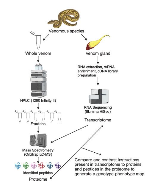
Toxins 2021, 13, 154 5 of 15
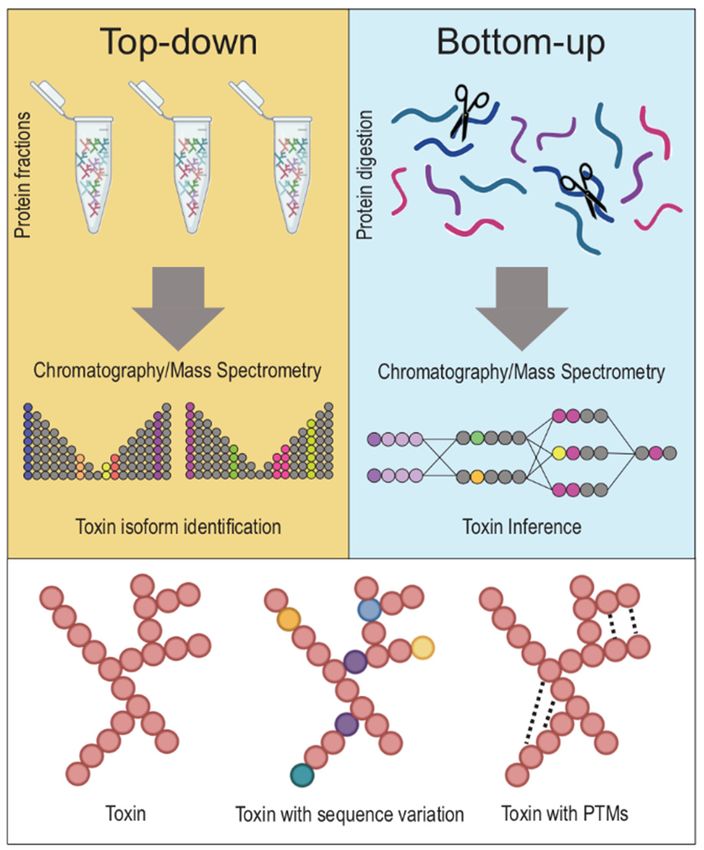
2.1.2. Separation Techniques
Most venoms are complex mixtures of proteins, making fractionation a necessary
task as mass spectrometry (MS) acquisition technologies can often not handle such diver-
sity [12]. These fractionation techniques are numerous, and some of the most common, to
be reviewed below in more detail, are: Reversed-phase high-performance liquid chromatog-
raphy (RP-HPLC, otherwise known as HPLC) (Figure 3B), capillary isoelectric focusing
Toxins 2021, 13, x FOR PEER REVIEW
(CIEF) (Figure 3C), size-exclusion chromatography (SEC) (Figure 3A), capillary zone6elec-
of 16
trophoresis (CZE) (Figure 3D), and 2D-Gel Electrophoresis (2D-GE) [20,31–39].
Figure3.3. Overview
Figure Overview of of most
mostcommonly
commonlyused usedsample
sample separation
separation techniques
techniquesin venomics.
in venomics. (A) A(A) A
molecular weight sieve is used for size exclusion chromatography to separate samples based on on
molecular weight sieve is used for size exclusion chromatography to separate samples based their
their
size orsize or molecular
molecular weight. weight. (B) High-performance
(B) High-performance liquid liquid chromatography
chromatography (HPLC)(HPLC)
is usedistoused to
separate
separate analytes in a sample based on their polarity. Analytes interact with
analytes in a sample based on their polarity. Analytes interact with a column (stationary phase)a column (stationary
phase) differently based on their polarity, causing them to elute at different speeds. The eluted
differently based on their polarity, causing them to elute at different speeds. The eluted molecules
molecules are detected by either a spectroscopic or an electrochemical detector, with a readout
are detected by either a spectroscopic or an electrochemical detector, with a readout available to the
available to the experimenter describing the eluate’s absorbance at different time points. (C) Capil-
experimenter
lary isoelectricdescribing the eluate’s
focusing (CIEF) absorbance
is a separation at different
technique that time points.
separates (C) Capillary
molecules based isoelectric
on their
focusing (CIEF)
isoelectric is ainseparation
points fused silicatechnique
capillarythat separates
tubes. (D) The molecules basedBioanalyzer
Agilent 2100 on their isoelectric points in
is an innovative
fused silica capillary tubes.technology
new “venom-on-a-chip” (D) The Agilent 2100 Bioanalyzer
that incorporates is an innovative
a variation of CIEF innewwhich“venom-on-a-chip”
microchannels
host an electrophoretic
technology separation
that incorporates of proteins,
a variation which
of CIEF are then
in which detected via host
microchannels fluorescence. The soft-
an electrophoretic
ware then of
separation transforms
proteins, this
whichdata
areinto
thengel-like images
detected and electropherograms
via fluorescence. The software forthen
easytransforms
interpretation.
this
data into gel-like images and electropherograms for easy interpretation.
Furthermore, varying the polarity of the stationary phase and varying the column’s
In HPLC,
length the sample
or the pump’s flowis rate,
prepared in vary
can all a solvent, a polar
the speed of mobile
samplephase,
elutionandandisultimately
pumped
through
affect thea column,
sample’sa retention
nonpolar time
stationary
in thephase, whose
stationary size and
phase. The packing material
slower flow rate may vary.
allows for
Based on the polarity of the sample’s components, they will each interact differently
a better separation of the sample’s components, especially if it is a medley of molecules as with
the
withstationary
venoms phase:
[41,42],Those thatcomes
but this are most
at polar will interact
the expense the least
of reduced with the stationary
separation resolution.
phase,
Lowerand flowthus,
rateselute
causefirst, with the
analytes opposite being
to increasingly truelongitudinally
diffuse for the sample’s mostthe
within nonpolar
column,
components. Theseresults
which ultimately separated
in acomponents
less precise can be detected
separation withanalytes
of these either spectroscopic
based on their detec-
po-
tors or electrochemical detectors to determine both elution time, as well as the
larity. Spectroscopic detection of these samples will typically show broader and muddled component’s
relative concentration
peaks, leading in the
to a lower sample [40].
resolution One
sample of the most important advances in HPLC
separation.
technology
CIEF is based on the technique of capillary gel(Toxins 2021, 13, 154 6 of 15
varying the center-to-center distance between peaks, ultimately improving a column’s
resolution and sensitivity [32].
Furthermore, varying the polarity of the stationary phase and varying the column’s
length or the pump’s flow rate, can all vary the speed of sample elution and ultimately
affect the sample’s retention time in the stationary phase. The slower flow rate allows for
a better separation of the sample’s components, especially if it is a medley of molecules
as with venoms [41,42], but this comes at the expense of reduced separation resolution.
Lower flow rates cause analytes to increasingly diffuse longitudinally within the column,
which ultimately results in a less precise separation of these analytes based on their polarity.
Spectroscopic detection of these samples will typically show broader and muddled peaks,
leading to a lower resolution sample separation.
CIEF is based on the technique of capillary gel electrophoresis (CGE), in which
molecules are separated in a gel, based on their different isoelectric points (Figure 3C). In
CIEF, however, instead of using gels, the separation is performed in fused silica capillary
tubes (internal diameter of 25–100 µm). Within the capillary, proteins migrate in response
to an electric field based on their isoelectric point and become focused at a point where
their net charge is balanced. Focused zones are then transported past a monitoring point
to detect the now separated proteins. By using a fused silica capillary tube, heat is effi-
ciently diffused, allowing higher voltages to separate a broader range of proteins. This is
essential, as introducing excessively high temperatures may introduce extraneous protein
denaturing [42–44].
CZE is another separation technique where sample components are separated based
on their charge. In this technique, a capillary column is immersed in two buffer-filled
reservoirs, to which a high voltage is applied. The sample is then injected into the capillary,
and its components are separated based on both the electrophoretic forces, as well as the
developing electro-osmotic forces within the capillary. This technique provides a higher
resolution, in terms of component separation, compared to HPLC, since peaks tend to be
very narrow. However, since flow rates tend to be low (nL per minute range), due to a very
small amount of starting material, CZE must be directly coupled with mass spectrometry
cannot be coupled with another downstream fractionation technique. To avoid potential
loss of material between fractionation and analytical steps, CZE is often directly interfaced
with nanospray MS, making it impossible to couple with another downstream fractionation
technique before MS. Due to this, CZE is not a reliable method for quantifying a given
component in a sample [45,46].
SEC is a chromatographic method in which hydrophilic molecules (proteins or other
solutes) are separated based on their size or molecular weight, either through molecular-
weight sieves or through gel-filtration chromatography [47]. It has proven to be very
useful in separating proteins, due to the technique’s ability to separate native proteins
(undigested) or protein complexes. This is essential in the field of venomics, as toxins are
often large proteins that may or may not exist in complexes with other proteins [48,49].
This technique can also be achieved using HPLC to perform a high-resolution fractionation
of molecules based on their molecular weight [47]. However, depending on the separation
technology used to achieve SEC, it is not a particularly high-resolution chromatographic
technique and has a limited dynamic range for protein separation. For example, the use
of molecular-weight sieves, while very user-friendly, has a far lower chromatographic
resolution than HPLC would [47].
2-dimensional gel electrophoresis (2DGE) is another common and highly accessible
method for groups to separate proteins in venoms and in order to characterize these
venoms. 2DGE separates proteins according to two different variables in the same gel.
In one dimension, proteins can be separated based on their isoelectric point, and in the
other by their relative molecular weight [50,51]. Based on the gel’s composition and buffers
used, 2DGE has the potential to resolve up to 10,000 proteins in a single gel, which can be
analyzed after capturing an image of the gel. The technique has its limitations: It cannot be
used to analyze a venom’s entire proteome as it has difficulty resolving a sample’s smallestToxins 2021, 13, 154 7 of 15
and largest proteins, as well as proteins that are highly acidic and highly basic. That
being said, 2DGE can separate and display up to thousands of proteins in one single lane
of a gel and ultimately enabling the visualization of numerous protein isoforms [51–54].
Furthermore, gel electrophoresis can be carried out in native or denaturing conditions
depending on the gel used. To carry out electrophoresis of non-denatured (intact) proteins
and protein complexes, one can use techniques such as Blue Native-PAGE (BN-PAGE) or
Clear Native electrophoresis to separation and isolation of protein complexes (of particular
interest in venom studies) [55,56]. With denaturing conditions, such as SDS-PAGE, one can
instead separate and isolate denature proteins, in which secondary structures have been
broken down. However, it should be noted that denaturing conditions tend to be preferable
when the experimental intention is to isolate proteins for protein sequencing [57].
A highly innovative hardware/software combination that automates the detection
and separation of proteins in venom is the Agilent 2100 Bioanalyzer, which enables rapid,
semi-automated “venom-on-a-chip” proteomic analyses. On a single chip, this technology
uses a variation of CIEF (Figure 3D). It uses microchannels that are etched into the chip,
allowing for the electrophoretic separation of proteins, and then detection via fluorescence.
The software transforms this data into gel-like images and electropherograms for easy
interpretation, allowing for complete separation and detection of protein components
with minimal effort from researchers [58–62]. Side-by-side comparisons of this technique
with pre-fractionated samples, and comparisons with other techniques, shows that it is
highly efficient at discriminating interspecies, as well as intraspecies venom variations [58].
Nonetheless, it is important to note that while this technique is very useful from an
electrophoretic point of view, it is currently impossible for a Bioanalyzer to return a
sample after separation. This makes the technique impossible to interface with MS to
subsequently identify separated peptides. Despite this, as the field of venomics is rapidly
growing, technologies, such as the Agilent 2100 Bioanalyzer will make venomic studies
more accessible to a broader range of laboratories, and will allow for simultaneous, high-
throughput studies of venoms from a variety of species and from multiple individuals.
2.1.3. Mass Spectrometry Techniques
Modern proteomics has embraced mass spectrometry to identify proteins based on
an analysis of the mass/charge ratio obtained from ionizing such proteins or peptides. In
order to perform an analysis of mass/charge ratio (m/z), proteins must be ionized. For this,
two techniques prevail as most efficient at preventing proteins from fragmenting in the
ionization process: Matrix-assisted laser desorption ionization (MALDI) and Electrospray
Ionization (ESI). In MALDI, a laser strikes a matrix of small molecules, in which an analyte
is embedded, sublimating the analyte molecule without fragmentation and ionizing it, since
the small matrix molecules can either protonate or deprotonate it (Figure 4A) [20,50,63–65].
In ESI, a high voltage, coupled with a parallel flow of nebulizing gas (usually nitrogen)
is used to vaporize a liquid solvent to create an aerosol. This solvent varies based on the
sample to be analyzed, but is typically composed of a polar solvent (for example, H2 O,
acetonitrile, tetrahydrofluoran) (Figure 4B). It is most unique from MALDI in that it is
better at preventing molecule fragmentation and can produce multiple-charged ions, which
can extend the ionizer’s range to accommodate very large molecules in the kDa to MDa
range [65–67].
Following ionization, the ions must be detected to determine the m/z. Currently,
the most common MS detection technique is time-of-flight (TOF), often coupled with
MALDI (Figure 5A). The main principle of TOF is that ions, in-flight following ionization,
travel at a rate proportional to their mass: Heavy ions take longer to reach the detector
than lighter ones, since they all begin their journey to the detector at the same time and
place [20,50,63–65]. However, in situations where MALDI is not the optimal ionization
technique (i.e., for the detection of larger peptides or proteins), ESI may also be interfaced
with a TOF analyzer (ESI-MS/MS). This can be done by coupling the ESI with quadrupole
MS to act as a mass filter, followed by an orthogonally placed TOF analyzer using aIonization (ESI). In MALDI, a laser strikes a matrix of small molecules, in which an analyte
is embedded, sublimating the analyte molecule without fragmentation and ionizing it,
since the small matrix molecules can either protonate or deprotonate it (Figure 4A)
[20,50,63–65]. In ESI, a high voltage, coupled with a parallel flow of nebulizing gas (usu-
ally nitrogen) is used to vaporize a liquid solvent to create an aerosol. This solvent varies
Toxins 2021, 13, 154 8 of 15
based on the sample to be analyzed, but is typically composed of a polar solvent (for ex-
ample, H2O, acetonitrile, tetrahydrofluoran) (Figure 4B). It is most unique from MALDI
in that it is better at preventing molecule fragmentation and can produce multiple-
charged ions,
reflectron to which
reflect can
the extend thetowards
ion beam ionizer’sthe
range
TOFtodetector
accommodate
[68]. ESIvery
maylarge
alsomolecules
be coupled
inwith
the kDa to MDa or
an Orbitrap range [65–67]. to increase resolving power over an ESI-TOF system [69].
a Q-Orbitrap
Figure
Figure 4. Ionization
4. Ionization techniques
techniques forspectrometry.
for mass mass spectrometry. (A) Matrix-assisted
(A) Matrix-assisted laser desorp-
laser desorption/ioniza-
tion/ionization (MALDI) is an ionization technique used for mass spectrometry
tion (MALDI) is an ionization technique used for mass spectrometry that involves sublimating that involvesansub-
limating
analyte an analyte
molecule molecule
embedded in aembedded in a matrix
matrix of small of small
molecules. molecules.
During During this
this sublimation, thesublimation,
analyte
itself
the is not fragmented,
analyte itself is notbut rather ionized
fragmented, but thanks to the protonating/deprotonating
rather ionized properties of
thanks to the protonating/deprotonating
the matrix molecules.
properties (B) Electrospray
of the matrix molecules. (B) ionization (ESI)ionization
Electrospray is another(ESI)
ionization technique
is another that technique
ionization consists
ofthat
the consists
use of high voltage and a nebulizing gas to vaporize a solvent, containing analytes, into
of the use of high voltage and a nebulizing gas to vaporize a solvent, containing analytes, an
aerosol. The remaining solvent in these droplets is evaporated, leaving a charged droplet, which
into an aerosol. The remaining solvent in these droplets is evaporated, leaving a charged droplet,
undergoes Coulomb fission resulting in charged progeny droplets. From these droplets, naked
which undergoes Coulomb fission resulting in charged progeny droplets. From these droplets, naked
charged analytes are detected by the device’s detector to investigate their m/z ratio.
charged analytes are detected by the device’s detector to investigate their m/z ratio.
Other detection techniques utilize frequency rather than time as a measure of an ion’s
mass; these include Orbitrap (Figure 5B) and FT-ICR (Fourier transform ion cyclotron
resonance) (Figure 5C). With more resolving power than TOF, the Orbitrap utilizes an ion
trap to analyze mass by trapping ions between an outer barrel-shaped electrode and an
inner rod-shaped electrode. Ions orbit around the inner electrode, and a Fourier transform
of the resulting charge frequency pattern reveals the ion’s mass spectrum [70,71]. In FT-ICR,
ions are instead trapped in a Penning trap (which uses a magnetic field to trap ions radially
and an electric field to confine particles axially), where they’re excited at their preferred
cyclotron frequency to a larger cyclotron radius by the axial oscillating electric field. Ions
rotate at their preferred cyclotron frequency in packets, producing a free induction decay
(FID) charge when they pass a pair of electrodes, which is essentially a superposition of
sine waves. Using a Fourier transform, the mass spectrum can be extracted from this
data sinusoidal FID charge data [63,72–74]. Since FT-ICR uses a superconducting magnet
instead of radio-frequency voltage, it is a much more stable device that can ultimately read
more accurate masses at a higher resolution.Toxins
Toxins2021,
2021,13,
13,x154
FOR PEER REVIEW 9 of
9 of1615
Detectiontechniques
Figure5.5.Detection
Figure techniquesforformass
massspectrometry.
spectrometry.(A) (A)Time-of-flight
Time-of-flight(TOF)
(TOF) is
is the
the most
most com-
common
detection technique in mass spectrometry and is often coupled with MALDI.
mon detection technique in mass spectrometry and is often coupled with MALDI. Ionized analyte Ionized analyte particles
travel at rates proportional to their mass. TOF utilizes this variable to infer particle
particles travel at rates proportional to their mass. TOF utilizes this variable to infer particle size size and (along
and
with (along with the reference
the reference database
database and andsoftware)
various various software) composition.
composition. (B) Orbitraps
(B) Orbitraps utilize
utilize ion traps to
ion traps to
identify anidentify
analyte’s anmass.
analyte’s
Thesemass.
ion These ion traps
traps consist of consist
trapping of ionized
trappingparticles
ionized particles
between be-an outer
tween an outer
electrode electrode
and inner and inner
electrode electrode and
and performing performing
a Fourier a Fourier
transform on thetransform on the charge
charge frequency pattern to
frequency pattern to produce the ion’s mass spectrum. (C) In a Fourier-transform
produce the ion’s mass spectrum. (C) In a Fourier-transform ion cyclotron resonance (FT-ICR) ion cyclotrondevice,
resonance (FT-ICR) device, ions are instead trapped in a Penning trap (which uses a magnetic field
ions are instead trapped in a Penning trap (which uses a magnetic field to trap ions radially and
to trap ions radially and an electric field to confine particles axially). Ions will rotate at their pre-
an electric field to confine particles axially). Ions will rotate at their preferred frequency in packets,
ferred frequency in packets, which produce a free induction decay (FID) charge as they pass a pair
which produce
of electrodes. Thisa FID
free is
induction decay (FID)
a time-domain charge
spectrum, as they
from which pass a pair of electrodes.
a frequency-domain This FID
spectrum canis a
time-domain spectrum, from which a frequency-domain spectrum can be extracted
be extracted via a Fourier transform. Following a mass correction, the sample’s mass spectrum can via a Fourier
betransform.
produced Following a mass correction,
from the frequency-domain the sample’s mass spectrum can be produced from the
spectrum.
frequency-domain spectrum.
Following ionization, the ions must be detected to determine the m/z. Currently, the
2.2. Transcriptomics
most common MS detection technique is time-of-flight (TOF), often coupled with MALDI
2.2.1. Transcriptomics:
(Figure Sequencing
5A). The main principle Techniques
of TOF is that ions, in-flight following ionization, travel
Early
at a rate on, polymerase
proportional chain
to their mass:reaction
Heavy(PCR) allowed
ions take longerfor to
thereach
studytheof detector
differentthan
com-
ponents
lighter of the
ones, sincevenom gland
they all begin at their
the transcriptome
journey to thelevel,
detectorwith at cDNA
the same libraries
time andidentifies
place
the numerous
[20,50,63–65]. venom components.
However, in situations However,
where MALDI the real game-changer
is not the optimal came whentech-
ionization high-
throughput sequencing technologies came along, such as Next Generation
nique (i.e., for the detection of larger peptides or proteins), ESI may also be interfaced with Sequencing,
aand
TOFall the platforms
analyzer that allow
(ESI-MS/MS). Thisforcan
it (pyrosequencing,
be done by coupling Illumina,
the ESI SOLiD, ion semiconduc-
with quadrupole MS
tor, DNA nanoball, etc.). All of these have proven to be of the utmost
to act as a mass filter, followed by an orthogonally placed TOF analyzer using a reflectron importance in the
togeneration
reflect theofion
debeam
novo venom
towardsglandthe TOFtranscriptomes
detector [68].[75].
ESI may also be coupled with an
Orbitrap or a Q-Orbitrap to increase resolving power over an early-stage
Most RNA-sequencing based assays begin with similar ESI-TOF systemworkflows:
[69]. RNA
is extracted, ribosomal RNA is depleted, or mRNA is enriched, cDNA is
Other detection techniques utilize frequency rather than time as a measure of an ion’s synthesized, and
an adaptor-ligated sequencing library is prepared. Depending on the
mass; these include Orbitrap (Figure 5B) and FT-ICR (Fourier transform ion cyclotron res- high-throughput
sequencing
onance) platform
(Figure utilized,
5C). With moretheresolving
library ispower
sequenced
thanto a read
TOF, thedepth of 20+
Orbitrap million
utilizes anreads
ion
per sample. The number of reads per sample may change from one sequencing platform
trap to analyze mass by trapping ions between an outer barrel-shaped electrode and an
to another, and it is a crucial variable in the quantitative capacity of an RNA-sequencing
inner rod-shaped electrode. Ions orbit around the inner electrode, and a Fourier transform
experiment. As the number of reads per sample increases, more transcripts can be detected,
of the resulting charge frequency pattern reveals the ion’s mass spectrum [70,71]. In FT-
increasing the sample’s sequencing depth and ultimately quantifying these transcripts to
ICR, ions are instead trapped in a Penning trap (which uses a magnetic field to trap ions
be more precise [76]. Finally, the sequencing reads are aligned and assembled into the
radially and an electric field to confine particles axially), where they’re excited at their
final transcriptome utilizing computational steps [77,78]. These data can be analyzed for
preferred cyclotron frequency to a larger cyclotron radius by the axial oscillating electric
a variety of purposes, with over 100 different RNA-Seq methods that have been derived
field. Ions rotate at their preferred cyclotron frequency in packets, producing a free induc-
from the standard protocol [77].Toxins 2021, 13, 154 10 of 15
Typically, most venom gland transcriptomes that have been sequenced to date have
been read using short-read sequencing technology, often Illumina sequencing, in part due
to its relatively low cost and easy to implement, but primarily due to the high quality,
quantitative data that can be obtained using this technique [79–93]. Generally, sequenced
cDNA fragments are under 200 bp in length with 20–30 million reads per sample, de-
pending on the technology and experimental limitations. This technique is very robust,
and has been verified using large-scale comparisons of short-read sequencing data, which
showed high intra-platform and inter-platform correlations [94]. However, multiple gene
isoforms pose an issue in transcriptome-wide analyses using short-read RNA-seq, since
transcript isoforms may be longer than 200 bp (for example, 50% of transcripts are longer
than 2500 bp in the human genome [95]). A single venom can contain multiple isoforms
of a single toxin, an important point to consider when performing short-read sequencing
on venom glands, especially when the source of such isoform variation is not yet known
(PTMs, SNPs, etc.).
While short-read RNA-seq can be achieved using Illumina technology, long-read
RNA-seq can be achieved using platforms, such as those from Pacific Biosciences and
Oxford Nanopore. These techniques can allow for sequencing of an entire, complete RNA
molecule following reverse transcription, circumventing the issues posed by short-read
sequencing techniques and reducing sequencing ambiguities of potential toxin isoforms in
a given sample [77,78,96–98]. However, long-read sequencing technologies are limited in
the number of reads they can perform per samples: While short-read sequencing allows
for 20–30 million reads per sample, long-read sequencing techniques are relatively low-
throughput and only allow for 500,000–10 million reads per sample, thus potentially
reducing the quantitative capacity of this technique and ultimately making it less valuable
for differential gene expression analyses. However, its ability to differentiate between
isoforms makes it ideal for de novo transcriptome assembly [77].
2.2.2. Current Gold Standards in Transcriptomics
The current gold standard for whole venom gland RNA-seq has been Illumina HiSeq
sequencing, using Trinity software to generate a de novo assembly and Trinotate software
to annotate the assembly [1–3,99–101]. This will certainly change as long-read RNA-
seq techniques are refined and upgraded. Improved resolution of proteomic techniques
has started to demonstrate the wide variety of toxin isoforms in a given venom sample,
and transcriptome sequencing can provide insights into how these isoforms evolved
from one another, thus offering a unique view into the evolution of prey and predators.
Gene duplication and isoform evolution are common events in the evolution of toxin
isoforms—to gain an understanding of how toxin isoforms go through this evolution, it
is essential for us to gain insights into what modifications provide toxins with a selective
advantage [102–105]. As mentioned above, short-read RNA-seq techniques are limited by
read length, which has been improved upon in the relatively young long-read techniques.
As these long-read techniques are upgraded, we will certainly be able to gain much higher
resolution, quantitative reads of long-read sequenced transcriptomes, ultimately providing
us with transcriptome-level insights into toxin isoform evolution.
2.3. Integrated Proteomic-Transcriptomic Techniques
The proteomic and transcriptomic techniques described in this review are powerful
tools in analyzing venoms and elucidation of individual toxins. However, integrating
proteomic and transcriptomic techniques in analyzing venoms presents an even stronger
approach that can allow for a rapid and thorough analysis of venom components, and
has become very common in the field of venomics [5,93,106,107]. While transcriptomics
provides a relatively unbiased representation of the diversity of transcripts in a given
sample, the technique also captures thousands of non-venom peptide encoding transcripts
(which may be part of other cellular processes) and cannot detect post-translational mod-
ifications, which have important consequences for a toxin peptide’s functionality [108].Toxins 2021, 13, 154 11 of 15
The combination of transcriptomics and proteomics allows for the transcriptomics arm of
experimentation to capture a sample’s high transcript diversity, and the proteomic arm
allows for the narrowing of this diversity to focus analyses only on toxin peptide-encoding
transcripts. Furthermore, following identification of toxin-encoding transcripts, many
transcriptomic techniques can provide insights into the relative expression level of these
transcripts, giving expression estimates for toxin-related genes of interest [108–110].
3. Conclusions
The sequencing of venom glands and the parallel sequencing of these venoms’ target
species, has allowed science to write out a so-called “molecular storyline”, hinting at how
much resistance mechanisms have evolved, and how venoms themselves have evolved
in response to maintain their evolutionary arms race [111–117]. For many years, our
understanding of the basic principles of evolution has relied on the idea that adaptations
to change are necessary for survival. An increasing number of discoveries in the study
of venoms and the evolution of their targets seem to confirm that one of the building
blocks of survival throughout evolution is indeed a species’ genomic thriftiness. Nature
has endowed species with the possibility for genomic plasticity in order to survive the
constant arms race of predator-prey interactions [118,119].
By gaining an understanding of how and why certain genes become plastic in the
evolution of venoms and their resistance mechanisms, we can begin to decipher nature’s
evolutionary methods and can start to apply these learned concepts to the treatment
of diseases.
Author Contributions: Conceptualization, R.S.-N. and S.M.-C.; writing—original draft preparation,
S.M.-C.; writing—review and editing, R.S.-N. and S.M.-C.; funding acquisition, R.S.-N. All authors
have read and agreed to the published version of the manuscript.
Funding: This work was supported by the Canadian Institutes of Health Research grant CIHR
PJT-173355 to RSN.
Institutional Review Board Statement: Not applicable.
Informed Consent Statement: Not applicable.
Data Availability Statement: Not applicable.
Conflicts of Interest: The authors declare no conflict of interest.
References
1. Von Reumont, B.M. Studying smaller and neglected organisms in modern evolutionary venomics implementing RNASEq
(transcriptomics)—A critical guide. Toxins 2018, 10, 292. [CrossRef]
2. Sunagar, K.; Morgenstern, D.; Reitzel, A.M.; Moran, Y. Ecological venomics: How genomics, transcriptomics and proteomics can
shed new light on the ecology and evolution of venom. J. Proteom. 2016, 135, 62–72. [CrossRef] [PubMed]
3. Wilson, D.; Daly, N.L. Venomics: A Mini-Review. High Throughput 2018, 7, 19–26. [CrossRef]
4. Wang, C.R.; Bubner, E.R.; Jovcevski, B.; Mittal, P.; Pukala, T.L. Interrogating the higher order structures of snake venom proteins
using an integrated mass spectrometric approach. J. Proteom. 2020, 216, 103680. [CrossRef] [PubMed]
5. Calvete, J.J. Venomics: Integrative venom proteomics and beyond. Biochem. J. 2017, 474, 611–634. [CrossRef] [PubMed]
6. Daltry, J.C.; Wüster, W.; Thorpe, R.S. Diet and snake venom evolution. Nat. Cell Biol. 1996, 379, 537–540. [CrossRef]
7. Holding, M.L.; Drabeck, D.H.; Jansa, S.A.; Gibbs, H.L. Venom resistance as a model for understanding the molecular basis of
complex coevolutionary adaptations. Integr. Comp. Biol. 2016, 56, 1032–1043. [CrossRef] [PubMed]
8. da Silva, N.J., Jr.; Aird, S.D. Prey specificity, comparative lethality and compositional differences of coral snake venoms. Comp.
Biochem. Physiol. C Toxicol. Pharmacol. 2001, 128, 425–456. [CrossRef]
9. Terlau, H.; Olivera, B.M. Conus venoms: A rich source of novel ion channel-targeted peptides. Physiol. Rev. 2004, 84, 41–68.
[CrossRef]
10. Terlau, H.; Shon, K.J.; Grilley, M.; Stocker, M.; Stühmer, W.; Olivera, B.M. Strategy for rapid immobilization of prey by a
fish-hunting marine sn. Nat. Commun. 1996, 381, 148–151. [CrossRef]
11. Olivera, B.M.; Gray, W.R.; Zeikus, R.; McIntosh, J.M.; Varga, J.; Rivier, J.; De Santos, V.; Cruz, L.J. Peptide neurotoxins from
fish-hunting cone snails. Science 1985, 230, 1338–1343. [CrossRef] [PubMed]Toxins 2021, 13, 154 12 of 15
12. Melani, R.D.; Nogueira, F.C.S.; Domont, G.B. It is time for top-down venomics. J. Venom. Anim. Toxins Incl. Trop. Dis. 2017, 23.
[CrossRef] [PubMed]
13. Lomonte, B.; Calvete, J.J. Strategies in ‘snake venomics’ aiming at an integrative view of compositional, functional, and
immunological characteristics of venoms. J. Venom. Anim. Toxins Incl. Trop. Dis. 2017, 23, 1–12. [CrossRef]
14. Nesvizhskii, A.I.; Aebersold, R. Interpretation of shotgun proteomic data: The protein inference problem. Mol. Cell. Proteom.
2005, 4, 1419–1440. [CrossRef]
15. Ainsworth, S.; Petras, D.; Engmark, M.; Süssmuth, R.D.; Whiteley, G.; Albulescu, L.-O.; Kazandjian, T.D.; Wagstaff, S.C.; Rowley,
P.; Wüster, W.; et al. The medical threat of mamba envenoming in sub-Saharan Africa revealed by genus-wide analysis of venom
composition, toxicity and antivenomics profiling of available antivenoms. J. Proteom. 2018, 172, 173–189. [CrossRef]
16. Petras, D.; Heiss, P.; Süssmuth, R.D.; Calvete, J.J. Venom proteomics of indonesian king cobra, ophiophagus hannah: Integrating
top-down and bottom-up approaches. J. Proteome Res. 2015, 14, 2539–2556. [CrossRef] [PubMed]
17. Petras, D.; Heiss, P.; Harrison, R.A.; Süssmuth, R.; Calvete, J.J. Top-down venomics of the East African green mamba, Dendroaspis
angusticeps, and the black mamba, Dendroaspis polylepis, highlight the complexity of their toxin arsenals. J. Proteom. 2016, 146,
148–164. [CrossRef]
18. Verano-Braga, T.; Dutra, A.A.; León, I.R.; Melo-Braga, M.N.; Roepstorff, P.; Pimenta, A.M.; Kjeldsen, F. Moving pieces in a venomic
puzzle: Unveiling post-translationally modified toxins from Tityus serrulatus. J. Proteome Res. 2013, 12, 3460–3470. [CrossRef]
19. Hempel, B.-F.; Damm, M.; Mrinalini, M.; Göçmen, B.; Karış, M.; Nalbantsoy, A.; Kini, R.M.; Suessmuth, R.D. Extended snake
venomics by top-down in-source decay: Investigating the newly discovered anatolian meadow viper subspecies, Vipera anatolica
senliki. J. Proteome Res. 2020, 19, 1731–1749. [CrossRef]
20. Calvete, J.J.; Juárez, P.; Sanz, L. Snake venomics. Strategy and applications. J. Mass Spectrom. 2007, 42, 1405–1414. [CrossRef]
21. Pla, D.; Petras, D.; Saviola, A.J.; Modahl, C.M.; Sanz, L.; Pérez, A.; Juárez, E.; Frietze, S.; Dorrestein, P.C.; Mackessy, S.P.; et al.
Transcriptomics-guided bottom-up and top-down venomics of neonate and adult specimens of the arboreal rear-fanged Brown
Treesnake, Boiga irregularis, from Guam. J. Proteom. 2018, 174, 71–84. [CrossRef]
22. Fukuyama, Y.; Iwamoto, S.; Tanaka, K. Rapid sequencing and disulfide mapping of peptides containing disulfide bonds by using
1,5-diaminonaphthalene as a reductive matrix. J. Mass Spectrom. 2005, 41, 191–201. [CrossRef] [PubMed]
23. Hughes, C.; Ma, B.; Lajoie, G. De Novo Sequencing Methods in Proteomics; Springer Nature: Berlin, Germany, 2010.
24. Barlow, A.; Pook, C.E.; Harrison, R.A.; Wüster, W. Coevolution of diet and prey-specific venom activity supports the role of
selection in snake venom evolution. Proc. R. Soc. B Biol. Sci. 2009, 276, 2443–2449. [CrossRef] [PubMed]
25. Harris, R.J.; Arbuckle, K. Tempo and mode of the evolution of venom and poison in tetrapods. Toxins 2016, 8, 193. [CrossRef]
[PubMed]
26. Davies, E.-L.; Arbuckle, K. Coevolution of snake venom toxic activities and diet: Evidence that ecological generalism favours
toxicological diversity. Toxins 2019, 11, 711. [CrossRef]
27. Modahl, C.M.; Mrinalini Frietze, S.; Mackessy, S.P. Adaptive evolution of distinct prey-specific toxin genes in rear-fanged snake
venom. Proc. R. Soc. B 2018, 285, 20181003. [CrossRef] [PubMed]
28. Amorim, F.G.; Costa, T.R.; Baiwir, D.; De Pauw, E.; Quinton, L.; Sampaio, S.V. Proteopeptidomic, functional and immunoreactivity
characterization of Bothrops moojeni snake venom: Influence of snake gender on venom composition. Toxins 2018, 10, 177.
[CrossRef]
29. Lyons, K.; Dugon, M.M.; Healy, K. Diet breadth mediates the prey specificity of venom potency in snakes. Toxins 2020, 12, 74.
[CrossRef]
30. Healy, K.; Carbone, C.; Jackson, A.L. Snake venom potency and yield are associated with prey-evolution, predator metabolism
and habitat structure. Ecol. Lett. 2019, 22, 527–537. [CrossRef]
31. Sousa, L.F.; Portes, J.A., Jr.; Nicolau, C.A.; Bernardoni, J.L.; Nishiyama, M.Y., Jr.; Amazonas, D.R.; Freitas-De-Sousa, L.A.; Mourão,
R.H.; Chalkidis, H.M.; Valente, R.H.; et al. Functional proteomic analyses of Bothrops atrox venom reveals phenotypes associated
with habitat variation in the Amazon. J. Proteom. 2017, 159, 32–46. [CrossRef]
32. Conlon, J.M. Purification of naturally occurring peptides by reversed-phase HPLC. Nat. Protoc. 2007, 2, 191–197. [CrossRef]
33. Biardi, J.E.; Ho, C.; Marcinczyk, J.; Nambiar, K. Isolation and identification of a snake venom metalloproteinase inhibitor from
California ground squirrel (Spermophilus beecheyi) blood sera. Toxicon 2011, 58, 486–493. [CrossRef]
34. Margres, M.J.; McGivern, J.J.; Wray, K.P.; Seavy, M.; Calvin, K.; Rokyta, D.R. Linking the transcriptome and proteome to
characterize the venom of the eastern diamondback rattlesnake (Crotalus adamanteus). J. Proteom. 2014, 96, 145–158. [CrossRef]
35. Prashanth, J.R.; Hasaballah, N.; Vetter, I. Pharmacological screening technologies for venom peptide discovery. Neuropharmacology
2017, 127, 4–19. [CrossRef] [PubMed]
36. Juárez, P.; Sanz, L.; Calvete, J.J. Snake venomics: Characterization of protein families in Sistrurus barbouri venom by cysteine
mapping, N-terminal sequencing, and tandem mass spectrometry analysis. Proteomics 2004, 4, 327–338. [CrossRef] [PubMed]
37. Nicolau, C.A.; Carvalho, P.C.; Junqueira-De-Azevedo, I.L.; Teixeira-Ferreira, A.; Junqueira, M.; Perales, J.; Neves-Ferreira, A.G.C.;
Valente, R.H. An in-depth snake venom proteopeptidome characterization: Benchmarking Bothrops jararaca. J. Proteom. 2017,
151, 214–231. [CrossRef] [PubMed]
38. Asakawa, M.; Matsumoto, T.; Umezaki, K.; Kaneko, K.; Yu, X.; Gomez-Delan, G.; Tomano, S.; Noguchi, T.; Ohtsuka, S.; Yu, X.
Toxicity and toxin composition of the greater blue-ringed octopus Hapalochlaena lunulata from Ishigaki Island, Okinawa Prefecture,
Japan. Toxins 2019, 11, 245. [CrossRef] [PubMed]Toxins 2021, 13, 154 13 of 15
39. Besenius, P.; Cormack, P.A.; Ludlow, R.F.; Otto, S.; Sherrington, D.C. Affinity chromatography in dynamic combinatorial libraries:
One-pot amplification and isolation of a strongly binding receptor. Org. Biomol. Chem. 2010, 8, 2414–2418. [CrossRef]
40. Vestal, M.L. High-performance liquid chromatography-mass spectrometry. Science 1984, 226, 275–281. [CrossRef]
41. Chen, H.; Horváth, C. High-speed high-performance liquid chromatography of peptides and proteins. J. Chromatogr. A 1995, 705,
3–20. [CrossRef]
42. Wang, C.; Lee, C.S. Capillary electrophoresis–mass spectrometry for proteomic and metabolic analysis. Proteom. Metab. Approaches
Biomark. Discov. 2013, 163–173, 163–173. [CrossRef]
43. Smoluch, M.; Mielczarek, P.; Drabik, A.; Silberring, J. Online and offline sample fractionation. Proteom. Profiling Anal. Chem. 2016,
63–99, 63–99. [CrossRef]
44. Trenerry, V.C.; Rochfort, S.J. Natural products research and metabolomics. Compr. Nat. Prod. II 2010, 2, 595–628. [CrossRef]
45. Nagy, K.; Vékey, K. Separation methods. Med Appl. Mass Spectrom. 2008, 61–92, 61–92. [CrossRef]
46. Moldoveanu, S.C.; David, V. Short overviews of the main analytical techniques containing a separation step. Sel. HPLC Method
Chem. Anal. 2017, 55–85, 55–85. [CrossRef]
47. Striegel, A. Size-exclusion chromatography. Liq. Chromatogr. 2013, 193–223, 193–223. [CrossRef]
48. Tasoulis, T.; Silva, A.; Veerati, P.M.; Baker, M.; Hodgson, W.C.; Dunstan, N.; Isbiter, G.K. Coastal taipan Oxyuranus scutellatus.
Toxins 2020, 12, 485. [CrossRef]
49. Kunalan, S.; Othman, I.; Hassan, S.S.; Hodgson, W.C. Proteomic characterization of two medically important malaysian snake
venoms, Calloselasma rhodostoma (Malayan Pit Viper) and Ophiophagus hannah (King Cobra). Toxins 2018, 10, 434. [CrossRef]
[PubMed]
50. Aslam, B.; Basit, M.; Nisar, M.A.; Khurshid, M.; Rasool, M.H. Proteomics: Technologies and their applications. J. Chromatogr. Sci.
2017, 55, 182–196. [CrossRef]
51. Bocian, A.; Urbanik, M.; Hus, K.K.; Łyskowski, A.; Petrilla, V.; Andrejčáková, Z.; Petrillová, M.; Legáth, J. Proteomic analyses of
agkistrodon contortrix contortrix venom using 2D electrophoresis and MS techniques. Toxins 2016, 8, 372. [CrossRef]
52. Klose, J.; Kobalz, U. Two-dimensional electrophoresis of proteins: An updated protocol and implications for a functional analysis
of the genome. Electrophoresis 1995, 16, 1034–1059. [CrossRef] [PubMed]
53. Martins-De-Souza, D. 2DE Gels: A story of love and hate in proteomics. Proteomics 2018, 18, e1700472. [CrossRef]
54. Viala, V.L.; Hildebrand, D.; Trusch, M.; Arni, R.K.; Pimenta, D.C.; Schlüter, H.; Betzel, C.; Spencer, P.J. Pseudechis guttatus venom
proteome: Insights into evolution and toxin clustering. J. Proteom. 2014, 110, 32–44. [CrossRef]
55. Barkan, N.P.; Bayazit, M.B.; Demiralp, D.O. Proteomic characterization of the venom of five bombus (Thoracobombus) species.
Toxins 2017, 9, 362. [CrossRef] [PubMed]
56. Wittig, I.; Karas, M.; Schägger, H. High resolution clear native electrophoresis for in-gel functional assays and fluorescence studies
of membrane protein complexes. Mol. Cell. Proteomics 2007, 6, 1215–1225. [CrossRef] [PubMed]
57. Wittig, I.; Braun, H.-P.; Schägger, H. Blue native PAGE. Nat. Protoc. 2006, 1, 418–428. [CrossRef]
58. Nowakowski, A.B.; Wobig, W.J.; Petering, D.H. Native SDS-PAGE: High resolution electrophoretic separation of proteins with
retention of native properties including bound metal ions. Metallomics 2014, 6, 1068–1078. [CrossRef]
59. Zancolli, G.; Sanz, L.; Calvete, J.J.; Wüster, W. Venom on-a-chip: A Fast and efficient method for comparative venomics. Toxins
2017, 9, 179. [CrossRef]
60. Kuschel, M.; Neumann, T.; Barthmaier, P.; Kratzmeier, M. Use of lab-on-a-chip technology for protein sizing and quantitation. J.
Biomol. Tech. 2002, 13, 172–178.
61. Ohashi, R.; Otero, J.M.; Chwistek, A.; Hamel, J.-F.P. Determination of monoclonal antibody production in cell culture using novel
microfluidic and traditional assays. Electrophoresis 2002, 23, 3623–3629. [CrossRef]
62. Schmut, O.; Horwath-Winter, J.; Zenker, A.; Trummer, G. The effect of sample treatment on separation profiles of tear fluid
proteins: Qualitative and semi-quantitative protein determination by an automated analysis system. Graefe’s Arch. Clin. Exp.
Ophthalmol. 2002, 240, 900–905. [CrossRef] [PubMed]
63. Nguyen, T.; Kwak, S.; Karpowicz, S.J. Re-use of commercial microfluidics chips for DNA, RNA, and protein electrophoresis.
Biotechniques 2014, 57, 267–271. [CrossRef] [PubMed]
64. Escoubas, P.; Quinton, L.; Nicholson, G.M. Venomics: Unravelling the complexity of animal venoms with mass spectrometry. J.
Mass Spectrom. 2008, 43, 279–295. [CrossRef] [PubMed]
65. Greco, V.; Piras, C.; Pieroni, L.; Ronci, M.; Putignani, L.; Roncada, P.; Urbani, A. Applications of MALDI-TOF mass spectrometry
in clinical proteomics. Expert Rev. Proteom. 2018, 15, 683–696. [CrossRef]
66. Andersen, J.S.; Svensson, B.; Roepstorff, P. Electrospray ionization and matrix assisted laser desorption/ionization mass
spectrometry: Powerful analytical tools in recombinant protein chemistry. Nat. Biotechnol. 1996, 14, 449–457. [CrossRef] [PubMed]
67. Yamashita, M.; Fenn, J.B. Electrospray ion source. Another variation on the free-jet theme. J. Phys. Chem. 1984, 88, 4451–4459.
[CrossRef]
68. Fenn, J.B.; Mann, M.; Meng, C.K.; Wong, S.F.; Whitehouse, C.M. Electrospray ionization for mass spectrometry of large
biomolecules. Science 1989, 246, 64–71. [CrossRef]
69. Nadler, W.M.; Waidelich, D.; Kerner, A.; Hanke, S.; Berg, R.; Trumpp, A.; Rösli, C. MALDI versus ESI: The Impact of the Ion
Source on Peptide Identification. J. Proteome Res. 2017, 16, 1207–1215. [CrossRef]You can also read