Myocarditis in Paediatric Patients: Unveiling the Progression to Dilated Cardiomyopathy and Heart Failure - MDPI
←
→
Page content transcription
If your browser does not render page correctly, please read the page content below
Journal of
           Cardiovascular
           Development and Disease

Review
Myocarditis in Paediatric Patients: Unveiling the
Progression to Dilated Cardiomyopathy and
Heart Failure
Inês Teixeira Farinha 1, * and Joana Oliveira Miranda 2,3
 1   Faculty of Medicine of Porto University, Porto 4200-319, Portugal
 2   Department of Paediatric Cardiology, Centro Hospitalar São João, Porto 4200-319, Portugal;
     joanam@gmail.com
 3   Department of Physiology and Cardiothoracic Surgery, Faculty of Medicine of Porto University,
     Porto 4200-319, Portugal
 *   Correspondence: i.t.farinha@gmail.com; Tel.: +351-91-4666-608

 Academic Editor: Joel D. Schilling
 Received: 29 August 2016; Accepted: 3 November 2016; Published: 8 November 2016

 Abstract: Myocarditis is a challenging and potentially life-threatening disease associated with high
 morbidity in some paediatric patients, due to its ability to present as an acute and fulminant disease
 and to ultimately progress to dilated cardiomyopathy. It has been described as an inflammatory
 disease of the myocardium caused by diverse aetiologies. Viral infection is the most frequent cause of
 myocarditis in developed countries, but bacterial and protozoal infections or drug hypersensitivity
 may also be causative agents. The prompt diagnosis in paediatric patients is difficult, as the spectrum
 of clinical manifestation can range from no myocardial dysfunction to sudden cardiac death. Recent
 studies on myocarditis pathogenesis have revealed a triphasic nature of this disease, which influences
 the diagnostic and therapeutic strategies to adopt in each patient. Endomyocardial biopsy remains
 the gold standard for diagnosing myocarditis, and several non-invasive diagnostic tools can be used
 to support the diagnosis. Intravenous immunoglobulin has become part of routine practice in the
 treatment of myocarditis in paediatric patients at many centres, but its true effect on the cardiac
 function has been the target of many studies. The aim of this review is to approach the recently
 discovered facets of paediatric myocarditis regarding its progression to dilated cardiomyopathy.

 Keywords: myocarditis; dilated cardiomyopathy; heart failure; inflammation; viral myocarditis

1. Introduction
     Myocarditis is a non-familial form of inflammatory heart muscle disease, in the absence of
predominant acute or chronic ischaemia [1–3]. It is, by definition, an inflammation of the myocardium,
which may also extend to the pericardium and endocardium [4]. Diagnosis is established by histological,
immunologic and immunohistochemical criteria, as it was defined by the World Health Organization
(WHO)/International Society and Federation of Cardiology (ISFC), in 1995 [5]. It is an important cause
of morbidity and mortality in children, due to its association with cardiac dysfunction and dilated
cardiomyopathy (DCM), which may represent the chronic phase of the disease [5,6].

2. Aetiology
    Acute myocarditis has multiple causes, including viruses, protozoa, bacteria, fungi, toxins, drugs,
metabolic abnormalities, hypersensitivity reactions and systemic autoimmune diseases [7]. Often the
underlying agent causing the disease is not identified [4].

J. Cardiovasc. Dev. Dis. 2016, 3, 31; doi:10.3390/jcdd3040031                     www.mdpi.com/journal/jcdd
J. Cardiovasc. Dev. Dis. 2016, 3, 31                                                               2 of 18

2.1. Infections
     More than 50% of paediatric acute myocarditises in Western Europe and North America are
caused by cardiotropic viruses [8,9]. The pattern of viral pathogens associated with myocarditis has
evolved over the last 20 years [10]. Enteroviruses (particularly coxsackieviruses) have traditionally
been considered the most frequent viral agents in myocarditis and DCM [11,12]. More recently,
new molecular techniques have revealed that the most commonly detected viral agents were
Parvovirus B19 and human herpesvirus type 6 [12–14]. Other previously unrecognized viruses
associated with myocarditis with varying degrees of frequency include adenovirus, cytomegalovirus,
Epstein–Barr, hepatitis C, herpes simplex type 2, influenza and parainfluenza viruses [9,11,13,15–19].
In 2010, Bratincsák et al. reported three cases of fulminant myocarditis in a paediatric population
associated with the pandemic H1N1 influenza A virus infection, and it is hypothesized that this
strain may be more associated with severe forms of myocarditis than other influenza strains [20].
Human immunodeficiency virus (HIV) has also been associated with myocarditis, which is often
asymptomatic [21]. HIV myocarditis may cause both systolic and diastolic dysfunction in paediatric
patients [22]. Some agents are thought to be responsible for the initiation of these processes and the
presence of viral nucleic acid in the myocardium, a common finding in HIV+ children, may be one of
them [23]. Infection of myocytes with other agents, like fungi, parasites or other viruses other than HIV
may be associated with the development of DCM and congestive heart failure [23,24]. Some drugs
used in highly active antiretroviral therapy (HAART) regimens, like Zidovudine, are cardiotoxic
themselves [22].
     Bacteria and other infectious causes, such as protozoa, fungi, and parasites, have been known to
cause myocarditis far less commonly than viruses [25]. Toxin-producing bacteria, including Clostridium
and Diphtheria, can cause severe myocardial damage, the latter causing myocarditis in countries
without widespread immunization [21,26]. Lyme disease, caused by the spirochete Borrelia burgdorferi,
can also result in acute myocarditis [7]. However, it is considered the least common manifestation
of early disseminated Lyme borreliosis in children and progression to DCM is rare [7,27]. In 2009,
Costello et al. reviewed 207 cases of children with early disseminated Lyme disease and found
that 16% had myocarditis, 42% of whom had advanced atrioventricular heart block, including 27%
with third-degree heart block [28]. In Latin-American countries, Chagas disease, which results from
an infection due to the protozoan Trypanosoma cruzi, is a common cause of acute myocarditis and
DCM [10,29]. In recent years, as a consequence of effective control of the transmission of the protozoan,
acute Chagas myocarditis has reduced in Brazil [7].

2.2. Hypersensitivity to Drugs and Other Substances
     Drugs may also cause myocardial inflammation via a direct cardiotoxic effect or by inducing
hypersensitivity reactions [4]. Hypersensitivity myocarditis is a rare form of inflammatory disease
of the myocardium [30]. The molecular basis is poorly understood, but it is thought to be a
hypersensitivity reaction to haptens from the cardiotoxic drug [31]. This diagnosis should always be
suspected when clinical presentation occurs in a setting consistent with drug allergy because of its
excellent prognosis after withdrawal of the causative toxin [32]. Some agents have been reported to
be implicated in hypersensitivity myocarditis in paediatric patients. These include chemotherapeutic
drugs, such as anthracyclines, dermatology drugs, like dapsone, and also tetanus and smallpox
vaccines [30,33–35].
     These drug-induced hypersensitivity responses are often associated with an eosinophilic
myocarditis, which is usually revealed after an endomyocardial biopsy (EMB) [21]. In this situation,
withdrawal of the offending medication may not always be enough and corticosteroid therapy may be
needed [36].

3. Myocarditis: A Triphasic Disease Process
     Since the majority of cases of myocarditis result from a viral infection, it has long been studied
in experimental murine models as a virus-induced autoimmune disease, which may progress to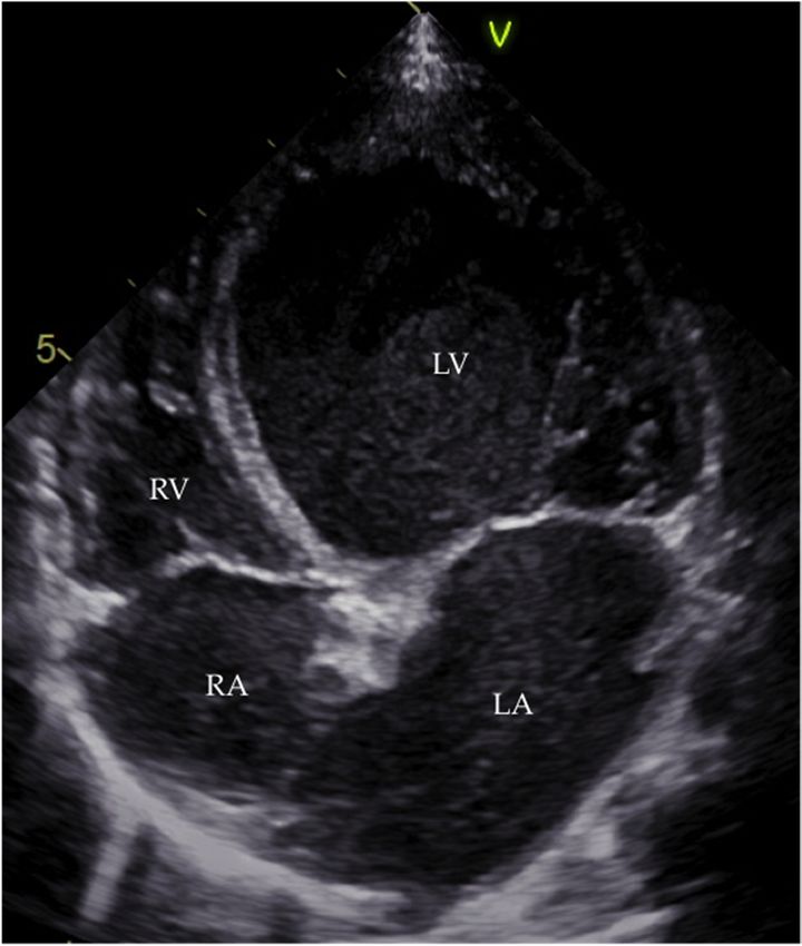
J. Cardiovasc. Dev. Dis. 2016, 3, 31 3 of 17
J. Cardiovasc. Dev. Dis. 2016, 3, 31 3 of 18
Since the majority of cases of myocarditis result from a viral infection, it has long been studied
in experimental murine models as a virus-induced autoimmune disease, which may progress to
gradual
gradualmyocardial
myocardialdilatation [3,37]. Myocarditis
dilatation [3,37]. Myocarditisprogresses
progresses through
through stages
stages withwith distinct
distinct processes
processes and
and manifestations.
manifestations. Thus,
Thus, it hasitbeen
hashelpful
been helpful
to set it intotheset it in the
structure of astructure
continuumofofathree
continuum of three
chronologically
chronologically successive
successive phases phases
of disease of disease
(Figure (Figure
1). For each phase,1). pathogenesis,
For each phase, pathogenesis,
diagnosis, diagnosis,
and treatment and
differ
treatment differ[37,38].
significantly significantly [37,38].
Figure 1. Myocarditis:
Figure AA
1. Myocarditis: triphasic disease.
triphasic In In
disease. recent
recentyears, myocarditis
years, myocarditishas
hasbeen
beenset
setininaaframework
framework of
of three
three successive
successive and distinct
and distinct phasesphases with undefined
with undefined borders.borders.
For eachFor eachaverage
phase, phase, duration
average duration
(regarding
the(regarding the case of coxsackievirus-mediated
case of coxsackievirus-mediated myocarditis), characteristics
myocarditis), characteristics and physiopathological
and physiopathological intervenients are
intervenients
indicated. are indicated.
CD, cluster CD, cluster
of differentiation; Th1,ofT differentiation;
helper 1; Th17, TTh1, T helper
helper 1; Th17,
17; Treg, T helper
regulatory T. 17; Treg,
regulatory T.
The three-phased course starts with the first phase, which is an induction stage where the virus
plays a The three-phased
major role in the course starts to
initial insult withthethe first phase, [6,38].
myocardium which Theis anentry
induction stage where
and active the virus
replication of the
plays
virus a major role
is followed in the initial insult
by cardiomyocyte lysistoand
the activation
myocardium [6,38].
of the hostThe entryimmune
innate and active replication
responses. of
In most
the virus is followed by cardiomyocyte lysis and activation of the host innate
patients, the immune system downregulates to a resting state after viral proliferation is brought under immune responses.
In most
control andpatients, the immune
they recover withoutsystem downregulates
significant sequelae to a resting
[3,37]. Thus, state
theafter viral
initial proliferation
insult often goesisunnoticed
brought
under
[38]. control and they recover without significant sequelae [3,37]. Thus, the initial insult often goes
unnoticed [38].
If host immune activation persists despite elimination of the virus, the second phase develops
If host of
as a result immune
both activation
adaptivepersists
immune despite
andelimination
autoimmune of theresponses
virus, the second
[38]. phase develops as of
The activation
a result of both adaptive immune and autoimmune responses [38]. The activation of antigen-specific
antigen-specific immunity involving T cells, B cells and antibody production contributes to
immunity involving T cells, B cells and antibody production contributes to additional myocardial
additional myocardial injury with deleterious effects [3,10]. Development of autoantibodies can be
injury with deleterious effects [3,10]. Development of autoantibodies can be induced by molecular
induced by molecular mimicry between myocardial antigens and viral peptides [10,38]. Clinically
mimicry between myocardial antigens and viral peptides [10,38]. Clinically overt congestive heart
overt congestive heart failure may develop in this phase [38]. In the majority of patients, the virus is
failure may develop in this phase [38]. In the majority of patients, the virus is eliminated and the
eliminated and the immune reaction is downregulated [3].
immune reaction is downregulated [3].
However,
However, a subset
a subsetofofpatients
patientsprogresses
progresses to to aa third phase in
third phase inwhich
whichthe theinflammatory
inflammatoryprocesses
processes
persist
persistandand
thethedisease develops
disease develops intointo
DCM DCM andandit may progress
it may despite
progress the ending
despite of theoffirst
the ending thephases
first
[3,38]. Viral myocarditis has become recognized as an important cause of
phases [3,38]. Viral myocarditis has become recognized as an important cause of DCM in children. DCM in children. Miranda
et al. performed
Miranda a retrospective
et al. performed review ofreview
a retrospective 61 paediatric patients
of 61 paediatric (37 female;
patients 24 male)
(37 female; diagnosed
24 male) diagnosed with
DCM and found that viral myocarditis was the aetiology in 18%
with DCM and found that viral myocarditis was the aetiology in 18% of cases [39]. of cases [39].
4. Pathogenic
4. Pathogenic Mechanisms
Mechanisms
TheThe causal
causal connectionbetween
connection betweenmyocarditis
myocarditis and
and its
its progression
progression to toDCM
DCMandandheart
heartfailure
failureis is
notnot
completely understood but has been hypothesized for many years [40]. Different
completely understood but has been hypothesized for many years [40]. Different studies usingstudies using murine
models
murine of myocarditis
models have contributed
of myocarditis to whattoiswhat
have contributed currently known known
is currently about the pathogenesis
about of the
the pathogenesis
disease
of the [4]. Viral
disease mediated
[4]. Viral cardiomyocyte
mediated injury, autoimmunity
cardiomyocyte and persistent
injury, autoimmunity viral infection
and persistent viralare three
infection
theories assumed to explain this process [41].
are three theories assumed to explain this process [41].
The disease begins with viral entry into cardiomyocytes [21]. Specific receptors have been
identified on human myocytes, such as the Coxsackie-adenoviral receptor (CAR) for
J. Cardiovasc. Dev. Dis. 2016, 3, 31                                                                4 of 18

     The disease begins with viral entry into cardiomyocytes [21]. Specific receptors have been
identified on human myocytes, such as the Coxsackie-adenoviral receptor (CAR) for Coxsackieviruses
of group B and some adenoviruses, and the erythrocyte P-antigen cellular receptor for Parvovirus
B19 [42–46]. Replicating viruses in the absence of an immune response causes myocytolysis, which
is the primary lesion responsible for focal necrosis and inflammation of the myocardium [40,47].
Subsequently, innate and adaptive immune responses are activated [7,38]. Many host pro-inflammatory
mediators are upregulated in an attempt to limit viral replication, including cytokines such as tumour
necrosis factor-α (TNF-α), interleukin (IL)-1 and IL-6, nitric oxide, macrophages and natural killer
(NK) cells [21,48]. Additionally, T lymphocyte (CD4+ and CD8+) activation was described to induce
cytokine production as well as apoptosis of targeted cells mediated by perforins, serine esterases and
the Fas ligand/Fas receptor pathway [49]. Even though these mechanisms are an essential path to
recovery, their cumulative effect is harmful since they also enhance myocardial tissue injury, therefore
contributing to the loss of virus-harbouring myocytes [21,38,50]. The decrease in the number of
contractile units and their inability to regenerate may lead to long-term remodelling and a clinical
picture consistent with DCM [6,7].
     Autoimmunity has also been recognized as having an important role in the progression of viral
myocarditis [41]. Secondarily to myocyte necrosis comes the exposure of heart muscle antigens [51].
Questions remain whether this mechanism is the only one triggering the autoimmune disease or if
molecular mimicry may also play a part [41]. When activated B lymphocytes produce antibodies that
cross-react with these antigens the autoimmune response begins and viral myocarditis progresses [7,41].
Anti-sarcolemmal, anti-myosin alpha and beta heavy chains, anti-mitochondrial proteins, and anti-β1
adrenergic receptor antibodies have all been identified in patients with DCM [7,52].
     According to Kuhl et al., persistent viral genome can be found in 67.4% of adult patients with
DCM, which is most likely caused by incomplete clearance of the pathogen [4,53]. The exact role of a
persistent cardiac infection in the development of DCM in paediatric patients has not been defined
yet [38]. It is hypothesized that continuous or intermittent virus replication may result in a chronic
myocyte damage and autoimmune injury [21,41].

5. Clinical Presentation
      Clinical manifestation of the disease in children has a variable spectrum, ranging from non-specific
systemic symptoms with no haemodynamic consequences (fever, myalgia, palpitations or dyspnoea)
to congestive heart failure, ventricular dysfunction, shock and life-threatening ventricular arrhythmias,
which consequently can lead to sudden cardiac death [6,7,54]. Syncope and chest pain may also
be presenting complaints [10]. The latter can mimic adult myocardial infarction with anterior chest
pressure pain radiating to the neck and arms and is typically seen in younger patients with few
cardiac risk factors [2,4,55]. This acute myocardial infarction-like syndrome can be associated with
electrocardiographic changes, such as ST segment elevation, and elevated biomarkers of myocardial
cell damage (creatine kinase and/or troponin I), in a context of normal coronary arteriography [2,7,56].
      Many patients may present with mild unspecific prodromal symptoms typical of a viral illness,
such as shortness of breath, vomiting, anorexia, abdominal pain, diarrhoea, fever, myalgia, lethargy,
syncope or seizures [4,7,57,58]. A history of respiratory tract infection or gastroenteritis may precede
the onset of myocarditis by several days to a few weeks [7]. In 2009, Durani et al. performed a
retrospective review of the presenting symptoms in patients ultimately diagnosed with myocarditis,
and the most frequently described were shortness of breath (69%), vomiting (48%), poor feeding (40%),
upper respiratory symptoms (39%), fever (36%), and lethargy (36%) [59].
      Clinical features also vary according to age. Infants may present with nonspecific symptoms
like anxiousness, malaise, fever, poor appetite, tachypnoea, tachycardia, and cyanosis [21]. Children
greater than two years of age may also complain of chest pain, shortness of breath, abdominal pain,
exercise intolerance, myalgia, arthralgia, fatigue, palpitations, cough or oedema [21,41,60].
J. Cardiovasc. Dev. Dis. 2016, 3, 31 5 of 18
The severity of symptoms is dependent on the age of the child [21]. Newborns and infants are
often more severely affected and, in contrast to older children and adults, they are more likely to
present with circulatory shock and acute DCM and may require advanced circulatory and respiratory
support in early stages of their disease [61,62].
6. Differential Diagnoses
Since myocarditis symptoms are not specific, other diseases can present in a similar clinical
pattern [6,63]. In the newborn or infant, sepsis, hypoxia, severe dehydration, hypoglycaemia or
anaemia must be ruled out. Other diagnoses should also be excluded, such as endocarditis and
endocardial fibroelastosis. Genetic X-linked disorders, like Barth syndrome, as well as congenital
structural heart lesions, such as critical coarctation of the aorta or anomalous origin of the left coronary
artery from the pulmonary artery or even a cerebral arteriovenous malformation may also be present.
In the older child, chronic tachyarrhythmia and pericarditis are two other differential diagnosed
conditions to add to the previously listed.
As far as DCM is concerned, only one-third of the patients enrolled in a Paediatric
Cardiomyopathy Registry study had a known cause of DCM at diagnosis [64]. On the subject
of differential diagnoses, it is very important to consider familial DCM, since it comprises 14%
of all paediatric DCM cases with a known cause and it carries a worse prognosis than other
aetiologies [65–67]. Many mutations with autosomal dominant, recessive, X-linked, and mitochondrial
inheritance patterns associated with the DCM phenotype have been identified [68]. Family cardiac
screening may carry some advantages if done quickly, particularly the possibility of avoiding the need
for the performance of other invasive and expensive diagnostic tools [65]. Other alternate diagnoses
consistent with clinical presentation of symptomatic heart failure should be excluded, such as metabolic
DCM (comprising 11% of paediatric DCM cases with a known cause), valvular and congenital heart
disease (occurring in only 3% of paediatric DCM cases with a known aetiology), bronchiolitis, thyroid
disease and chemotherapy-related cardiomyopathy [66,69–71]. Some genetic disorders, involving 26%
of paediatric DCM cases with a known cause, like Emery–Dreifuss muscular dystrophy, Laing distal
myopathy or Duchenne and Becker muscular dystrophy should be investigated in idiopathic DCM,
after exclusion of all other identifiable causes [66,70].
7. Diagnosis and Treatment: A Triphasic Approach
Having in mind the triphasic framework of myocarditis, it is helpful to systematize its diagnostic
and therapeutic strategies, which are considerably different for each stage of disease [38].
7.1. First phase: Viral Replication
7.1.1. Diagnosis
Initially, the diagnosis of myocarditis is very dependent on clinical suspicion based on history
and clinical picture of the disease, especially when cardiac involvement is apparent [6,7]. However,
the wide range of clinical manifestations of paediatric myocarditis may easily transpire unnoticed by
the clinician [59].
Definite diagnosis of viral myocarditis can only be proved by finding evidence of active viral
infection, using histological or serological identification of virus [6,38]. EMB used to be required to
define myocarditis according to the Dallas criteria [4]. Since it is not performed routinely in some
centres, there are several non-invasive tests that can support the suspected diagnosis, something that
is particularly important in paediatric patients [72].
The 12-lead electrocardiogram (ECG) is an easily available initial test [4]. Retrospective studies
have found abnormal ECGs in 93% to 100% of paediatric patients with myocarditis [59,73]. However, a
normal ECG does not exclude the possibility of the disease [57]. The most common findings in the acute
phase of clinical myocarditis are sinus tachycardia with low-voltage QRS complexes and nonspecificJ. Cardiovasc. Dev. Dis. 2016, 3, 31 6 of 18
T-wave changes, but these are not specific markers [4,6,21]. The ECG may also reveal ST-segment
decrease or elevation, occasionally resembling a pattern seen in acute myocardial infarction [74,75].
Although commonly used as a screening tool, an ECG has the sensitivity of only 47% for the diagnosis
of myocarditis in paediatric patients [76,77].
Echocardiography is still the most useful imaging technique to be performed in paediatric
suspected cases to assess left ventricular structure, wall motion abnormalities, regional or global
ventricular dysfunction and valvular insufficiency, since these abnormalities are often present in
acute myocardial inflammation [7,10,78]. However, there are no specific echocardiographic features
of myocarditis and they are usually insufficient to differentiate this disease from other forms of
cardiomyopathy [6,51].
Cardiac MRI (cMRI) is proving to be the most attractive imaging tool for diagnosing myocardial
inflammation and myocyte injury, especially if performed within 14 days of the beginning of
symptoms, when the sensitivity for the diagnosis is higher (Figure 2) [6,79]. In addition to its
unique potential for providing anatomic and morphologic information, cMRI has the ability
to characterize tissue by measuring T1 and T2 relaxation times and spin densities, which are
dependent on water content [80,81]. Detection of myocardial oedema, the initial change in myocardial
tissue during the first phase of inflammation, is measured with T2-weighted imaging [51,80].
Gadolinium diethylenetriaminepentacetate (Gd-DTPA) is used as an extracellular contrast agent
in contrast-enhanced cMRIs, and it distributes differently according to the type of cardiac tissue,
inflamed or normal tissue [7]. Early enhancement in T1-weighted images obtained minutes after
Gd-DTPA infusion may assess hyperaemia, but it is not the most specific finding for the diagnosis
due to artefacts [82,83]. On the other hand, delayed Gd-DTPA enhancement suggests the presence
of myocardial fibrosis and necrosis associated with myocarditis, lesions that are often histologically
patchy in nature [7,84]. One cMRI approach to diagnose myocarditis is the Lake Louise criteria
(LLC) [85]. The diagnosis is made if two out of three LLC criteria are met: (i) increased myocardial
early Gd-DTPA enhancement ratio or absolute myocardial enhancement of ≥45% on T1-wheighted
images; (ii) increased myocardial signal intensity on T2-weighted images (increased T2-ratio or
regional increase in T2 signal intensity); and (iii) non-ischaemic lesions at late Gd-DTPA enhancement
imaging [83]. Even though the LLC have shown some utility, particularly in patients with acute onset
of symptoms, their qualitative nature and bad performance when applied in patients with heart
failure and chronic symptoms has contributed to the search for novel quantitative T1 and T2 mapping
techniques, including the quantification of extracellular volume [85]. A recent study performed in
adult patients showed that myocardial T1 and T2 mapping and T1-derived extracellular volume
fraction significantly improved the diagnostic accuracy of cMRI compared with conventional LLC [86].
By identifying areas of myocardial inflammation globally, cMRI offers an advantage in the diagnosis
of this disease when compared to EMB and may even be used to direct it, consequently improving
its diagnostic sensitivity [6,87]. However, cMRI with delayed Gd-DTPA enhancement alone has been
reported to have a relatively low sensitivity in visualizing areas of myocarditis in adult patients,
ranging from 27% to 95% [75]. Additionally, the use of cMRI in routine practice is limited by the lack
of availability in most emergency settings and the need for general anaesthesia in young paediatric
patients [88].
EMB is still the gold standard of diagnosing acute myocarditis even though in the past its
routine clinical use for the diagnosis of myocarditis was not recommended [7,10]. The 2013 position
statement of the European Society of Cardiology Working Group recommended that all patients with
clinically suspected myocarditis should be considered for selective coronary angiography and EMB [89].
The role of EMB is undeniable, since immunohistochemistry and immunofluorescence techniques,
viral DNA or RNA identification by PCR and real-time PCR amplification of viral genome, used in
conjunction with standard EMB, provide an increase in diagnostic sensitivity [6,89]. However, EMB is
not routinely performed to diagnose myocarditis [90]. The sensitivity of this diagnostic tool is limited
due to sampling error and the rate of major complications associated with this procedure is variable,patients with heart failure and chronic symptoms has contributed to the search for novel
quantitative T1 and T2 mapping techniques, including the quantification of extracellular volume
[85]. A recent study performed in adult patients showed that myocardial T1 and T2 mapping and
T1-derived extracellular volume fraction significantly improved the diagnostic accuracy of cMRI
compared
J. Cardiovasc. with conventional
Dev. Dis. 2016, 3, 31 LLC [86]. By identifying areas of myocardial inflammation globally, 7 of 18
cMRI offers an advantage in the diagnosis of this disease when compared to EMB and may even be
used to direct it, consequently improving its diagnostic sensitivity [6,87]. However, cMRI with
ranging Gd-DTPA
delayed fromJ. Cardiovasc. Dev. Dis. 2016, 3, 31 8 of 18
inflammatory cells can be distinguished by cluster differentiation (CD) [101]. Monoclonal antibodies
to CD3 and CD68 or CD11 allow the detection and localization of T cells and activated macrophages,
respectively [10]. CD20 stands for B-cells [101]. Human leukocyte antigen (HLA) can be used to
detect HLA class II expression in professional antigen-presenting immune cells [10,37]. The addition
of immunohistochemical criteria in the analysis of EMBs has made it possible to quantitatively define
myocardial inflammation as focal and diffuse mononuclear infiltrates with >14 leucocytes/mm2 [5,101].
T cell subpopulations may include CD4 (helper), CD8 (suppressor) and CD45R0 (memory or activated
T-cells) [101]. Having these subpopulations in mind, inflammation may be more specifically diagnosed
by >7.0 CD3+ lymphocytes/mm2 and/or >35.0 CD11b+/Mac-1+ macrophages/mm2 [103].
Viral serologies may be abnormal as well [6]. However, IgG antibodies for cardiotropic viruses
are often positive in the blood stream without associated cardiac involvement, a fact that limits its
significance in the diagnostic process [6,104].
7.2.2. Treatment
Since autoimmunity myocardial inflammatory processes may take place despite virus elimination,
immunomodulatory therapies targeting the host immune system can be efficacious [4,7]. However,
this topic remains controversial in the paediatric age group due to the reduced number of studies
performed and the lack of respective controls [7]. Some administered immunomodulatory strategies
include immunosuppression with corticosteroids, intravenous immunoglobulin (IVIG), azathioprine,
and cyclosporine [105].
Corticosteroid treatment remains controversial when it comes to its benefit in children with
myocarditis [106]. While some recent studies have shown improvement in left ventricular ejection fraction
(LVEF) and symptoms among children treated with corticosteroids and/or other immunosuppressive
agents, like cyclosporine, others found no differences in the final outcome [16,107–109]. The only
aspect that seems to be commonly accepted is that immunosuppression with corticosteroids should
not be used in the acute phase of myocarditis since it interferes with its natural eradication and,
therefore, prolongs the disease [109].
IVIG has become part of routine immunomodulatory therapy for treating children with acute
myocarditis at many centres, in a standard high-dose of 2 g/kg per 24 h [4,110]. There is a possibility
that paediatric patients are more likely to respond to IVIG than adults due to a higher chance of a viral
myocarditis [110]. Drucker et al. performed a retrospective study to assess the effect of a 2 mg/kg
dose of IVIG therapy in 21 consecutive children with presumed acute myocarditis on survival and
recovery of left ventricular function [111]. Their experience found that the use of high-dose IVIG was
associated with improvements in recovery of left ventricular systolic function and a tendency toward
better overall survival during the first year after presentation [10,111]. Nigro et al. reported a case of
a seven-month-old girl with a severe myocarditis and persistent Parvovirus B19 DNA in the blood,
who received IVIG therapy containing neutralizing antibodies specific to that virus [112]. The clinical
improvement and viral clearing suggested the beneficial role of this therapeutic agent. A paediatric
trial conducted by Bhatt et al. in India evaluated the efficacy of IVIG among 83 children (ages ranging
from two months to 12 years) with suspected viral encephalitis and associated myocarditis [113].
They found a lower death rate among children receiving IVIG than in those who did not, and it is
suggested that IVIG may be useful in the select group of children beyond the neonatal period who
have the association of both viral diseases. Thus, a high risk of bias is present in this study, since
it is not clear whether this benefit extends to children with myocarditis alone [110]. On the other
hand, a retrospective analysis revealed that the treatment with IVIG provided no advantage in overall
survival and time to recovery of normal left ventricular function in comparison to steroid therapy
alone [114,115]. The question of IVIG role in paediatric myocarditis remains to be answered [114].
The insufficient evidence to support its routine use may require a prospective randomized study in
order to truly assess the advantage of this treatment in this specific population [6,10,110].J. Cardiovasc. Dev. Dis. 2016, 3, 31 9 of 18
Immunoadsorption is a therapeutic concept currently under discussion [6]. Its aim is to eliminate
cardiotoxic autoantibodies together with cytokines, which have been associated with DCM [116,117].
Repetitive plasmapheresis sessions allow the removal of IgG primarily and little amount of IgA and
IgM, and have been associated with improvement in cardiac function and reduction of oxidative
stress [118–120]. In some studies, polyclonal IgG has been given at the end of the procedure, in order to
J. Cardiovasc.
provide Dev. Dis. 2016, 3,immunomodulatory
an additional 31 effect [6]. The exact mechanism of action is still not known,9 of 17
but the benefit of this process may come from the removal of circulating autoantibodies, elimination of
Jahns etoral.
cytokines have provided
unloading direct
of the heart and evidence of specific
the circulation circulating
by the temporary β1-adrenoceptor
removal antibodies
of proteins [119,121].
[122,123].JahnsThis et
autoimmune attack, which
al. have provided alsoevidence
direct plays a causal role incirculating
of specific DCM, can β1-adrenoceptor
be eradicated with
the antibodies
elimination of theThis
[122,123]. β-receptor
autoimmune antibody [6]. However,
attack, which also plays a this
causalpathophysiologically appealing
role in DCM, can be eradicated
concept lacks
with the randomized
elimination clinical
of the trials antibody
β-receptor [6,119]. [6]. However, this pathophysiologically appealing
concept lacks randomized clinical trials [6,119].
7.3. Third Phase: Dilated Cardiomyopathy
7.3. Third Phase: Dilated Cardiomyopathy
7.3.1. Diagnosis
7.3.1. Diagnosis
This phase
This is is
phase identified
identifiedbybythe
theusual
usualimaging tools in
imaging tools inorder
ordertotoexclude
exclude other
other causes
causes of dilatation
of dilatation
(Figures
(Figures 3 and 4) [6]. When it comes to echocardiography, it is important to acknowledge thatthat
3 and 4) [6]. When it comes to echocardiography, it is important to acknowledge eveneven
though
though thethemost frequentechocardiographic
most frequent echocardiographic finding
finding associated
associated with myocarditis
with myocarditis is a DCM
is a DCM phenotype
phenotype of left dilatation
of left ventricular ventricular dilatation
and reduced and fraction,
ejection reduced ejection
other fraction,
parameters other parameters
of ventricular remodelling of
ventricular remodelling
may be present may be present
in histologically in histologically
proven myocarditis, proven
including myocarditis,
regional hypertrophyincluding regional
and regional
wall motion abnormalities [7,124]. The left ventricle of a patient with acute myocarditis
hypertrophy and regional wall motion abnormalities [7,124]. The left ventricle of a patient with may even lack
acute
significantmay
myocarditis dilatation [4]. significant dilatation [4].
even lack
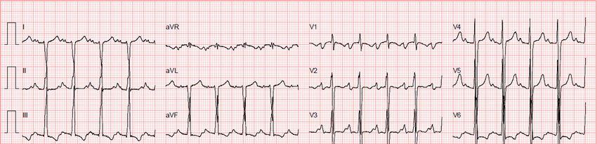
Figure 3. 3.ECG
Figure ECG of
of aa14-year-old
14-year-oldgirlgirl
withwith familial
familial DCM showing
DCM showing sinus tachycardia,
sinus tachycardia, right axis
right axis deviation
deviation and nonspecific
and nonspecific ST-waveST-wave
changes. changes.J. Cardiovasc. Dev. Dis. 2016, 3, 31 10 of 18
J. Cardiovasc. Dev. Dis. 2016, 3, 31 10 of 17
Figure 4.
Figure 4. Four-chamber
Four-chamber apical
apical view
view echocardiogram
echocardiogram of an 11-month-old
of an 11-month-old boy with DCM.
boy with DCM. RA,
RA, right
right
atrium; LA, left atrium; RV, right ventricle; LV, left ventricle.
atrium; LA, left atrium; RV, right ventricle; LV, left ventricle.
7.3.2. Treatment
7.3.2. Treatment
Therapy after
Therapy after absence
absence of of both
both infectious
infectious and and autoimmune
autoimmune stagesstages isis focused
focused on on prevention
prevention and
and
reversal ofofthethe
reversal unfavourable
unfavourable remodelling
remodelling processprocess of the myocardium
of the myocardium and reductionand reduction of
of haemodynamic
haemodynamic stress by using angiotensin converting enzyme (ACE) inhibitors,
stress by using angiotensin converting enzyme (ACE) inhibitors, β-blockers and spironolactone [6,37,38]. β-blockers and
spironolactone [6,37,38]. β-blockers like carvedilol can be cautiously introduced
β-blockers like carvedilol can be cautiously introduced only in stable patients and the dose should be only in stable
patientsslowly
titrated and the dose
[125]. should
These be must
agents titrated slowly [125].
be stopped Theseinotropes
whenever agents must be stopped
are needed [121]. whenever
inotropes are needed [121].
Anticoagulation with warfarin in DCM stage is difficult in young infants and many clinicians
preferAnticoagulation
to use aspirin inwith warfarin
patients with in DCM stage
severely is difficult
compromised in young
cardiac outputinfants anddysrhythmias,
or atrial many cliniciansin
prefer to use aspirin in patients with severely compromised cardiac output
order to prevent the formation of mural thrombi [126,127]. However, aspirin and other non-steroidalor atrial dysrhythmias, in
order to prevent the formation of mural thrombi [126,127]. However, aspirin
anti-inflammatory agents have been associated with exacerbation of heart failure, probably due to the and other non-steroidal
anti-inflammatory
inhibition agents have
of prostaglandin been which
synthesis, associated with exacerbation
ultimately may lead to aof heart failure,
decrease in renalprobably
perfusiondue to
[121].
the inhibition of prostaglandin synthesis, which ultimately may lead to a decrease
Their use is not recommended in adult patients with reduced left ventricular function according to in renal perfusion
[121].
the Their
“2016 use is Society
European not recommended
of Cardiology in Guidelines
adult patients with
for the reduced
diagnosis and left ventricular
treatment function
of acute and
according to the “2016 European Society of Cardiology Guidelines for the diagnosis
chronic heart failure” [128]. Prophylactic aspirin should be reserved for paediatric patients with DCM and treatment of
acute and chronic heart failure” [128]. Prophylactic aspirin should be reserved
who have not experienced any previous thromboembolic event or intracardiac thrombus, with LVEF for paediatric patients
with DCM
between 25%who andhave30%notorexperienced any previous
fractional shortening (FS)thromboembolic
between 15% and event or intracardiac
20%. Treatment thrombus,
should be
with LVEF
stopped between
whenever 25% and
systolic 30% or
function fractional
improves shortening (FS) between 15% and 20%. Treatment
[129].
should be stopped
Although whenever
paediatric systolicoften
myocarditis function improves
improves [129].
within the first months of presentation, a minority
Although paediatric myocarditis often improves
of patients may progress to severe heart failure, which is unresponsive within the first to months
optimalofmedical
presentation, a
therapy,
minority of patients may progress to severe
therefore requiring cardiac transplantation [7,10,21]. heart failure, which is unresponsive to optimal medical
therapy,
Thesetherefore
patientsrequiring
need to be cardiac transplantation
frequently monitored[7,10,21].
throughout their lives and a complete history,
examination, and echocardiography should be undertaken [37]. their lives and a complete history,
These patients need to be frequently monitored throughout
examination, and echocardiography should be undertaken [37].
8. Prognosis
8. Prognosis
Since a significant number of cases of myocarditis do not get diagnosed due to the unspecific
Since a its
symptoms, significant numberand
true incidence of cases
outcome of myocarditis do nottoget
remain difficult diagnosed [7,61,97,130].
characterize due to the unspecific
A lack
symptoms, its true incidence and outcome remain difficult to characterize [7,61,97,130]. A lack ofJ. Cardiovasc. Dev. Dis. 2016, 3, 31 11 of 18
of studies aiming to assess the prognosis of paediatric myocarditis adds to the difficulty and the
extrapolation of outcomes from the adult to the paediatric population should not be assumed [97].
The main outcomes paediatric patients can experience include complete recovery, progression
to DCM and death or transplantation [4,61,91]. According to Peta and co-workers’ 20-year study of
175 children with myocarditis, survival free from death or transplantation was 74% at one year, 65% at
five years, 62% at 10 years, and 56% at 20 years. At 15 years after diagnosis, 60% of the participants
remained free from transplantation, of whom 69% achieved echocardiographic normalization of
left ventricular function, 96% were free of cardiac symptoms, 80% were not receiving long-term
medical therapy, and 4% had an implantable cardioverter-defibrillator [131]. Another study including
222 paediatric patients with DCM due to myocarditis revealed a five-year rate of freedom from
transplantation of 81% and an estimated survival of 92% at one year and 90% at two and five years [119].
In an apparently paradoxical way, patients with fulminant myocarditis have a more favourable
long-term survival than patients with acute myocarditis [61]. The initial clinical presentation of
abrupt-onset haemodynamic instability experienced by these patients requires an aggressive intensive
care management, which probably accounts for the better prognosis [4,61]. According to a Paediatric
Cardiomyopathy Registry study, most patients experience normalization of ventricular size and
systolic function within several months and it is more likely to occur in patients with a normal left
ventricular diastolic diameter or with a greater left ventricular posterior wall thickness [132].
9. New Perspectives
Throughout the past few decades, our understanding of the molecular basis of myocarditis has
grown, which has allowed the development of many diagnostic and therapeutic innovations [90,118,132].
New molecular technologies have recently been used to characterize upregulations in micro-RNA
profiles during the pathological progression of myocarditis [76]. Additionally, prevention of
remodelling of the myocardium by matrix metalloproteinases and identification of potent novel
antivirals and biological medications seem to be appealing treatment concepts and are currently on
the radar of some recent studies [118]. These promising avenues may lead to future development of
interesting and accurate targets for the diagnosis and treatment of myocarditis in paediatric patients.
10. Conclusions
Myocarditis is a potentially life-threatening disease often clinically disguised as a benign one.
Its tendency to occur in young patients makes it one of the most frequent causes of DCM in this age
group. The amount of research made in experimental animal models in the last few decades has
allowed a better understanding of this disease, particularly its triphasic nature. However, assessing an
unambiguous prompt diagnosis of myocarditis and correctly staging it probably remains the hardest
challenge for clinicians, as this information is useful to initiate a specific treatment and improve the
outcome. Therefore, further clinical trials are needed in a quest for novel efficient diagnosis and
treatment strategies, which could eventually lead to a reduction in paediatric morbidity and mortality.
Conflicts of Interest: The authors declare no conflict of interest.
References
1. Elliott, P.; Andersson, B.; Arbustini, E.; Bilinska, Z.; Cecchi, F.; Charron, P.; Dubourg, O.; Kuhl, U.; Maisch, B.;
McKenna, W.J.; et al. Classification of the cardiomyopathies: A position statement from the european
society of cardiology working group on myocardial and pericardial diseases. Eur. Heart J. 2008, 29, 270–276.
[CrossRef] [PubMed]
2. Ellis, C.R.; Di Salvo, T. Myocarditis: Basic and clinical aspects. Cardiol. Rev. 2007, 15, 170–177. [CrossRef]
[PubMed]
3. Shauer, A.; Gotsman, I.; Keren, A.; Zwas, D.R.; Hellman, Y.; Durst, R.; Admon, D. Acute viral myocarditis:
Current concepts in diagnosis and treatment. Isr. Med. Assoc. J. 2013, 15, 180–185. [PubMed]J. Cardiovasc. Dev. Dis. 2016, 3, 31 12 of 18
4. Durani, Y.; Giordano, K.; Goudie, B.W. Myocarditis and pericarditis in children. Pediatr. Clin. N. Am. 2010,
57, 1281–1303. [CrossRef] [PubMed]
5. Richardson, P.; McKenna, W.; Bristow, M.; Maisch, B.; Mautner, B.; O’Connell, J.; Olsen, E.; Thiene, G.;
Goodwin, J.; Gyarfas, I.; et al. Report of the 1995 world health organization/international society and
federation of cardiology task force on the definition and classification of cardiomyopathies. Circulation 1996,
93, 841–842. [PubMed]
6. Stiller, B. Management of myocarditis in children: The current situation. Adv. Exp. Med. Biol. 2008, 609,
196–215. [PubMed]
7. Kuhl, U.; Schultheiss, H.P. Myocarditis in children. Heart Fail. Clin. 2010, 6, 483–496. [CrossRef] [PubMed]
8. Bowles, N.E.; Bowles, K.R.; Towbin, J.A. Viral genomic detection and outcome in myocarditis. Heart Fail. Clin.
2005, 1, 407–417. [CrossRef] [PubMed]
9. Martin, A.B.; Webber, S.; Fricker, F.J.; Jaffe, R.; Demmler, G.; Kearney, D.; Zhang, Y.H.; Bodurtha, J.; Gelb, B.;
Ni, J.; et al. Acute myocarditis. Rapid diagnosis by PCR in children. Circulation 1994, 90, 330–339. [CrossRef]
[PubMed]
10. Canter, C.E.; Simpson, K.E. Diagnosis and treatment of myocarditis in children in the current era. Circulation
2014, 129, 115–128. [CrossRef] [PubMed]
11. Dettmeyer, R.; Baasner, A.; Schlamann, M.; Haag, C.; Madea, B. Coxsackie B3 myocarditis in 4 cases of
suspected sudden infant death syndrome: Diagnosis by immunohistochemical and molecular-pathologic
investigations. Pathol. Res. Pract. 2002, 198, 689–696. [CrossRef] [PubMed]
12. Mahrholdt, H.; Wagner, A.; Deluigi, C.C.; Kispert, E.; Hager, S.; Meinhardt, G.; Vogelsberg, H.; Fritz, P.;
Dippon, J.; Bock, C.T.; et al. Presentation, patterns of myocardial damage, and clinical course of viral
myocarditis. Circulation 2006, 114, 1581–1590. [CrossRef] [PubMed]
13. Comar, M.; D’Agaro, P.; Campello, C.; Poli, A.; Breinholt, J.P., 3rd; Towbin, J.A.; Vatta, M. Human herpes
virus 6 in archival cardiac tissues from children with idiopathic dilated cardiomyopathy or congenital heart
disease. J. Clin. Pathol. 2009, 62, 80–83. [CrossRef] [PubMed]
14. Molina, K.M.; Garcia, X.; Denfield, S.W.; Fan, Y.; Morrow, W.R.; Towbin, J.A.; Frazier, E.A.; Nelson, D.P.
Parvovirus B19 myocarditis causes significant morbidity and mortality in children. Pediatr. Cardiol. 2013, 34,
390–397. [CrossRef] [PubMed]
15. Bowles, N.E.; Ni, J.; Kearney, D.L.; Pauschinger, M.; Schultheiss, H.P.; McCarthy, R.; Hare, J.; Bricker, J.T.;
Bowles, K.R.; Towbin, J.A. Detection of viruses in myocardial tissues by polymerase chain reaction. Evidence
of adenovirus as a common cause of myocarditis in children and adults. J. Am. Coll. Cardiol. 2003, 42,
466–472. [CrossRef]
16. Camargo, P.R.; Okay, T.S.; Yamamoto, L.; Del Negro, G.M.; Lopes, A.A. Myocarditis in children and detection
of viruses in myocardial tissue: Implications for immunosuppressive therapy. Int. J. Cardiol. 2011, 148,
204–208. [CrossRef] [PubMed]
17. Matsumori, A.; Shimada, T.; Chapman, N.M.; Tracy, S.M.; Mason, J.W. Myocarditis and heart failure
associated with hepatitis c virus infection. J. Card. Fail. 2006, 12, 293–298. [CrossRef] [PubMed]
18. Savon, C.; Acosta, B.; Valdes, O.; Goyenechea, A.; Gonzalez, G.; Pinon, A.; Mas, P.; Rosario, D.; Capo, V.;
Kouri, V.; et al. A myocarditis outbreak with fatal cases associated with adenovirus subgenera c among
children from havana city in 2005. J. Clin. Virol. 2008, 43, 152–157. [CrossRef] [PubMed]
19. Talsma, M.D.; Kroos, M.A.; Visser, G.; Kimpen, J.L.; Niezen, K.E. A rare presentation of childhood pompe
disease: Cardiac involvement provoked by epstein-barr virus infection. Pediatrics 2002, 109, e65. [CrossRef]
[PubMed]
20. Bratincsak, A.; El-Said, H.G.; Bradley, J.S.; Shayan, K.; Grossfeld, P.D.; Cannavino, C.R. Fulminant myocarditis
associated with pandemic H1N1 influenza a virus in children. J. Am. Coll. Cardiol. 2010, 55, 928–929.
[CrossRef] [PubMed]
21. Blauwet, L.A.; Cooper, L.T. Myocarditis. Prog. Cardiovasc. Dis. 2010, 52, 274–288. [CrossRef] [PubMed]
22. Singh, P.; Hemal, A.; Agarwal, S.; Kumar, D. Cardiac manifestations in hiv infected children. Indian J. Pediatr.
2015, 82, 230–234. [CrossRef] [PubMed]
23. Keesler, M.J.; Fisher, S.D.; Lipshultz, S.E. Cardiac manifestations of hiv infection in infants and children.
Ann. N. Y. Acad. Sci. 2001, 946, 169–178. [CrossRef] [PubMed]J. Cardiovasc. Dev. Dis. 2016, 3, 31 13 of 18
24. Bowles, N.E.; Kearney, D.L.; Ni, J.; Perez-Atayde, A.R.; Kline, M.W.; Bricker, J.T.; Ayres, N.A.; Lipshultz, S.E.;
Shearer, W.T.; Towbin, J.A. The detection of viral genomes by polymerase chain reaction in the myocardium
of pediatric patients with advanced hiv disease. J. Am. Coll. Cardiol. 1999, 34, 857–865. [CrossRef]
25. Cooper, L.T., Jr. Myocarditis. N. Engl. J. Med. 2009, 360, 1526–1538. [CrossRef] [PubMed]
26. Galazka, A. The changing epidemiology of diphtheria in the vaccine era. J. Infect. Dis. 2000, 181 (Suppl. 1),
S2–S9. [CrossRef] [PubMed]
27. Sood, S.K. Lyme disease in children. Infect. Dis. Clin. N. Am. 2015, 29, 281–294. [CrossRef] [PubMed]
28. Costello, J.M.; Alexander, M.E.; Greco, K.M.; Perez-Atayde, A.R.; Laussen, P.C. Lyme carditis in children:
Presentation, predictive factors, and clinical course. Pediatrics 2009, 123, e835–e841. [CrossRef] [PubMed]
29. Punukollu, G.; Gowda, R.M.; Khan, I.A.; Navarro, V.S.; Vasavada, B.C. Clinical aspects of the chagas’ heart
disease. Int. J. Cardiol. 2007, 115, 279–283. [CrossRef] [PubMed]
30. Pereira, C.M.; Vaz, M.; Kotha, S.; Santosh, N.H. Dapsone hypersensitivity syndrome with myocarditis.
J. Assoc. Physi. India 2014, 62, 728–731. [PubMed]
31. Teo, R.Y.; Tay, Y.K.; Tan, C.H.; Ng, V.; Oh, D.C. Presumed dapsone-induced drug hypersensitivity syndrome
causing reversible hypersensitivity myocarditis and thyrotoxicosis. Ann. Acad. Med. Singap. 2006, 35,
833–836. [PubMed]
32. Park, Y.; Ahn, S.G.; Ko, A.; Ra, S.H.; Cha, J.; Jee, Y.G.; Lee, J.H. Hypersensitivity myocarditis confirmed
by cardiac magnetic resonance imaging and endomyocardial biopsy. Korean J. Int. Med. 2014, 29, 236–240.
[CrossRef] [PubMed]
33. Cassimatis, D.C.; Atwood, J.E.; Engler, R.M.; Linz, P.E.; Grabenstein, J.D.; Vernalis, M.N. Smallpox vaccination
and myopericarditis: A clinical review. J. Am. Coll. Cardiol. 2004, 43, 1503–1510. [CrossRef] [PubMed]
34. Dilber, E.; Karagoz, T.; Aytemir, K.; Ozer, S.; Alehan, D.; Oto, A.; Celiker, A. Acute myocarditis associated
with tetanus vaccination. Mayo Clin. Proc. 2003, 78, 1431–1433. [CrossRef] [PubMed]
35. Simmons, A.; Vacek, J.L.; Meyers, D. Anthracycline-induced cardiomyopathy. Postgrad. Med. 2008, 120,
67–72. [CrossRef] [PubMed]
36. Taliercio, C.P.; Olney, B.A.; Lie, J.T. Myocarditis related to drug hypersensitivity. Mayo Clin. Proc. 1985, 60,
463–468. [CrossRef]
37. Liu, P.P.; Mason, J.W. Advances in the understanding of myocarditis. Circulation 2001, 104, 1076–1082.
[CrossRef] [PubMed]
38. Mason, J.W. Myocarditis and dilated cardiomyopathy: An inflammatory link. Cardiovasc. Res. 2003, 60, 5–10.
[CrossRef]
39. Miranda, J.O.; Costa, L.; Rodrigues, E.; Teles, E.L.; Baptista, M.J.; Areias, J.C. Paediatric dilated
cardiomyopathy: Clinical profile and outcome. The experience of a tertiary centre for paediatric cardiology.
Cardiol. Young 2015, 25, 333–337. [CrossRef] [PubMed]
40. Kawai, C. From myocarditis to cardiomyopathy: Mechanisms of inflammation and cell death: Learning from
the past for the future. Circulation 1999, 99, 1091–1100. [CrossRef] [PubMed]
41. Calabrese, F.; Thiene, G. Myocarditis and inflammatory cardiomyopathy: Microbiological and molecular
biological aspects. Cardiovasc. Res. 2003, 60, 11–25. [CrossRef]
42. Bergelson, J.M. Receptors mediating adenovirus attachment and internalization. Biochem. Pharmacol. 1999,
57, 975–979. [CrossRef]
43. Bergelson, J.M.; Cunningham, J.A.; Droguett, G.; Kurt-Jones, E.A.; Krithivas, A.; Hong, J.S.; Horwitz, M.S.;
Crowell, R.L.; Finberg, R.W. Isolation of a common receptor for coxsackie b viruses and adenoviruses 2 and
5. Science 1997, 275, 1320–1323. [CrossRef] [PubMed]
44. Bultmann, B.D.; Klingel, K.; Sotlar, K.; Bock, C.T.; Baba, H.A.; Sauter, M.; Kandolf, R. Fatal parvovirus
b19-associated myocarditis clinically mimicking ischemic heart disease: An endothelial cell-mediated
disease. Hum. Pathol. 2003, 34, 92–95. [CrossRef] [PubMed]
45. Koehl, B.; Oualha, M.; Lesage, F.; Rambaud, C.; Canioni, D.; Hubert, P.; Leruez-Ville, M. Fatal parvovirus
B19 myocarditis in children and possible dysimmune mechanism. Pediatr. Inf. Dis. J. 2012, 31, 418–421.
[CrossRef] [PubMed]
46. Kuhl, U.; Pauschinger, M.; Bock, T.; Klingel, K.; Schwimmbeck, C.P.; Seeberg, B.; Krautwurm, L.; Poller, W.;
Schultheiss, H.P.; Kandolf, R. Parvovirus B19 infection mimicking acute myocardial infarction. Circulation
2003, 108, 945–950. [CrossRef] [PubMed]J. Cardiovasc. Dev. Dis. 2016, 3, 31 14 of 18
47. Liu, P.P. New concepts in myocarditis: Crossroads in the 1990s. Prog. Pediatr. Cardiol. 1992, 1, 37–47.
[CrossRef]
48. Matsumori, A.; Yamada, T.; Suzuki, H.; Matoba, Y.; Sasayama, S. Increased circulating cytokines in patients
with myocarditis and cardiomyopathy. Br. Heart J. 1994, 72, 561–566. [CrossRef] [PubMed]
49. Binah, O. Cytotoxic lymphocytes and cardiac electrophysiology. J. Mol. Cell. Cardiol. 2002, 34, 1147–1161.
[CrossRef] [PubMed]
50. Liu, P.P.; Opavsky, M.A. Viral myocarditis: Receptors that bridge the cardiovascular with the immune
system? Circ. Res. 2000, 86, 253–254. [CrossRef] [PubMed]
51. Kindermann, I.; Barth, C.; Mahfoud, F.; Ukena, C.; Lenski, M.; Yilmaz, A.; Klingel, K.; Kandolf, R.; Sechtem, U.;
Cooper, L.T.; et al. Update on myocarditis. J. Am. Coll. Cardiol. 2012, 59, 779–792. [CrossRef] [PubMed]
52. Caforio, A.L.; Mahon, N.J.; Tona, F.; McKenna, W.J. Circulating cardiac autoantibodies in dilated
cardiomyopathy and myocarditis: Pathogenetic and clinical significance. Eur. J. Heart Fail. 2002, 4, 411–417.
[CrossRef]
53. Kuhl, U.; Pauschinger, M.; Noutsias, M.; Seeberg, B.; Bock, T.; Lassner, D.; Poller, W.; Kandolf, R.;
Schultheiss, H.P. High prevalence of viral genomes and multiple viral infections in the myocardium of adults
with “idiopathic” left ventricular dysfunction. Circulation 2005, 111, 887–893. [CrossRef] [PubMed]
54. Sinagra, G.; Anzini, M.; Pereira, N.L.; Bussani, R.; Finocchiaro, G.; Bartunek, J.; Merlo, M. Myocarditis in
clinical practice. Mayo Clin. Proc. 2016, 91, 1256–1266. [CrossRef] [PubMed]
55. Angelini, A.; Calzolari, V.; Calabrese, F.; Boffa, G.M.; Maddalena, F.; Chioin, R.; Thiene, G. Myocarditis
mimicking acute myocardial infarction: Role of endomyocardial biopsy in the differential diagnosis. Heart
2000, 84, 245–250. [CrossRef] [PubMed]
56. Sugimoto, M.; Kuwata, S.; Kurishima, C.; Kim, J.H.; Iwamoto, Y.; Senzaki, H. Cardiac biomarkers in children
with congenital heart disease. World J. Pediatr. WJP 2015, 11, 309–315. [CrossRef] [PubMed]
57. Chang, Y.J.; Chao, H.C.; Hsia, S.H.; Yan, D.C. Myocarditis presenting as gastritis in children.
Pediatr. Emerg. Care 2006, 22, 439–440. [CrossRef] [PubMed]
58. Chavda, K.K.; Dhuper, S.; Madhok, A.; Chowdhury, D. Seizures secondary to a high-grade atrioventricular
block as a presentation of acute myocarditis. Pediatr. Emerg. Care 2004, 20, 387–390. [CrossRef] [PubMed]
59. Durani, Y.; Egan, M.; Baffa, J.; Selbst, S.M.; Nager, A.L. Pediatric myocarditis: Presenting clinical
characteristics. Am. J. Emerg. Med. 2009, 27, 942–947. [CrossRef] [PubMed]
60. Da Silva, M.A.; da Silva, R.P.; de Morais, S.C.; Fragata Filho, A.A.; Correia Ede, B. Clinical aspects and
development of dilated cardiomyopathy in infants and children. Arq. Bras. Cardiol. 1991, 56, 213–218.
[PubMed]
61. Amabile, N.; Fraisse, A.; Bouvenot, J.; Chetaille, P.; Ovaert, C. Outcome of acute fulminant myocarditis in
children. Heart 2006, 92, 1269–1273. [CrossRef] [PubMed]
62. Kleinert, S.; Weintraub, R.G.; Wilkinson, J.L.; Chow, C.W. Myocarditis in children with dilated
cardiomyopathy: Incidence and outcome after dual therapy immunosuppression. J. Heart Lung Transplant.
1997, 16, 1248–1254. [PubMed]
63. Dancea, A.B. Myocarditis in infants and children: A review for the paediatrician. Paediatr. Child Health 2001,
6, 543–545. [PubMed]
64. Cox, G.F.; Sleeper, L.A.; Lowe, A.M.; Towbin, J.A.; Colan, S.D.; Orav, E.J.; Lurie, P.R.; Messere, J.E.;
Wilkinson, J.D.; Lipshultz, S.E. Factors associated with establishing a causal diagnosis for children with
cardiomyopathy. Pediatrics 2006, 118, 1519–1531. [CrossRef] [PubMed]
65. Herath, V.C.; Gentles, T.L.; Skinner, J.R. Dilated cardiomyopathy in children: Review of all presentations to a
children’s hospital over a 5-year period and the impact of family cardiac screening. J. Paediatr. Child Health
2015, 51, 595–599. [CrossRef] [PubMed]
66. Towbin, J.A.; Lowe, A.M.; Colan, S.D.; Sleeper, L.A.; Orav, E.J.; Clunie, S.; Messere, J.; Cox, G.F.; Lurie, P.R.;
Hsu, D.; et al. Incidence, causes, and outcomes of dilated cardiomyopathy in children. JAMA 2006, 296,
1867–1876. [CrossRef] [PubMed]
67. Wilkinson, J.D.; Westphal, J.A.; Bansal, N.; Czachor, J.D.; Razoky, H.; Lipshultz, S.E. Lessons learned from
the pediatric cardiomyopathy registry (PCMR) study group. Cardiology Young 2015, 25 (Suppl. 2), 140–153.
[CrossRef] [PubMed]
68. Williams, G.D.; Hammer, G.B. Cardiomyopathy in childhood. Curr. Opin. Anaesthesiol. 2011, 24, 289–300.
[CrossRef] [PubMed]You can also read