The Potential Beneficial Effects of Resveratrol on Cardiovascular Complications in Marfan Syndrome Patients-Insights from Rodent-Based Animal ...
←
→
Page content transcription
If your browser does not render page correctly, please read the page content below
International Journal of
           Molecular Sciences

Review
The Potential Beneficial Effects of Resveratrol on
Cardiovascular Complications in Marfan Syndrome
Patients–Insights from Rodent-Based Animal Studies
Mitzi M. van Andel 1 , Maarten Groenink 1,2 , Aeilko H. Zwinderman 3 , Barbara J.M. Mulder 1 and
Vivian de Waard 4, *
 1    Department of Cardiology, Amsterdam UMC, Academic Medical Center, Amsterdam Cardiovascular
      Sciences, University of Amsterdam, 1105 AZ Amsterdam, The Netherlands;
      m.m.vanandel@amc.uva.nl (M.M.v.A.); m.groenink@amc.uva.nl (M.G.); b.j.mulder@amc.uva.nl (B.J.M.M.)
 2    Department of Radiology, Amsterdam UMC, Academic Medical Center, University of Amsterdam,
      1105 AZ Amsterdam, The Netherlands
 3    Department of Clinical Epidemiology, Amsterdam UMC, Academic Medical Center,
      University of Amsterdam, 1105 AZ Amsterdam, The Netherlands; a.h.zwinderman@amc.uva.nl
 4    Department of Medical Biochemistry, Amsterdam UMC, Academic Medical Center, Amsterdam
      Cardiovascular Sciences, University of Amsterdam, 1105 AZ Amsterdam, The Netherlands
 *    Correspondence: v.dewaard@amc.uva.nl; Tel.: +31-020-5665129

 Received: 2 February 2019; Accepted: 1 March 2019; Published: 5 March 2019

 Abstract: Marfan syndrome (MFS) patients are at risk for cardiovascular disease. In particular,
 for aortic aneurysm formation, which ultimately can result in a life-threatening aortic dissection
 or rupture. Over the years, research into a sufficient pharmacological treatment option against
 aortopathy has expanded, mostly due to the development of rodent disease models for aneurysm
 formation and dissections. Unfortunately, no optimal treatment strategy has yet been identified for
 MFS. The biologically-potent polyphenol resveratrol (RES), that occurs in nuts, plants, and the skin of
 grapes, was shown to have a positive effect on aortic repair in various rodent aneurysm models. RES
 demonstrated to affect aortic integrity and aortic dilatation. The beneficial processes relevant for MFS
 included the improvement of endothelial dysfunction, extracellular matrix degradation, and smooth
 muscle cell death. For the wide range of beneficial effects on these mechanisms, evidence was found
 for the following involved pathways; alleviating oxidative stress (change in eNOS/iNOS balance and
 decrease in NOX4), reducing protease activity to preserve the extracellular matrix (decrease in MMP2),
 and improving smooth muscle cell survival affecting aortic aging (changing the miR21/miR29
 balance). Besides aortic features, MFS patients may also suffer from manifestations concerning the
 heart, such as mitral valve prolapse and left ventricular impairment, where evidence from rodent
 models shows that RES may aid in promoting cardiomyocyte survival directly (SIRT1 activation) or
 by reducing oxidative stress (increasing superoxide dismutase) and increasing autophagy (AMPK
 activation). This overview discusses recent RES studies in animal models of aortic aneurysm
 formation and heart failure, where different advantageous effects have been reported that may
 collectively improve the aortic and cardiac pathology in patients with MFS. Therefore, a clinical study
 with RES in MFS patients seems justified, to validate RES effectiveness, and to judge its suitability as
 potential new treatment strategy.

 Keywords: resveratrol; Marfan syndrome; aortic aneurysms

Int. J. Mol. Sci. 2019, 20, 1122; doi:10.3390/ijms20051122                     www.mdpi.com/journal/ijms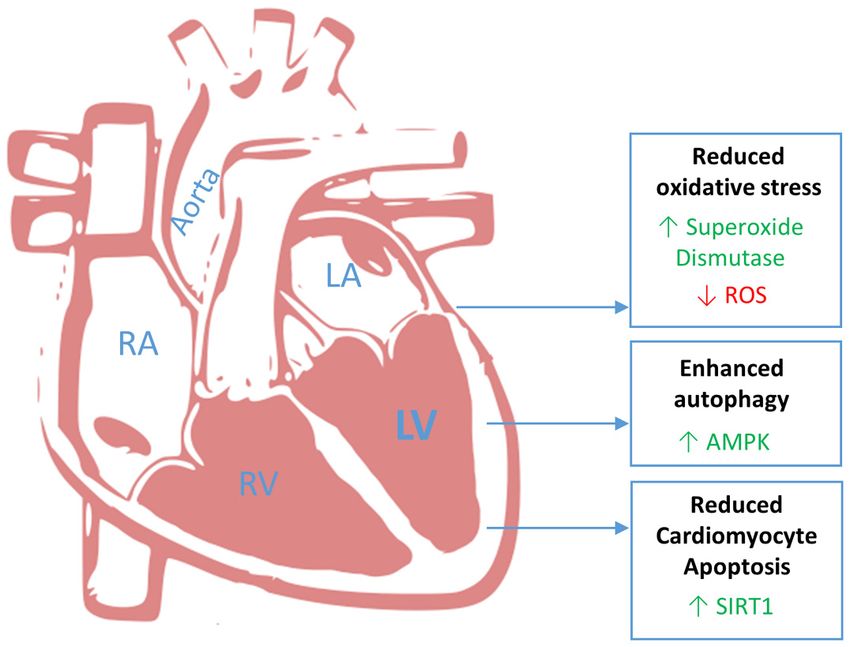
Int. J. Mol. Sci. 2019, 20, 1122                                                                      2 of 17

1. Introduction
     Marfan syndrome (MFS) is an autosomal dominant inherited disorder of the connective tissue,
characterized by mutations in the Fibrillin-1 gene (FBN1). Patients with MFS predominantly suffer
from skeletal, ocular and cardiovascular disease. The cardiovascular complications, in particular
aortic aneurysm formation, ultimately result in aortic dissection or rupture, leading to reduced life
expectancy [1]. For a rare disease, MFS is relatively common with an estimated prevalence of 1:5000 [2].
     Standard management of the cardiovascular disease in MFS is to surgically resect the enlarged
part of the aorta, which is then replaced by a synthetic graft (with or without an artificial aortic valve),
when the aneurysm has reached certain dimensions (4.5–5 cm aortic diameter). Although this strategy
has increased survival in MFS significantly, this surgery which is on average performed at a relatively
young age (20–50 years), is a heavy burden. Moreover, prophylactic surgery of the proximal aorta is
associated with progression of aneurysm formation and aortic dissection in the more distal aorta [3].
     It is intriguing that in this era of drug development, no pharmacological treatment strategy has
been identified that can inhibit aortic disease in MFS patients. Thus far, pharmacological treatment is
based on blood pressure lowering drugs, using mostly β-blockers and the angiotensin II type-1 receptor
(AGTR1) blocker, losartan. While these drugs slow down the aortic disease somewhat in MFS patients [4],
evidence for the efficacy of these drugs on aortic root dilatation in patients is limited, as well as the
evidence for these drugs to target the underlying cause of the progressive aortic degradation.
     MFS research has expanded over the last 15 years because of development of MFS mouse
models, however, this has not yet translated into novel treatment approaches to compensate for
the FBN1 gene defect that causes cardiovascular disease [5]. Part of the problem is that MFS is
complicated to study because already 1847 different FBN1 mutations have been reported, according
to the Universal Mutation Database (last update 2014), causing heterogeneity in MFS phenotype [6].
Moreover, even within MFS families with the same mutation there is a large variation in disease
pathogenesis [7], probably caused by genetic modifiers, such as common or rare genetic variants
(polymorphisms). So to reach effectiveness in all different MFS patients, it is essential to find common
ground as therapeutic approach. In that light, we found that the biologically-potent polyphenol
resveratrol (RES) promoted aortic repair in one of the MFS mouse models (Fbn1C1039G/+ mice) [8].
While cardiovascular disease in MFS may be caused by different FBN1 defects and modifying factors,
promoting cardiovascular repair would benefit all types of MFS patients.
     RES is a dietary supplement found in certain nuts and plants, best known in the skins of grapes.
RES is usually produced by plants in response to stressors, such as pathogens. A wide range of
beneficial effects has been shown in rodent models of disease, such as reducing oxidative stress,
improved calcium handling and inhibition of pathological hypertrophic signaling [9]. We have shown
in the Fbn1C1039G/+ MFS mice that RES is also effective at inhibiting the aortic root dilatation rate by
affecting a mechanism different from AGTR1 or transforming growth factor beta (TGF-β) signaling,
which is prominent in MFS [8].
     The present overview will discuss recent RES studies in animal models of aortic aneurysm formation
and heart failure, to investigate the different effects reported for RES on cardiovascular pathology relevant
for MFS patients, to explore if a clinical study with RES in MFS patients is supportable.

2. Aorta
      Among all clinical complications in MFS patients, aortic complications are the leading cause of
morbidity and mortality. The aorta, which is the largest artery in the human body, supplies all vital
organs with oxygenated blood containing nutrients. At the site of the aortic root, the heart pumps blood
into the aorta under high pressure. Ordinarily, the aortic wall can withstand these pressures due to the
well-organized structural extracellular matrix (ECM) protein network it contains. However, in patients
with MFS, genetic defects in these structural proteins, mostly FBN1 mutations, cause fundamental
changes in this network of ECM proteins, rendering the patients vulnerable for aortic disease.
Int. J. Mol. Sci. 2019, 20, x 3 of 17
Int. J. Mol. Sci. 2019, 20, 1122 3 of 17
cause fundamental changes in this network of ECM proteins, rendering the patients vulnerable for
aortic disease.
Various studies have been conducted to analyze the effect of RES on different cardiovascular
diseases [9,10]. We
We will here discuss primarily the studies related to the effect of RES on aortopathy
in relation to what is found in MFS patients. Interestingly,
Interestingly, RES was beneficial against aortopathy in
four different aortic aneurysm models; namely in the local periaortic application of calcium chloride
(CaCl22)-model, inin the
the local
local inter-aortic elastase infusion model, in the systemic chronic infusion of
angiotensin-II (AngII)model
angiotensin-II (AngII) modelandandininthe
thegenetic
genetic FBN1-mutation
FBN1-mutation (Fbn1C1039G/+)
(Fbn1C1039G/+) model
model of MFS
of MFS [8,11–
[8,11–13].
13]. effect
The The effect of on
of RES RES on aneurysm
aneurysm development
development is examined
is examined in theseinstudies,
these studies,
and theand the
effect oneffect
aorticon aortic
features
features relevant for MFS will be validated by literature
relevant for MFS will be validated by literature and summarized. and summarized.
2.1. Aortic
Aortic Aneurysm
Aneurysm
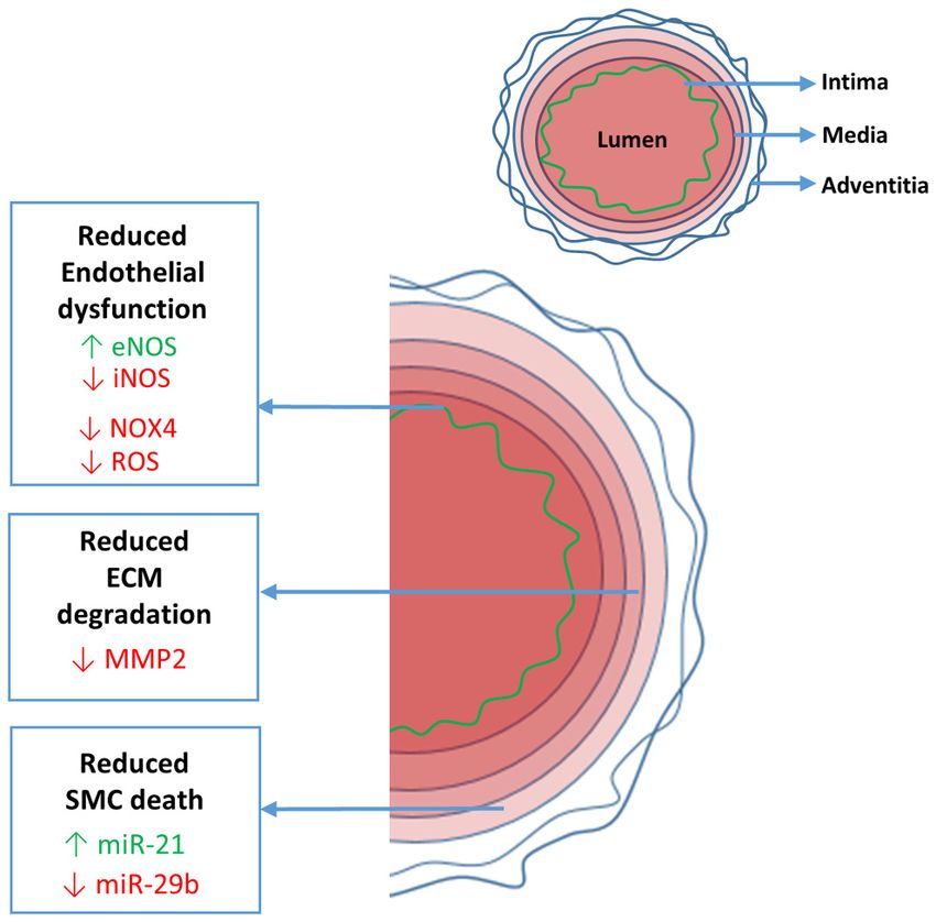
The aortic wall consists of three well-defined layers; the internal layer (intima), the middle layer
(media) and
and the
the outer
outerlayer
layer(adventitia)
(adventitia)(Figure
(Figure1).1).TheThe intima
intima mainly
mainly consists
consists of endothelial
of an an endothelial
cell
cell layer
layer (and(and a small
a small numbernumber of smooth
of smooth muscle
muscle cell (SMC)
cell (SMC) layerslayers
in thein adult
the adult
human human aorta).
aorta). The
The endothelial
endothelial cells form
cells form the barrier
the tight tight barrier
between between
the blood the and
blood
theand thewall,
vessel vessel
andwall, and the
provide provide
cues
the SMCs,
for cues fordependent
SMCs, dependent
on shearon shearmechanical
stress, stress, mechanical
stress andstress and circulating
circulating moleculesmolecules thatsense.
that they they
sense. Between the intima and the media there is the internal elastic lamina that separates,
Between the intima and the media there is the internal elastic lamina that separates, and at the same and at the
sameconnects,
time time connects, thelayers.
the two two layers.
Figure 1.1.Resveratrol
Resveratrolimproves
improvespathological aortic
pathological features
aortic in Marfan
features syndrome
in Marfan by enhancing
syndrome beneficial
by enhancing
(green) eNOS
beneficial andeNOS
(green) miR-21 and
and decreasing
miR-21 detrimental
and decreasing (red) iNOS,
detrimental NOX4,
(red) ROS,
iNOS, MMP2
NOX4, and
ROS, miR-29b.
MMP2 and
eNOS or iNOS: endothelial or inducible nitric oxide synthase; miR-21 and miR-29b: microRNA-21
miR-29b. eNOS or iNOS: endothelial or inducible nitric oxide synthase; miR-21 and miR-29b: and
-29b; NOX4: NADPH
microRNA-21 oxidase
and -29b; NOX4:4; ROS: reactive
NADPH oxygen
oxidase species;
4; ROS: MMP2:oxygen
reactive matrix species;
metalloproteinase 2.
MMP2: matrix
metalloproteinase 2.
The media occupies around 80% of the entire vessel wall surface and consists of concentric fenestrated
lamellae
Theofmedia
elasticoccupies
fibers, together
around with
80%SMCs embedded
of the in a fine
entire vessel network
wall of ECM,
surface such as collagen
and consists fibrils.
of concentric
The media is mainly responsible for the contractility and distensibility (elasticity) of
fenestrated lamellae of elastic fibers, together with SMCs embedded in a fine network of ECM, such the aorta.
The adventitia
as collagen is the
fibrils. The external
media stent of
is mainly the aorta, offering
responsible a strong tensile
for the contractility andsupport with thick
distensibility layers
(elasticity)
of the
of collagen
aorta.fibers maintained by fibroblasts, and it contains the vasa vasorum. From the adventitia,
the vasa
The vasorum
adventitiaprotrudes into the
is the external stentouter layers
of the aorta,ofoffering
the media to sufficiently
a strong provide
tensile support thethick
with necessary
layers
oxygen
of and fibers
collagen nutrients to the aortic
maintained SMCs. Via the
by fibroblasts, andexternal elastic
it contains the lamina the adventitia
vasa vasorum. From is connected
the adventitia,to
the media. While fibrilline-1 is mostly known for its presence in elastic fibers in the
the vasa vasorum protrudes into the outer layers of the media to sufficiently provide the necessary media, it is also
abundantly
oxygen and present
nutrients andto associated with collagen
the aortic SMCs. Via thefibers in the
external adventitia
elastic lamina[14].
the adventitia is connectedInt. J. Mol. Sci. 2019, 20, 1122 4 of 17
The normal aortic diameter decreases when moving away from the aortic valve. In the ascending
aorta the normal diameter isInt. J. Mol. Sci. 2019, 20, 1122 5 of 17
developed for 2 weeks, whereafter the mice were divided into treatment groups. One group received
RES supplemented high fat diet (0.05 g RES/100 g diet, thus approximately 1 mg RES/mouse/day) and
the other group high fat diet alone. RES treatment reduced AAA progression with induced SIRT1 activity,
as observed in the MFS mice as well. Proinflammatory pathways were reduced upon RES, as well as
MMP2 and -9. The authors were interested in the protective role of angiotensin-converting enzyme 2
(ACE2), which was increased in the mice and in cultured SMC by RES in a SIRT1-dependent manner.
Taken together, RES treatment showed multiple different mechanisms by which the aorta integrity
and aortic dilatation was preserved. Below a more detailed discussion will be provided on the potential
mechanisms of RES related to endothelial and SMC function in MFS.
2.2. Endothelial Dysfunction
RES reduced inflammation, angiogenesis and miR-29b in the above mentioned aneurysm models,
which all related to diminishing endothelial cell activation. Interestingly, it has been shown that
MFS patients have disturbed endothelial function, as measured by flow mediated dilatation, which
strongly correlated with enhanced aortic diameter [28], posing the question if endothelial dysfunction
is causally related to aneurysm formation.
2.2.1. eNOS/iNOS Balance
The vascular endothelium determines the permeability of the aorta for macromolecules and
leukocytes, prevents coagulation, and regulates vascular tone. The endothelium regulates vascular
tone by communicating with SMCs via endothelial cell-derived nitric oxide (NO), which reduces
SMC contractility [29]. Endothelial dysfunction is described as an impaired endothelium-dependent
vasorelaxation caused by the loss or overproduction of NO bioavailability [30]. RES has been shown to
regulate intracellular calcium in endothelial and SMCs differently. In endothelial cells it triggers NO
synthesis [31]. NO is a potent vasodilator synthesized by endothelial NO synthase (eNOS), which is
impaired in a MFS mouse model [29]. However, there are three isoforms of NOS; the endothelial, the
neuronal and the inducible forms [30]. In contrast to eNOS production in endothelial cells, inducible
NOS (iNOS) is increased in MFS in the SMC [32]. Excessive iNOS driven NO production causes
oxidative stress and cellular damage via accumulation of peroxynitrites (ONOO-) [33]. The aortic
iNOS and NO levels in the Fbn1C1039G/+ mouse model of MFS could be neutralized, which protected
mice from aortic dilatation and medial degeneration [34]. The relevance of iNOS as driver of aneurysm
formation also becomes clear in a novel MFS-like model with metalloproteinase ADAMTS1-deficiency,
being essential to restrain iNOS activity [34]. Interestingly, in the MFS aorta ADAMTS1 was almost
absent, which may explain the iNOS phenotype in the MFS mice. The effect of RES on ADAMTS
proteins has not been investigated yet in the context of vascular disease.
The transcription factor KLF2 is essential in endothelial cell health by promoting a quiescent
flow sensitive phenotype, in part by increasing eNOS [35]. Moreover, induction of KLF2 can reduce
TGF-β–mediated activation of endothelium [36]. Since TGF-β is enhanced in MFS [37], this may be the
cause of endothelial dysfunction in MFS. We and others demonstrated that RES enhances KLF2 [8]
via SIRT1 activation [38], and multiple studies showed that RES increased eNOS [39–41], thereby
promoting healthy endothelial cell function. Interestingly, miR-21 overexpression in endothelial cells
also induced eNOS [42], so the induction of miR-21 upon RES in the MFS mice [8] probably played a
role in endothelial de-activation as well.
2.2.2. NOX4 and ROS
Apart from the reactive nitrogen species, there is also enhanced oxidative stress in MFS via other
mechanisms [32,43,44]. Oxidative stress in the vessel wall may also be caused by the nicotinamide
adenine dinucleotide phosphate (NADPH) oxidase, which is a membrane-bound enzyme complex
consisting of different NOX subunits. This enzyme complex can become activated in all vascular cell
types, endothelial cells, SMCs, and fibroblasts, upon inflammatory conditions [45]. Careful regulationInt. J. Mol. Sci. 2019, 20, 1122 6 of 17
of NADPH oxidase activity is crucial to maintain a healthy level of reactive oxygen species (ROS) in the
vasculature. Since inflammatory cells have enhanced NADPH oxidase activity, causing excessive ROS,
the protective effect of RES in the CaCl2 -induced AAA model is presumably in part caused by inhibition
of NADPH oxidase-mediated ROS from inflammatory cells and inflammation-induced activation of
vascular cells. However, there is also accumulation of oxidative stress observed in MFS mice, which
have limited inflammation. The excessive oxidative stress caused vasomotor dysfunction in the thoracic
aorta [43]. RES is known to reduce oxidative stress, which is mostly ascribed to activation of SIRT1,
causing inhibition of NADPH oxidase activation and thereby protecting endothelial function [46].
Inhibition of SIRT1 significantly increased vascular superoxide production, enhanced NADPH oxidase
activity, and mRNA expression of its subunits p22(phox) and NOX4, which was prevented by RES [46].
Interestingly, NOX4 is highly increased in the human and murine MFS aorta. Moreover, in MFS mice
deficient for NOX4, aortic aneurysm formation was attenuated and elastin degradation reduced [47].
Apart from beneficial SMC effects, endothelial dysfunction in the MFS ascending aorta was prevented
by NOX4-deficiency or ROS inhibition [47].
While SIRT1 activation seems key in these pathways, we experimented with SIRT1 activation and
SIRT1 inhibition in the Fbn1C1039G/+ MFS model and did not observe a beneficial or detrimental
effect on the aorta; aortic dilatation was still observed [8], suggesting that perhaps multiple signaling
pathways induced by RES act in concert to exert its beneficial function. In that light, we also described
an endothelial-dependent effect of RES on the downregulation of detrimental miR-29b in SMC, thereby
increasing SMC survival [8].
2.3. Medial Degeneration
The strict architecture of the medial aorta normally consists of well-organized elastic fibers,
surrounded by circumferentially oriented SMCs and collagen fibers embedded in a complex ECM [48].
The main functions of the aortic media are contractility and distensibility. Mutations in SMC contractile
proteins and ECM(-related) proteins are known to cause aortic aneurysm disease, showing the
importance of both SMC contractility and ECM structure for aortic health [49].
Medial degeneration or cystic medial necrosis is a process observed in the aging aorta [50], patient
with hypertension [50], bicuspid aortic valves [51] and Fallot’s tetralogy [52], but also specifically
encountered in patients with aortic aneurysms, such as in MFS patients [53]. Medial degeneration in
MFS is characterized by accumulation of proteoglycans and glucosaminoglycans within SMC-depleted
areas of the aortic media, fragmentation or depletion of elastic fibers and SMC death [54]. The effect of
RES on these features will be extracted below.
2.3.1. ECM Degeneration
In all the aneurysm studies using RES, the ECM was protected or restored. Interestingly, miR-29b
downregulation in different aneurysm models also resulted in improved ECM structure and prevented
aneurysm growth [22–24]. We showed that RES decreased miR-29b in the MFS aorta in mice and
promoted SMC survival [8]. However, others showed that miR-29b directly regulated ECM remodeling
because many ECM genes are targets of miR-29b [22]. In addition, in the MFS mice, RES decreased
elastin breaks while losartan treatment did not [8], which is in line with a RES-mediated miR-29b
function in ECM protection. Thus increasing ECM production and remodeling by RES may overcome
excessive ECM damage and thereby limit aneurysm growth.
Apart from ECM production, preventing ECM destruction by proteases may be a feasible strategy
to reduce aortic disease. In AAA, the aortic ECM is destroyed by the abundance of aortic inflammation,
where inflammatory cells secrete proteolytic enzymes to invade the vessel wall and to clear the
surroundings from ECM debris. A well-studied class of proteases is the MMPs, which are abundant
in human aneurysm tissue. MMPs form a family of enzymes that degrade ECM components and
are released from mesenchymal cells and leukocytes [55]. High levels of MMPs result in a decreased
stability of the ECM, and eventually in reduction of elasticity and rupture of the vessel wall. In allInt. J. Mol. Sci. 2019, 20, 1122 7 of 17
three AAA models using RES, MMP9 was reduced, which is mostly related to reduced macrophage
influx, because these cells produce excessive MMP9 [11–13]. Since inflammation is very mild in MFS
aortic tissue and inhibition of inflammation did not reduce aortic dilatation in MFS mice [26,27],
most MMPs in MFS will be produced by the SMCs. Interestingly, in two of the AAA models using
RES [11,13], also MMP2 was reduced upon RES treatment. Indeed, in a severe murine model of MFS,
the Fbn1mgR/mgR mice, enhanced MMP2 production was observed in aortic SMCs, which was
responsible for the detrimental induction of ERK1/2 and aortic dilatation [56]. The same authors
generated MMP2 deficient MFS mice, which had prolonged lifespan due to decreased aortic rupture.
Thus MMP2 inhibition seems a favorable feature of RES in MFS.
In the aneurysm research field doxycycline, an antibiotic, is often used as golden standard to reduce
aneurysm formation in mice, which is ascribed to MMP inhibition. Yet, doxycycline actually inhibits
mitochondrial function of all mammalian cells [57], since mitochondria are of bacterial origin. Thus
apart from preventing MMP production, doxycycline changes cellular metabolism, which makes MMP
inhibition not a specific function of doxycycline. Since mitochondrial dysfunction is characteristic for
aneurysm tissue and causes oxidative stress by enhanced ROS [58], further suppression of mitochondrial
respiration by long-term doxycycline treatment in humans does not seem constructive. Indeed, a human
AAA trial using doxycycline was terminated prematurely due to enhance AAA growth in the doxycycline
treated patients [59]. The mitochondrial dysfunction in the aneurysm tissue could however be reversed
by promoting peroxisome proliferator-activated receptor gamma coactivator 1-alpha (PGC1a)-mediated
mitochondrial biogenesis [58], which is also a well-known target and function of RES [60].
2.3.2. SMC Death
SMCs are highly specialized cells that regulate blood pressure and blood flow through contraction
and regulation of ECM structure [61]. Aortic SMCs exhibit a differentiated phenotype characterized by
the expression of contractile markers specific to smooth muscle [62]. A unique and essential quality of
the SMC is its ability to move between a synthetic and contractile phenotype. This is not only critical
for normal development but also for response to injury through proliferation, migration and ECM
synthesis. The relevance of proper SMC function is also exemplified by mutations in a number of SMC
contractile genes and genes in the TGF-β pathway, causing aneurysm formation through impaired
SMC response [49]. Moreover, the study where calcium channel blockers display increased aneurysm
development in MFS patients and mice is also related to SMC contractility [20]. As has been discussed
already, there is enhanced oxidative stress in the MFS aorta. Adrenergic contraction experiments with
MFS aortas revealed that impaired vasomotor function was caused by oxidative stress due to enhanced
iNOS and NADPH-oxidases and reduced superoxide-eliminating enzymes superoxide dismutase-1
and -2 [43]. Interestingly, SMC dysfunction precedes the elastin degradation [63], and increased
SMC loss leads to aortic wall weakness in MFS [51]. This suggests that improving SMC health may
increase aortic repair. In fact, in all four aneurysm manuscripts with RES treatment, the aortic elastin
integrity was preserved, indicative of better SMC function, since these cells maintain the ECM in the
aortic media.
There have been various observations concerning MFS SMC. They have been described as less
differentiated leading to an immature vessel wall [64] or a premature switch from a synthetic to a
contractile phenotype [65]. SMCs cultured from MFS aortas actually have an extremely differentiated
SMC phenotype because of a TGF-β-induced SMC contractile gene expression profile [66]. This reveals
that there are so many more SMC phenotypes apart from the two mostly described as contractile or
synthetic. The MFS SMCs are stiff cells and impaired in regenerative capacity [66]. Thus it seems that
the flexibility to switch phenotype is lost, so they are not responsive to signals to promote aortic repair.
In line with these data, we found that the SMCs in the dilated aorta in MFS mice were senescent [8].
Senescence is characterized as a state of premature termination of the proliferative response often
induced by oxidative stress. The presence of enhanced oxidative stress in MFS aortas has been
discussed earlier. RES is known to counteract senescence and indeed changed SMC phenotype in MFSInt. J. Mol. Sci. 2019, 20, 1122 8 of 17
mice by increasing miR-21, which is known to stimulate SMC proliferation [25,67]. One of the targets
of miR-21 is the phosphatase and tensin homolog (PTEN) protein expression. Increased miR-21 results
in decreased PTEN, leading to increased activation of AKT, a cell survival factor.
Obviously, next to enhanced proliferation, decreased SMC death is important to prevent SMC
depletion in the aorta. Interestingly, we previously observed that anti-inflammatory treatments in MFS
mice did not reduce aortic dilatation. Actually, prednisolone treatment induced glucosaminoglycan
accumulation in the aorta [27]. Since this is characteristic at sites of SMC depletion [54] it suggests
that prednisolone promoted SMC death. In the RES treated MFS mice we showed increased
pro-inflammatory nuclear factor kappa-light-chain-enhancer of activated B cells (NFkB) signaling,
which was probably responsible for the reduction in detrimental miR-29b, and therefore decreased SMC
apoptosis [8,24]. The specific anti-apoptotic target transcripts of miR-29b were Bcl-2 and Mcl-1, which
were increased in the RES treated MFS aorta, while SMC apoptosis was diminished. So where excessive
inflammation is harmful in the AAA models, degrading the aorta faster than its repair capacity, modest
inflammation may activate the SMC to enhance their repair function in MFS, overcoming the senescent
state by promoting SMC proliferation and preventing death. RES reduced inflammation in the AAA
models and decreased SMC death in MFS, showing again that RES influences multiple mechanisms.
In conclusion, RES improved endothelial dysfunction, ECM degradation and SMC death by
alleviating oxidative stress (eNOS/iNOS balance; NOX4 and ROS), MMP2 and the miR-29b/miR-21
balance, respectively (Figure 1).
3. Heart
Apart from aorta pathology, other established features in MFS patients concern the heart; namely
mitral valve prolapse (MVP), mild left ventricular (LV) dilatation (i.e., increased LV end diastolic
dimension) and/or mild impairment of both LV systolic and diastolic function. The latter two can even
occur in patients without significant valvular abnormalities or previous aortic surgery [68]. In addition,
pulmonary artery dilatation may occur, often presenting simultaneously with dilatation of the aortic
root [69]. The latter feature however, seldom results in symptoms in the patient. Due to a lack of
specificity and incomplete knowledge of threshold values for dilatation of the pulmonary artery,
this minor criterion was omitted from the revised criteria (Ghent 2) [70]. For this reason, we will only
discuss MVP and LV dysfunction.
3.1. Mitral Valve Prolapse
Mitral dysfunction is present in 80% of patients with MFS, identified with auscultatory or
echocardiographic evidence [71]. In contrast to idiopathic mitral valve regurgitation, the onset in MFS
patients occurs at extremely young age and increases steadily with age [71,72]. MVP increases from
43% at 30 years of age to 77% at 60 years of age [72]. MVP is characterized by the systolic displacement
or billowing of the mitral leaflets into the left atrium. MVP usually causes minor clinical symptoms in
patients, but is a known risk factor for heart failure, arrhythmia, endocarditis, and sudden death [73].
Moreover, it is the most common cause of isolated mitral regurgitation requiring surgical repair.
While MVP is an important clinical feature in MFS patients, it has not been studied consistently
in MFS rodent models. Moreover, there is no other model for MVP which included studies with RES.
This is clearly a research opportunity. The only relevant data on mitral valves development in MFS was
obtained from Fbn1-deficient MFS mice, where postnatal architectural changes in mitral valves were
observed. The leaflets showed excess cell proliferation, reduced apoptosis, and increased TGF-β pathway
activation [74]. The effect of RES on mitral valve architecture and function has not been studied yet.
3.2. Left Ventricular Impairment
Impairment of the left ventricular function in patients with MFS may occur as a consequence
of significant valvular disease, but can also appear due to other severe vascular manifestations of
MFS, like aortic dilatation. The latter can lead to increased hemodynamic stress and, combinedInt. J. Mol. Sci. 2019, 20, 1122 9 of 17
with altered cardiac mechanobiology, result in cardiac dilatation [75]. However, in a small subset
of MFS patients, impaired LV function, as expressed by increased LV diameters, has also been
observed without a clear cause [76], suggesting an intrinsic cardiac defect due to the FBN1 mutation.
Indeed, it was demonstrated that a fibrillin-1-deficient ECM compromises the physical properties of
myocardial tissue, resulting in abnormal mechanosignaling by muscle cells and subsequently leading
to spontaneous dilated cardiomyopathy in MFS mice [77]. ECM defects thus make the heart vulnerable
to cardiac dysfunction.
Fibrillin-1 is present as long fibers in the apex, mid-ventricles and atria. Collagen had a similar
arrangement to that of fibrillin-1, whereas elastic fibers were primarily present in the atria and the
blood vessels [78]. It is known that microfibrillins in the ECM are expressed in the left ventricle of the
heart [78]. The Fbn1C1039G/+ mice demonstrated mild LV contractile dysfunction. Both structural
ECM and molecular signaling alterations are implicated in MFS-related cardiomyopathy [79].
Since most of the rodent heart models where the effect of RES has been examined, are based
on myocardial infarction or cardiac hypertrophy, the model representing dilated cardiomyopathy,
occasionally observed in MFS, is the chemical toxicity model of doxorubicin (DOX)-induced thinning of
the left ventricle. DOX is used as an antitumor drug, but its clinical application is limited because of its
cardiotoxicity. The characteristics of this model are thinning and dilatation of the ventricular wall, causing
reduced cardiac function presented with a reduced ejection fraction [80,81]. Therefore, we will use these
DOX studies to extract the potential effects of RES on dilated cardiomyopathy known in MFS patients.
Several mechanisms have been proposed to explain the cardiotoxic side effects of DOX, including
cardiomyocyte apoptosis, myofibrillar damage, impaired calcium handling, impaired mitochondrial
activity, and increased generation of ROS [82]. As described previously, cell death, ECM damage,
and increased oxidative stress were also observed in the aorta of MFS patients and mice, therefor it is
likely that these pathological processes play an important role in the pathology of the heart as well.
In multiple rodent DOX-models, the effect of RES on cardiac function was determined, where RES
protected the cardiomyocytes against structural damage and apoptosis [83–86]. This effect was mainly
associated with the upregulation of SIRT1. In two studies, this upregulation led to reduced expression
of tumor suppressor protein p53, which subsequently de-activated the apoptotic pathway [83,85].
Another model demonstrated that the SIRT1 upregulation due to RES, reduced mitogen-activated
protein kinase p38 (p38MAPK) activation, which is the primary pathological signaling pathway in
oxidative stress-induced heart injury, and in this way protected cardiomyocyte function [86]. Apart
from alleviating DOX-induced cardiotoxicity through inhibiting p38MAPK/p53-mediated apoptosis,
RES also improved autophagy by increasing AMP-activated protein kinase (AMPK) activation [87].
AMPK is an enzyme that plays a role in cellular metabolism when cellular energy is low. Autophagy is
the process of intracellular vesicles engulfing protein aggregates or damaged mitochondria to fuse with
lysosomes to degrade the contents and reuse the building blocks. This AMPK-mediated self-cleaning
promotes cellular health [88]. This pathway may actually also apply to the aorta, since in endothelial
cells, RES improved cellular health by promoting autophagy in an AMPK-dependent manner as
well [89–91]. The effect of RES on AMPK-dependent autophagy in SMC remains to be determined.
DOX induces mitochondrial dysfunction and thereby increases oxidative stress in cardiomyocytes
by enhanced ROS production, which also occurs in MFS, and is thought to cause cellular and ECM
damage. RES was shown to decrease ROS production in the DOX-induced heart injury model [86].
The proposed mechanism for the decrease in ROS accumulation is the antioxidant properties of RES,
by increasing the activity of the antioxidant enzyme superoxide dismutase, which converts ROS.
The capacity of RES to reduce oxidative stress has similarly been observed in AAA [11].
In general, the data on the effect of RES that could be of interest for the heart features observed in
MFS patients is limited. However, the main protective mechanisms of RES in the context of cardiac
function concern SIRT1-mediated downregulation of p38MAPK activity to reduce cardiomyocyte
apoptosis, AMPK-mediated enhanced autophagy and increased antioxidant superoxide dismutase to
decrease oxidative stress/ROS (Figure 2).In general, the data on the effect of RES that could be of interest for the heart features observed
in MFS patients is limited. However, the main protective mechanisms of RES in the context of cardiac
function concern SIRT1-mediated downregulation of p38MAPK activity to reduce cardiomyocyte
apoptosis,
Int. J. Mol. Sci.AMPK-mediated
2019, 20, 1122 enhanced autophagy and increased antioxidant superoxide dismutase 10 of 17
to decrease oxidative stress/ROS (Figure 2).
Figure 2. Resveratrol
Resveratrol improves
improves pathological
pathological cardiac
cardiac features
features in
in Marfan
Marfan syndrome by increasing
beneficial (green) superoxide
superoxide dismutase,
dismutase, AMPK
AMPK andand SIRT1
SIRT1 and
and decreasing
decreasing detrimental
detrimental (red)
(red) ROS.
ROS.
ROS: reactive
reactiveoxygen
oxygenspecies; AMPK:
species; AMPK: AMP-activated
AMP-activated protein kinase;
protein SIRT1:SIRT1:
kinase; sirtuin-1; RA: right
sirtuin-1; RA:atrium;
right
RV rightRV
atrium; ventricle; LA: left atrium;
right ventricle; LA: left LV: left ventricle.
atrium; LV: left ventricle.
4. Exercise
4. Exercise Mimetic
Mimetic
Exercise is
Exercise is widely
widely accepted
accepted to to prevent
prevent or or mitigate
mitigate metabolic
metabolic disease
disease such such as as obesity,
obesity, type
type 22
diabetes, and
diabetes, and cardiovascular
cardiovascular disease.
disease. For patients with
For patients with MFSMFS itit is
is advised
advised to to refrain
refrain from
from doing
doing intense
intense
sports to prevent a rise in blood pressure and thereby aortic aneurysm
sports to prevent a rise in blood pressure and thereby aortic aneurysm expansion and rupture, expansion and rupture, although
hard evidence
although hard is lacking is
evidence to lacking
supporttothis advice.this
support However, exercise mimetics
advice. However, exercise have been have
mimetics identified,
been
and may thus
identified, and bemay of thus
valuebeasofpharmacological agents. Interestingly,
value as pharmacological RES is considered
agents. Interestingly, an exercise
RES is considered an
mimetic mimetic
exercise [92,93], which puts
[92,93], which exercise
puts in the context
exercise in theofcontext
MFS inof another
MFS inlight. Recently,
another light. two studies
Recently, in
two
MFS mice using an exercise protocol have shown to prevent aortic dilatation
studies in MFS mice using an exercise protocol have shown to prevent aortic dilatation [94,95]. [94,95].
In the
In the MFS
MFS Fbn1C1039G/+
Fbn1C1039G/+mice miceat atfour
fourmonths
monthsof ofage,
age,there
thereare are already
already differences
differences between
between the the
MFS mice and the wild type littermates in aorta dilatation, elastic lamina
MFS mice and the wild type littermates in aorta dilatation, elastic lamina ruptures, aortic stiffness, as ruptures, aortic stiffness,
as well
well as as cardiac
cardiac hypertrophy,
hypertrophy, leftleft ventricular
ventricular fibrosis,
fibrosis, andand intramyocardial
intramyocardial vessel
vessel remodeling.
remodeling. A
A moderate exercise protocol reduced aorta dilatation and cardiomyopathy
moderate exercise protocol reduced aorta dilatation and cardiomyopathy in the MFS mice, while in the MFS mice, while the
the
other pathological
other pathological characteristics
characteristics remained
remained unaltered
unaltered [94]. The exercise
[94]. The exercise training
training consisted
consisted of of running
running
on aa treadmill
on treadmill for for 11hhaaday,
day,fivefivedays
daysa aweek
week forfor
five months.
five months. However,
However, thethe
study could
study not not
could reveal the
reveal
underlying mechanisms for the beneficial effects of exercise on the
the underlying mechanisms for the beneficial effects of exercise on the MFS aorta and heart. MFS aorta and heart.
In the
In the same
same MFSMFS mouse
mouse model,
model, yetyet another
another research
research group
group performed
performed exercise
exercise experiments
experiments withwith
two different protocols; voluntary cage-wheel exercise or forced treadmill
two different protocols; voluntary cage-wheel exercise or forced treadmill exercise, and a sedentary exercise, and a sedentary
lifestyle as
lifestyle as reference
reference forfor aorta
aorta pathology,
pathology, for for five
five months
months starting
starting at at four
four weeks
weeks of of age
age [95]. They
[95]. They
demonstrated that
demonstrated that exercise
exercise could
could prevent
prevent aorta
aorta dilatation,
dilatation, preserve
preserve the the elastic
elastic fibers
fibers and
and improve
improve the the
tensile strength of the aortic wall as compared to the sedentary MFS mice.
tensile strength of the aortic wall as compared to the sedentary MFS mice. With the voluntary exercise With the voluntary exercise
regimen, optimal
regimen, optimal aortic
aortic improvements
improvements were were measured
measured due due toto reduced
reduced MMP2 MMP2 and and MMP9
MMP9 activity.
activity.
If the main beneficial effect of RES is related to the exercise effect, then other exercise mimetics
If the main beneficial effect of RES is related to the exercise effect, then other exercise mimetics
may also
may alsobe be beneficial in MFS.
beneficial in TheMFS. anti-diabetic drug metformin,
The anti-diabetic and AMP analog
drug metformin, and 5-aminoimidazole-
AMP analog 5-
4-carboxamide ribonucleotide (AICAR),
aminoimidazole-4-carboxamide and nuclear
ribonucleotide receptor
(AICAR), andperoxisome proliferator-activated
nuclear receptor peroxisome
receptor delta (PPARδ) agonists have also been described as exercise mimetics [92,93], which may thus
affect cardiovascular pathology in MFS similarly. The exercise signature, using these exercise mimetics,
consists of enhanced mitochondrial biogenesis in which SIRT1, AMPK, and PGC1α play an important
role [92,93], and were all mentioned earlier as protectors in MFS studies.Int. J. Mol. Sci. 2019, 20, 1122 11 of 17
Interestingly, prescription for metformin is indeed associated with a decrease in AAA enlargement [96],
already suggesting a beneficial role for metformin in aneurysm progression. This effect deserves more
extensive follow up in prospective clinical trials in AAA patients and perhaps also in MFS.
Overall, the mechanisms during mild/moderate exercise or RES treatment that protect MFS mice
from cardiovascular pathology may be the same and thus setting up an exercise training protocol for
MFS patients could be considered. In the meantime, a clinical study using RES in MFS patients seems
justified and feasible.
5. Conclusion
The cardiovascular pathology in MFS patients is complicated. Aortic pathology mainly consists
of uncontrolled aortic growth, endothelial dysfunction, and medial degeneration. Besides aorta
pathology, MFS patients may present with other cardiovascular manifestations, such as left ventricular
dysfunction. Given the multifactorial nature of MFS, traditional therapies like β-blockers and losartan
likely do not adequately target all pathways involved in cardiovascular dysfunction in MFS. In the
literature we found evidence of beneficial effects of RES on multiple levels to combat aneurysm
formation and left ventricular dysfunction in multiple rodent-based animal models, suggesting that
RES may have therapeutic potential in MFS patients by reducing oxidative stress and promoting cell
survival. Although, clinical trials with RES have been performed, these were not in MFS patients. While
in rodents RES is very potent to combat a plethora of diseases, the translation to human cardiovascular
disease seems less efficient [97]. Yet, the rapid implementation of RES in a well-designed trial, over a
longer period of time than previously performed with RES, seems attractive to determine whether
treatment with RES is beneficial for MFS patients.
With the variable results from different ARB trials in mind over the last decade, careful consideration
on the setup of a new clinical trial in MFS patients is required. A meaningful strategy is largely
determined by the number of patients available, the chosen primary outcome, the duration of the study
and the dosage of RES. The collective data in this manuscript shows that a feasibility study could be
considered to monitor specific RES effects in MFS patients as prerequisite for a novel clinical RES trial.
Funding: This research was funded by AMC Foundation and Horstingstuit Foundation.
Conflicts of Interest: The authors declare no conflict of interest.
Abbreviations
AAA Abdominal aortic aneurysm
ACE2 Angiotensin-converting enzyme 2
AGTR1 Angiotensin type-1 receptor (Agtr1 [mouse])
AMPK AMP-activated protein kinase
ARB Angiotensin II receptor blocker
ECM Extracellular matrix
eNOS Endothelial nitric oxide synthase
FBN1 Fibrillin-1 (Fbn1 [mouse])
iNOS Inducible nitric oxide synthase
MFS Marfan syndrome
MMP Matrix metalloproteinase
NFκB Nuclear factor kappa light chain enhancer of activated B cells
PGC1a Peroxisome proliferator-activated receptor gamma coactivator 1-alpha
RES Resveratrol
ROS Reactive oxygen species
SIRT1 NAD-dependent deacetylase sirtuin-1
SMC Smooth muscle cell
TGF-β Transforming growth factor betaInt. J. Mol. Sci. 2019, 20, 1122 12 of 17
References
1. Silverman, D.I.; Burton, K.J.; Gray, J.; Bosner, M.S.; Kouchoukos, N.T.; Roman, M.J.; Boxer, M.; Devereux, R.B.;
Tsipouras, P. Life expectancy in the Marfan syndrome. Am. J. Cardiol. 1995, 75, 157–160. [CrossRef]
2. Judge, D.P.; Dietz, H.C. Marfan’s syndrome. Lancet 2005, 366, 1965–1976. [CrossRef]
3. den Hartog, A.W.; Franken, R.; Zwinderman, A.H.; Timmermans, J.; Scholte, A.J.; van den Berg, M.P.;
de Waard, V.; Pals, G.; Mulder, B.J.; Groenink, M. The risk for type B aortic dissection in Marfan syndrome.
J. Am. Coll. Cardiol. 2015, 65, 246–254. [CrossRef] [PubMed]
4. Teixido-Tura, G.; Forteza, A.; Rodriguez-Palomares, J.; Gonzalez Mirelis, J.; Gutierrez, L.; Sanchez, V.;
Ibanez, B.; Garcia-Dorado, D.; Evangelista, A. Losartan Versus Atenolol for Prevention of Aortic Dilation in
Patients With Marfan Syndrome. J. Am. Coll. Cardiol. 2018, 72, 1613–1618. [CrossRef] [PubMed]
5. Milewicz, D.M.; Ramirez, F. Therapies for Thoracic Aortic Aneurysms and Acute Aortic Dissections.
Arterioscler. Thromb. Vasc. Biol. 2019, 39, 126–136. [CrossRef] [PubMed]
6. Collod-Beroud, G.; Le Bourdelles, S.; Ades, L.; Ala-Kokko, L.; Booms, P.; Boxer, M.; Child, A.; Comeglio, P.;
De Paepe, A.; Hyland, J.C.; et al. Update of the UMD-FBN1 mutation database and creation of an FBN1
polymorphism database. Hum. Mutat. 2003, 22, 199–208. [CrossRef] [PubMed]
7. De Backer, J.; Loeys, B.; Leroy, B.; Coucke, P.; Dietz, H.; De Paepe, A. Utility of molecular analyses in the
exploration of extreme intrafamilial variability in the Marfan syndrome. Clin. Genet. 2007, 72, 188–198.
[CrossRef] [PubMed]
8. Hibender, S.; Franken, R.; van Roomen, C.; Ter Braake, A.; van der Made, I.; Schermer, E.E.; Gunst, Q.; van
den Hoff, M.J.; Lutgens, E.; Pinto, Y.M.; et al. Resveratrol Inhibits Aortic Root Dilatation in the Fbn1C1039G/+
Marfan Mouse Model. Arterioscler. Thromb. Vasc. Biol. 2016, 36, 1618–1626. [CrossRef] [PubMed]
9. Sung, M.M.; Dyck, J.R. Therapeutic potential of resveratrol in heart failure. Ann. N. Y. Acad. Sci. 2015, 1348,
32–45. [CrossRef] [PubMed]
10. Bonnefont-Rousselot, D. Resveratrol and Cardiovascular Diseases. Nutrients 2016, 8, 250. [CrossRef]
[PubMed]
11. Kaneko, H.; Anzai, T.; Morisawa, M.; Kohno, T.; Nagai, T.; Anzai, A.; Takahashi, T.; Shimoda, M.; Sasaki, A.;
Maekawa, Y.; et al. Resveratrol prevents the development of abdominal aortic aneurysm through attenuation
of inflammation, oxidative stress, and neovascularization. Atherosclerosis 2011, 217, 350–357. [CrossRef]
[PubMed]
12. Palmieri, D.; Pane, B.; Barisione, C.; Spinella, G.; Garibaldi, S.; Ghigliotti, G.; Brunelli, C.; Fulcheri, E.;
Palombo, D. Resveratrol counteracts systemic and local inflammation involved in early abdominal aortic
aneurysm development. J. Surg. Res. 2011, 171, e237–e246. [CrossRef] [PubMed]
13. Moran, C.S.; Biros, E.; Krishna, S.M.; Wang, Y.; Tikellis, C.; Morton, S.K.; Moxon, J.V.; Cooper, M.E.;
Norman, P.E.; Burrell, L.M.; et al. Resveratrol Inhibits Growth of Experimental Abdominal Aortic Aneurysm
Associated With Upregulation of Angiotensin-Converting Enzyme 2. Arterioscler. Thromb. Vasc. Biol. 2017,
37, 2195–2203. [CrossRef] [PubMed]
14. Lindeman, J.H.; Ashcroft, B.A.; Beenakker, J.W.; van Es, M.; Koekkoek, N.B.; Prins, F.A.; Tielemans, J.F.;
Abdul-Hussien, H.; Bank, R.A.; Oosterkamp, T.H. Distinct defects in collagen microarchitecture underlie
vessel-wall failure in advanced abdominal aneurysms and aneurysms in Marfan syndrome. Proc. Natl. Acad.
Sci. USA 2010, 107, 862–865. [PubMed]
15. Adams, J.N.; Trent, R.J. Aortic complications of Marfan’s syndrome. Lancet 1998, 352, 1722–1723. [CrossRef]
16. Hartog, A.W.; Franken, R.; Zwinderman, A.H.; Groenink, M.; Mulder, B.J. Current and future
pharmacological treatment strategies with regard to aortic disease in Marfan syndrome. Expert Opin
Pharmacother, Expert Opin. Pharmacother. 2012, 13, 647–662. [CrossRef] [PubMed]
17. Groenink, M.; den Hartog, A.W.; Franken, R.; Radonic, T.; de Waard, V.; Timmermans, J.; Scholte, A.J.; van
den Berg, M.P.; Spijkerboer, A.M.; Marquering, H.A.; et al. Losartan reduces aortic dilatation rate in adults
with Marfan syndrome: a randomized controlled trial. Eur. Heart J. 2013, 34, 3491–3500. [CrossRef] [PubMed]
18. Pereira, L.; Andrikopoulos, K.; Tian, J.; Lee, S.Y.; Keene, D.R.; Ono, R.; Reinhardt, D.P.; Sakai, L.Y.; Biery, N.J.;
Bunton, T.; et al. Targetting of the gene encoding fibrillin-1 recapitulates the vascular aspect of Marfan
syndrome. Nat. Genet. 1997, 17, 218–222. [CrossRef] [PubMed]Int. J. Mol. Sci. 2019, 20, 1122 13 of 17
19. Holm, T.M.; Habashi, J.P.; Doyle, J.J.; Bedja, D.; Chen, Y.; van Erp, C.; Lindsay, M.E.; Kim, D.; Schoenhoff, F.;
Cohn, R.D.; et al. Noncanonical TGFbeta signaling contributes to aortic aneurysm progression in Marfan
syndrome mice. Science 2011, 332, 358–361. [CrossRef] [PubMed]
20. Doyle, J.J.; Doyle, A.J.; Wilson, N.K.; Habashi, J.P.; Bedja, D.; Whitworth, R.E.; Lindsay, M.E.; Schoenhoff, F.;
Myers, L.; Huso, N.; et al. A deleterious gene-by-environment interaction imposed by calcium channel
blockers in Marfan syndrome. Elife 2015, 4, e08648. [CrossRef] [PubMed]
21. Wanga, S.; Hibender, S.; Ridwan, Y.; van Roomen, C.; Vos, M.; van der Made, I.; van Vliet, N.; Franken, R.;
van Riel, L.A.; Groenink, M.; et al. Aortic microcalcification is associated with elastin fragmentation in
Marfan syndrome. J. Pathol. 2017, 243, 294–306. [CrossRef] [PubMed]
22. Boon, R.A.; Seeger, T.; Heydt, S.; Fischer, A.; Hergenreider, E.; Horrevoets, A.J.; Vinciguerra, M.; Rosenthal, N.;
Sciacca, S.; Pilato, M.; et al. MicroRNA-29 in aortic dilation: implications for aneurysm formation. Circ. Res.
2011, 109, 1115–1119. [CrossRef] [PubMed]
23. Zampetaki, A.; Attia, R.; Mayr, U.; Gomes, R.S.; Phinikaridou, A.; Yin, X.; Langley, S.R.; Willeit, P.; Lu, R.;
Fanshawe, B.; et al. Role of miR-195 in aortic aneurysmal disease. Circ. Res. 2014, 115, 857–866. [CrossRef]
[PubMed]
24. Merk, D.R.; Chin, J.T.; Dake, B.A.; Maegdefessel, L.; Miller, M.O.; Kimura, N.; Tsao, P.S.; Iosef, C.; Berry, G.J.;
Mohr, F.W.; et al. miR-29b participates in early aneurysm development in Marfan syndrome. Circ. Res. 2012,
110, 312–324. [CrossRef] [PubMed]
25. Maegdefessel, L.; Azuma, J.; Toh, R.; Deng, A.; Merk, D.R.; Raiesdana, A.; Leeper, N.J.; Raaz, U.;
Schoelmerich, A.M.; McConnell, M.V.; et al. MicroRNA-21 blocks abdominal aortic aneurysm development
and nicotine-augmented expansion. Sci. Transl. Med. 2012, 4, 122ra22. [CrossRef] [PubMed]
26. Radonic, T.; de Witte, P.; Groenink, M.; de Waard, V.; Lutter, R.; van Eijk, M.; Jansen, M.; Timmermans, J.;
Kempers, M.; Scholte, A.J.; et al. Inflammation aggravates disease severity in Marfan syndrome patients.
PLoS ONE 2012, 7, e32963. [CrossRef] [PubMed]
27. Franken, R.; Hibender, S.; den Hartog, A.W.; Radonic, T.; de Vries, C.J.; Zwinderman, A.H.; Groenink, M.;
Mulder, B.J.; de Waard, V. No beneficial effect of general and specific anti-inflammatory therapies on aortic
dilatation in Marfan mice. PLoS ONE 2014, 9, e107221. [CrossRef] [PubMed]
28. Takata, M.; Amiya, E.; Watanabe, M.; Omori, K.; Imai, Y.; Fujita, D.; Nishimura, H.; Kato, M.; Morota, T.;
Nawata, K.; et al. Impairment of flow-mediated dilation correlates with aortic dilation in patients with
Marfan syndrome. Heart Vessels 2014, 29, 478–485. [CrossRef] [PubMed]
29. Chung, A.W.; Au Yeung, K.; Cortes, S.F.; Sandor, G.G.; Judge, D.P.; Dietz, H.C.; van Breemen, C. Endothelial
dysfunction and compromised eNOS/Akt signaling in the thoracic aorta during the progression of Marfan
syndrome. Br. J. Pharmacol. 2007, 150, 1075–1083. [CrossRef] [PubMed]
30. Moncada, S.; Palmer, R.M.; Higgs, E.A. Nitric oxide: physiology, pathophysiology, and pharmacology.
Pharmacol. Rev. 1991, 43, 109–142. [PubMed]
31. Buluc, M.; Demirel-Yilmaz, E. Resveratrol decreases calcium sensitivity of vascular smooth muscle and
enhances cytosolic calcium increase in endothelium. Vascul. Pharmacol. 2006, 44, 231–237. [CrossRef]
[PubMed]
32. Soto, M.E.; Soria-Castro, E.; Lans, V.G.; Ontiveros, E.M.; Mejia, B.I.; Hernandez, H.J.; Garcia, R.B.; Herrera, V.;
Perez-Torres, I. Analysis of oxidative stress enzymes and structural and functional proteins on human aortic
tissue from different aortopathies. Oxid. Med. Cell. Longev. 2014, 2014, 760694. [CrossRef] [PubMed]
33. Lomeli, O.; Perez-Torres, I.; Marquez, R.; Criales, S.; Mejia, A.M.; Chiney, C.; Hernandez-Lemus, E.;
Soto, M.E. The Evaluation of Flow-Mediated Vasodilation in the Brachial Artery Correlates With Endothelial
Dysfunction Evaluated by Nitric Oxide Synthase Metabolites in Marfan Syndrome Patients. Front. Physiol.
2018, 9, 965. [CrossRef] [PubMed]
34. Oller, J.; Mendez-Barbero, N.; Ruiz, E.J.; Villahoz, S.; Renard, M.; Canelas, L.I.; Briones, A.M.; Alberca, R.;
Lozano-Vidal, N.; Hurle, M.A.; et al. Nitric oxide mediates aortic disease in mice deficient in the
metalloprotease Adamts1 and in a mouse model of Marfan syndrome. Nat. Med. 2017, 23, 200–212.
[CrossRef] [PubMed]
35. Marin, T.; Gongol, B.; Chen, Z.; Woo, B.; Subramaniam, S.; Chien, S.; Shyy, J.Y. Mechanosensitive
microRNAs-role in endothelial responses to shear stress and redox state. Free Radic. Biol. Med. 2013,
64, 61–68. [CrossRef] [PubMed]Int. J. Mol. Sci. 2019, 20, 1122 14 of 17
36. Boon, R.A.; Fledderus, J.O.; Volger, O.L.; van Wanrooij, E.J.; Pardali, E.; Weesie, F.; Kuiper, J.; Pannekoek, H.;
ten Dijke, P.; Horrevoets, A.J. KLF2 suppresses TGF-beta signaling in endothelium through induction of
Smad7 and inhibition of AP-1. Arterioscler. Thromb. Vasc. Biol. 2007, 27, 532–539. [CrossRef] [PubMed]
37. Franken, R.; den Hartog, A.W.; de Waard, V.; Engele, L.; Radonic, T.; Lutter, R.; Timmermans, J.; Scholte, A.J.;
van den Berg, M.P.; Zwinderman, A.H.; et al. Circulating transforming growth factor-beta as a prognostic
biomarker in Marfan syndrome. Int. J. Cardiol. 2013, 168, 2441–2446. [CrossRef] [PubMed]
38. Gracia-Sancho, J.; Villarreal, G., Jr.; Zhang, Y.; Garcia-Cardena, G. Activation of SIRT1 by resveratrol induces
KLF2 expression conferring an endothelial vasoprotective phenotype. Cardiovasc. Res. 2010, 85, 514–519.
[CrossRef] [PubMed]
39. Wallerath, T.; Deckert, G.; Ternes, T.; Anderson, H.; Li, H.; Witte, K.; Forstermann, U. Resveratrol, a
polyphenolic phytoalexin present in red wine, enhances expression and activity of endothelial nitric oxide
synthase. Circulation 2002, 106, 1652–1658. [CrossRef] [PubMed]
40. Leikert, J.F.; Rathel, T.R.; Wohlfart, P.; Cheynier, V.; Vollmar, A.M.; Dirsch, V.M. Red wine polyphenols
enhance endothelial nitric oxide synthase expression and subsequent nitric oxide release from endothelial
cells. Circulation 2002, 106, 1614–1617. [CrossRef] [PubMed]
41. Dolinsky, V.W.; Chakrabarti, S.; Pereira, T.J.; Oka, T.; Levasseur, J.; Beker, D.; Zordoky, B.N.; Morton, J.S.;
Nagendran, J.; Lopaschuk, G.D.; et al. Resveratrol prevents hypertension and cardiac hypertrophy in
hypertensive rats and mice. Biochim. Biophys. Acta 2013, 1832, 1723–1733. [CrossRef] [PubMed]
42. Weber, M.; Baker, M.B.; Moore, J.P.; Searles, C.D. MiR-21 is induced in endothelial cells by shear stress and
modulates apoptosis and eNOS activity. Biochem. Biophys. Res. Commun. 2010, 393, 643–648. [CrossRef]
[PubMed]
43. Yang, H.H.; van Breemen, C.; Chung, A.W. Vasomotor dysfunction in the thoracic aorta of Marfan syndrome
is associated with accumulation of oxidative stress. Vascul. Pharmacol. 2010, 52, 37–45. [CrossRef] [PubMed]
44. Soto, M.E.; Iturriaga Hernandez, A.V.; Guarner Lans, V.; Zuniga-Munoz, A.; Aranda Fraustro, A.; Velazquez
Espejel, R.; Perez-Torres, I. Participation of oleic acid in the formation of the aortic aneurysm in Marfan
syndrome patients. Prostag. Other Lipid Mediat. 2016, 123, 46–55. [CrossRef] [PubMed]
45. Marinkovic, G.; Heemskerk, N.; van Buul, J.D.; de Waard, V. The Ins and Outs of Small GTPase Rac1 in the
Vasculature. J. Pharmacol. Exp. Ther. 2015, 354, 91–102. [CrossRef] [PubMed]
46. Zarzuelo, M.J.; Lopez-Sepulveda, R.; Sanchez, M.; Romero, M.; Gomez-Guzman, M.; Ungvary, Z.;
Perez-Vizcaino, F.; Jimenez, R.; Duarte, J. SIRT1 inhibits NADPH oxidase activation and protects endothelial
function in the rat aorta: implications for vascular aging. Biochem. Pharmacol. 2013, 85, 1288–1296. [CrossRef]
[PubMed]
47. Jimenez-Altayo, F.; Meirelles, T.; Crosas-Molist, E.; Sorolla, M.A.; Del Blanco, D.G.; Lopez-Luque, J.;
Mas-Stachurska, A.; Siegert, A.M.; Bonorino, F.; Barbera, L.; et al. Redox stress in Marfan syndrome:
Dissecting the role of the NADPH oxidase NOX4 in aortic aneurysm. Free Radic. Biol. Med. 2018, 118, 44–58.
[CrossRef] [PubMed]
48. Wagenseil, J.E.; Mecham, R.P. Elastin in large artery stiffness and hypertension. J. Cardiovasc. Transl. Res.
2012, 5, 264–273. [CrossRef] [PubMed]
49. Milewicz Dianna, M.; Trybus Kathleen, M.; Guo, D.-c.; Sweeney, H.L.; Regalado, E.; Kamm, K.; Stull James, T.
Altered Smooth Muscle Cell Force Generation as a Driver of Thoracic Aortic Aneurysms and Dissections.
Arterioscler. Thromb. Vasc. Biol. 2017, 37, 26–34. [CrossRef] [PubMed]
50. De Backer, J. Cardiovascular characteristics in Marfan syndrome and their relation to the genotype. Verh. K.
Acad. Geneeskd. Belg. 2009, 71, 335–371. [PubMed]
51. Nataatmadja, M.; West, M.; West, J.; Summers, K.; Walker, P.; Nagata, M.; Watanabe, T. Abnormal extracellular
matrix protein transport associated with increased apoptosis of vascular smooth muscle cells in marfan
syndrome and bicuspid aortic valve thoracic aortic aneurysm. Circulation 2003, 108 (Suppl. 1), Ii329–Ii334.
[CrossRef] [PubMed]
52. Tan, J.L.; Davlouros, P.A.; McCarthy, K.P.; Gatzoulis, M.A.; Ho, S.Y. Intrinsic histological abnormalities of
aortic root and ascending aorta in tetralogy of Fallot: evidence of causative mechanism for aortic dilatation
and aortopathy. Int. J. Cardiol. 2019, 278, 65–69. [CrossRef] [PubMed]
53. Yan, J.; Lehsau, A.C.; Sauer, B.; Pieper, B.; Mohamed, S.A. Comparison of biomechanical properties in ascending
aortic aneurysms of patients with congenital bicuspid aortic valve and Marfan syndrome. Int. J. Cardiol. 2019,
278, 65–69. [CrossRef] [PubMed]You can also read