Harnessing the Membrane Translocation Properties of AB Toxins for Therapeutic Applications - Infoscience
←
→
Page content transcription
If your browser does not render page correctly, please read the page content below
toxins
Review
Harnessing the Membrane Translocation Properties of AB
Toxins for Therapeutic Applications
Numa Piot, F. Gisou van der Goot and Oksana A. Sergeeva *

                                           Global Health Institute, School of Life Sciences, EPFL, 1015 Lausanne, Switzerland; numa.piot@epfl.ch (N.P.);
                                           gisou.vandergoot@epfl.ch (F.G.v.d.G.)
                                           * Correspondence: oksana.sergeeva@epfl.ch

                                           Abstract: Over the last few decades, proteins and peptides have become increasingly more com-
                                           mon as FDA-approved drugs, despite their inefficient delivery due to their inability to cross the
                                           plasma membrane. In this context, bacterial two-component systems, termed AB toxins, use various
                                           protein-based membrane translocation mechanisms to deliver toxins into cells, and these mechanisms
                                           could provide new insights into the development of bio-based drug delivery systems. These toxins
                                           have great potential as therapies both because of their intrinsic properties as well as the modular
                                           characteristics of both subunits, which make them highly amenable to conjugation with various drug
                                           classes. This review focuses on the therapeutical approaches involving the internalization mecha-
                                           nisms of three representative AB toxins: botulinum toxin type A, anthrax toxin, and cholera toxin. We
                                           showcase several specific examples of the use of these toxins to develop new therapeutic strategies
                                           for numerous diseases and explain what makes these toxins promising tools in the development of
                                           drugs and drug delivery systems.

                                           Keywords: botulinum toxin; anthrax toxin; cholera toxin; membrane translocation; endocytosis;
                                           therapeutic applications; drug delivery


                                           Key Contribution: This paper showcases the mechanisms and several therapeutic applications of
Citation: Piot, N.; van der Goot, F.G.;
                                           botulinum toxin, anthrax toxin, and cholera toxin.
Sergeeva, O.A. Harnessing the
Membrane Translocation Properties
of AB Toxins for Therapeutic
Applications. Toxins 2021, 13, 36.
                                           1. Introduction
https://doi.org/10.3390/
toxins13010036                                  The FDA is increasingly approving biological drugs. In 2018, these protein-based
                                           drugs made up 25% of FDA approvals and included antibodies, growth factors, hormones,
Received: 4 November 2020                  and enzymes that target a broad range of diseases [1]. The market for such drugs is expected
Accepted: 1 January 2021                   to increase over the next few years due to their interesting properties [2]. Compared to
Published: 6 January 2021                  traditional small-molecule drugs, protein- or peptide-based drugs generally show high
                                           specificity, high efficacy and high selectivity, and allow the development of drugs for
Publisher’s Note: MDPI stays neu-          a broad range of targets, notably in cancer treatment [3]. The increasing market share
tral with regard to jurisdictional clai-   for biologics is even more impressive considering the inability of most of these drugs
ms in published maps and institutio-       to cross the cellular plasma membrane and reach the cytosol, making their delivery a
nal affiliations.                          huge challenge that currently hinders the field [4]. Indeed, tools that could improve the
                                           delivery of biologics, especially those that could be applied broadly, would see immediate
                                           application and greatly benefit the drug-delivery field. Furthermore, if delivery were
Copyright: © 2021 by the authors. Li-
                                           sufficiently efficient, the range of potential drug targets would be drastically broadened
censee MDPI, Basel, Switzerland.
                                           due to the increased accessibility of intracellular proteins.
This article is an open access article
                                                Fortunately, a naturally evolved system for delivering functional proteins into cells
distributed under the terms and con-       is provided by bacterial toxins, which have evolved to hijack cellular internalization
ditions of the Creative Commons At-        mechanisms and developed membrane translocation devices for exactly this purpose. AB
tribution (CC BY) license (https://        toxins are a family of bacterial toxins that include diphtheria toxin, cholera toxin, anthrax
creativecommons.org/licenses/by/           toxin, Shiga toxin, and botulinum toxin, among others [5–9]. They are named for their two
4.0/).                                     components: an active part (A) that is responsible for the catalytic activity of the toxin, and

Toxins 2021, 13, 36. https://doi.org/10.3390/toxins13010036                                                      https://www.mdpi.com/journal/toxins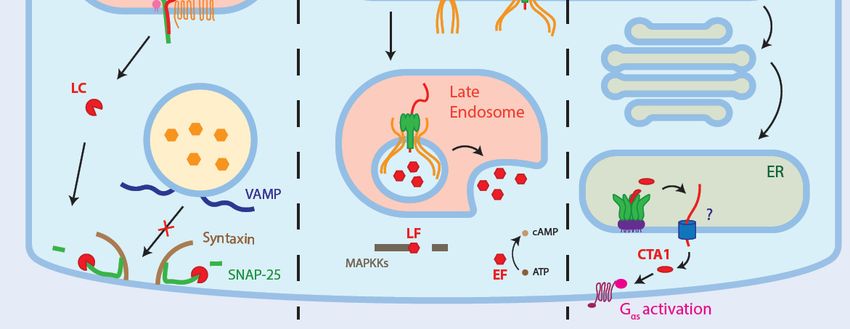
Toxins 2021, 13, 36                                                                                          2 of 17

                      a binding part (B) that is involved in binding to the cell membrane and escorting the A
                      subunit to its destination. AB toxins have finely tuned their cellular entry to evade host
                      defenses, providing hints and tools for protein-based drug development. In this review, we
                      focus on how the internalization mechanisms of three AB toxins—botulinum toxin type A,
                      anthrax toxin, and cholera toxin—can be used in different therapeutic approaches (Table 1).
                      We decided to focus on these three toxins based on the strong modular potential of anthrax
                      toxin, on the already approved use of botulinum toxin type A in several neurological
                      disorders and on the wide variety of therapeutical applications of cholera toxin. The
                      practical approaches presented in this review take advantage of both the intrinsic properties
                      of the toxins as well as the modularity of both the A and B subunits, all aspects that can be
                      further extended to other AB toxins.
Toxins 2021, 13, 36                                                                                                                                                                                                3 of 17

                  Table 1. Summary of the internalization mechanism steps of botulinum toxin type A, anthrax toxin, and cholera toxin as well as their use in therapeutic applications.

                                                         Botulinum Toxin Type A                                       Anthrax Toxin                                                Cholera Toxin
                 Bacterium                              Clostridium bacteria family                                  Bacillus anthracis                                              Vibrio cholerae
                 A subunit                                 BoNT light chain (LC)                                     Lethal factor (LF)                              Cholera toxin A1 and A2 (CTA1 and CTA2)
                 B subunit                                BoNT heavy chain (HC)                                   Protective Antigen (PA)                                        Cholera toxin B (CTB)
                 Receptors                         Polysialogangliosides and SV2 [10,11]                         CMG2 and TEM8 [12,13]                                                 GM1 [14]
              Oligomerization                                      None                                            A3 B7 or A4 B8 [15,16]                                            AB5 [5,17,18]
                                                                                                   From plasma membrane to early endosomes and late               From plasma membrane to early endosomes and
           Cellular compartments           From synaptic membrane to synaptic vesicles [19,20]
                                                                                                                      endosomes [21]                                retro-translocation to the Golgi and ER [22,23]
     Membrane translocation mechanism          HC translocates LC through the membrane [24]        PA pore translocating LF across the membrane [25,26]        Uses the ERAD-associated translocation mechanism [27]
             Cytosolic target                     SNAP-25 (of the SNARE complex) [28]                   LF cleaves MAPKK family members [29]                                      Activates Gαs [27]
         Therapeutic applications
                                                                                                                                                           •      Use as adjuvant in mucosal vaccines [34,35]
                                           •    Use in the treatment of dystonic and spastic                                                               •      Use of an inactive CT to temporarily occupy the
                                                disorders [30]                                     •   Treatment of cancer relying on the                         ERAD-associated translocation machinery in the
 •     Intrinsic properties
                                           •    Use in the treatment of autonomic disorders [31]       MAPKK-associated pathways [32,33]                          treatments of genetic protein misfolding diseases [36]
                                           •    Use in pain therapy [7]                                                                                    •      Use of CTB retro-translocation property to image
                                                                                                                                                                  neuronal cells [37]

                                           •    Cytosolic transport of LCE using LCA -HCA as a     •   Delivery of non-native protein or small molecule    •      Vaccines development consisting of antigens fused
 •     A subunit modularity
                                                delivery system [38]                                   cargos using PA-LFN delivery system [39–45]                with CTA2 and delivered using CTB [46–48]

                                                                                                                                                           •      Conjugation of self and non-self antigens to CTB to
                                                                                                                                                                  increase antigen uptake in the development of
                                                                                                   •   Cell-specific delivery of cargos using non-native
                                           •    Use of other botulinum toxin HC (i.e HCB ) to                                                                     therapies against pathogens, autoimmune and
                                                                                                       protease-specific PA [52–54]
                                                increase LCA uptake [49]                                                                                          inflammatory diseases [18,59]
 •     B subunit modularity                                                                        •   Targeting to non-native receptors using
                                           •    Targeting of non-native receptors to inhibit                                                               •      CTB-KDEL fusion protein to induce ER retention and
                                                                                                       Affibodies, DARPins or receptors ligand for the
                                                protein secretion [50,51]                                                                                         UPR-mediated wound healing response in colon [60]
                                                                                                       cytosolic delivery of non-native cargos [55–58]
                                                                                                                                                           •      Use of CTB ability to cross the blood-brain barrier and
                                                                                                                                                                  target neuronal cells to deliver drugs [61]
Toxins 2021, 13, 36 4 of 17
2. Botulinum Toxin Type A
2.1. Botulinum Toxin Type A Internalization Mechanism
As part of the AB toxin family, botulinum neurotoxins consist of seven different
toxin types (termed A–G) and are produced by the Clostridium family of bacteria [7].
Generally, the toxin reaches the bloodstream by using transcytosis to cross the epithelial
layer of the lungs or the gastrointestinal tract [62]. This review will briefly depict the
internalization mechanism of botulinum toxin type A (BoNT/A), which is the most-studied
toxin subtype with the most FDA-approved therapeutical applications. For more detailed
informations about this particular process, the readers are referred to previously published
literature [7,63]
BoNT/A is composed of a catalytic subunit, the 50-kDa light chain (LC), connected
by a disulfide bridge to the binding subunit, a 100-kDa heavy chain (HC), responsible for
the binding and translocation of the catalytic subunit into the cytosol (Figure 1A) [10]. The
HC first recognizes polysialogangliosides (PSGs) at the nerve terminal and then stabilizes
the binding by a high-affinity interaction with synaptic vesicle protein 2 (SV2) [11,19].
Endocytosis of BoNT/A targets it to small synaptic vesicles, which was shown to be
enhanced by synaptic vesicle recycling induced by neuronal activity [20,24].
The exact membrane translocation mechanism remains unsolved, though experiments
with lipid bilayer modeling this process were reviewed by Pirazzini et al. [64]. According
to the models, the acidic environment of synaptic vesicles induces a structural change of
the HC, converting it into a chaperone and an ion channel that unfolds LC and shuttles it
across the membrane of the vesicle to the cytosol [64]. In the cytosol, LC refolds into a Zn2+ -
dependent metalloprotease that hydrolyses a specific peptide bond in SNAP-25, which is
part of the cytosolic membrane fusion complex, termed SNARE [28,65,66]. This complex
consists of three different proteins: SNAP-25, syntaxin, and vesicle-associated membrane
protein (VAMP), the latter two being targets of the other botulinum toxin types [67,68]. The
cleavage of SNAP-25 by BoNT/A in neurons inhibits the fusion of synaptic vesicles, and
thus the release of neurotransmitters in the synapses, leading to paralysis.
While the half-life of the toxin in the bloodstream is approximately four hours, the
lifetime of BoNT/A is drastically increased once it reaches the cytosol due to its high stability
and resistance to proteasomal degradation [69,70]. The very long lifetime of the toxin explains
how it can induce paralysis for up to 6 months in humans. Although botulinum toxin subtypes
seem to internalize using the same pathway, they bind to different receptors with variable
expression in the different neuronal cell types and their catalytic subunits target different
proteins of the SNARE complex, inducing variations in the inhibition of synaptic vesicle
fusion. These two aspects of botulinum toxin, aside from its intrinsic therapeutic properties,
allow for the development of new therapeutic strategies for numerous diseases.
2.2. Botulinum Toxin Type A Therapeutic Applications
Under the name of Botox® , botulinum toxin is well known for its use in cosmetic
treatments, as its effect on acetylcholine release by motoneurons at the neuromuscular
junction leads to muscle relaxation. This is of great interest in muscle hyperactivation
disorders. In this context, BoNT/A was first approved by the FDA in 1989 for the treatment
of blepharospasm, a hyperactivation of the eyelid muscles that leads to repetitive and
uncontrolled eyelid quivering. The long-term effect of the toxin, which can last for months,
and the low diffusion rate in the extracellular fluid, which allows restricted and precise
treatment in targeted body areas, make BoNT/A an invaluable therapeutic tool [30]. Since
its initial approval, botulinum toxin has shown promise in the treatment of many other
dystonic and spastic disorders which are associated with the dysfunction of the skeletal
nervous system and voluntary body movements, such as cervical dystonia, laryngeal
dystonia, or upper and lower limb spasticity [31]. BoNT/A also inhibits the autonomic
nervous system, which controls unconscious muscular contractions, and was used in
the treatment of several autonomic disorders, such as hyperhidrosis, hypersalivation,the toxin reaches the bloodstream by using transcytosis to cross the epithelial layer of the
lungs or the gastrointestinal tract [62]. This review will briefly depict the internalization
mechanism of botulinum toxin type A (BoNT/A), which is the most-studied toxin subtype
with the most FDA-approved therapeutical applications. For more detailed informations
about this particular process, the readers are referred to previously published literature
[7,63]
Toxins 2021, 13, 36 5 of 17
BoNT/A is composed of a catalytic subunit, the 50-kDa light chain (LC), connected
by a disulfide bridge to the binding subunit, a 100-kDa heavy chain (HC), responsible for
the binding and translocation of the catalytic subunit into the cytosol (Figure 1A) [10]. The
HC first recognizes polysialogangliosides (PSGs) at the nerve terminal and then stabilizes
allergic
therhinitis,
binding byasa well as some
high-affinity urologic
interaction and
with gastrointestinal
synaptic vesicle protein 2disorders characterized
(SV2) [11,19]. En- by
docytosis of BoNT/A
hyperactivation targets
of smooth it to small[71].
muscles synaptic vesicles, which was shown to be enhanced
by synaptic vesicle recycling induced by neuronal activity [20,24].
Figure 1. Internalization mechanisms of botulinum toxin type A, anthrax toxin, and cholera toxin. (A) Botulinum toxin
Figure 1. Internalization mechanisms(PSGs)
binds to polysialogangliosides of botulinum
and then totoxin type
synaptic A, protein
vesicle anthrax toxin,which
2 (SV2), andleads
cholera
to thetoxin. (A) Botulinum
internalization of toxin
binds to polysialogangliosides
the toxin in small synaptic (PSGs) and
vesicles. Thethen
low pHto induces
synaptic vesicle change
a structural protein 2 (SV2), toxin
of botulinum which leads
heavy to(HC)
chain the that
internalization
of the toxin leads to the unfolding of the light chain (LC) and its translocation through the membrane. Once in the cytosol, the disulfide
in small synaptic vesicles. The low pH induces a structural change of botulinum toxin heavy chain (HC)
bond between the HC and LC is reduced, and the LC refolds. LC cleaves SNAP-25 and impairs synaptic vesicle fusion.
that leads to(B) the unfolding
Anthrax of the
toxin binds light
to its chainCMG2
receptors, (LC) or
and its translocation
TEM8, and is cleaved bythrough the membrane.
a furin-family protease. In thisOnce
form, in
PAthe
oli- cytosol, the
disulfide bond gomerizes
between and the
clusters
HCinand lipidLC
raftsisatreduced,
the plasmaand
membrane.
the LCThe oligomeric
refolds. LC form of PASNAP-25
cleaves recruits LFand
or EF.impairs
The receptor-
synaptic vesicle
fusion. (B) Anthrax toxin binds to its receptors, CMG2 or TEM8, and is cleaved by a furin-family protease. In this form,
PA oligomerizes and clusters in lipid rafts at the plasma membrane. The oligomeric form of PA recruits LF or EF. The
Toxins 2021, 13, x. https://doi.org/10.3390/xxxxx www.mdpi.com/journal/toxins
receptor-PA complex is endocytosed and is targeted to early endosomes. While some PA pores start to form at the limiting
membrane of endosomes (a), some are sorted in intraluminal vesicles (ILVs) and targeted to lysosomes (b). On the way to
lysosomes, the PA oligomers undergo pH-dependent PA pore formation in the membrane of ILVs. The pores allow the
translocation of unfolded LF through the membrane. These vesicles fuse with the limiting membrane of late endosomes and
release their content in the cytosol, where LF cleaves MAPKKs and EF converts ATP into cAMP. (C) The cholera toxin B
subunit binds in a pentameric form to the membrane on GM1 in lipid raft domains of the plasma membrane. CTA2 interacts
with the pentamer and links the catalytically active CTA1 subunit via a disulfide bond. Once endocytosed in endosomes, the
toxin is transported to the trans-Golgi network (TGN) and then to the endoplasmic reticulum (ER) using retro-translocation.
The reductive environment of the ER frees CTA1 by breaking the disulfide bond, which is then translocated through the ER
membrane using ERAD-associated mechanisms. In the cytosol, CTA1 constitutively activates Gαs, increasing cAMP levels.
The development of therapeutic BoNT/A also showed a nociceptive effect that was
first considered to be a consequence of muscle relaxation [72], though it was later shown that
the reduction in pain was a direct effect of BoNT/A on the nociceptor system. This effect
was induced by a combination of the inhibition of neuropeptides and the release of anti-
inflammatory mediators with a decrease in the transport of pain sensors, such as TRPV1
and TRPA1, at the plasma membrane in a process that relies on SNAP-25 [7]. Furthermore,
several publications showed a distant action of the toxin in CNS regions following localized
peripheral injections, indicating that the nociceptive function of botulinum toxin might
arise from retro and anterograde axonal transport, which is responsible for the movement
of different organelles to and from the neuronal cell body, towards the central nervousToxins 2021, 13, 36 6 of 17
system [73,74]. Indeed, BoNT/A has been FDA-approved for the treatment of chronic
migraines and also showed efficacy in the treatment of chronic headaches and certain
neuropathies, such as postherpetic neuralgia, post-traumatic neuralgia, and painful diabetic
neuropathy [63].
The intrinsic properties of botulinum toxin have made it an effective therapeutic for
many seemingly unrelated disorders, though the major therapeutic potential of BoNT/A
lies in its modularity. For example, the seven currently recognized serotypes (A-G) of
botulinum toxin each have several subtypes (A1, C2, etc.) [75]. All these toxins have the
same general structure consisting of one catalytic domain (LC), one binding domain, and
one translocation domain (HC), but they each have specific binding affinities for different
receptors and distinctive cleavage sites on various targets [68,76]. This modularity is
normally used by bacteria to target different neuronal membranes and induce various
deleterious effects, though it has also been elegantly exploited by Rummel et al. They
swapped the N-terminal and C-terminal domain of the HC of several botulinum toxin
types and showed that these domains can modulate toxin affinity for unique neuronal
membranes [77]. In this context, a botulinum toxin chimera was designed with the HC of
BoNT/B (HCB ), due to higher concentration of the HCB receptor, Synaptotagmin II (SytII),
over HCA receptors for an increased uptake in synaptic vesicles, and the LC of BoNT/A
(LCA ), due to its longer lifetime in the cytosol compared to LCB . This chimera induced a
prolonged neuromuscular paralysis in mice of 50 days, compared to 30 days when using
the full-length BoNT/A [49].
Similarly, Wang et al. made a chimeric botulinum toxin to target and suppress the
release of the pain signaling peptide, calcitonin gene-related peptide (CGRP), by sensory
neurons. This distinctive specificity was achieved due to the properties of the three different
chains of the chimera, which was composed of LCE fused to a mutated inactive form of
LCA (mLCA ), both connected to the HCA that internalized the fused LCs in the cytosol [38].
In this chimera, internalization was achieved because sensory neurons express the HCA
receptor isoform SV2C, but not the HCE receptor isoforms SV2A and B [19,78]. A long-
lasting effect was due to the presence of a dileucine motif in mLCA that plays a role in
its protection from proteasomal degradation [79]. Finally, the strong inhibition of CGRP
release is due to the LCE -induced cleavage of 26 amino acids from the C-terminal of SNAP-
25, while LCA cleaves only 9. Taken together, this LCE -mLCA -HCA chimera showed strong
nociceptive inhibition both in vitro in trigerminal ganglion neurons and in vivo in mice [38].
These two chimera examples perfectly illustrate how the modularity of the different types
of botulinum toxin can affect their therapeutic applications.
Using the same strategy, fusion proteins of botulinum toxin with other proteins were
created in order to modulate the targeted receptor and, thus, the targeted cell type. To
develop a treatment for acromegaly, an endocrine disorder characterized by an increased
secretion of pituitary growth hormone (GH), Somm et al. made a modified fusion construct
of the GH-releasing hormone with the translocation domain of HCD and the LCD , which
cleaves one of the protein of the SNARE complex responsible for GH secretion, VAMP2 [50].
This construct decreased GH production and secretion in vivo, which reduced the body
weight and body size of juvenile rats. Similarly, a study using a botulinum toxin fusion
construct with wheat germ agglutinin inhibited insulin secretion in hamster pancreatic
cells [51]. Together, these examples further illustrate the extraordinarily broad spectrum of
therapeutic applications of AB toxins and how the properties of the bacterial toxins can be
exploited to achieve a targeted therapeutic strategy.
3. Anthrax Toxin
3.1. Anthrax Toxin Internalization Mechanism
An important concern for animal health and human public safety in the context of
bioterrorism, anthrax toxin is an AB toxin produced by the gram-positive spore-forming
bacterium Bacillus anthracis. This toxin consists of a B subunit, protective antigen (PA), and
two catalytic A subunits, lethal factor (LF) and edema factor (EF). PA is an 83-kDa proteinToxins 2021, 13, 36 7 of 17
that is responsible for the binding of the toxin to its main receptors, capillary morphogen-
esis 2 (CMG2) and tumor endothelial marker 8 (TEM8) [12,13]. LF is an 91-kDa matrix
metalloprotease that cleaves the MAPKK family members, which impairs the associated
signaling pathways and eventually leads to apoptosis, especially in macrophages [29,80].
EF is a calmodulin-dependent adenylyl cyclase that increases the cytosolic cAMP lev-
els. This review briefly describes the internalization process of anthrax toxin and, for a
more in-depth understanding of this mechanism, readers are oriented towards previously
published reviews [6].
Initially in LF and EF internalization, extracellular PA binds to one of its receptors,
CMG2 or TEM8, and then is cleaved by furin-family proteins (Figure 1B). This cleavage
allows PA to oligomerize into heptamers or octamers, also called pre-pores [15,16,81],
which can then recruit three or four LF or EF subunits, respectively, for internalization.
On the cytosolic side, PA binding to the TEM8 or CMG2 receptor causes it to release
from the actin cytoskeleton [82,83], allowing ubiquitination of the receptor, which triggers
endocytosis of the receptor-anthrax toxins complex [82].
Anthrax toxin and its receptors are then targeted to early endosomes where they are
sorted in endosomal intraluminal vesicles (ILVs) and trafficked through the endocytic
pathway towards late endosomes [21]. On the way to late endosomes, the acidification
of the microenvironment induces a conformational change in the PA pore [25], and this
low pH is also required for the translocation of LF [26]. Pores can form at the limiting
membrane of the endosomes, translocating LF or EF directly into the cytosol, though most
pores form in the membrane of ILVs [21,84]. These pores allow the translocation of LF
or EF to the lumen of ILVs and, by back-fusion of ILVs with the limiting membrane of
late endosomes, LF or EF eventually reaches the cytosol [21]. In opposition to BoNT/A,
evidence suggest that LF has a very short half-life in the cytosol and its long-term effect
relies on its ability to remain dormant in ILVs which stochastically back-fuse with the
membrane of endosomes over a long period of time [21].
3.2. Anthrax Toxin Therapeutic Applications
The therapeutic potential of anthrax lethal toxin was originally exploited in anti-cancer
treatments due to its inhibitory effect on the MAPKK-associated pathway. Unlike normal
cells, cancer cells usually rely on only a few dysregulated pathways to increase their
growth, survival, or motility. Accordingly, some cancers, such as melanoma bearing the
V600E BRAF mutation, mostly rely on the constitutively activated MAPK pathway for cell
growth and survival, and anthrax toxin was shown to decrease both these processes in this
particular cell line [32]. Similarly, anthrax lethal toxin was shown to reduce cell growth and
tumor angiogenesis in renal cell carcinoma and to reduce cell motility and invasiveness in
astrocytes by targeting the MAPK pathway [33].
Although anthrax lethal toxin showed interesting intrinsic anti-tumor properties, most
of its potential in therapy relies on its modular properties, like its ability to translocate
different non-native proteins, drugs, and other molecules. As mentioned previously,
PA oligomers create a pore in endosomes, allowing LF to eventually reach the cytosol,
suggesting that LF fusion proteins could go through the pore as well—as long as they can
successfully unfold while passing through the pore and refold later in the cytosol. In the
1990s, the first attempts to fuse proteins to the N-terminus of the LF subunit were done to
target proteins to the cytosol and confirm the potential of anthrax toxin as a delivery system.
FP59, a fusion between the N-terminus of LF (LFN ) with the ADP-ribosylation domains of
Pseudomonas exotoxin A, was the first successful translocation of a foreign protein into the
cytosol [39]. Shortly after, both catalytic domains of the Shiga and diphtheria toxins reached
the cytosol when fused to LFN , further supporting that the N-terminal residues of LF
were sufficient to translocate complicated polypeptide chains through the PA pore [40,41].
However, Blanke et al. later showed that a simple positively-charged polycationic peptide
could replace LFN for the delivery of diphtheria toxin to the cytosol [42].Toxins 2021, 13, 36 8 of 17
Besides bacterial toxins, the LFN delivery system was shown to be useful in other
applications, such as the development of a potential HIV vaccine and the treatment of
neurodegenerative diseases [43,44]. In a broader perspective, Rabideau et al. assessed
the feasibility of translocation through the PA pore for many different cargo molecules,
from short or cyclic peptides to small molecule drugs. They concluded that while non-
canonical peptides and small-molecule drugs, such as doxorubicin, can be translocated,
cyclic peptides and the small molecule docetaxel cannot, which they hypothesized was due
to rigidity of the cargo [45]. These examples provide strong evidence that many different
cargo proteins can be delivered to the cytosol both in vitro and in vivo using anthrax toxin,
which can be used for the targeted delivery of vaccines, drugs, and other proteins.
Besides its ability to translocate different non-native cargos, another modular char-
acteristic of PA lies in the specificity of the protease that processes it, thereby allowing it
to oligomerize. In the last two decades, several groups focused on unraveling the best
combinations of mutations in PA that would allow more targeted and less toxic tumor ther-
apies. The two PA mutants, PA-L1 and PA-U2, were programmed to be specific for several
cancer cell lines in vitro by changing the cleavage site from furin to matrix metallopro-
teases (MMPs) and urokinase plasminogen activator (uPA), respectively [52,53], which are
overexpressed in many cancer types while not very abundant at the surface of normal cells.
In particular, PA-U2 showed a strong anti-tumor activity and specificity when combined
with FP59 in mice [54]. To make the tumor targeting more specific, PA-L1 and PA-U2 were
mutated on their homo-oligomerization domain to render them complementary, making
them even more specific to cancer cells expressing both proteases. This approach was
shown to be efficient with different sets of PA mutants both in vitro and in vivo [85–88].
In addition to their use as anti-tumor drugs, the protease-specific PA mutants were also
used in combination with radioactively labelled LF or LFN -β-lactamase fusion protein to
develop methods of imaging plasma membrane protease activity in tumors or in cancer
cell lines, respectively [89,90].
Using the potential of PA to internalize molecules, several research groups adapted
this technology to allow cancer-specific receptors to bind and internalize PA-fusions specific
for those receptors. Varughese et al. were the first to unravel the potential of this strategy
by targeting FP59 to a c-Myc-specific 9E10 hybridoma cell line using a PA-c-Myc fusion
protein [55]. McCluskey et al. used a similar approach containing a mutated PA (mPA)
that cannot bind its natural receptors fused with a high-affinity Affibody, ZHER2, targeting
the HER2 receptor [56]. They showed that both mPA-EGF and mPA-ZHER2 could deliver
an LFN -fused diphtheria toxin catalytic domain (DTA) to kill several cancer cell lines
depending on the presence of their respective receptors [56]. Based on these observations,
PA can form pores and deliver cargos as long as the targeted receptor is able to internalize,
broadening the number of potential targets at the cell surface of cancer cells.
Additionally, Loftis et al. used an mPA fused with the single-chain variable fragment
(scFv) of an antibody to internalize and deliver LFN -DTA through EGFR or carcinoem-
bryonic antigen, which could kill pancreatic cancer cells overexpressing the two receptors
at the plasma membrane. For additional specificity towards their pancreatic cancer cell
line, they made an LF-RRSP fusion protein which targets the Ras–ERK signaling pathway,
crucial for many pancreatic cancer cells [57]. Similarly, Becker et al. used designed ankyrin
repeat proteins (DARPins) fused to a PA-CMG2-based construct to specifically target trans-
membrane glycoprotein epithelial cell adhesion molecule (EpCAM) at the surface of cells.
These engineered constructs were shown to target EpCAM-expressing cells with a high
specificity and to deliver LFN -based constructs to the cytosol [58]. Overall, these engineered
proteins show that both the A and B subunits of anthrax toxin have strong potential as a
protein delivery system, and they open many new routes for investigating the development
of therapeutics. However, the immunogenicity of anthrax toxin subunits, as illustrated
by the use of PA in anthrax vaccines, for example, remain a challenge to address in its
therapeutical applications [91].Toxins 2021, 13, 36 9 of 17
4. Cholera Toxin
4.1. Cholera Toxin Internalization Mechanism
Cholera toxin (CT) is an AB toxin of the heat-labile enterotoxin family and is produced
by the bacterium Vibrio cholerae. The functional B subunit is actually a 55-kDa pentameric
ring of individual B subunits (CTB) that tightly bind to its glycolipid receptor, GM1. The A
subunit consists of two parts: an 11-kDa catalytically active CTA1 subunit and a 18-kDa CTA2
subunit, whose role is to anchor CTA1 in the lumen of the B-pentameric ring [5,17,18]. CTA1
is an ADP-ribosyltransferase that constitutively activates the heterotrimeric G-protein, Gαs.
CTA1 and CTA2 are connected through a flexible linker containing a disulfide bridge. For a
more detailed description of the cholera toxin internalization process, readers are referred to
the following reviews [5,14].
Once bound to its receptor, CT associates with the GM1- and cholesterol-rich lipid
rafts at the plasma membrane, which are necessary for efficient endocytosis of the toxin
(Figure 1C) [22]. Once endocytosed, the toxin reaches early endosomes where it is targeted
to the trans-Golgi network (TGN) via retrograde transport [23]. From there, CT bypasses
the Golgi stacks and directly reaches the reductive environment of the ER [92], wherein
the disulfide bridge between CTA1 and CTA2 is reduced and protein disulfide isomerase
finishes the separation of both CTA subunits [27,93]. CTA1 is then thought to spontaneously
unfold at physiological temperature. At that stage, it is thought to mimic a misfolded
protein leading to its recognition by the ER-associated degradation (ERAD)-dependent
pathway and its retro-translocation into the cytosol [14]. The C-terminus of CTA1 contains
a KDEL motif that is not necessary for endosome to ER retrograde transport, but it is
thought to play a role in ER retention once CTA1 dissociates from CTA2 and CTB [94]. In
the cytosol, the low number of lysines in CTA1 most likely protects it from ubiquitination
and further degradation by the proteasome [95]. Its ADP-ribosyltransferase activity then
activates Gαs, which in turn increases cAMP levels in the cell, impairing sodium uptake
and increasing chloride extrusion. Eventually, this induces the secretion of water and leads
to intense diarrhea [14].
4.2. Cholera Toxin Therapeutic Applications
CT has been known for decades to have immunogenic properties. As early as 1984,
it was used as an adjuvant in mucosal vaccines, as it was able to trigger both a mucosal
and systemic antibody response [34,35]. It was also shown that the CTA-induced toxicity
could be avoided by triggering the immune response through the use of only CTB [96].
Besides co-injection of the CTB adjuvant with different antigens, the immune response
could be improved by conjugating CTB with an antigen [96]. This improvement is likely
due to the broad presence of GM1 in many immune cells (B cells, T cells, macrophages,
dendritic cells), as well as in epithelial cells and neurons, which would increase the uptake
of the antigen-conjugated CTB in those cells [97]. This strategy has been used for the
development of mucosal vaccines against a wide range of bacteria, viruses, and parasites
in mice, as reviewed in previous publications [59,98]. Additionally, several other groups
used the non-toxic CTA2 subunit as a fusion protein, co-injected with CTB, to develop
their mucosal vaccine [46,47]. For example, Tinker et al. developed a mucosal vaccine
against West Nile Virus using domain III of the virus envelope conjugated to CTA2 and the
CTB subunit. The fusion protein was shown to efficiently bind to the plasma membrane,
internalize into the perinuclear region of Vero and DC2.4 dendritic cells in vitro, and induce
an increased production of IgG and IgM in mice after several injections [48]. Although the
immunogenic effect of CTB has been exploited as previously mentioned, one drawback of
this immunogenicity lies in the production of neutralizing antibodies towards CTB, which
can be an issue if used as a therapeutic or as a drug delivery system. This particular topic
will be discussed further below.
In addition to immunogenicity, CT was also shown to have immunosuppressive and
anti-inflammatory properties [99]. When conjugated to different antigens, CTB induced
immune tolerance towards autoantigens in the context of autoimmune diseases or allergies,Toxins 2021, 13, 36 10 of 17
such as type I diabetes, asthma, Behcet’s disease, atherosclerosis, or Crohn’s disease [18].
These immunosuppressive properties of CT were shown to rely on several different mecha-
nisms: modulation of cytokine production, mucosal generation of regulatory T cells, and
induction of tolerogenic antigen-presenting cells and B cells [100]. For example, a treatment
for type I diabetes using a CTB-GAD (glutamic acid decarboxylase) fusion protein was
shown to suppress the activation of human umbilical cord blood dendritic cells through
the down-regulation of pro-inflammatory cytokines (IL-6 and IL-12) and up-regulation
of immunosuppressive cytokine IL-10 [101]. Similarly, Denes et al. used a recombinant
vaccinia virus rVV-CTB-GAD to treat non-obese diabetic (NOD) mice, which showed a
significant decrease in hyperglycemia and pancreatic β islet inflammation [102]. This
underlines the potential of CT in antigen uptake and its role in the modulation of the
immune response.
Another very interesting therapeutic approach provided by CT consists of targeting
and temporarily occupying the ERAD pathway, thus rescuing deleterious phenotypes in
genetic diseases with mutations that lead to the premature degradation of a misfolded
protein. Adnan et al. illustrated this strategy using inactivated Shiga toxin and CT in cells
derived from patients with an F508 deletion in cystic fibrosis transmembrane conductance
regulator (CFTR) bronchiolar epithelia, a mutation in a plasma membrane chloride channel
that leads to cystic fibrosis [36]. The inactivated toxins were able to induce 5–10-fold
increases in protein levels, 20-fold increases in cell surface expression, and 2-fold chloride
transport through the membrane with no apparent cytotoxicity. Similarly, they were also
able to increase glucocerebrocidase (GCC) by 3-fold in N370SGCC Gaucher’s disease cells,
the mutation of which leads to the accumulation of glucocerebrosides in lysosomes. An
advantage of this strategy over the use of ERAD inhibitors is that inactivated CT doesn’t
induce any ER stress and unfolded protein response (UPR), which can lead to apoptosis.
Using a relatively similar approach, Royal et al. designed a CTB subunit with a KDEL
ER-retention motif that would induce an UPR response [60]. This UPR response led to
TGF-β secretion and increased the wound healing response in vitro in Caco2 cells and
in colon explants from patients with inflammatory bowel disease as well as in vivo in a
dextran sodium sulfate-induced acute colitis mouse model. These two examples strongly
illustrate the potential for hijacking the CT membrane translocation mechanism or its
ability to trigger ER stress to treat diseases based on genetic protein misfolding and for
mucosal healing in intestinal inflammatory diseases.
In addition to these therapeutic strategies, CT has interesting potential for the treat-
ment of neurological disorders due to its ability to cross the blood-brain barrier (BBB) and
internalize into neuronal cells. It has been shown to be particularly efficient in the treatment
of glioblastoma in mice [61]. CTB subunits conjugated with paclitaxel-loaded nanoparticles
induced apoptosis of intracranial glioma cells and suppressed neovasculature in vivo.
Furthermore, this ability of CT to enter neuronal cells has been exploited to develop new
neural imaging techniques. Once internalized, the toxin is able to reach the cell body and
its dendrites via retrograde transport, which makes it useful for nerve visualization and
potentially drug delivery. For example, CTB was conjugated to fluorescent gold nanodots
and injected in the sciatic nerve of rats [37]. After 2 days, the fluorescent signal was visible
in the spinal cord and was stable for 10 days. This tool could bring an interesting novel vi-
sualization technique for the detection of neuronal lesions, further supporting the potential
of CT in the development of therapeutic tools.
5. Discussion
In this review, we have illustrated the outstanding diversity of therapeutical strategies
provided by the use of botulinum toxin type A, anthrax toxin, and cholera toxin. In addition
to the intrinsic therapeutic properties provided by these AB toxins, their modularity in
terms of receptor recognition, protease specificity, and non-native cargo delivery allowed
the development of many treatments (Figure 2). While the intrinsic properties alone of the
three toxins could be therapeutic against specific diseases, their huge potential lies in theToxins 2021, 13, 36 11 of 17
possibility of modifying both the A and B subunits of the toxins. The A subunit allows
the internalization of non-native cargos into different cell types and in vivo, while the B
subunit allows targeting of different receptors and cell types. Several groups have even
modulated both subunits of these toxins to deliver drugs or proteins to cells expressing
specific non-native receptors, showing the potential of AB toxins as intracellular delivery
systems. However, some challenges linked to the immunogenicity and toxicity of these
toxins remain to be addressed.
As exogenous proteins, toxins often induce the production of neutralizing antibodies
that can interfere with treatments, especially for repeated injections of a drug over a long
period of time, like with autoinflammatory diseases [103,104]. In this context, due to its
low immunogenicity and, likely also, to the localized mode of administration, BoNT/A
triggers the production of neutralizing antibodies in only up to 3% of patients [105]. However,
both CT and anthrax toxin were shown to induce the formation of neutralizing antibodies,
potentially decreasing the efficiency of an associated drug in long-term therapies [103,104].
This issue could be addressed by investigating potential mutations in the antigens or by using
immunosuppressive drugs to decrease the production of neutralizing antibodies [106,107]. In
this context, Liu et al. used a combination of cyclophosphamide and pentostatin, two drugs to
prevent host-versus-graft rejections, to successfully suppress the antibody production induced
by an anthrax-based cancer treatment in mice [87]. However, the risk and benefits have to be
carefully weighed when attempting to deliver these therapies together.
Another issue linked to the use of toxins in therapy would be toxicity. While BoNT/A
and cholera toxin B are not toxic when used properly, anthrax toxin PA might have a toxic
effect as illustrated by its potential role in the Gulf War Illness, in which multi-systemic
disease was first observed in Gulf War veterans vaccinated with the PA-based anthrax
vaccine [108]. However, this observation needs further validation, as many other chemical
or biological factors might have played a role in the development of the disease.
The three bacterial toxins reviewed here have interesting modular properties that
could allow their development into various elegant therapeutic strategies. Overall, these
toxins have shown new potential therapeutic alternatives in autoimmune and inflammatory
diseases, cancer, genetic protein misfolding diseases, movement disorders, and in vaccine
development. Although many examples used these three highlighted toxins, several other
AB toxins have been shown to have similar characteristics in therapy, such as Shiga toxin
and diphtheria toxin, further widening the range of therapeutic possibilities [109,110].
For example, these toxins target different cell types depending on the expression of their
receptor. Such specificity can be hijacked to deliver drugs or non-native proteins conjugated
to AB toxins to very specific targets in the human organism, as long as the cargo can unfold
or is flexible enough to be translocated across the membrane by the B subunit. In addition,
one can imagine various ways to target non-native receptors using fusion constructs of
the B subunit of AB toxins with Affibodies, DARPins or the natural ligand of the targeted
receptor, among others. As described for botulinum toxin and for anthrax toxin in the
previous chapters, this elegant strategy has shown promising results and allows for the
delivery of cargos to several different cell types with high specificity. Importantly, such
systems provide new solutions for the delivery of proteins and peptides which are unable
to efficiently translocate through membranes, thereby potentially further increasing the
number of new biologics on the market in the coming years.Toxins 2021, 13, 36 12 of 17
Toxins 2021, 13, x FOR PEER REVIEW 9 of 19
Figure 2. Schematic representation of the different constructs described in this study and brief description of their prop-
Figure 2. Schematic representation of the different constructs described in this study and brief description of their properties.
erties. The three original toxins at the top of their respective compartments are highlighted. A and B domains of each
Thetoxin’s
three subunits
original are
toxins at the top
represented of their
in red respective
and green, compartments
respectively. arethe
The text on highlighted.
right brieflyA and B
depict domains
either of each toxin’s
the internalization
subunits
processare
ofrepresented in redorand
the original toxin thegreen, respectively.
therapeutic Theoftext
properties the on the right
chimeric briefly depict either the internalization process
constructs.
of the original toxin or the therapeutic properties of the chimeric constructs.Toxins 2021, 13, 36 13 of 17
Author Contributions: Conceptualization, N.P. and O.A.S.; visualization, N.P.; writing—original
draft preparation, N.P. and O.A.S.; writing—review and editing, N.P., F.G.v.d.G. and O.A.S. All
authors have read and agreed to the published version of the manuscript.
Funding: This research was funded by the Swiss National Science Foundation (www.snf.ch), grant
number 310030B_176393.
Acknowledgments: We would like to thank the members of the van der Goot laboratory for com-
pelling discussions that helped shape this manuscript.
Conflicts of Interest: The authors declare no conflict of interest.
References
1. FDA Center for Drug Evaluation and Research. Novel Drug Approvals for 2018. 2020. Available online: https://www.fda.gov/
drugs/new-drugs-fda-cders-new-molecular-entities-and-new-therapeutic-biological-products/novel-drug-approvals-2018 (ac-
cessed on 12 October 2020).
2. Bioengineered Protein Drugs Market Research Report. Available online: https://www.bccresearch.com/market-research/
biotechnology/bioengineered-protein-drugs-report.html (accessed on 12 October 2020).
3. Craik, D.J.; Fairlie, D.P.; Liras, S.; Price, D. The Future of Peptide-based Drugs. Chem. Biol. Drug Des. 2013, 81, 136–147. [CrossRef]
[PubMed]
4. Beilhartz, G.L.; Sugiman-Marangos, S.N.; Melnyk, R.A. Repurposing bacterial toxins for intracellular delivery of therapeutic
proteins. Biochem. Pharmacol. 2017, 142, 13–20. [CrossRef] [PubMed]
5. Sanchez, J.; Holmgren, J. Cholera toxin—A foe & a friend. Indian J. Med. Res. 2011, 133, 153–163. [PubMed]
6. Friebe, S.; van der Goot, F.G.; Bürgi, J. The Ins and Outs of Anthrax Toxin. Toxins 2016, 8, 69. [CrossRef] [PubMed]
7. Pirazzini, M.; Rossetto, O.; Eleopra, R.; Montecucco, C. Botulinum Neurotoxins: Biology, Pharmacology, and Toxicology. Pharmacol.
Rev. 2017, 69, 200–235. [CrossRef]
8. Sharma, N.C.; Efstratiou, A.; Mokrousov, I.; Mutreja, A.; Das, B.; Ramamurthy, T. Diphtheria. Nat. Rev. Dis. Primers 2019, 5, 1–18.
[CrossRef]
9. Beddoe, T.; Paton, A.W.; Le Nours, J.; Rossjohn, J.; Paton, J.C. Structure, Biological Functions and Applications of the AB5 Toxins.
Trends Biochem. Sci. 2010, 35, 411–418. [CrossRef]
10. Lacy, D.B.; Tepp, W.; Cohen, A.C.; DasGupta, B.R.; Stevens, R.C. Crystal structure of botulinum neurotoxin type A and implications
for toxicity. Nat. Struct. Biol. 1998, 5, 898–902. [CrossRef]
11. Kitamura, M.; Iwamori, M.; Nagai, Y. Interaction between Clostridium botulinum neurotoxin and gangliosides. Biochim. Biophys.
Acta 1980, 628, 328–335. [CrossRef]
12. Bradley, K.A.; Mogridge, J.; Mourez, M.; Collier, R.J.; Young, J.A.T. Identification of the cellular receptor for anthrax toxin. Nature
2001, 414, 225–229. [CrossRef]
13. Scobie, H.M.; Rainey, G.J.A.; Bradley, K.A.; Young, J.A.T. Human capillary morphogenesis protein 2 functions as an anthrax toxin
receptor. Proc. Natl. Acad. Sci. USA 2003, 100, 5170–5174. [CrossRef]
14. Wernick, N.L.B.; Chinnapen, D.J.-F.; Cho, J.A.; Lencer, W.I. Cholera toxin: An intracellular journey into the cytosol by way of the
endoplasmic reticulum. Toxins 2010, 2, 310–325. [CrossRef]
15. Milne, J.C.; Furlong, D.; Hanna, P.C.; Wall, J.S.; Collier, R.J. Anthrax protective antigen forms oligomers during intoxication of
mammalian cells. J. Biol. Chem. 1994, 269, 20607–20612. [PubMed]
16. Kintzer, A.F.; Thoren, K.L.; Sterling, H.J.; Dong, K.C.; Feld, G.K.; Tang, I.I.; Zhang, T.T.; Williams, E.R.; Berger, J.M.; Krantz,
B.A. The protective antigen component of anthrax toxin forms functional octameric complexes. J. Mol. Biol. 2009, 392, 614–629.
[CrossRef] [PubMed]
17. Holmgren, J.; Lönnroth, I.; Svennerholm, L. Tissue receptor for cholera exotoxin: Postulated structure from studies with GM1
ganglioside and related glycolipids. Infect. Immun. 1973, 8, 208–214. [CrossRef] [PubMed]
18. Royal, J.M.; Matoba, N. Therapeutic Potential of Cholera Toxin B Subunit for the Treatment of Inflammatory Diseases of the
Mucosa. Toxins 2017, 9, 379. [CrossRef] [PubMed]
19. Dong, M.; Yeh, F.; Tepp, W.H.; Dean, C.; Johnson, E.A.; Janz, R.; Chapman, E.R. SV2 Is the Protein Receptor for Botulinum
Neurotoxin A. Science 2006, 312, 592–596. [CrossRef]
20. Harper, C.B.; Martin, S.; Nguyen, T.H.; Daniels, S.J.; Lavidis, N.A.; Popoff, M.R.; Hadzic, G.; Mariana, A.; Chau, N.; McCluskey,
A.; et al. Dynamin inhibition blocks botulinum neurotoxin type A endocytosis in neurons and delays botulism. J. Biol. Chem.
2011, 286, 35966–35976. [CrossRef]
21. Abrami, L.; Brandi, L.; Moayeri, M.; Brown, M.J.; Krantz, B.A.; Leppla, S.H.; van der Goot, F.G. Hijacking Multivesicular Bodies
Enables Long-Term and Exosome-Mediated Long-Distance Action of Anthrax Toxin. Cell Rep. 2013, 5, 986–996. [CrossRef]
22. Fujinaga, Y.; Wolf, A.A.; Rodighiero, C.; Wheeler, H.; Tsai, B.; Allen, L.; Jobling, M.G.; Rapoport, T.; Holmes, R.K.; Lencer,
W.I. Gangliosides that associate with lipid rafts mediate transport of cholera and related toxins from the plasma membrane to
endoplasmic reticulm. Mol. Biol. Cell 2003, 14, 4783–4793. [CrossRef]Toxins 2021, 13, 36 14 of 17
23. Chinnapen, D.J.-F.; Chinnapen, H.; Saslowsky, D.; Lencer, W.I. Rafting with cholera toxin: Endocytosis and tracking from plasma
membrane to ER. FEMS Microbiol. Lett. 2007, 266, 129–137. [CrossRef] [PubMed]
24. Colasante, C.; Rossetto, O.; Morbiato, L.; Pirazzini, M.; Molgó, J.; Montecucco, C. Botulinum Neurotoxin Type A is Internalized
and Translocated from Small Synaptic Vesicles at the Neuromuscular Junction. Mol. Neurobiol. 2013, 48, 120–127. [CrossRef]
[PubMed]
25. Rainey, G.J.A.; Wigelsworth, D.J.; Ryan, P.L.; Scobie, H.M.; Collier, R.J.; Young, J.A.T. Receptor-specific requirements for anthrax
toxin delivery into cells. Proc. Natl. Acad. Sci. USA 2005, 102, 13278–13283. [CrossRef] [PubMed]
26. Krantz, B.A.; Finkelstein, A.; Collier, R.J. Protein translocation through the anthrax toxin transmembrane pore is driven by a
proton gradient. J. Mol. Biol. 2006, 355, 968–979. [CrossRef]
27. Tsai, B.; Rodighiero, C.; Lencer, W.I.; Rapoport, T.A. Protein disulfide isomerase acts as a redox-dependent chaperone to unfold
cholera toxin. Cell 2001, 104, 937–948. [CrossRef]
28. Pirazzini, M.; Rossetto, O.; Bolognese, P.; Shone, C.C.; Montecucco, C. Double anchorage to the membrane and intact inter-chain
disulfide bond are required for the low pH induced entry of tetanus and botulinum neurotoxins into neurons. Cell Microbiol.
2011, 13, 1731–1743. [CrossRef]
29. Duesbery, N.S.; Webb, C.P.; Leppla, S.H.; Gordon, V.M.; Klimpel, K.R.; Copeland, T.D.; Ahn, N.G.; Oskarsson, M.K.; Fukasawa, K.;
Paull, K.D.; et al. Proteolytic inactivation of MAP-kinase-kinase by anthrax lethal factor. Science 1998, 280, 734–737. [CrossRef]
30. Tang-Liu, D.D.-S.; Aoki, K.R.; Dolly, J.O.; de Paiva, A.; Houchen, T.L.; Chasseaud, L.F.; Webber, C. Intramuscular injection of
125I-botulinum neurotoxin-complex versus 125I-botulinum-free neurotoxin: Time course of tissue distribution. Toxicon 2003,
42, 461–469. [CrossRef]
31. Thenganatt, M.A.; Fahn, S. Botulinum Toxin for the Treatment of Movement Disorders. Curr. Neurol. Neurosci. Rep. 2012,
12, 399–409. [CrossRef]
32. Abi-Habib, R.J.; Urieto, J.O.; Liu, S.; Leppla, S.H.; Duesbery, N.S.; Frankel, A.E. BRAF status and mitogen-activated pro-
tein/extracellular signal-regulated kinase kinase 1/2 activity indicate sensitivity of melanoma cells to anthrax lethal toxin. Mol.
Cancer Ther. 2005, 4, 1303–1310. [CrossRef]
33. Huang, D.; Ding, Y.; Luo, W.-M.; Bender, S.; Qian, C.-N.; Kort, E.; Zhang, Z.-F.; VandenBeldt, K.; Duesbery, N.S.; Resau, J.H.; et al.
Inhibition of MAPK kinase signaling pathways suppressed renal cell carcinoma growth and angiogenesis in vivo. Cancer Res.
2008, 68, 81–88. [CrossRef] [PubMed]
34. Elson, C.O.; Ealding, W. Generalized systemic and mucosal immunity in mice after mucosal stimulation with cholera toxin. J.
Immunol. 1984, 132, 2736–2741. [PubMed]
35. Jackson, R.J.; Fujihashi, K.; Xu-Amano, J.; Kiyono, H.; Elson, C.O.; McGhee, J.R. Optimizing oral vaccines: Induction of systemic
and mucosal B-cell and antibody responses to tetanus toxoid by use of cholera toxin as an adjuvant. Infect. Immun. 1993,
61, 4272–4279. [CrossRef] [PubMed]
36. Adnan, H.; Zhang, Z.; Park, H.-J.; Tailor, C.; Che, C.; Kamani, M.; Spitalny, G.; Binnington, B.; Lingwood, C. Endoplasmic
Reticulum-Targeted Subunit Toxins Provide a New Approach to Rescue Misfolded Mutant Proteins and Revert Cell Models of
Genetic Diseases. PLoS ONE 2016, 11, e0166948. [CrossRef] [PubMed]
37. Zhao, Y.; Maharjan, S.; Sun, Y.; Yang, Z.; Yang, E.; Zhou, N.; Lu, L.; Whittaker, A.K.; Yang, B.; Lin, Q. Red fluorescent AuNDs with
conjugation of cholera toxin subunit B (CTB) for extended-distance retro-nerve transporting and long-time neural tracing. Acta
Biomater. 2020, 102, 394–402. [CrossRef]
38. Wang, J.; Meng, J.; Lawrence, G.W.; Zurawski, T.H.; Sasse, A.; Bodeker, M.O.; Gilmore, M.A.; Fernández-Salas, E.; Francis, J.;
Steward, L.E.; et al. Novel chimeras of botulinum neurotoxins A and E unveil contributions from the binding, translocation, and
protease domains to their functional characteristics. J. Biol. Chem. 2008, 283, 16993–17002. [CrossRef]
39. Arora, N.; Klimpel, K.R.; Singh, Y.; Leppla, S.H. Fusions of anthrax toxin lethal factor to the ADP-ribosylation domain of
Pseudomonas exotoxin A are potent cytotoxins which are translocated to the cytosol of mammalian cells. J. Biol. Chem. 1992,
267, 15542–15548.
40. Arora, N.; Leppla, S.H. Fusions of anthrax toxin lethal factor with shiga toxin and diphtheria toxin enzymatic domains are toxic
to mammalian cells. Infect. Immun. 1994, 62, 4955–4961. [CrossRef]
41. Arora, N.; Leppla, S.H. Residues 1-254 of anthrax toxin lethal factor are sufficient to cause cellular uptake of fused polypeptides.
J. Biol. Chem. 1993, 268, 3334–3341. [CrossRef]
42. Blanke, S.R.; Milne, J.C.; Benson, E.L.; Collier, R.J. Fused polycationic peptide mediates delivery of diphtheria toxin A chain to the
cytosol in the presence of anthrax protective antigen. Proc. Natl. Acad. Sci. USA 1996, 93, 8437–8442. [CrossRef]
43. Goletz, T.J.; Klimpel, K.R.; Arora, N.; Leppla, S.H.; Keith, J.M.; Berzofsky, J.A. Targeting HIV proteins to the major histocom-
patibility complex class I processing pathway with a novel gp120-anthrax toxin fusion protein. Proc. Natl. Acad. Sci. USA 1997,
94, 12059–12064. [CrossRef] [PubMed]
44. Liu, X.H.; Collier, R.J.; Youle, R.J. Inhibition of axotomy-induced neuronal apoptosis by extracellular delivery of a Bcl-XL fusion
protein. J. Biol. Chem. 2001, 276, 46326–46332. [CrossRef] [PubMed]
45. Rabideau, A.E.; Liao, X.; Akçay, G.; Pentelute, B.L. Translocation of Non-Canonical Polypeptides into Cells Using Protective
Antigen. Sci. Rep. 2015, 5, 11944. [CrossRef] [PubMed]
46. Hajishengallis, G.; Hollingshead, S.K.; Koga, T.; Russell, M.W. Mucosal immunization with a bacterial protein antigen genetically
coupled to cholera toxin A2/B subunits. J. Immunol. 1995, 154, 4322–4332. [PubMed]Toxins 2021, 13, 36 15 of 17
47. Lee, S.F.; Halperin, S.A.; Salloum, D.F.; MacMillan, A.; Morris, A. Mucosal Immunization with a Genetically Engineered Pertussis
Toxin S1 Fragment-Cholera Toxin Subunit B Chimeric Protein. Infect. Immun. 2003, 71, 2272–2275. [CrossRef]
48. Tinker, J.K.; Yan, J.; Knippel, R.J.; Panayiotou, P.; Cornell, K.A. Immunogenicity of a West Nile virus DIII-cholera toxin A2/B
chimera after intranasal delivery. Toxins 2014, 6, 1397–1418. [CrossRef]
49. Wang, J.; Zurawski, T.H.; Bodeker, M.O.; Meng, J.; Boddul, S.; Aoki, K.R.; Dolly, J.O. Longer-acting and highly potent chimaeric
inhibitors of excessive exocytosis created with domains from botulinum neurotoxin A and B. Biochem. J. 2012, 444, 59–67.
[CrossRef]
50. Somm, E.; Bonnet, N.; Martinez, A.; Marks, P.M.H.; Cadd, V.A.; Elliott, M.; Toulotte, A.; Ferrari, S.L.; Rizzoli, R.; Hüppi, P.S.; et al.
A botulinum toxin–derived targeted secretion inhibitor downregulates the GH/IGF1 axis. J. Clin. Investig. 2012, 122, 3295–3306.
[CrossRef]
51. Chaddock, J.A.; Purkiss, J.R.; Friis, L.M.; Broadbridge, J.D.; Duggan, M.J.; Fooks, S.J.; Shone, C.C.; Quinn, C.P.; Foster, K.A.
Inhibition of vesicular secretion in both neuronal and nonneuronal cells by a retargeted endopeptidase derivative of Clostridium
botulinum neurotoxin type A. Infect. Immun. 2000, 68, 2587–2593. [CrossRef]
52. Liu, S.; Netzel-Arnett, S.; Birkedal-Hansen, H.; Leppla, S.H. Tumor Cell-selective Cytotoxicity of Matrix Metalloproteinase-
activated Anthrax Toxin. Cancer Res. 2000, 60, 6061–6067.
53. Liu, S.; Bugge, T.H.; Leppla, S.H. Targeting of tumor cells by cell surface urokinase plasminogen activator-dependent anthrax
toxin. J. Biol. Chem. 2001, 276, 17976–17984. [CrossRef] [PubMed]
54. Liu, S.; Aaronson, H.; Mitola, D.J.; Leppla, S.H.; Bugge, T.H. Potent antitumor activity of a urokinase-activated engineered anthrax
toxin. Proc. Natl. Acad. Sci. USA 2003, 100, 657–662. [CrossRef] [PubMed]
55. Varughese, M.; Chi, A.; Teixeira, A.V.; Nicholls, P.J.; Keith, J.M.; Leppla, S.H. Internalization of a Bacillus anthracis protective
antigen-c-Myc fusion protein mediated by cell surface anti-c-Myc antibodies. Mol. Med. 1998, 4, 87–95. [CrossRef] [PubMed]
56. McCluskey, A.J.; Olive, A.J.; Starnbach, M.N.; Collier, R.J. Targeting HER2-positive cancer cells with receptor-redirected anthrax
protective antigen. Mol. Oncol. 2013, 7, 440–451. [CrossRef]
57. Loftis, A.R.; Santos, M.S.; Truex, N.L.; Biancucci, M.; Satchell, K.J.F.; Pentelute, B.L. Anthrax Protective Antigen Retargeted with
Single-Chain Variable Fragments Delivers Enzymes to Pancreatic Cancer Cells. Chembiochem 2020, 21, 2772–2776. [CrossRef]
58. Becker, L.; Verdurmen, W.P.R.; Plückthun, A. Reengineering anthrax toxin protective antigen for improved receptor-specific
protein delivery. BMC Biol. 2020, 18, 100. [CrossRef]
59. Baldauf, K.J.; Royal, J.M.; Hamorsky, K.T.; Matoba, N. Cholera Toxin B: One Subunit with Many Pharmaceutical Applications.
Toxins 2015, 7, 974–996. [CrossRef]
60. Royal, J.M.; Oh, Y.J.; Grey, M.J.; Lencer, W.I.; Ronquillo, N.; Galandiuk, S.; Matoba, N. A modified cholera toxin B subunit
containing an ER retention motif enhances colon epithelial repair via an unfolded protein response. FASEB J. 2019, 33, 13527–13545.
[CrossRef]
61. Guan, J.; Zhang, Z.; Hu, X.; Yang, Y.; Chai, Z.; Liu, X.; Liu, J.; Gao, B.; Lu, W.; Qian, J.; et al. Cholera Toxin Subunit B Enabled
Multifunctional Glioma-Targeted Drug Delivery. Adv. Healthc. Mater. 2017, 6. [CrossRef]
62. Couesnon, A.; Pereira, Y.; Popoff, M.R. Receptor-mediated transcytosis of botulinum neurotoxin A through intestinal cell
monolayers. Cell. Microbiol. 2008, 10, 375–387. [CrossRef]
63. Matak, I.; Lacković, Z. Botulinum toxin A, brain and pain. Prog. Neurobiol. 2014, 119–120, 39–59. [CrossRef] [PubMed]
64. Pirazzini, M.; Tehran, D.A.; Leka, O.; Zanetti, G.; Rossetto, O.; Montecucco, C. On the translocation of botulinum and tetanus
neurotoxins across the membrane of acidic intracellular compartments. Biochim. Biophys. Acta (BBA) Biomembr. 2016, 1858, 467–474.
[CrossRef] [PubMed]
65. Fischer, A.; Montal, M. Single molecule detection of intermediates during botulinum neurotoxin translocation across membranes.
Proc. Natl. Acad. Sci. USA 2007, 104, 10447–10452. [CrossRef] [PubMed]
66. Kalandakanond, S.; Coffield, J.A. Cleavage of SNAP-25 by botulinum toxin type A requires receptor-mediated endocytosis,
pH-dependent translocation, and zinc. J. Pharmacol. Exp. Ther. 2001, 296, 980–986. [PubMed]
67. Südhof, T.C. A molecular machine for neurotransmitter release: Synaptotagmin and beyond. Nat. Med. 2013, 19, 1227–1231.
[CrossRef]
68. Rossetto, O.; Schiavo, G.; Montecucco, C.; Poulain, B.; Deloye, F.; Lozzi, L.; Shone, C.C. SNARE motif and neurotoxins. Nature
1994, 372, 415–416. [CrossRef] [PubMed]
69. Ravichandran, E.; Gong, Y.; Saleem, F.H.A.; Ancharski, D.M.; Joshi, S.G.; Simpson, L.L. An Initial Assessment of the Systemic
Pharmacokinetics of Botulinum Toxin. J. Pharmacol. Exp. Ther. 2006, 318, 1343–1351. [CrossRef]
70. Tsai, Y.C.; Maditz, R.; Kuo, C.; Fishman, P.S.; Shoemaker, C.B.; Oyler, G.A.; Weissman, A.M. Targeting botulinum neurotoxin
persistence by the ubiquitin-proteasome system. Proc. Natl. Acad. Sci. USA 2010, 107, 16554–16559. [CrossRef] [PubMed]
71. Dressler, D. Botulinum toxin therapy: Its use for neurological disorders of the autonomic nervous system. J. Neurol. 2013,
260, 701–713. [CrossRef]
72. Tsui, J.C.; Stoessl, A.J.; Eisen, A.; Calne, S.; Calne, D. Double-Blind Study of Botulinum Toxin in Spasmodic Torticollis. Lancet
1986, 328, 245–247. [CrossRef]
73. Restani, L.; Antonucci, F.; Gianfranceschi, L.; Rossi, C.; Rossetto, O.; Caleo, M. Evidence for Anterograde Transport and
Transcytosis of Botulinum Neurotoxin A (BoNT/A). J. Neurosci. 2011, 31, 15650–15659. [CrossRef] [PubMed]You can also read