Antibody Conjugates for Sarcoma Therapy: How Far along Are We? - MDPI
←
→
Page content transcription
If your browser does not render page correctly, please read the page content below
biomedicines
Review
Antibody Conjugates for Sarcoma Therapy: How Far along
Are We?
Letizia Polito *,† , Giulia Calafato † , Massimo Bortolotti                       , Cecilia Chiarelli Olivari, Stefania Maiello
and Andrea Bolognesi

                                          Department of Experimental Diagnostic and Specialty Medicine-DIMES, General Pathology Section,
                                          Alma Mater Studiorum—University of Bologna, 40126 Bologna, Italy; giulia.calafato2@unibo.it (G.C.);
                                          massimo.bortolotti2@unibo.it (M.B.); cecilia.chiarelli@studio.unibo.it (C.C.O.); stefania.maiello2@unibo.it (S.M.);
                                          andrea.bolognesi@unibo.it (A.B.)
                                          * Correspondence: letizia.polito@unibo.it
                                          † These authors equally contributed to this work.

                                          Abstract: Sarcomas are one of the most difficult type of cancer to manage and treat because of their
                                          extremely heterogeneous molecular and morphological features. Despite the progress made over the
                                          years in the establishment of standard protocols for high and low grading/staging sarcoma patients,
                                          mostly with chemotherapy and/or radiotherapy, 50% of treated patients experience relapse episodes.
                                          Because of this, in the last 20 years, new therapeutic approaches for sarcoma treatment have been
                                          evaluated in preclinical and clinical studies. Among them, antibody-based therapies have been the
                                          most studied. Immunoconjugates consist of a carrier portion, frequently represented by an antibody,
                                          linked to a toxic moiety, i.e., a drug, toxin, or radionuclide. While the efficacy of immunoconjugates
                                          is well demonstrated in the therapy of hematological tumors and more recently also of epithelial

                                   ones, their potential as therapeutic agents against sarcomas is still not completely explored. In
                                          this paper, we summarize the results obtained with immunoconjugates targeting sarcoma surface
Citation: Polito, L.; Calafato, G.;
Bortolotti, M.; Chiarelli Olivari, C.;
                                          antigens, considering both preclinical and clinical studies. To date, the encouraging results obtained
Maiello, S.; Bolognesi, A. Antibody       in preclinical studies allowed nine immunoconjugates to enter clinical trials, demonstrating the
Conjugates for Sarcoma Therapy:           validity of immunotherapy as a promising pharmacological tool also for sarcoma therapy.
How Far along Are We?. Biomedicines
2021, 9, 978. https://doi.org/            Keywords: sarcoma; cancer therapy; immunoconjugates; immunotherapy; antibody; drug delivery;
10.3390/biomedicines9080978               ribosome-inactivating proteins; bacterial toxins; radionuclides

Academic Editor: Hiroshi Wakao

Received: 1 July 2021                     1. Introduction
Accepted: 4 August 2021
                                                Sarcomas are a heterogeneous-low-incidence group of malignancies that arise from
Published: 8 August 2021
                                          mesenchymal tissue. They comprehend more than 50 histotypes with different molecular
                                          biology, epigenetic landscape, and variable response to treatments. Although sarcomas
Publisher’s Note: MDPI stays neutral
                                          can develop anywhere in the body, they are found mostly in the arms, legs, chest, and
with regard to jurisdictional claims in
published maps and institutional affil-
                                          abdomen. According to their tissue-origin, sarcomas are classified in two major groups:
iations.
                                          soft tissue sarcoma (STS) and bone sarcoma (BS).
                                                In 2021, American Cancer Society’s estimates show that about 3610 new cases of BS
                                          and 13,460 of STS will be diagnosed, with 2060 and 5350 deaths expected, respectively [1].
                                          Epidemiology data indicate that sarcomas have not the same incidence in all age groups,
                                          but it is possible to identify two peaks in people
Biomedicines 2021, 9, 978 2 of 17
Over the years, many progresses have been made on STS patients with localized dis-
ease at diagnosis, achieving a 5-year relative survival rate of 81.3%. Unfortunately, this rate
dramatically drops to 16% in patients with metastasized STS at diagnosis. The 5-year rela-
tive survival of patients diagnosed with bone and joint cancer is 66.8% [2]. Approximately
50% of patients with high-grade STS experienced relapse, progression and metastasis after
the first-line standard treatment [4]. These data and the heterogeneous nature of sarcomas
support the idea that using a personalized therapy instead of a standardized protocol could
be a valid strategy to improve the patient’s outcome.
Immunotherapy is one of the most promising individualized therapeutic approaches
for the treatment of cancer that uses immune system components to fight the disease. Over
the years, many clinical trials have reported the effects of antibody-based therapies on a
variety of tumors, including sarcoma, in terms of improved overall survival compared to
conventional chemotherapy drugs.
2. Immunoconjugates for Targeted Cancer Therapy
Many studies have been conducted to assess the efficacy of monoclonal antibodies
(mAbs) in targeted cancer therapy. The specificity of immunotherapy depends on the
surface antigen expression of target cells and its cytotoxicity is independent from the
parameters that determine the toxicity of chemotherapy and radiotherapy. The selected
antigens should have precise characteristics: easy accessibility, high expression on targeted
malignant cells, and low or no expression on non-target healthy cells. The main cytotoxic
pathways that can be activated after mAbs-antigen binding are: complement-dependent
cytotoxicity (CDC) and antibody-dependent cellular cytotoxicity (ADCC), mediated by
the Fcγ receptors on effector cells, such as granulocytes, macrophages, and natural killers.
Moreover, some antibodies can directly kill the target cell by triggering the apoptotic
pathway. However, the antibody cytotoxicity is often limited because of phenomena of
CDC and ADCC resistance or selection of apoptosis resistant tumor clones. Two main
strategies can be adopted to overcome these obstacles, thus enhancing mAbs efficacy. First,
mAbs can be used in combination with standard chemotherapy or administered to patients
with highly responding cancer subtypes [5,6]. Second, antibodies can be linked to pharma-
cologically active molecules, combining the antibody specificity to the therapeutic effects
of such molecules. This concept paved the way for the development of immunoconjugates
(ICs), which contain anticancer drugs, toxins (from plants or bacteria), or radionuclides.
ICs have been evaluated in numerous preclinical studies and in various clinical trials, either
administered individually or in combination with conventional chemotherapy [7–9]. ICs
are composed of three elements: a carrier molecule (i.e., an antibody or its fragment), a
toxic payload, and a linker. After binding to the targeted antigen, the IC is internalized and
the toxic payload can exert its pharmacological effect [10]. Choosing an antigen and an
antibody should satisfy certain rules. The targeted antigen must be extracellularly exposed
and expressed higher on cancer cells rather than healthy ones. The antibody should have
high affinity and avidity toward antigen and efficient internalization after binding [11,12].
Various anticancer molecules have been considered for antibody–drug conjugate
(ADC) production. The most used agents are distinguished into: (i) DNA-targeting drugs,
which lead to DNA alkylation or double-strand break (i.e., duocarmycins, calicheamicins,
pyrrolobenzodiazepines, anthracycline, and camptothecin derivatives) and (ii) tubulin-
targeting drugs, which block tubulin depolymerization, thus determining cell-cycle ar-
rest into G2/M phase (i.e., monomethyl auristatin E and F, MMAE and MMAF, respec-
tively) [13,14]. Four requirements are essential in addressing the drug choice: potency,
stability, water solubility, and easy conjugation. It is crucial to find a balance between
drug toxicity, generally with effective concentrations in the nM range, and in vivo systemic
tolerability. Moreover, the stability in blood circulation and water solubility of the molecule
are necessary to guarantee a proper distribution in body fluids. Lastly, the molecular
and chemical structure of the drug should allow the conjugation with the linker, thereby
facilitating drug-linker binding to the antibody [15].Biomedicines 2021, 9, 978 3 of 17
In addition to common anticancer drugs, plant or bacteria toxins can be used in IC
construction. Ribosome-inactivating proteins (RIPs) are plant toxins able to deadenylate
rRNA, thus irreversibly blocking protein synthesis and inducing cell death. Beside ribo-
somes, RIPs can act on other substrates such as DNA, mRNA, tRNA, and poly(A), whose
damage causes the activation of multiple cell death pathways (i.e., apoptosis, necroptosis)
and oxidative stress [16,17]. Bacterial toxins are other powerful tools that can be used as
payload in IC construction. The most used ones are Pseudomonas aeruginosa exotoxin A
(PE) and diphtheria toxin, and their truncated forms, which arrest protein synthesis by
inactivating the elongation factor 2 through ADP-ribosylation. RIPs or bacterial toxins can
be conjugated to an antibody (or its fragment), constituting the so-called immunotoxins
(ITs). Over the years, many ITs have been constructed and tested in preclinical and clinical
studies in different cancer models, showing promising results both in hematological and
solid tumors [18,19]. The use of toxins rather than anticancer drugs in the construction of an
IC has some advantages. Being enzymes, toxins act in a catalytic and not in a stoichiometric
way as drugs do. Moreover, toxins do not induce drug resistance, a phenomenon that
is often observed in patients treated with chemotherapeutics [20]. Lastly, toxins can act
on both dividing and non-dividing cells, while most chemotherapeutic drugs only act on
proliferating cells [21].
Radionuclides represent another type of payload type used in targeted therapy. In
this case, the antibody is (radio)labeled with a radioisotope that emits ionizing particles to
obtain a radioimmunoconjugate (RIC). Each particle (α and β- particles and Auger elec-
trons) is characterized by a specific linear energy transfer, physical half-life, and penetration
depth in tumor tissue, thus offering different possibilities of use according to the physical
characteristic of the tumor (large tumors, micro-metastasis, single cancer cells) [22,23].
Radioimmunotherapy (RIT) advantages in cancer therapy are represented by the stability
and low dimension of (radio)labeled conjugates. RICs can easily reach cancer sites and
kill target cells without the typical chemotherapy side effects [24,25]. RICs can act not
only on target cells but also on the surrounding ones. This characteristic represents an
advantage because also tumor stromal cells and cancer cells with low antigen expression, or
expressing mutated antigens, will be eliminated. At the same time, this RIC property is also
potentially dangerous because of its aspecific toxicity to normal tissues. Other difficulties
are related to RIC manipulation and stability as well as radionuclide half-life. For this
reason, it is essential in clinical practice to manage properly radiation intensity, time of
exposure, and administration protocol, in order to maximize efficacy and reduce possible
damages on radio-sensitive organs such as bone marrow.
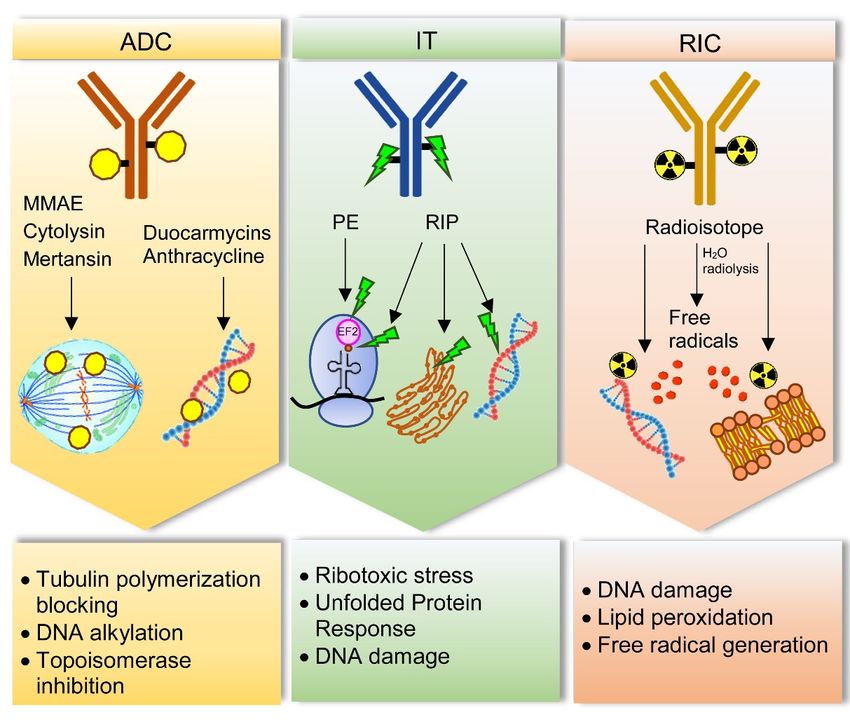
The main mechanisms by which drugs, toxins, and radionuclides can damage cancer
cells are schematized in Figure 1.
To date, many clinical trials have been conducted to investigate the efficacy and safety
of ICs in patients with hematological and solid cancers, administered alone or in combi-
nation with other therapeutic agents. In the last 20 years, Food and Drug Administration
approved 10 ADCs, 1 IT and 2 RICs for targeted cancer therapy [26–29].Biomedicines 2021,
Biomedicines 9, 978
2021, 9, 978 44of
of17
17
Figure 1. Main cell damage mechanisms induced by immunoconjugates for sarcoma treatment. Antibody–drug conjugates
Figure 1. Main cell damage mechanisms induced by immunoconjugates for sarcoma treatment. Antibody–drug conjugates
(ADC)
(ADC) cancan contain
contain drugs
drugs acting
acting with
with different
different mechanisms.
mechanisms. Monomethyl
Monomethyl auristatin
auristatin EE (MMAE),
(MMAE), cytolysin
cytolysin and
and mertansin
mertansin
block tubulin
block tubulin polymerization,
polymerization, thusthus hampering
hamperingcell
cellcycle.
cycle.Duocarmycins
Duocarmycinsand andanthracycline
anthracycline target DNA,
target DNA, inducing
inducing alkylation
alkyla-
and and
tion topoisomerase
topoisomerase inhibition, respectively.
inhibition, Immunotoxins
respectively. Immunotoxins (IT)(IT)
cancan
be constructed
be constructedwith Pseudomonas
with Pseudomonas exotoxin
exotoxin(PE) or
(PE)
ribosome-inactivating
or ribosome-inactivating proteins
proteins(RIPs).
(RIPs).PE
PEinhibits
inhibitselongation
elongationfactor-2
factor-2(EF2)
(EF2)through
through its
its ADP-ribosylation, thus
thus inducing
inducing
ribotoxic
ribotoxic stress
stress and
and protein
protein synthesis
synthesis blocking.
blocking. RIPs
RIPs can
can inhibit
inhibit protein
protein synthesis
synthesis through
through rRNA-N-glycosylase
rRNA-N-glycosylase activity
activity
removing
removing aa specific
specific adenine
adenine in
in aa stem–loop
stem–loop region
region ofof the
the main
main ribosomal
ribosomal RNA.
RNA. InIn addition,
addition, RIPs
RIPs can
can act
act on
on different
different
substrates
substratessuch
suchas as endoplasmic
endoplasmicreticulum,
reticulum,through
throughunfolded
unfoldedprotein
proteinresponse,
response,and
andDNA,
DNA,by bydirectly
directlydamage.
damage.Radioim-
Radioim-
munoconjugates (RIC) contain radioisotopes, which can damage DNA and cause lipid peroxidation in cell membranes.
munoconjugates (RIC) contain radioisotopes, which can damage DNA and cause lipid peroxidation in cell membranes. This
This effect can occur directly or indirectly through free radicals produced by water radiolysis.
effect can occur directly or indirectly through free radicals produced by water radiolysis.
3.3.Immunoconjugates
Immunoconjugatesfor forSarcoma
SarcomaTherapy
Therapy
Although
AlthoughICs ICshave
haveshowed
showedrelevant
relevant effects
effects mainly
mainly on on hematological
hematological malignancies,
malignancies,
numerous
numerous studies
studies have
have paved
paved the
the way
way to
to their
their application
application for for solid
solid tumors,
tumors, including
including
sarcomas.
sarcomas.Unlike
Unlikehematological
hematologicaltumors,
tumors,sarcomas
sarcomasas aswell
wellas asall
all solid
solid cancers
cancers have
have some
some
molecular
molecularand andmorphological
morphologicalcharacteristics
characteristicsthat
thatmake
makethem
themmoremoredifficult
difficultto
to treat
treat with
with
IC-based
IC-based therapy [30]. In particular, the difficulty of penetration inside tumor massre-
therapy [30]. In particular, the difficulty of penetration inside tumor mass is is
lated to the
related abundant
to the extracellular
abundant matrix,
extracellular disorganized
matrix, vasculature
disorganized and absence
vasculature of func-
and absence of
tional lymphatic
functional vessels
lymphatic thatthat
vessels causes increased
causes increasedinterstitial fluid
interstitial fluidpressure
pressure[31].
[31].However,
However,
protocol optimization, progressive
protocol progressive reduction
reductionofofICICsize
sizeand
andimplementation
implementation of of
penetration
penetra-
efficacy
tion are expected
efficacy to significantly
are expected improve
to significantly improvetargeted therapy
targeted therapyof solid tumors
of solid tumors[32,33].
[32,33].
3.1.Antibody–Drug
3.1. Antibody–DrugConjugates
Conjugatesfor
forSarcoma
Sarcoma
Many studies
Many studies have
havereported
reportedthe
theefficacy
efficacyof ADCs towards
of ADCs specific
towards antigens
specific expressed
antigens ex-
in different types of sarcoma.
pressed in different types of sarcoma.
Theendosialin/CD248/TEM1
The endosialin/CD248/TEM1 receptor
receptor is aistransmembrane
a transmembrane glycoprotein
glycoprotein expressed
expressed on
on pericytes and fibroblasts during embryogenesis. In adults, its presence dramatically
pericytes and fibroblasts during embryogenesis. In adults, its presence dramatically drops
drops in normal tissues while it is expressed in mesenchymal tumors, such as sarcoma,Biomedicines 2021, 9, 978 5 of 17
neuroblastoma, as well as in perivascular and tumor-associated stroma [34]. Moreover, it is
associated with tumor angiogenesis and inflammation [35,36]. In sarcomas, this antigen is
highly expressed on the surface of malignant, perivascular, and stromal cells, even on high
grade and advanced sarcoma [37,38]. Two anti-endosialin ADCs were tested in preclinical
models of sarcoma. The antitumor efficacy of the anti-endosialin-MC-VC-PABC-MMAE
was tested on two endosialin-positive human cell lines and one sarcoma xenograft model.
Inhibiting concentration 50 (IC50 ) values were 0.5 µg/mL for the Ewing sarcoma (ES) cell
line A-673 and 1.5 µg/mL for the osteosarcoma (OS) cells SJSA-1, without any correlation
between the extent of cell growth inhibition and endosialin expression levels. The antitumor
activity of this ADC was also tested in nude mice bearing A-673 cells xenografts. The
dose of 15 mg/kg of anti-endosialin-MC-VC-PABC-MMAE determined a marked and
durable inhibition of tumor growth leading to mice survival of 80% after day 150, thus
demonstrating the antitumor efficacy of this ADC [39]. The anti-endosialin ENDOS/ADC
was tested in sarcoma preclinical models. This IC is composed of a humanized anti-
endosialin mAb hMP-E-8.3 linked to a duocarmycin derivative alkylating agent. In this
case, SJSA-1 cells resulted more sensitive to ENDOS/ADC than A-673 cells, displaying
IC50 values of 0.8 nM and 8.6 nM, respectively. Moreover, in a SJSA-1 derived xenograft
model, mice treated with ENDOS/ADC showed a strong reduction of tumor volume [40].
Glycoprotein non-metastatic b (GPNMB) is a transmembrane protein involved in
bone differentiation and remodeling [41,42]. Different cancer cell types are characterized
by high levels of this glycoprotein, among them OS cells [43,44]. In addition, GPNMB
is involved in cancer migration, invasion, progression, and metastasis, as well as poor
patient prognosis [45,46]. The fully human IgG2 mAb CR011, which recognizes GPNMB
extracellular domain, was linked to MMAE in the Glembatumumab vedotin ADC. This
IC was tested in preclinical models of OS: 10 short-term cell cultures from patient-derived
OS, 5 standard OS cell lines and 4 xenograft cell lines. Glembatumumab vedotin had
a significant cytotoxic activity, displaying IC50 values lower than 55 µg/mL in most of
the treated cells. Moreover, ADC effect correlates with GPNMB expression levels [47].
Glembatumumab vedotin entered phase 2 clinical trial involving 22 patients (ranging
12–50 years) with recurrent or refractory OS. This ADC was administered intravenously
at 1.9 mg/kg/dose over 90 min on day 1; the treatment was repeated every 21 days for
up to 18 courses. The results showed a limited efficacy. In fact, only one patient had a
partial response and two maintained a stable tumor disease. No correlation was observed
between GPNMB expression and clinical response (NCT02487979).
Leucine-rich repeat containing 15 (LRRC15), a member of the Leucine-Rich Repeat
superfamily, is another target evaluated for ADC-based sarcoma therapy. LRRC15 is
overexpressed in cancer-associated fibroblasts and cancer cells from many epithelial and
mesenchymal solid tumors. In particular, it was reported that OS tissue samples had high
LRRC15 expression both on cancer and stroma cells [48]. ABBV-085 ADC is composed
of the anti-LRRC15 humanized IgG1 kappa antibody Ab1 conjugated to the antimitotic
drug MMAE. The antitumor efficacy of ABBV-085 was evaluated in a cancer+/stromal+
patient-derived xenograft (PDX) of OS. Results showed that ABBV-085 was extremely
effective in terms of tumor growth inhibition, in comparison to other standard OS therapies
(doxorubicin, ifosfamide, gemcitabine, cisplatin) [49,50]. A multicenter phase 1 dose-
escalation clinical trial of ABBV-085 is currently under investigation in patients with
advanced solid tumors, including undifferentiated pleomorphic sarcoma (NCT02565758).
CD56, also called Neural Cell Adhesion Molecule (NCAM), is a homophilic binding
glycoprotein present on the surface of neurons and glia where it has a prominent role
in neuronal adhesion and migration ability, neurite outgrowth, synapse formation and
synaptic plasticity [51,52]. CD56 can also be found in hematopoietic cells, above all in
natural killer cells, where it acts as an adhesion molecule [53]. CD56 is over-expressed
in different cancer types, like neuroblastoma, rhabdomyosarcoma (RMS) and most of the
STS, Wilms tumor, acute myeloid leukemia, glioma, and astrocytoma, as well as in several
carcinomas [54–56]. Lorvotuzumab mertansine (IMGN901) is an ADC composed of an anti-Biomedicines 2021, 9, 978 6 of 17
CD56 humanized N901 mAb conjugated to the maytansinoid DM1, via a stable disulfide
linker. IMGN901 was tested in vitro on two RMS and two ES cell lines showing a great
sensitivity, which is not always correlated to CD56 expression intensity. In vivo studies
were conducted in tumor xenograft models; stable complete responses were observed
in two out of seven RMS xenografts; even in this case there was not a strong correlation
between CD56 expression levels and treatment response. The response variability might
be due to factors other than CD56 expression, such as mitotic rate, chemoresistance to
tubulin targeting agents and/or intracellular processing of IMGN901 [57]. This conjugate
in a phase 1 trial on myeloma patients had demonstrated ample evidence of safety and
signals of clinical activity [58]. These results paved the way for a phase 2 clinical trial
where IMGN901 was evaluated in patients with relapsed or refractory Wilms tumor, RMS,
neuroblastoma, pleuropulmonary blastoma, malignant peripheral nerve sheath tumor,
or synovial sarcoma (SS). Patients received lorvotuzumab mertansine intravenously at
110 mg/m2 over 1–1.5 h on days 1 and 8; the treatment was repeated every 21 days for up to
17 courses in the absence of disease progression or unacceptable toxicity. Despite the high
level of CD56 found in all treated pediatric tumors, only few patients had a relevant clinical
response. This might be due to many factors: limited penetration of the conjugate/payload
into solid tumor cancer cells, presence of unrecognized CD56 isoforms that could interfere
with the binding/internalization process and tumor resistance to DM1 (NCT02452554).
Endoglin (ENG or CD105) is a homodimeric glycoprotein, expressed in endothelial
cells, bone marrow cells, and macrophages, and involved in embryogenesis, angiogenesis,
and vascular establishment as well as homeostasis [59,60]. In tumors its presence is
associated with neo-angiogenesis, which represents a key feature of malignant cancer. ENG
can be both a transmembrane protein acting as a co-receptor for transforming growth
factor-β, and a soluble extracellular matrix protein after cleavage by metalloproteinase
14 occurs. In sarcomas ENG is associated with poor outcome in ES patients, being a
key point in tumor cell plasticity, tumor progression and invasiveness [61]. OMTX703 is
an ADC composed of the anti-ENG mAb OMTX003, which recognize ENG extracellular
domain, linked to cytolysin. The antitumor efficacy of this ADC was evaluated in cell
lines, cell line-derived xenografts, and PDX of ES. After OMTX703 treatment, a potent
anti-proliferative effect was reported in the ES8 cell line with an IC50 value of 260.6 nM.
In the same cell line, it was observed a correlation between ENG expression level, which
was extremely high, and ADC internalization ability and cytotoxic effect. OMTX703
efficacy was assessed in a ES8 xenograft model (immunocompromised NOD-SCID-IL-
2Rgnull/null mice), where tumor growth was strikingly reduced with a 60 mg/kg dose
of ADC. Interestingly, immunohistochemistry studies confirmed that ENG expression
levels in xenograft tumors were quite similar to those found in parental cell lines in vitro.
Lastly, OMTX703 antineoplastic effect was evaluated in PDX models, which display the
highest ENG expression and better represent the typical heterogeneity of these tumors. It
was observed that OMTX703 (30 and 60 mg/kg) was able to produce a dose-dependent
antitumor response. A complete response rate of 60% was achieved at the end of the
treatment with the highest dose (60 mg/kg) [62].
The urokinase plasminogen activator receptor–associated protein uPARAP/Endo180
plays a crucial role in the process of collagen turnover. The receptor acts through the
endocytosis of extracellular matrix collagen, which is further addressed to lysosomal
degradation [63,64]. In normal tissues, this receptor is expressed in a limited range of
cell types involved in tissue development, such as fibroblasts and osteogenesis-associated
mesenchymal cells [65,66]. In cancer cells its expression was found to be high, especially
in OS and STS. In a preclinical study the antitumor efficacy of 2h9-vc-MMAE, an ADC
composed of an anti-uPARAP mAb linked to MMAE was evaluated in fibrosarcoma (FS)
and RMS cell lines (HT1080 and RD, respectively), expressing high levels of uPARAP. The
ADC cytotoxic mechanism is related to the binding and internalization properties as well
as to ADC lysosomal cleavage. The ADC was able to significantly reduce cell viability on
sarcoma cell lines even if to a lesser extent than on hematological cell lines. In addition,Biomedicines 2021, 9, 978 7 of 17
ADC efficacy was evaluated in mice xenografted with human leukemic cells. Complete
rescue of all treated animals was observed with no sign of adverse effects [67].
Receptor tyrosine kinase-like orphan receptors (ROR) are a family of transmembrane
tyrosine kinases. ROR1 is a Wnt5a receptor expressed during embryonic development
and in several hematologic and solid malignancies [68]. NBE-002 is an ADC consisting
of a humanized anti-ROR1 mAb conjugated to a derivative of the potent anthracycline
PNU-159682 [69]. NBE-002 is currently under evaluation in a phase 1/2 clinical trial in
patients (age ≥ 18 years) with advanced solid tumors, including sarcoma; NBE-002 has
been given intravenously on day 1 of repeated 21-day courses (NCT04441099).
ROR2 is one of the non-canonical Wnt receptors, which plays significant roles during
early embryonic development in several tissue types [70]. The protein may be involved in
the early formation of chondrocytes and in osteoblastogenesis [71]. ROR2 is overexpressed
during embryonic development and in several important cancer types, including sarcoma,
where its levels are strongly correlated with worst prognosis of patient [70]. CAB-ROR2-
ADC or BA3021 is a ROR2-targeting ADC composed of a conditionally active biologic (CAB)
anti-ROR2 antibody conjugated to an undisclosed payload [72]. This ADC is currently being
evaluated in a phase 1/2 clinical trial in patients (age ≥ 18 years) with locally advanced
unresectable or metastatic solid tumors, including STS (NCT03504488—recruiting status).
AXL is a member of receptor tyrosine kinases TAM family. AXL is widely expressed
in healthy cells and tissues, where it is involved in cell survival, phagocytic clearance
of dying cells, natural killer cell differentiation, and cell aggregation [73]. AXL is also
highly expressed on a variety of cancer types, OS included, where it plays a central role
in tumor proliferation, survival, stem cell phenotype, metastasis, and resistance to cancer
therapy [74,75]. CAB-AXL-ADC or BA3011 is composed of a CAB anti-AXL antibody
conjugated to an undisclosed payload [76]. This ADC is currently under investigation in a
phase 1/2 clinical trial in patients (age ≥ 18 years in phase 1; age ≥ 12 years in phase 2)
with advanced solid tumors including different types of sarcoma. In phase 1 trial all
patients will receive BA3011, while in phase 2 trial all patients will receive either BA3011
alone or in combination with nivolumab (NCT03425279—recruiting status). Enapotamab
Vedotin or HuMax-AXL-ADC is another anti-AXL ADC, firstly tested in vitro and in vivo
in preclinical models of non-small cell lung cancer [77]. It consists of a human AXL-
specific IgG1 conjugated to the cytotoxic agent MMAE. Enapotamab Vedotin is now being
evaluated in a phase 1/2 clinical trial in patients (age ≥ 18 years) with selected, relapsed
and advanced or metastatic solid tumors, sarcoma included, which no longer respond to
standard therapy (NCT02988817).
CD70 is a transmembrane antigen belonging to the tumor necrosis factor (TNF) lig-
and super family. Its interaction with CD27 receptor enhances cellular proliferation and
induces anti-apoptotic proteins playing a major role in T-cell costimulation. CD70 is often
upregulated in T- and B- cell lymphomas and in various solid tumors even if its exact role
during the disease onset and progression remains unknown [78]. CD70 was identified as a
specific and highly expressed surface protein in uterine leiomyosarcoma cell lines and in
clinical samples of this rare and aggressive gynecologic malignancy. The antihuman-CD70
mAb vorsetuzumab was conjugated to MMAF towards uterine leiomyosarcoma cell line
and its antitumor effects were evaluated in vitro and in vivo. This anti-CD70 ADC showed
a significant cytotoxicity on SK-LMS-1 cells, displaying IC50 equal to 0.120 nM and it
strongly inhibited tumor growth in SK-LMS-1 xenograft mouse models and in uterine
leiomyosarcoma PDX mouse models with a relative tumor reduction of 54.5% and 84.7%,
respectively [79].
ADCs tested in preclinical studies and in clinical trials are reported in Table 1.Biomedicines 2021, 9, 978 8 of 17
Table 1. Antibody–drug conjugates tested for sarcoma therapy.
ADC Target Antibody Drug Tumor In Vitro In Vivo Clinical Trial Ref.
anti-endosialin- Fully √ √
MC-VC-PABC- Endosialin human MMAE ES, OS - [39]
MMAE mAb
Duocarmycin √ √
ENDOS/ADC Endosialin hMP-E-8.3 ES, OS - [40]
derivative
Glemtumumab √ √ NCT02487979
GPNMB CR011 MMAE OS [47]
vedotin Phase 2
LRRC15 √ √ NCT02565758
ABBV-085 LRRC15 MMAE OS [49,50]
Ab1 Phase 1
√ √ NCT02452554
IMGN901 NCAM N901 DM1 RMS, ES [57]
Phase 2
√ √
OMTX703 Endoglin OMTX003 Cytolysin ES - [62]
√
2h9-vc-MMAE uPARAP 2h9 MMAE FS, RMS - - [67]
humanized PNU- √ NCT04441099
NBE-002 ROR1 S - [69]
mAb 159682 Phase 1/2
√ √ NCT03504488
BA3021 ROR2 CAB undisclosed STS [72]
Phase 1/2
√ √ NCT03425279
BA3011 AXL CAB undisclosed S [76]
Phase 1/2
Enapotamab human √ √ NCT02988817
AXL MMAE S [77]
vedotin IgG1-κ Phase 1/2
√ √
CD70-ADC CD70 vorsetuzumab MMAF uLMS - [79]
√
The symbols and - mean tested and not tested, respectively, in vitro, in vivo or in a clinical trial. Abbreviations: ES, Ewing sarcoma;
FS, fibrosarcoma; GPNMB, glycoprotein non-metastatic b; LRRC15, leucine-rich repeat containing 15; MMAE, monomethyl auristatin E;
MMAF, monomethyl auristatin F; NCAM, neural cell adhesion molecule; OS, osteosarcoma; RMS, rhabdomyosarcoma; ROR, receptor
tyrosine kinase-like orphan receptor; S, sarcoma (unspecified type); STS, soft tissue sarcoma; uLMS, uterine leiomyosarcoma; uPARAP,
urokinase plasminogen activator receptor–associated protein.
3.2. Immunotoxins for Sarcoma
Various ITs have been tested for sarcoma therapy, evaluating their binding, internal-
ization ability, and anti-tumor effect.
Chondroitin sulfate proteoglycan 4 (CSPG4) is a tumor-associated surface antigen,
firstly found on human melanoma cells [80]. It is used as a marker of proliferation and
metastasis in poor prognosis tumor types such as breast cancer and STS, whilst its expres-
sion is very low in healthy tissues [81]. The CSPG4-specific PE-based IT, αMCSP-ETA’, was
tested for RMS adjuvant therapy. Multiple parameters were evaluated in vitro on three
embryonal RMS cell lines (RD, FL-OH1 and TE-671) and one alveolar RMS cell line (Rh30).
IT binding was specific on CSPG4+ cells and IT internalization was rapid; αMCSP-ETA’
inhibited RMS cell proliferation with IC50 values ranging from 0.02 to 50 nM and induced
apoptosis. The binding was also evaluated ex vivo on three patient-derived paraffin-
embedded RMS tumor sections, exhibiting good specificity. Although preliminary, these
results highlighted the therapeutic potential of this IT (alone or combined with standard
drugs) for RMS treatments [82].
The well-known epidermal growth factor receptor (EGFR) is a member of the ErbB
tyrosine kinase receptor family. It is involved in routine cellular processes such as pro-
liferation, differentiation, and cellular development [83]. EGFR is highly expressed in
several solid cancers [84]. EGFR is overexpressed in up to 76% of embryonal RMS cases,
so it is considered a suitable target for RMS immunotherapy [85]. The EGFR-specific
recombinant IT 425(scFv)-ETA0 was tested in vitro on three different embryonal RMS cell
lines RD, FL-OH1 and TE-671. Experiments demonstrated binding specificity and valuable
internalization. Moreover, 425(scFv)-ETA0 was able to reduce cell viability (IC50 valuesBiomedicines 2021, 9, 978 9 of 17
in picomolar range) and to strongly activate apoptotic pathway. The EGFR+ cell binding
activity of the IT 425(scFv)-ETA0 was also demonstrated ex vivo on two patient-derived
formalin-fixed paraffin-embedded RMS specimens [86]. EGFR was also used as target of
an indirect IT, consisting of a primary EGFR specific mAb followed by a secondary F(ab’)2
anti-mouse Ig linked to saporin-S6. The indirect IT caused a significant inhibition of cell
growth and protein synthesis (IC50 0.95 nM) and a strong increase in apoptosis in RD/18
RMS cell line. The toxic activity of the anti-EGFR IT was also observed on RMS cell lines
expressing low levels of EGFR [87].
The glycoprotein gp72 is a tumor-associated cell surface antigen present in melanoma,
bladder and breast carcinoma and osteogenic sarcoma [88]. 791T/36-RTA derives from
the conjugation of the murine anti-gp72 mAb 791T/36 with ricin A chain (RTA) and it
specifically inhibited tumor cell growth in vitro in the OS cell line 791T. The cytotoxicity
of this IT depended primarily on its very rapid cell surface binding, endocytosis and
intracellular processing leading to the release of the toxic payload in the cytoplasm to
inhibit protein synthesis [89].
B7H3 is a cell surface glycoprotein expressed on cancer cells and not found on normal
tissues [90]. It is involved in natural killer and T cell inhibition, as well as in tumor cell
migration and invasion [91]. The recombinant IT 8H9(scFv)-PE38 was constructed with
the truncated form of PE (PE38) conjugated to the single-chain fragment variable (scFv) of
the anti-B7H3 mAb 8H9. The IT had a cytotoxic effect in vitro on three B7H3+ human OS
cell lines (U2OS, CRL1427, and OHS-M1), with IC50 values of 0.03, 0.05, and 0.02 µg/mL,
respectively. 8H9(scFv)-PE38 was also tested in vivo in xenograft SCID mice bearing OHS-
M1 cells. The results indicated that tumor regression was achievable using 0.15 mg/kg IT
without significant systemic toxicity for animals [92].
Another sarcoma-associated antigen is an 80 kDa surface glycoprotein recognized by
TP-3, a mAb that particularly reacts with OS. This antigen was found to be highly expressed
on OS and in some hemangiopericytoma, chondrosarcoma, malignant fibrous histiocytoma,
and synovial sarcoma. Healthy tissues exhibited a very low expression of this antigen [93].
TP-3 mAb was conjugated to pokeweed antiviral protein (PAP) and the cytotoxic effect
of this IT was tested in vitro on the human OS cell line OHS. TP-3-PAP was able to kill
TP-3+ cells in a specific and efficient manner, with IC50 values in the picomolar range.
Furthermore, the antitumor activity of this IT was assayed in vivo in a TP-3+ mouse model
bearing human sarcoma lung metastases with good results in terms of number and size
reduction of the metastases in a dose dependent manner [94]. Two recombinant TP-3 based
ITs were produced combining the toxin PE38 with the monovalent and bivalent disulfide-
stabilized Fv of the antibody, TP-3(dsFv)-PE38 and TP-3(dsFv)2 -PE38, respectively. These
ITs were tested in vitro on three human OS cell lines (OHS-M1, OHS, and SaOS). Results
indicated a specific effect for TP-3+ cells, with a great binding affinity. Bivalent IT was more
cytotoxic than monovalent IT, with IC50 of 4–42 ng/mL and 30–235 ng/mL, respectively.
The antitumor activity was tested in vivo in SCID mice bearing human OHS-M1 cells;
TP-3(dsFv)2 -PE38 showed a twofold increased effect compared to monovalent IT [95].
CD133 is a transmembrane glycoprotein, also known as AC133 or prominin-1, which
is used as a cellular marker of cancer stem cells (CSCs) in many different malignancies,
including sarcomas [96]. CSCs are usually a small subpopulation of cancer cells that
are responsible for chemoresistance, relapsed disease and metastasis [97]. Unfortunately,
normal stem cells, including hematopoietic, endothelial, and neuronal stem cells are CD133+
too. For this reason, new biotechnological strategies are fundamental to selectively kill
CSCs, rescuing other CD133+ cells. Photochemical internalization is a site-specific and
light-dependent drug delivery method that relies on the activation of a molecule, called
photosensitizer, which co-localizes with the therapeutic agent of interest in endo-lysosomal
compartments of the cells. The photosensitizer meso-tetraphenyl chlorin disulfonate
(TPCS2a) was used to perform photochemical internalization of two anti-CD133 ITs. These
ITs were obtained conjugating the biotinylated anti-CD133/1 (AC133) and anti-CD133/2
(293C) mAbs to streptavidin–saporin. The efficacy of this method was assessed on theBiomedicines 2021, 9, 978 10 of 17
undifferentiated human sarcoma cell line SW872, on the human FS cell line HT-1080 and
on SW872-derived mouse xenografts cells, obtaining specific cytotoxic effects. Moreover,
in vitro and in vivo experiments revealed a strong decrease in colony forming ability and a
great tumor initiation inhibition of the surviving cells after photochemical internalization
of the anti-CD133-saporin [98].
As discussed above, TEM1/endosialin/CD248 is a cell surface receptor highly ex-
pressed on human sarcomas that is considered a valid target for immunotherapeutic
treatments. The human scFv-Fc fusion protein (78Fc) specifically bound TEM1+ sarcoma
cell lines in vitro (SJSA-1, A673, MES-SA, and HOS) and sarcoma cells in xenografted
nude mice. The 78Fc was chemically conjugated to the plant toxin saporin to augment
its cytotoxicity. In vitro experiments revealed that 78Fc-Sap was able to specifically kill
TEM+ sarcoma cells with a significantly higher effect in comparison with saporin alone.
In vivo antitumor activity of 78Fc-Sap was assessed on SJSA-1 and A673 derived xenografts
showing a high and specific tumor growth inhibition with no systemic toxicity even at the
highest dose (0.2 mg/kg) [99].
As previously reported, the anti-endoglin mAb OMTX003 is a valid carrier to construct
therapeutic ICs against ES. OMTX003 was also conjugated to nigrin b A chain. OMTX503
IT was highly stable, its cell surface binding ability was specific for endoglin+ cells and
the cellular internalization was efficient. It showed an antiproliferative activity in vitro on
three ES cell lines with different level of endoglin expression (RM82, TC71 and CADO)
with IC50 values of 0.118 nM, 9.155 nM, and 17.38 nM, respectively. Thus, the cytotoxic
effect was related to the endoglin expression level. OMTX503 was also tested in vivo on
RM82-derived mouse xenografts at 0.5 mg/kg, obtaining good and significant results in
terms of tumor growth inhibition and cell viability reduction [62].
ITs tested in preclinical studies are reported in Table 2.
Table 2. Immunotoxins tested for sarcoma therapy.
Target Antibody Toxin Tumor In Vitro In Vivo Clinical Trial Ref.
√
CSPG4 αMCSP ETA’ RMS - - [82]
√
425 (scFv) ETA’ RMS - - [86]
EGFR murine mAb (clone √
Saporin RMS - - [87]
528)
√
gp72 791T/36 RTA OS - - [89]
√ √
B7H3 8H9 (scFv) PE38 OS - [92]
80 kDa sarcoma √ √
TP-3 PAP OS - [94]
associated antigen
80 kDa sarcoma TP-3 (dsFv) √ √
PE38 OS - [95]
associated antigen TP-3 (dsFv)2
AC133 √ √
CD133 Saporin S - [98]
293C
√ √
TEM1 78Fc Saporin S - [99]
Nigrin-b A √ √
Endoglin OMTX003 ES - [62]
chain
√
The symbols and - mean tested and not tested, respectively, in vitro, in vivo or in a clinical trial. Abbreviations: CSPG4, chondroitin
sulfate proteoglycan 4; dsFv, disulfide-linked fragment variable; EGFR, epidermal growth factor receptor; ES, Ewing sarcoma; ETA’,
truncated version of Pseudomonas exotoxin A; OS, osteosarcoma; PE, Pseudomonas exotoxin; RMS, rhabdomyosarcoma; RTA, ricin toxin
A-chain; S, sarcoma (unspecified type); scFv, single-chain fragment variable.Biomedicines 2021, 9, 978 11 of 17
3.3. Radioimmunoconjugates for Sarcoma
The first RIC approved in clinical practice was ibritumomab tiuxetan, in which the anti-
CD20 mAb rituximab is (radio)labeled with Yttrium-90 for the treatment of non-Hodgkin’s
lymphoma [100].
Despite most studies reported in literature describe the use of RICs for diagnostic
purposes, some works depict the attempts to apply RIT to the treatment of sarcomas.
CD146 is a cancer associated cell adhesion molecule (CAM) overexpressed in several
cancer types, including OS; it is associated with tumor progression, neoangiogenesis,
and vascular development [101]. Anti-CD146 murine mAb OI-3 was (radio)labeled with
Lutetium-177 or Iodine-125 and was tested in biodistribution/dosimetry experiments.
Results showed promising data in terms of RIC tumor uptake in nude mice bearing OHS
xenografts [102].
Insulin-like growth factor 2 receptor (IGF2R) is another valid target for OS treatment,
because it is overexpressed in several cell lines and patient-derived OS cells [103]. A novel
murine anti-IGF2R mAb, named 2G11, was (radio)labeled with Indium-111 to determine
biodistribution and tumor uptake in OS tumor bearing SCID mice. Successively, 2G11 was
(radio)labeled with both Lutetium-177 and Bismuth-213, obtaining good results in vivo in
terms of slowing down tumor growth, without local or systemic toxicity referred [104].
Frizzled homologue 10 (FZD10) is the main target used for synovial sarcoma (SS)
RIT. FZD10 is a transmembrane receptor of the Wnt signaling pathway whose gene is
upregulated specifically in SS, but not expressed in any normal human tissue except for
placenta [105]. The murine mAb 92–13 (radio)labeled with Yttrium-90 showed specific
binding ability to FZD10 in vitro and in vivo, good internalization into FZD10+ cells and
strong antitumor activity in SS mouse xenografts [106,107]. The humanized chimeric anti-
FZD10 mAb OTSA101 was (radio)labeled with Indium-111 or Yttrium-90. In a phase 1
clinical trial, 20 patients with advanced/recurrent SS received an injection of the Indium-
111-OTSA101 RIC to determine tumor uptake and biodistribution. Successively, only those
patients (n = 10) that showed a significant tumor uptake, were treated with Yttrium-90-
OTSA10. Unfortunately, no patients showed an objective tumor regression. In fact, best
overall response was a stable disease in 3 patients [108]. Adsorbed dose simulations can
explain tumor response on treated patients. The estimated biodistribution and dosimetry of
(radio)labeled anti-FZD10, in normal tissue and tumor, was evaluated through Monte Carlo-
based 3D simulations [109]. In a comparative preclinical study, OTSA101 was (radio)labeled
with both Yttrium-90 and Astatine-211. Astatine-211-OTSA101, an α-emitting anti-FZD10
RIC, suppressed tumor growth of SS mouse xenografts more efficiently than the same
dose of the Yttrium-90-OTSA101, a β-emitting anti FZD10 RIC, without remarkable toxic
side effects [110]. This confirmed that α-RIT is superior to β-RIT in treating solid tumors
because α-particles, with higher linear energy transfer, may have more advantages in terms
of cytotoxicity compared to β-particles, with lower linear energy transfer [111].
The glycoprotein B7H3 is expressed on desmoplastic small round tumor cells (DSRCT),
a rare sarcoma that affects adolescents and young adults involving the peritoneum. RIT
treatment against DSRCT was tested in a phase 1 clinical trial (NCT01099644) on 52 patients.
Murine anti-B7H3 mAb omburtamab (8H9) linked to Iodine-131 was administrated with
an intraperitoneal injection. Related toxicity was mild and transient in almost all patients
and adsorbed dose was low in normal tissues [112]. To date, Iodine-131-omburtamab is on
a phase 2 clinical trial to improve patient survival (NCT04022213—recruiting status).
RICs tested in preclinical studies and clinical trials are reported in Table 3.Biomedicines 2021, 9, 978 12 of 17
Table 3. Radioimmunoconjugates tested for sarcoma therapy.
In In Clinical
Target Antibody Radionuclide Half-life Emission Tumor Ref.
Vitro Vivo Trial
OI-3 125 I √ √
59.5 days Auger
CD146 CHOI-3.1 OS - [102]
177 Lu 6.7 days β- , Auger
CHOI-3.3
111 In 67.4 h γ √ √
IGF2R 2G11 177 Lu 6.7 days β- , Auger OS - [104]
213 Bi 46 min α
√ √
FZD10 92–13 90 Y 64.1 h β- SS - [106,107]
111 In 67.4 h γ √
FZD10 OTSA101 SS - Phase 1 [108,109]
90 Y 64.1 h β-
90 Y 64.1 h β- √
FZD10 OTSA101 211 At SS - - [110]
7.2 h α, Auger
√
B7H3 8H9 131 I 8.0 days β- DSRCT - Phase 2 [112]
√
The symbols and - mean tested and not tested, respectively, in vitro, in vivo or in a clinical trial. Abbreviations: DSRCT, desmoplastic
small round cell tumor; FZD10, frizzled homologue 10; IGF2R, insulin-like growth factor 2 receptor; OS, osteosarcoma; SS, synovial
sarcoma.
4. Conclusions
This review aims to provide a comprehensive overview of the latest advances in
sarcoma immunotherapy and their impact on clinical oncology. The IC studies reported
in this review show efficacy and clinical potential in sarcoma therapy. Although rare in
adults, sarcomas are more frequent among pediatric tumors. Sarcomas are characterized
by molecular and morphological complexity; the rarity and heterogeneity of sarcomas
induce clinicians and researchers to seek and validate personalized therapeutic approaches.
IC-based immunotherapy has been showing increasingly interesting results in terms of
anti-tumor efficacy beside to a reduction of side effects. These positive results depend
mainly on the possibility to select new engineered carrier moieties characterized by stability,
binding specificity and reduced immunogenicity. The results obtained in preclinical studies
with ICs in sarcoma models encouraged the translation from bench to bed.
The clinical studies over the last 20 years allowed nine ADCs to be approved by the
FDA and many others are in phase 3 clinical trial [113] in different neoplastic diseases.
Currently, seven ADCs and two RICs are under phase 1–2 clinical trials for sarcoma therapy
and many other ICs have been evaluated in preclinical studies.
We believe that, in the near future, antibody-based therapeutic approaches could
improve sarcoma patient outcome by overcoming some difficulties associated to standard
therapy, such as the tumor resistance to the anticancer drugs, leading to patient relapse,
and the onset of secondary malignancies [114,115].
Author Contributions: All the authors wrote the paper and collected the literature. All authors have
read and agreed to the published version of the manuscript.
Funding: This work was supported by funds for selected research topics from the Alma Mater
Studiorum, University of Bologna, by the Pallotti Legacies for Cancer Research and by Fondazione
CARISBO, Project 2019.0539.
Institutional Review Board Statement: Not applicable.
Informed Consent Statement: Not applicable.
Data Availability Statement: Not applicable.
Conflicts of Interest: The authors declare no conflict of interest.Biomedicines 2021, 9, 978 13 of 17
References
1. American Cancer Society–Cancer Statistics Center. Available online: https://cancerstatisticscenter.cancer.org/#!/ (accessed on 29
June 2021).
2. NCI—The Surveillance, Epidemiology, and End Results (SEER) Program. Available online: https://seer.cancer.gov/ (accessed on
29 June 2021).
3. Meyer, M.; Seetharam, M. First-Line Therapy for Metastatic Soft Tissue Sarcoma. Curr. Treat. Options Oncol. 2019, 20, 6. [CrossRef]
4. In, G.K.; Hu, J.S.; Tseng, W.W. Treatment of advanced, metastatic soft tissue sarcoma: Latest evidence and clinical considerations.
Ther. Adv. Med. Oncol. 2017, 9, 533–550. [CrossRef]
5. Van Cutsem, E.; Köhne, C.H.; Hitre, E.; Zaluski, J.; Chang Chien, C.R.; Makhson, A.; D’Haens, G.; Pintér, T.; Lim, R.; Bodoky, G.;
et al. Cetuximab and chemotherapy as initial treatment for metastatic colorectal cancer. N. Engl. J. Med. 2009, 360, 1408–1417.
[CrossRef] [PubMed]
6. Hochster, H.S.; Hart, L.L.; Ramanathan, R.K.; Childs, B.H.; Hainsworth, J.D.; Cohn, A.L.; Wong, L.; Fehrenbacher, L.; Abubakr, Y.;
Saif, M.W.; et al. Safety and efficacy of oxaliplatin and fluoropyrimidine regimens with or without bevacizumab as first-line
treatment of metastatic colorectal cancer: Results of the TREE Study. J. Clin. Oncol. 2008, 26, 3523–3529, Erratum in: J. Clin. Oncol.
2008, 26, 4697. [CrossRef]
7. Bolognesi, A.; Polito, L. Immunotoxins and other conjugates: Pre-clinical studies. Mini Rev. Med. Chem. 2004, 4, 563–583.
[CrossRef]
8. Polito, L.; Bortolotti, M.; Pedrazzi, M.; Bolognesi, A. Immunotoxins and other conjugates containing saporin-s6 for cancer therapy.
Toxins 2011, 3, 697–720. [CrossRef]
9. Bolognesi, A.; Bortolotti, M.; Maiello, S.; Battelli, M.G.; Polito, L. Ribosome-Inactivating Proteins from Plants: A Historical
Overview. Molecules 2016, 21, 1627. [CrossRef] [PubMed]
10. Khongorzul, P.; Ling, C.J.; Khan, F.U.; Ihsan, A.U.; Zhang, J. Antibody-Drug Conjugates: A Comprehensive Review. Mol. Cancer
Res. 2020, 18, 3–19. [CrossRef] [PubMed]
11. Thomas, A.; Teicher, B.A.; Hassan, R. Antibody-drug conjugates for cancer therapy. Lancet Oncol. 2016, 17, 254–262. [CrossRef]
12. Sievers, E.L.; Senter, P.D. Antibody-drug conjugates in cancer therapy. Annu. Rev. Med. 2013, 64, 15–29. [CrossRef] [PubMed]
13. Nepali, K.; Ojha, R.; Lee, H.Y.; Liou, J.P. Early investigational tubulin inhibitors as novel cancer therapeutics. Expert Opin. Investig.
Drugs 2016, 25, 917–936. [CrossRef]
14. Hoffmann, R.M.; Coumbe, B.G.T.; Josephs, D.H.; Mele, S.; Ilieva, K.M.; Cheung, A.; Tutt, A.N.; Spicer, J.F.; Thurston, D.E.;
Crescioli, S.; et al. Antibody structure and engineering considerations for the design and function of Antibody Drug Conjugates
(ADCs). Oncoimmunology 2017, 7, 1395127. [CrossRef] [PubMed]
15. Chau, C.H.; Steeg, P.S.; Figg, W.D. Antibody-drug conjugates for cancer. Lancet 2019, 394, 793–804. [CrossRef]
16. Polito, L.; Bortolotti, M.; Farini, V.; Battelli, M.G.; Barbieri, L.; Bolognesi, A. Saporin induces multiple death pathways in
lymphoma cells with different intensity and timing as compared to ricin. Int. J. Biochem. Cell Biol. 2009, 41, 1055–1061. [CrossRef]
[PubMed]
17. Polito, L.; Bortolotti, M.; Pedrazzi, M.; Mercatelli, D.; Battelli, M.G.; Bolognesi, A. Apoptosis and necroptosis induced by
stenodactylin in neuroblastoma cells can be completely prevented through caspase inhibition plus catalase or necrostatin-1.
Phytomedicine 2016, 23, 32–41. [CrossRef]
18. Weidle, U.H.; Tiefenthaler, G.; Schiller, C.; Weiss, E.H.; Georges, G.; Brinkmann, U. Prospects of bacterial and plant protein-based
immunotoxins for treatment of cancer. Cancer Genom. Proteom. 2014, 11, 25–38.
19. Gilabert-Oriol, R.; Weng, A.; von Mallinckrodt, B.; Melzig, M.F.; Fuchs, H.; Thakur, M. Immunotoxins constructed with ribosome-
inactivating proteins and their enhancers: A lethal cocktail with tumor specific efficacy. Curr. Pharm. Des. 2014, 20, 6584–6643.
[CrossRef]
20. Mansoori, B.; Mohammadi, A.; Davudian, S.; Shirjang, S.; Baradaran, B. The Different Mechanisms of Cancer Drug Resistance: A
Brief Review. Adv. Pharm. Bull. 2017, 7, 339–348. [CrossRef]
21. Polito, L.; Djemil, A.; Bortolotti, M. Plant Toxin-Based Immunotoxins for Cancer Therapy: A Short Overview. Biomedicines 2016, 4,
12. [CrossRef]
22. Gill, M.R.; Falzone, N.; Du, Y.; Vallis, K.A. Targeted radionuclide therapy in combined-modality regimens. Lancet Oncol. 2017, 18,
414–423. [CrossRef]
23. Steiner, M.; Neri, D. Antibody-radionuclide conjugates for cancer therapy: Historical considerations and new trends. Clin. Cancer
Res. 2011, 17, 6406–6416. [CrossRef]
24. Pandit-Taskar, N. Targeted Radioimmunotherapy and Theranostics with Alpha Emitters. J. Med. Imaging Radiat. Sci. 2019, 50,
S41–S44. [CrossRef]
25. Gopal, A.K.; Gooley, T.A.; Maloney, D.G.; Petersdorf, S.H.; Eary, J.F.; Rajendran, J.G.; Bush, S.A.; Durack, L.D.; Golden, J.;
Martin, P.J.; et al. High-dose radioimmunotherapy versus conventional high-dose therapy and autologous hematopoietic stem
cell transplantation for relapsed follicular non-Hodgkin lymphoma: A multivariable cohort analysis. Blood 2003, 102, 2351–2357.
[CrossRef]
26. Biochempeg. Available online: https://www.biochempeg.com/article/74.html (accessed on 29 June 2021).
27. ClinicalTrials.gov. Available online: https://clinicaltrials.gov/ct2/results?cond=&term=immunoconjugate&cntry=&state=
&city=&dist= (accessed on 29 June 2021).Biomedicines 2021, 9, 978 14 of 17
28. Drugbank. Available online: https://go.drugbank.com/unearth/q?utf8=%E2%9C%93&query=immunoconjugate&searcher=
drugs (accessed on 29 June 2021).
29. Kim, E.G.; Kim, K.M. Strategies and Advancement in Antibody-Drug Conjugate Optimization for Targeted Cancer Therapeutics.
Biomol. Ther. (Seoul) 2015, 23, 493–509. [CrossRef] [PubMed]
30. Govindan, S.V.; Sharkey, R.M.; Goldenberg, D.M. Prospects and progress of antibody-drug conjugates in solid tumor therapies.
Expert Opin. Biol. Ther. 2016, 16, 883–893. [CrossRef] [PubMed]
31. Minchinton, A.I.; Tannock, I.F. Drug penetration in solid tumours. Nat. Rev. Cancer 2006, 6, 583–592. [CrossRef] [PubMed]
32. Trédan, O.; Galmarini, C.M.; Patel, K.; Tannock, I.F. Drug resistance and the solid tumor microenvironment. J. Natl. Cancer Inst.
2007, 99, 1441–1454. [CrossRef]
33. Tiller, K.E.; Tessier, P.M. Advances in Antibody Design. Annu. Rev. Biomed. Eng. 2015, 17, 191–216. [CrossRef]
34. Thway, K.; Robertson, D.; Jones, R.L.; Selfe, J.; Shipley, J.; Fisher, C.; Isacke, C.M. Endosialin expression in soft tissue sarcoma as a
potential marker of undifferentiated mesenchymal cells. Br. J. Cancer 2016, 115, 473–479. [CrossRef]
35. Naylor, A.J.; Azzam, E.; Smith, S.; Croft, A.; Poyser, C.; Duffield, J.S.; Huso, D.L.; Gay, S.; Ospelt, C.; Cooper, M.S.; et al. The
mesenchymal stem cell marker CD248 (Endosialin) is a negative regulator of bone formation in mice. Arthritis Rheum. 2012, 64,
3334–3343. [CrossRef]
36. Simonavicius, N.; Ashenden, M.; van Weverwijk, A.; Lax, S.; Husoi, D.L.; Buckley, C.D.; Huijbers, I.J.; Yarwood, H.; Isacke, C.M.
Pericytes promote selective vessel regression to regulate vascular patterning. Blood 2012, 120, 1516–1527. [CrossRef]
37. Rouleau, C.; Curiel, M.; Weber, W.; Smale, R.; Kurtzberg, L.; Mascarello, J.; Berger, C.; Wallar, G.; Bagley, R.; Honma, N.; et al.
Endosialin protein expression and therapeutic target potential in human solid tumors: Sarcoma versus carcinoma. Clin. Cancer
Res. 2008, 14, 7223–7236. [CrossRef]
38. Rouleau, C.; Smale, R.; Fu, Y.S.; Hui, G.; Wang, F.; Hutto, E.; Fogle, R.; Jones, C.M.; Krumbholz, R.; Roth, S.; et al. Endosialin is
expressed in high grade and advanced sarcomas: Evidence from clinical specimens and preclinical modeling. Int. J. Oncol. 2011,
39, 73–89. [CrossRef] [PubMed]
39. Rouleau, C.; Gianolio, D.A.; Smale, R.; Roth, S.D.; Krumbholz, R.; Harper, J.; Munroe, K.J.; Green, T.L.; Horten, B.C.; Schmid, S.M.;
et al. Anti-Endosialin Antibody-Drug Conjugate: Potential in Sarcoma and Other Malignancies. Mol. Cancer Ther. 2015, 14,
2081–2089. [CrossRef] [PubMed]
40. Capone, E.; Piccolo, E.; Fichera, I.; Ciufici, P.; Barcaroli, D.; Sala, A.; De Laurenzi, V.; Iacobelli, V.; Iacobelli, S.; Sala, G. Generation
of a novel Antibody-Drug Conjugate targeting endosialin: Potent and durable antitumor response in sarcoma. Oncotarget 2017, 8,
60368–60377. [CrossRef]
41. Selim, A.A.; Abdelmagid, S.M.; Kanaan, R.A.; Smock, S.L.; Owen, T.A.; Popoff, S.N.; Safadi, F.F. Anti-osteoactivin antibody
inhibits osteoblast differentiation and function in vitro. Crit. Rev. Eukaryot. Gene Expr. 2003, 13, 265–275. [CrossRef]
42. Sheng, M.H.; Wergedal, J.E.; Mohan, S.; Lau, K.H. Osteoactivin is a novel osteoclastic protein and plays a key role in osteoclast
differentiation and activity. FEBS Lett. 2008, 582, 1451–1458. [CrossRef] [PubMed]
43. Kubista, B.; Klinglmueller, F.; Bilban, M.; Pfeiffer, M.; Lass, R.; Giurea, A.; Funovics, P.T.; Toma, C.; Dominkus, M.; Kotz, R.; et al.
Microarray analysis identifies distinct gene expression profiles associated with histological subtype in human osteosarcoma. Int.
Orthop. 2011, 35, 401–411. [CrossRef]
44. Taya, M.; Hammes, S.R. Glycoprotein Non-Metastatic Melanoma Protein B (GPNMB) and Cancer: A Novel Potential Therapeutic
Target. Steroids 2018, 133, 102–107. [CrossRef]
45. Rich, J.N.; Shi, Q.; Hjelmeland, M.; Cummings, T.J.; Kuan, C.T.; Bigner, D.D.; Counter, C.M.; Wang, X.F. Bone-related genes
expressed in advanced malignancies induce invasion and metastasis in a genetically defined human cancer model. J. Biol. Chem.
2003, 278, 15951–15957. [CrossRef]
46. Zhou, L.T.; Liu, F.Y.; Li, Y.; Peng, Y.M.; Liu, Y.H.; Li, J. Gpnmb/osteoactivin, an attractive target in cancer immunotherapy.
Neoplasma 2012, 59, 1–5. [CrossRef]
47. Roth, M.; Barris, D.M.; Piperdi, S.; Kuo, V.; Everts, S.; Geller, D.; Houghton, P.; Kolb, E.A.; Hawthorne, T.; Gill, J.; et al. Targeting
Glycoprotein NMB With Antibody-Drug Conjugate, Glembatumumab Vedotin, for the Treatment of Osteosarcoma. Pediatr. Blood
Cancer 2016, 63, 32–38. [CrossRef] [PubMed]
48. Cui, J.; Dean, D.; Wei, R.; Hornicek, F.J.; Ulmert, D.; Duan, Z. Expression and clinical implications of leucine-rich repeat containing
15 (LRRC15) in osteosarcoma. J. Orthop. Res. 2020, 38, 2362–2372. [CrossRef] [PubMed]
49. Purcell, J.W.; Tanlimco, S.G.; Hickson, J.; Fox, M.; Sho, M.; Durkin, L.; Uziel, T.; Powers, R.; Foster, K.; McGonigal, T.; et al. LRRC15
Is a Novel Mesenchymal Protein and Stromal Target for Antibody-Drug Conjugates. Cancer Res. 2018, 78, 4059–4072. [CrossRef]
[PubMed]
50. Ben-Ami, E.; Perret, R.; Huang, Y.; Courgeon, F.; Gokhale, P.C.; Laroche-Clary, A.; Eschle, B.K.; Velasco, V.; Le Loarer, F.;
Algeo, M.P.; et al. LRRC15 Targeting in Soft-Tissue Sarcomas: Biological and Clinical Implications. Cancers 2020, 12, 757.
[CrossRef] [PubMed]
51. Thiery, J.P.; Duband, J.L.; Rutishauser, U.; Edelman, G.M. Cell adhesion molecules in early chicken embryogenesis. Proc. Natl.
Acad. Sci. USA 1982, 79, 6737–6741. [CrossRef]
52. Huang, R.; Yuan, D.J.; Li, S.; Liang, X.S.; Gao, Y.; Lan, X.Y.; Qin, H.M.; Ma, Y.F.; Xu, G.Y.; Schachner, M.; et al. NCAM regulates
temporal specification of neural progenitor cells via profilin2 during corticogenesis. J. Cell Biol. 2020, 219, e201902164. [CrossRef]You can also read