Automating multimodal microscopy with NanoJ-Fluidics - bioRxiv
←
→
Page content transcription
If your browser does not render page correctly, please read the page content below

bioRxiv preprint first posted online May. 14, 2018; doi: http://dx.doi.org/10.1101/320416. The copyright holder for this
preprint (which was not peer-reviewed) is the author/funder. All rights reserved. No reuse allowed without permission.

          Automating multimodal microscopy with
                     NanoJ-Fluidics
Pedro Almada1,2 * , Pedro M. Pereira1-3 * , Siân Culley1-3 , Ghislaine Caillol4 , Fanny Boroni-Rueda4 , Christina L. Dix1 , Romain F.
           Laine1,3 , Guillaume Charras5,6 , Buzz Baum1,6 , Christophe Leterrier4 , and Ricardo Henriques1-3

                                          1
                                            MRC-Laboratory for Molecular Cell Biology. University College London, London, UK
                                     2
                                         Department of Cell and Developmental Biology, University College London, London, UK
                                                                    3
                                                                      The Francis Crick Institute, London, UK
                                                      4
                                                        Aix Marseille Université, CNRS, INP UMR7051, Marseille, France
                                                               5
                                                                 London Centre for Nanotechnology, London, UK
                                           6
                                             Institute for the Physics of Living Systems, University College London, London, UK
                                                                           *
                                                                             Equal contributing authors

Fluorescence microscopy can reveal all aspects of cellular mech-
anisms, from molecular details to dynamics, thanks to ap-
proaches such as super-resolution and live-cell imaging. Each
of its modalities requires specific sample preparation and imag-
ing conditions to obtain high-quality, artefact-free images, ulti-
mately providing complementary information. Combining and
multiplexing microscopy approaches is crucial to understand
cellular events, but requires elaborate workflows involving mul-
tiple sample preparation steps. We present a robust fluidics ap-
proach to automate complex sequences of treatment, labelling
and imaging of live and fixed cells. Our open-source NanoJ-
Fluidics system is based on low-cost LEGO hardware controlled
by ImageJ-based software and can be directly adapted to any
                                                                                      Fig. 1. Schematics of the NanoJ-Fluidics system a) 3D side view of a single
microscope, providing easy-to-implement high-content, multi-
                                                                                      LEGO syringe pump unit with syringe attached. b) 2D top view of a syringe pump
modal imaging with high reproducibility. We demonstrate its                           array (representing 4 pumps out of 128 maximum) and a fluid extraction peristaltic
capacity to carry out complex sequences of experiments such                           pump, both controlled by an Arduino® UNO. c) Example of possible workflow.
as super-resolved live-to-fixed imaging to study actin dynamics;
highly-multiplexed STORM and DNA-PAINT acquisitions of                                hampers their reproducibility and throughput, limiting their
multiple targets; and event-driven fixation microscopy to study
                                                                                      appeal for quantitative work (8).
the role of adhesion contacts in mitosis.
                                                                                      Automated fluid handling using microfluidic chips presents
Automation | Fluidics | LEGO | Live-cell imaging | Super-Resolution Mi-               an attractive alternative, but adds specific constrains on cul-
croscopy | High-Content | Correlative Imaging | STORM | DNA-PAINT
Correspondence: christophe.leterrier@univ-amu.fr, r.henriques@ucl.ac.uk
                                                                                      turing conditions and sample preparation (5, 9). A more
                                                                                      simple and tractable method would automate fluid exchange
                                                                                      in commonly used open imaging chambers, in a manner
Introduction                                                                          similar to standard laboratory protocols, while being eas-
Fluorescence microscopy is ubiquitously used to observe cel-                          ily adaptable to existing microscope stages. For this, we
lular processes, thanks to its ease of use, exquisite sensi-                          devised an easy-to-implement open-source system called
tivity and molecular specificity. It is generally performed                           NanoJ-Fluidics (Fig. 1a-b), which primarily consists of an
using dedicated sample preparation procedures, tailored to                            automated computer-controlled syringe pump array capa-
achieve optimal imaging conditions for each chosen tech-                              ble of rapidly and robustly exchanging sample conditions.
nique. Obtaining the best possible temporal and spatial reso-                         This allows the automation of the protocols of sample treat-
lution, while keeping a high signal, capacity for deep imaging                        ment, labelling and preparation directly on the microscope
and live-cell compatibility is nearly impossible with a "one-                         stage (Fig. 1c and S1). Our approach makes complex multi-
size fits all” approach. As such, each microscopy method en-                          modal imaging protocols highly accessible to researchers. To
tails a compromise between some of these features (1). Al-                            demonstrate this, we performed a range of complex multi-
ternatively, unique insights can be gained by combining in-                           step experiments: first, we devised live-to-fixed experiments
formation from multiple approaches, but at the cost of com-                           with time-lapse imaging of living cells expressing reporter
plex correlative workflows (2, 3). Recent developments to-                            probes, followed by in-situ online fixation, labelling and
ward high-resolution imaging of a large number of molecular                           super-resolved imaging, thus connecting the dynamics and
targets have further broadened the variety and complexity of                          structural dimensions of cellular processes (Fig. 2 and Movie
imaging procedures, with the use of multiple rounds of la-                            S1). Next, we used NanoJ-Fluidics to automate a highly-
belling and imaging (4–7). These elaborate protocols involve                          multiplexed STORM and DNA-PAINT acquisition to obtain
sequences of sample imaging, washing and labelling. Car-                              a five-colour nanoscopic image of distinct cellular targets
rying these protocols in a non-automated manner critically                            (Fig. 3 and Movie S2). Finally, we showcased the capacity

                                                                                                      Almada & Pereira et al.     |   bioR‰iv   |   May 14, 2018   |   1–12
bioRxiv preprint first posted online May. 14, 2018; doi: http://dx.doi.org/10.1101/320416. The copyright holder for this
preprint (which was not peer-reviewed) is the author/funder. All rights reserved. No reuse allowed without permission.
Live-to-fixed super-resolution microscopy. Although
super-resolution microscopy in living cells is achievable with
techniques such as SIM (12), RESOLFT (13) and SRRF (14),
near molecular scale super-resolution microscopy using
single-molecule localisation microscopy (SMLM) is typi-
cally limited to fixed-cell imaging . This is due to the require-
ment for non-physiological conditions, including the use of
anti-fading media and high-intensity illumination (15, 16)
typically required for commonly used approaches such as
STORM(17, 18) and DNA-PAINT (19). NanoJ-Fluidics ad-
dresses this issue, by allowing the combination of both dy-
namic live-cell and subsequent fixed-cell high-resolution ob-
servations on the same set of cells.
To demonstrate this, we carried out a sequence of live-cell
imaging, fixation, labelling and STORM imaging of actin
within COS cells (Fig. 2a). Thanks to its compatibility with
most standard microscopes and cell friendly low-illumination
Fig. 2. Super-resolution live-to-fixed cell imaging of actin in COS-7 cells a)
requirements, the SRRF approach was chosen to achieve
NanoJ-Fluidics workflow used for live-to-fixed super-resolution imaging. b) HILO
and SRRF microscopy images of a COS-7 cell expressing UtrCH-GFP imaged ev- live-cell super-resolution imaging of GFP labelled utrophin
ery 10 min for 150 min (a zoomed region of the imaged cell is shown at 30 minutes calponin homology domain, a validated actin probe (20)
intervals, Sup. Movie S1 shows extended time-lapse and field-of-view). c) HILO
(Fig. 2b). SRRF achieved an improved resolution (≥175 nm,
and SRRF microscopy images of UtrCH-GFP at t = 150 min and the corresponding
STORM image after fixation and staining with phalloidin-AF647. Scale bars are 10 S4) over the standard wide-field images (HILO, ≥270 nm,
µm. S4), resolving the assembly and disassembly of actin bun-
dles during cell-shape changes (Fig. 2b and Sup. Movie S1).
for high-content correlative imaging with event-driven fixa- We then used NanoJ-Fluidics to perform automated fixation,
tion, studying the role of adhesion contacts for cells in mito- washing, permeabilisation and phalloidin-AF647 staining, a
sis (Fig. 4 and Movie S3). sequence lasting 100 min (Fig. 2a). We finally performed a
STORM acquisition on the previously imaged cell after per-
fusing the optimised imaging buffer (Fig. 2c). The structural
Results detail of the actin organisation observed by STORM is con-
The NanoJ-Fluidics framework. NanoJ-Fluidics is a com- siderably higher than the live-cell observation, with an esti-
plete system, using off-the-shelf components, as well as mated resolution of ≥43 nm. An extended characterisation of
open-source control software. The hardware component con- the resolutions achieved here is described in Sup. Note 2 and
sists of compact LEGO® syringe pumps (Fig. 1a) that can Fig. S4, using NanoJ-SQUIRREL (21). Our system thus pro-
be configured as a multiplexed array of up to 128 syringes vides easy correlative live-cell and fixed-cell super-resolution
(Fig. 1b), plus a peristaltic pump and an open-source Ar- imaging. Interestingly, these observations also make it pos-
duino® electronic controller to interface with the microscope sible to evaluate if there are unwanted structural changes to
acquisition computer (Fig. 1b). By using LEGO parts, the cells caused by the fixation process (shown and discussed in
system is cost-effective, robust and repeatable thanks to the Sup. Note 3).
high-quality standards and low tolerances of the LEGO man-
Multiplexed super-resolution microscopy with STORM
ufacturing process. The system performs labelling and treat-
and DNA-PAINT. Obtaining a super-resolution, high-quality
ment protocols that were traditionally done on the bench, but
multi-channel image has long been a challenge in SMLM as a
these steps can be directly automated on the microscope stage
consequence of the difficulty to optimise labelling and imag-
(Fig. S1). For instance, a wash step will completely remove
ing for many labels simultaneously (22). NanoJ-Fluidics
liquid from the imaging chamber and replenish it with wash
is ideally suited for large channel multiplexing via sequen-
buffer. The system is easy to set up (Sup. Note 1), highly
tial exchange of fluorescent labels and/or imaging buffers.
modular and compatible with most microscopes and exper-
Furthermore, recent DNA-PAINT modalities can reach more
imental workflows (Fig. S1). NanoJ-Fluidics does not re-
than the typical 2-3 label channels by using antibodies cou-
quire any design and microfabication process and simply uses
pled to orthogonal DNA strands, and sequential labelling
common labware (Fig. S2). The software component is pro-
combined with imaging sequences of PAINT imagers (23–
vided as an ImageJ/Micro-Manager plugin (10, 11) as well as
25).
a stand-alone package for independent fluidics control (Sup.
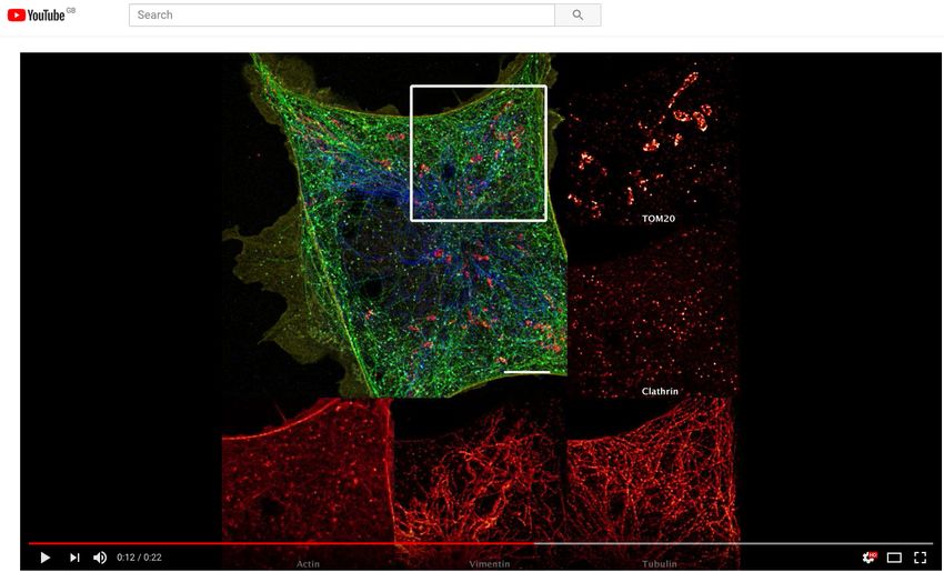
Here, we demonstrate how NanoJ-Fluidics easily handles se-
Software). By allowing precise control of each steps in the
quential STORM and DNA-PAINT acquisitions in a sim-
protocol (Fig. S3), NanoJ-Fluidics provides a highly repeat-
ple and optimal manner. Fixed cells were labelled using
able and robust way to carry out imaging experiments directly
DNA-coupled primary antibodies targeting mitochondria, vi-
onto the microscope.
mentin, microtubules and clathrin, as well as fluorescent
phalloidin to label actin. NanoJ-Fluidics was set up to carry
2 | bioR‰iv Almada & Pereira et al. | NanoJ-Fluidics
bioRxiv preprint first posted online May. 14, 2018; doi: http://dx.doi.org/10.1101/320416. The copyright holder for this
preprint (which was not peer-reviewed) is the author/funder. All rights reserved. No reuse allowed without permission.

Fig. 3. Automated DNA-PAINT and STORM imaging a) NanoJ-Fluidics workflow used for STORM and DNA-PAINT imaging. b) Left, full view of a cell showing 5-channel
merge of STORM and DNA-PAINT with actin (yellow), vimentin (blue), tubulin (green), clathrin (cyan) and mitochondria (red). Right, zoom of the boxed area. c) Single-channel
image of each imaged target (top), with insets (bottom) showing a zoom of the boxed area. A movie corresponding to this experiment is available as Sup. Movie S2. Scale
bar corresponds to 10 µm for full images and 2 µm for zoom.

out imaging buffer exchange for STORM imaging of actin                                   high-resolution.
(26), followed by two rounds of washing and labelling for
2-colour DNA-PAINT imaging of the other targets (Fig. 3a).
The first round imaged mitochondria and vimentin whereas
the second round imaged clathrin and tubulin. Fig. 3b-c re-                              We first blocked asynchronous cells in G2 via treatment with
spectively show the resulting 5-colour image and the individ-                            a CDK1 inhibitor (RPE1 cells expressing zyxin-GFP). Next,
ual channels, along with zooms on cellular regions highlight-                            the cell cycle was released by exchanging the inhibitor by
ing the high resolution obtained by this combined STORM                                  growth media using NanoJ-Fluidics (Fig. 4a) and imaged
and DNA-PAINT scheme (≥68 nm minimum resolution, ex-                                     by live-cell time-lapse microscopy (Fig. 4b). When the ob-
cept actin rendering which has a 97 nm minimum, character-                               served cells reached a minimal area (an indicator of mitotic
isation shown in Sup. Note 2 and Fig. S5).                                               rounding), the fixative was injected (Fig. 4a-b). A consid-
                                                                                         erable portion of cells (≥10%) were found to be fixed in a
Fixation-on-event imaging. NanoJ-Fluidics has also the                                   similar rounding state due to the simultaneous release of the
advantage of allowing sample treatments, such as fixation,                               G2 block (insets of Fig. 4c I, Sup. Movie S3). The sample
at precise times during the experiment. Thanks to the in-                                was then immunolabelled for b1-integrin, plus co-stained for
tegration of NanoJ-Fluidics with the image acquisition, de-                              F-actin and DNA, then imaged (Fig. 4 c III-VI). Both actin
termining the time of treatment can be triggered by visual                               and DNA staining allowed a secondary visual validation of
queues obtained from the imaging. To demonstrate this ca-                                pre-mitotic cell rounding. As we recently described using
pacity, we carried out an experiment observing the state of                              comparable experiments (28), b1-integrin is shown to retain
focal adhesions, as mammalian cells progress into division.                              a similar spatial pattern similar to zyxin when the cells were
Fixation was triggered by the observation of the rounding of                             spread on the substrate, in their pre-division shape (Fig. 4d).
the cells as they approach mitosis (27). Also, in order to fully                         We observed that, while zyxin retracts during cell rounding
exploit the fluidics automation of NanoJ-Fluidics, we com-                               (Fig. 4b), b1-integrin remains in its original position (Fig. 4d
bined it with tiling imaging and image stitching in order to                             and Sup. Movie S3), helping guide daughter cell migration
obtain fields-of-view of several millimetres while conserving                            (28).

Almada & Pereira et al.   |   NanoJ-Fluidics                                                                                                                 bioR‰iv   |   3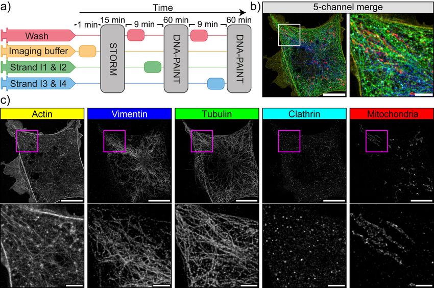
bioRxiv preprint first posted online May. 14, 2018; doi: http://dx.doi.org/10.1101/320416. The copyright holder for this
preprint (which was not peer-reviewed) is the author/funder. All rights reserved. No reuse allowed without permission.

Fig. 4. Event-driven fixation of cells upon mitotic rounding a) NanoJ-Fluidics workflow of the protocol event-driven treatment performed here. b) Stills of RPE1 zyxin-GFP
live-cell time-lapse during mitotic rounding. Scale bar, 20 µm.c) Stitched mosaic (17x17 individual regions) of: I- First frame of the live-cell time-lapse; II- Last frame of the
live-cell time-lapse; III- RPE1 zyxin-GFP cells immunolabelled for active b1-integrin; IV- Overlay of RPE1 zyxin-GFP cells immunolabelled for active b1-integrin and stained
for F-actin (with phalloidin-TRITC) and DNA (with DAPI). Insets represent cells where mitotic rounding was observed (Sup. Movie S3), dashed inset is the cell in b) and d).
Scale bar is 1 mm. d) I - Maximum intensity projection of the first 12 min in b); II - Active b1-integrin staining; III - Overlay of both panels. Scale bar, 20 µm.

Discussion                                                                                  imaging (Fig. 2) enables a synergy between the dynamic
                                                                                            but less spatially resolved information obtained from live-cell
We introduce NanoJ-Fluidics (Fig. 1) and demonstrate its ap-                                imaging and the near molecular-scale structural information
plicability in multiple experimental contexts: in-situ correla-                             provided by SMLM. The NanoJ-Fluidics framework allows
tive live-to-fixed super-resolution imaging (Fig. 2); multi-                                to do this in-situ without the laborious requirement of relo-
modal multi-label super-resolution imaging (Fig. 3); and                                    cating multiple cells or performing manual immunolabelling
high-content, event-driven correlative live-to-fixed imaging                                steps. The live-to-fixed transition, and more generally sam-
(Fig.4). The reliability provided by the computer-controlled                                ple treatment, can also be based on specific biological cues,
fluidic experiments overcomes the issues of repeatability and                               allowing for event-driven correlative live-cell and fixed-cell
low throughput of traditional, non-automated approaches.                                    imaging (Fig. 4). This showcases the broad scope of the
Effortless correlative live-cell and fixed-cell super-resolution                            NanoJ-Fluidics in multiple and increasingly complex exper-

4   |   bioR‰iv                                                                                                                      Almada & Pereira et al.   |   NanoJ-Fluidics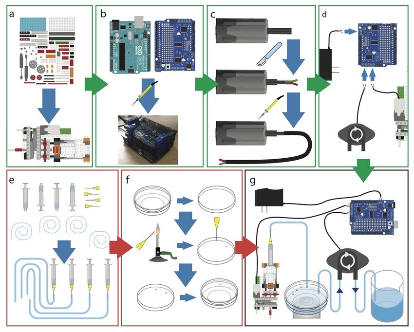
bioRxiv preprint first posted online May. 14, 2018; doi: http://dx.doi.org/10.1101/320416. The copyright holder for this
preprint (which was not peer-reviewed) is the author/funder. All rights reserved. No reuse allowed without permission.

imental settings, and highlights its potential in combination         already allows for NanoJ-Fluidics to be fully integrated into
with recent high-throughput and/or high-content approaches            microscopy acquisition software, enabling a seamless combi-
(29–33).                                                              nation between the imaging and fluid exchange protocol. In
Besides this potential for complex biological interrogation,          conclusion, NanoJ-Fluidics makes high-content multi-modal
the simplicity and robustness of the NanoJ-Fluidics facili-           microscopy experiments tractable and available to a large au-
tates experimental optimisation (Sup. Note 3) (21). To ob-            dience of researchers, while improving reliability, optimisa-
tain optimal SMLM images, for example, the type, timing               tion and repeatability of imaging protocols.
and concentration of fixation, permeabilisation and blocking
reagents, the antibody concentration, the fluorophores used           Software and Hardware Availability. NanoJ-Fluidics fol-
and the composition of the imaging buffer should be opti-             lows open-source software and hardware standards, it is
mised. However, tuning each step individually and especially          part of the NanoJ project (14, 21, 43). The steps to
in combination is a daunting task. The NanoJ-fluidics au-             assemble a complete functioning system are described in
tomation and its integration with imaging allows for a more           https://github.com/HenriquesLab/NanoJ-Fluidics/wiki.
comprehensive and reliable exploration of the many options            ACKNOWLEDGEMENTS
available (21, 34–38).                                                We thank Prof. Ralf Jungmann at Max Planck Institute (MPI) of Biochemistry
                                                                      Munich for reagents and advice. This work was funded by grants from the
Fluorescent microscopy is widely used for its molecular               UK Biotechnology and Biological Sciences Research Council (BB/M022374/1;
specificity of labelling and its potential for multi-colour           BB/P027431/1; BB/R000697/1) (R.H., P.M.P. and R.F.L.), the UK Medical Research
                                                                      Council (MR/K015826/1) (R.H.), the Wellcome Trust (203276/Z/16/Z) (S.C. and
imaging. However, SMLM microscopy approaches tend                     R.H.) and the Centre Nationnal de la Recherche Scientifique (CNRS ATIP-AVENIR
to be restricted to a couple of colours due to fluorophore            program AO2016) (C.L.). P.A. was supported by a PhD fellowship from the UK’s
                                                                      Biotechnology and Biological Sciences Research Council. C.L.D. was supported
photophysics requiring specific imaging buffers. A flexi-             by PhD funding from the Medical Research Council, UK (1214605). Research
ble and easy-to-use fluidics system, such as NanoJ-Fluidics           by B.B. was supported by UCL, Cancer Research UK (C1529/A17343), and MRC
                                                                      (MC_CF12266).
is a significant advance in this context: the easy combina-
                                                                      AUTHOR CONTRIBUTIONS
tion of STORM (22) with DNA-PAINT (23–25, 39), as we                  P.A. and R.H. devised the hardware and wrote the software. P.A., P.M.P., C.L. and
demonstrate in Fig. 3, and the potential to perform sequen-           R.H. planned experiments. Experimental data sets were acquired by P.M.P. (Fig.
                                                                      2), G.C, F.B.R. and C.L. (Fig. 3), P.A. and C.L.D. (Fig. 4). Data was analysed by
tial labelling protocols (e.g. bleaching or antibody removal          P.A., P.M.P. and S.C. while G.C., B.B., C.L. and R.H. provided research advice. The
(40, 41)) can extend high spatial resolution multiplexing far         paper was written by P.A. P.M.P., R.F.L., C.L. and R.H. with editing contributions of
                                                                      all the authors.
beyond what live-cell fluorescent microscopy can currently
                                                                      COMPETING FINANCIAL INTERESTS
achieve.                                                              The authors declare no competing financial interests.
NanoJ-Fluidics is easy (and fun) to set up and use. Its highly
modular design means that the system can be easily adapted
for specific experiments, such as by detaching syringe pump           Bibliography
modules to be kept at different temperature (Fig. S1). There            1. Zhe Liu, Luke D Lavis, and Eric Betzig. Imaging live-cell dynamics and structure at the
                                                                           single-molecule level. Molecular cell, 58(4):644–659, 2015.
are several advantages to the NanoJ-Fluidics design com-                2. Štefan Bálint, Ione Verdeny Vilanova, Ángel Sandoval Álvarez, and Melike Lakadamyali.
pared to standard flow cells: glass-bottom chambers are eas-               Correlative live-cell and superresolution microscopy reveals cargo transport dynamics at
                                                                           microtubule intersections. Proceedings of the National Academy of Sciences, 110(9):3375–
ier to prepare in comparison to microfluidic chips; unwanted               3380, 2013.
air bubbles formed in the tubing are easily dissipated be-              3. Meghan Hauser, Michal Wojcik, Doory Kim, Morteza Mahmoudi, Wan Li, and Ke Xu. Cor-
                                                                           relative super-resolution microscopy: new dimensions and new opportunities. Chemical
fore corrupting the sample; being a non-pressurised system                 reviews, 117(11):7428–7456, 2017.
means that there is higher tolerance to flow-rate fluctuations.         4. Ralf Jungmann, Maier S Avenda ñ o, Johannes B Woehrstein, Mingjie Dai, William M
                                                                           Shih, and Peng Yin. Multiplexed 3D cellular super-resolution imaging with DNA-PAINT
Notwithstanding, flow cells have the general advantage of                  and Exchange-PAINT. . Nature methods, 11(3):313–8, 3 2014. ISSN 1548-7105. doi:
requiring smaller volumes of reagents. Automated pipet-                    10.1038/nmeth.2835.
                                                                        5. Johnny Tam, Guillaume Alan Cordier, Joseph Steven Borbely, Ángel Sandoval Álvarez, and
ting robots are also a good alternative, being particularly ef-            Melike Lakadamyali. Cross-talk-free multi-color storm imaging using a single fluorophore.
ficient in multiwell high-content experiments (42), but are                PloS one, 9(7):e101772, 2014.
                                                                        6. Christopher C Valley, Sheng Liu, Diane S Lidke, and Keith A Lidke. Sequential superres-
generally restricted to highly specialised laboratories. The               olution imaging of multiple targets using a single fluorophore. PloS one, 10(4):e0123941,
open-source software component of the NanoJ-Fluidics pro-                  2015.
                                                                        7. Tai Kiuchi, Makio Higuchi, Akihiro Takamura, Masahiro Maruoka, and Naoki Watanabe.
vides an Application Programming Interface (API) and plu-                  Multitarget super-resolution microscopy with high-density labeling by exchangeable probes.
gin framework that allows third party syringe pumps, such as               Nature methods, 12(8):743, 2015.
                                                                        8. Ann Wheeler and Ricardo Henriques. Standard and Super-resolution Bioimaging Data Anal-
those commercially available, to be controlled by the com-                 ysis: A Primer. John Wiley & Sons, 2017.
mon NanoJ-Fluidics graphical user interface (GUI, Fig. S3).             9. Melike Lakadamyali. Correlative live-cell and super-resolution microscopy and its biological
                                                                           applications. Super-Resolution Imaging in Biomedicine, page 281, 2016.
This means that the automation protocols established here are          10. Caroline a Schneider, Wayne S Rasband, and Kevin W Eliceiri. NIH Image to ImageJ: 25
directly applicable to pre-existing or alternative hardware.               years of image analysis . Nature Methods, 9(7):671–675, 2012. ISSN 1548-7091. doi:
                                                                           10.1038/nmeth.2089.
In this work we have chosen to focus mainly on the applica-            11. Arthur D Edelstein, Mark a Tsuchida, Nenad Amodaj, Henry Pinkard, Ronald D Vale, and
                                                                           Nico Stuurman. Advanced methods of microscope control using uManager software .
tion of NanoJ-Fluidics to live-cell imaging and SMLM ap-                   Journal of Biological Methods, 1(2):1–10, 2014. doi: 10.14440/jbm.2014.36.
proaches, as each SMLM imaging requires a finely-tuned                 12. M G Gustafsson. Surpassing the lateral resolution limit by a factor of two using structured
                                                                           illumination microscopy. . Journal of microscopy, 198(Pt 2):82–87, 5 2000. ISSN 0022-2720.
chemical environments to produce high-resolution images.                   doi: 10.1046/j.1365-2818.2000.00710.x.
However, our fluidic exchange system can be used in any                13. M. Hofmann, C. Eggeling, S. Jakobs, and S. W. Hell. Breaking the diffraction barrier in
                                                                           fluorescence microscopy at low light intensities by using reversibly photoswitchable proteins
imaging experiments that would benefit from an automated                   . Proceedings of the National Academy of Sciences, 102(49):17565–17569, 2005. ISSN
exchange of the sample media. Our Micro-Manager plugin                     0027-8424. doi: 10.1073/pnas.0506010102.

Almada & Pereira et al.   |   NanoJ-Fluidics                                                                                                          bioR‰iv      |   5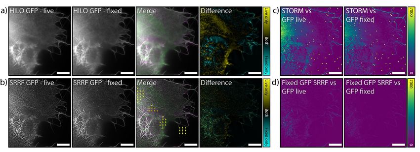
bioRxiv preprint first posted online May. 14, 2018; doi: http://dx.doi.org/10.1101/320416. The copyright holder for this
preprint (which was not peer-reviewed) is the author/funder. All rights reserved. No reuse allowed without permission.

 14. Nils Gustafsson, Siân Culley, George Ashdown, Dylan M. Owen, Pedro Matos Pereira, and         39. Syuan-Ming Guo, Remi Veneziano, Simon Gordonov, Li Li, Demian Park, Anthony B Kulesa,
     Ricardo Henriques. Fast live-cell conventional fluorophore nanoscopy with ImageJ through          Paul C Blainey, Jeffrey R Cottrell, Edward S Boyden, and Mark Bathe. Multiplexed confocal
     super-resolution radial fluctuations . Nature Communications, 7(12471):12471, 8 2016.             and super-resolution fluorescence imaging of cytoskeletal and neuronal synapse proteins.
     ISSN 2041-1723. doi: 10.1038/ncomms12471.                                                         bioRxiv, page 111625, 2017.
 15. Sébastien Herbert, Helena Soares, Christophe Zimmer, and Ricardo Henriques. Single-           40. Jason Yi, Asit Manna, Valarie A Barr, Jennifer Hong, Keir C Neuman, and Lawrence E
     Molecule Localization Super-Resolution Microscopy: Deeper and Faster . Microscopy                 Samelson. madSTORM: a Super-Resolution Technique for Large-Scale Multiplexing at
     and Microanalysis, 18(06):1419–1429, 2012.             ISSN 1431-9276.       doi: 10.1017/        Single Molecule Accuracy. . Molecular biology of the cell, 27, 2016. ISSN 1939-4586. doi:
     S1431927612013347.                                                                                10.1091/mbc.E16-05-0330.
 16. Ricardo Henriques, Caron Griffiths, E. Hesper Rego, and Musa M. Mhlanga. PALM and             41. Christopher C. Valley, Sheng Liu, Diane S Lidke, and Keith a. Lidke. Sequential superres-
     STORM: Unlocking live-cell super-resolution . Biopolymers, 95(5):322–331, 2011. ISSN              olution imaging of multiple targets using a single fluorophore. . PloS one, 10(4):e0123941,
     00063525. doi: 10.1002/bip.21586.                                                                 2015. ISSN 1932-6203. doi: 10.1371/journal.pone.0123941.
 17. Michael J Rust, Mark Bates, and Xiaowei W Zhuang. Sub-diffraction-limit imaging by            42. Ross C Lagoy and Dirk R Albrecht. Automated fluid delivery from multiwell plates to mi-
     stochastic optical reconstruction microscopy (STORM). . Nature methods, 3(10):793–5,              crofluidic devices for high-throughput experiments and microscopy. Scientific reports, 8(1):
     10 2006. ISSN 1548-7091. doi: 10.1038/nmeth929.                                                   6217, 2018.
 18. Mike Heilemann, Sebastian van de Linde, Mark Sch ü ttpelz, Robert Kasper, Britta              43. Robert D. M. Gray, Corina Beerli, Pedro Matos Pereira, Kathrin Maria Scherer, Jerzy
     Seefeldt, Anindita Mukherjee, Philip Tinnefeld, and Markus Sauer. Subdiffraction-resolution       Samolej, Christopher Karl Ernst Bleck, Jason Mercer, and Ricardo Henriques. VirusMap-
     fluorescence imaging with conventional fluorescent probes. . Angewandte Chemie (Interna-          per: open-source nanoscale mapping of viral architecture through super-resolution mi-
     tional ed. in English), 47(33):6172–6, 2008. ISSN 1521-3773. doi: 10.1002/anie.200802376.         croscopy. . Scientific reports, 6:29132, 7 2016. ISSN 2045-2322. doi: 10.1038/srep29132.
 19. Ralf Jungmann, Christian Steinhauer, Max Scheible, Anton Kuzyk, Philip Tinnefeld, and
     Friedrich C. Simmel. Single-molecule kinetics and super-resolution microscopy by fluo-
     rescence imaging of transient binding on DNA origami . Nano Letters, 10(11):4756–4761,
     2010. ISSN 15306984. doi: 10.1021/nl103427w.
 20. Brian M Burkel, George von Dassow, and William M Bement. Versatile fluorescent probes
     for actin filaments based on the actin-binding domain of utrophin. . Cell motility and the
     cytoskeleton, 64(11):822–32, 11 2007. ISSN 0886-1544. doi: 10.1002/cm.20226.
 21. Siân Culley, David Albrecht, Caron Jacobs, Pedro Matos Pereira, Christophe Leterrier, Ja-
     son Mercer, and Ricardo Henriques. Quantitative mapping and minimization of super-
     resolution optical imaging artifacts. Nature Methods, 2 2018. ISSN 1548-7091. doi:
     10.1038/nmeth.4605.
 22. Graham T Dempsey, Joshua C Vaughan, Kok Hao Chen, Mark Bates, and Xiaowei Zhuang.
     Evaluation of fluorophores for optimal performance in localization-based super-resolution
     imaging . Nature Methods, 8(12):1027–1036, 12 2011. ISSN 1548-7091. doi: 10.1038/
     nmeth.1768.
 23. Ralf Jungmann, Maier S Avendaño, Johannes B Woehrstein, Mingjie Dai, William M
     Shih, and Peng Yin. Multiplexed 3D cellular super-resolution imaging with DNA-PAINT
     and Exchange-PAINT. . Nature methods, 11(3):313–8, 2014. ISSN 1548-7105. doi:
     10.1038/nmeth.2835.
 24. Sarit S. Agasti, Yu Wang, Florian Schueder, Aishwarya Sukumar, Ralf Jungmann, and
     Peng Yin. DNA-barcoded labeling probes for highly multiplexed Exchange-PAINT imag-
     ing . Chem. Sci., 8(4):3080–3091, 2017. ISSN 2041-6520. doi: 10.1039/C6SC05420J.
 25. Joerg Schnitzbauer, Maximilian T. Strauss, Thomas Schlichthaerle, Florian Schueder, and
     Ralf Jungmann. Super-resolution microscopy with DNA-PAINT . Nature Protocols, 12(6):
     1198–1228, 2017. ISSN 17502799. doi: 10.1038/nprot.2017.024.
 26. Christophe Leterrier, Jean Potier, Ghislaine Caillol, Claire Debarnot, Fanny Rueda Boroni,
     and Bénédicte Dargent. Nanoscale Architecture of the Axon Initial Segment Reveals an
     Organized and Robust Scaffold . Cell Reports, pages 2781–2793, 2015. ISSN 22111247.
     doi: 10.1016/j.celrep.2015.11.051.
 27. Nitya Ramkumar and Buzz Baum. Coupling changes in cell shape to chromosome segre-
     gation , 2016. ISSN 14710080.
 28. Christina L. Dix, Helen K. Matthews, Marina Uroz, Susannah McLaren, Lucie Wolf, Nicholas
     Heatley, Zaw Win, Pedro Almada, Ricardo Henriques, Michael Boutros, Xavier Trepat, and
     Buzz Baum. The Role of Mitotic Cell-Substrate Adhesion Re-modeling in Animal Cell
     Division . Developmental Cell, 45(1):132–144, 2018. ISSN 18781551. doi: 10.1016/j.
     devcel.2018.03.009.
 29. Anne Beghin, Adel Kechkar, Corey Butler, Florian Levet, Marine Cabillic, Olivier
     Rossier, Gregory Giannone, Rémi Galland, Daniel Choquet, and Jean Baptiste Sibarita.
     Localization-based super-resolution imaging meets high-content screening . Nature Meth-
     ods, 14(12):1184–1190, 2017. ISSN 15487105. doi: 10.1038/nmeth.4486.
 30. Pedro M. Pereira, Pedro Almada, and Ricardo Henriques. High-content 3D multicolor super-
     resolution localization microscopy . Methods in Cell Biology, 125:95–117, 1 2015. ISSN
     0091-679X. doi: 10.1016/BS.MCB.2014.10.004.
 31. Wei Ouyang, Andrey Aristov, Mickaël Lelek, Xian Hao, and Christophe Zimmer. Deep learn-
     ing massively accelerates super-resolution localization microscopy. Nature biotechnology,
     2018.
 32. Martin Weigert, Uwe Schmidt, Tobias Boothe, M Andreas, Alexander Dibrov, Akanksha Jain,
     Benjamin Wilhelm, Deborah Schmidt, Coleman Broaddus, Siân Culley, et al. Content-aware
     image restoration: Pushing the limits of fluorescence microscopy. bioRxiv, page 236463,
     2017.
 33. Pedro Almada, Siân Culley, and Ricardo Henriques. PALM and STORM: Into large fields
     and high-throughput microscopy with sCMOS detectors. . Methods (San Diego, Calif.), 88:
     109–21, 10 2015. ISSN 1095-9130. doi: 10.1016/j.ymeth.2015.06.004.
 34. Juri N Bach, Giacomo Giacomelli, and Marc Bramkamp. Sample preparation and choice of
     fluorophores for single and dual color photo-activated localization microscopy (palm) with
     bacterial cells. Light Microscopy: Methods and Protocols, pages 129–141, 2017.
 35. Donna R Whelan and Toby D M Bell. Image artifacts in Single Molecule Localization
     Microscopy: why optimization of sample preparation protocols matters. . Scientific reports,
     5:7924, 2015. ISSN 2045-2322. doi: 10.1038/srep07924.
 36. Daniela Leyton-Puig, Katarzyna M. Kedziora, Tadamoto Isogai, Bram van den Broek, Kees
     Jalink, and Metello Innocenti. PFA fixation enables artifact-free super-resolution imaging
     of the actin cytoskeleton and associated proteins . Biology Open, 5(7):1001–1009, 2016.
     ISSN 2046-6390. doi: 10.1242/bio.019570.
 37. Aaron R Halpern, Marco D Howard, and Joshua C Vaughan. Point by point: An introductory
     guide to sample preparation for single-molecule, super-resolution fluorescence microscopy.
     Current protocols in chemical biology, 7(2):103–120, 2017.
 38. Ulrike Schnell, Freark Dijk, Klaas A Sjollema, and Ben NG Giepmans. Immunolabeling
     artifacts and the need for live-cell imaging. Nature methods, 9(2):152, 2012.

6   |   bioR‰iv                                                                                                                                 Almada & Pereira et al.       |   NanoJ-Fluidics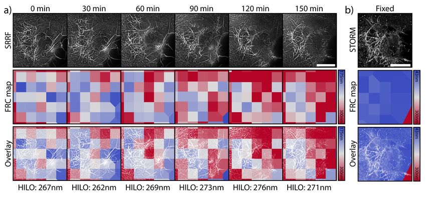
bioRxiv preprint first posted online May. 14, 2018; doi: http://dx.doi.org/10.1101/320416. The copyright holder for this
preprint (which was not peer-reviewed) is the author/funder. All rights reserved. No reuse allowed without permission.

Methods                                                               N-STORM microscope equipped with 405, 488, 561 and
                                                                      647 nm lasers (20, 50, 50 and 125 mW at the optical fiber
Cell lines. COS7 cells were cultured in phenol-red free
                                                                      output). One individual syringe pump module containing the
DMEM (Gibco) supplemented with 2 mM GlutaMAX
                                                                      fixative was kept within the incubator of the microscope at
(Gibco), 50 U/ml penicillin, 50 µg/ml streptomycin (Pen-
                                                                      37°C. All steps after cell transfection were performed on the
strep, Gibco) and 10% fetal bovine serum (FBS; Gibco).
                                                                      microscope, using NanoJ-Fluidics. COS7 cells were seeded
hTERT-RPE1 cells stably expressing Zyxin-GFP (1) were
                                                                      on ultraclean (5) 25 mm diameter thickness 1.5H coverslips
cultured in DMEM F-12 Glutamax (Gibco), with 10% FBS,
                                                                      (Marienfeld) at a density of 0.3–0.9x10⁵ cells/cm2 . One day
3.4% sodium bicarbonate (Gibco), 1% Penstrep. All cells
                                                                      after splitting, cells were transfected with a plasmid encoding
were grown at 37°C in a 5% CO2 humidified incubator. Cell
                                                                      the calponin homology domain of utrophin fused to GFP
lines have not been authenticated.
                                                                      (GFP-UtrCH) using Lipofectamin 2000 (Thermo Fisher Sci-
                                                                      entific) according to the manufacturer’s recommendations.
Plasmids. GFP-UtrCH was a gift from William Bement (2)
                                                                      Cells were imaged 1-2 days post transfection in culture
(Addgene plasmid #26737).
                                                                      medium using an Attofluor cell chamber (Thermofisher),
                                                                      covered with the lid of a 35 mm dish (Thermofisher), that
Antibody conjugation. Secondary antibodies (see bellow                was kept in place using black non-reflective aluminum tape
for details) were labelled with DNA strands (see bellow for           (T205-1.0—AT205, THORLABs).
details) as previously described (3). In short, secondary             Cells were fixed at 37°C for 15 minutes with 4%
antibodies were concentrated via amicon 100 kDa spin filters          paraformaldehyde in cytoskeleton-preserving buffer (1X
to 2-6 mg/ml. 50-100 µg of antibody was labelled using a              PEM, 80 mM PIPES pH 6.8, 5 mM EGTA, 2 mM MgCl2)
Maleimide-Peg2-succinimidyl ester for 90 min at 40x molar             (6). After fixation cells were permeabilised (1X PEM with
excess at 4°C on a shaker. Crosslinker stocks of 10 mg/ml             0.25% Triton-X) for 20 min, blocked with blocking buffer
in DMF were diluted in 1x PBS to reach 40x molar excess               (5% Bovine Serum Albumin (BSA) in 1X PEM) for 30
in 5 µl, which were subsequently added to the antibody.               minutes, and stained with Phalloidin-AF647 (Molecular
After the reaction had been done, unreacted crosslinker               Probes, 4 units/mL) for 30 minutes.
was removed via a zeba spin column. Thiolated DNA was                 Laser-illumination Highly Inclined and Laminated Optical
reduced using DTT for 2h at room temperature. DTT was                 sheet (HILO) imaging of Utrophin-GFP in live COS7 cells
separated from the reduced DNA via a Nap5 column and                  was performed at 37 °C and 5% CO2 on a Nikon N-STORM
fractions containing DNA were concentrated via 3 kDa                  microscope. A 100x TIRF objective (Plan-APOCHROMAT
amicon spin filters. The reduced DNA was then added to                100x/1.49 Oil, Nikon) with additional 1.5x magnification
the antibody bearing a functional maleimide group in 25x              was used to collect fluorescence onto an EMCCD camera
molar excess and incubated over night at 4°C on a shaker in           (iXon Ultra 897, Andor), yielding a pixel size of 107 nm. For
the dark. Antibody-DNA constructs were finally purified via           timelapse imaging, 100 raw frames (33 ms exposure) were
100 kDa amicon spin filters. DNA-PAINT labelling:                     acquired once every 10 minutes (with the illumination shut-
- For Mitochondria: Goat anti-Mouse (A28174, Ther-                    ter closed between acquisitions) for 150 minutes with 488
moFisher) with I1 (docking: 5’-TTATACATCTA-3’; imager:                nm laser illumination at 4% of maximum output. STORM
5’-CTAGATGTAT-ATTO655-3’);                                            HILO imaging of Alexa Fluor 647-phalloidin in fixed cells
- For Vimentin: Goat anti-chicken (Abcam, ab7113)                     was performed on the same system. 50,000 frames were
with I2 (docking: 5’-TTAATTGAGTA-3’; imager: 5’-                      acquired with 33 ms exposure and 642 nm laser illumination
GTACTCAATT-Cy3B-3’);                                                  at maximum output power with 405 nm pumping when
- For Clathrin: Goat anti-Rabbit (A27033, ThermoFisher).              required. STORM imaging was performed in GLOX buffer
Goat anti-Chicken (ab7113, Abcam) with I3 (docking: 5’-               (150mM Tris, pH 8, 1% glycerol, 1% glucose, 10mM NaCl,
TTTCTTCATTA-3’; imager: 5’-GTAATGAAGA-Cy3B-3’);                       1% b-mercaptoethanol, 0.5 mg/ml glucose oxidase, 40 µg/ml
- For alpha-tubulin: Donkey anti-Rat (A18747, Ther-                   catalase) supplemented with Phalloidin-Alexa Fluor 647 (1
moFisher) with I4 (docking: 5’-TTTATTAAGCT-3’; imager:                U/mL).
5’-CAGCTTAATA-ATTO655-3’).
Sequences for DNA-PAINT strands were obtained from (4).
Both thiolated and fluorophore conjugated DNA strands                 Multiplexed Super-Resolution microscopy with Ex-
were obtained from Metabion.                                          change-PAINT and STORM. COS-7 cells (obtained from
                                                                      ATCC) were seeded on 18mm, 1.5H glass coverslips
                                                                      (Menzel-Gläser). 24 hours after seeding, they were fixed
NanoJ-Fluidics framework. We provide the detailed in-                 using 4% PFA, 4% sucrose in PEM buffer at 37°C (6).
structions to easily build and use the system in a regular bi-        After blocking in phosphate buffer with 0.022% gelatin,
ology lab, as well as the software enabling its control and           0.1% Triton-X100 for 1.5 hours, cells were incubated with
automation (Sup. Note 1).                                             primary antibodies overnight at 4°C: mouse monoclonal
                                                                      anti-TOM20 (BD Bioscience # 612278), rabbit polyclonal
Live-to-Fixed Super-Resolution imaging. The NanoJ-                    anti-clathrin heavy chain (abcam ab21679), chicken poly-
Fluidics syringe pump array was installed on a Nikon                  clonal anti-vimentin (BioLegend # 919101) and rat anti-

Almada & Pereira et al.   |   NanoJ-Fluidics                                                                                bioR‰iv   |   7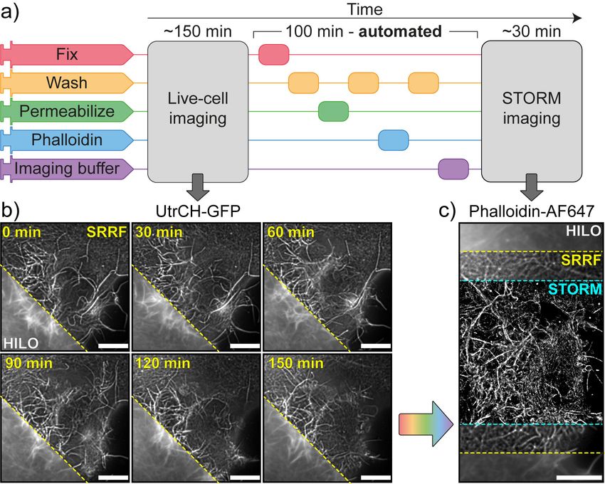
bioRxiv preprint first posted online May. 14, 2018; doi: http://dx.doi.org/10.1101/320416. The copyright holder for this
preprint (which was not peer-reviewed) is the author/funder. All rights reserved. No reuse allowed without permission.

alpha-tubulin (mix of clone YL1/2 abcam # 6160 and clone                drift between acquisition passes were aligned manually on
YOL1/34 Millipore CBL270). After rinses, they were incu-                high-resolution reconstructions.
bated with Exchange-PAINT secondary antibodies coupled                  FRC values were obtained using NanoJ-SQUIRREL after re-
to DNA sequences: goat anti-mouse I1, goat anti-chicken                 construction of original data separated into two two different
I2, goat anti-rabbit I3 and donkey anti-rat I4 for 1.5 hours at         stacks composed of odd or even images (9). NanoJ-SRRF,
RT (see Ab conjugation section for antibody and sequence                NanoJ-SQUIRREL and ThunderSTORM are available in
details). After rinses, they were incubated in phalloidin-              Fiji (10).
Atto488 (Sigma) at 12.5 µM for 90 min at RT and imaged
within a few days. For STORM/PAINT imaging, the NanoJ-
Fluidics array was installed on an N-STORM microscope                   Bibliography
(Nikon) equipped with 405, 488, 561 and 647 nm lasers
                                                                         1. Christina L. Dix, Helen K. Matthews, Marina Uroz, Susannah McLaren, Lucie Wolf, Nicholas
(25, 80, 80 and 125 mW at the optical fiber output). First,                 Heatley, Zaw Win, Pedro Almada, Ricardo Henriques, Michael Boutros, Xavier Trepat, and
a STORM image of phalloidin-ATTO488 was performed in                        Buzz Baum. The Role of Mitotic Cell-Substrate Adhesion Re-modeling in Animal Cell
                                                                            Division . Developmental Cell, 45(1):132–144, 2018. ISSN 18781551. doi: 10.1016/j.
buffer C (PBS 0.1M pH7.2, 500 mM NaCl) using 30,000                         devcel.2018.03.009.
frames at 30 ms/frame at 50% power of the 488 nm laser.                  2. Brian M. Burkel, George Von Dassow, and William M. Bement. Versatile fluorescent probes
                                                                            for actin filaments based on the actin-binding domain of utrophin . Cell Motility and the
After injection of the I1-ATTO655 and I2-CY3B imagers in                    Cytoskeleton, 64(11):822–832, 2007. ISSN 08861544. doi: 10.1002/cm.20226.
buffer C, 60,000 frames were aquired in an alternating way               3. Joerg Schnitzbauer, Maximilian T. Strauss, Thomas Schlichthaerle, Florian Schueder, and
                                                                            Ralf Jungmann. Super-resolution microscopy with DNA-PAINT . Nature Protocols, 12(6):
(647 nm at 60% and 561 nm at 30%) to image TOM20 and                        1198–1228, 2017. ISSN 17502799. doi: 10.1038/nprot.2017.024.
vimentin, respectively. After three rinses with buffer C, I3-            4. Ralf Jungmann, Maier S Avenda ñ o, Johannes B Woehrstein, Mingjie Dai, William M
                                                                            Shih, and Peng Yin. Multiplexed 3D cellular super-resolution imaging with DNA-PAINT
Cy3B and I4-ATTO655 were in buffer C were injected, and                     and Exchange-PAINT. . Nature methods, 11(3):313–8, 3 2014. ISSN 1548-7105. doi:
60,000 frames were aquired in an alternating way (561 nm                    10.1038/nmeth.2835.
                                                                         5. Pedro M. Pereira, Pedro Almada, and Ricardo Henriques. High-content 3D multicolor super-
at 30% 647 nm at 60%) to image clathrin and microtubules,                   resolution localization microscopy . Methods in Cell Biology, 125:95–117, 1 2015. ISSN
respectively. All imager strands were used at concentration                 0091-679X. doi: 10.1016/BS.MCB.2014.10.004.
                                                                         6. Daniela Leyton-Puig, Katarzyna M. Kedziora, Tadamoto Isogai, Bram van den Broek, Kees
between 0.25 and 2 nM.                                                      Jalink, and Metello Innocenti. PFA fixation enables artifact-free super-resolution imaging
                                                                            of the actin cytoskeleton and associated proteins . Biology Open, 5(7):1001–1009, 2016.
                                                                            ISSN 2046-6390. doi: 10.1242/bio.019570.
Event Detection and Live-to-Fix Imaging. hTERT-RPE1                      7. Nils Gustafsson, Siân Culley, George Ashdown, Dylan M. Owen, Pedro Matos Pereira, and
cells stably expressing Zyxin-GFP were incubated with 9                     Ricardo Henriques. Fast live-cell conventional fluorophore nanoscopy with ImageJ through
                                                                            super-resolution radial fluctuations . Nature Communications, 7(12471):12471, 8 2016.
mM Ro-3306 (Enzolife Sciences ALX-270-463) to inhibit                       ISSN 2041-1723. doi: 10.1038/ncomms12471.
CDK1 activity for 15-20 hours. Inhibition was released                   8. Martin Ovesn ý , Pavel K ř í ž ek, Josef Borkovec, Zdeněk Š vindrych, and Guy M.
                                                                            Hagen. ThunderSTORM: A comprehensive ImageJ plug-in for PALM and STORM data
by replacing drug containing media by fresh media at the                    analysis and super-resolution imaging . Bioinformatics, 30(16):2389–2390, 2014. ISSN
microscope immediately before imaging. Cells were im-                       14602059. doi: 10.1093/bioinformatics/btu202.
                                                                         9. Siân Culley, David Albrecht, Caron Jacobs, Pedro Matos Pereira, Christophe Leterrier, Ja-
aged using a Nikon Eclipse Ti microscope (Nikon) equipped                   son Mercer, and Ricardo Henriques. Quantitative mapping and minimization of super-
with an Neo-Zyla sCMOS camera (Andor), LED illumina-                        resolution optical imaging artifacts. Nature Methods, 2 2018. ISSN 1548-7091. doi:
                                                                            10.1038/nmeth.4605.
tion (CoolLED) and a 60X objective (Plan Apo 60X/1.4 Oil,               10. Johannes Schindelin, Ignacio Arganda-Carreras, Erwin Frise, Verena Kaynig, Mark Longair,
Nikon). Images were acquired every 3 min until enough cells                 Tobias Pietzsch, Stephan Preibisch, Curtis Rueden, Stephan Saalfeld, Benjamin Schmid,
                                                                            Jean-Yves Tinevez, Daniel James White, Volker Hartenstein, Kevin Eliceiri, Pavel Toman-
had underwent mitotic rounding. At that point, 16% warmed                   cak, and Albert Cardona. Fiji: an open-source platform for biological-image analysis .
PFA was added to cells in media to a final concentration of                 Nature Methods, 9(7):676–682, 7 2012. ISSN 1548-7091. doi: 10.1038/nmeth.2019.

4%, and incubated at room temperature for 20 minutes. They
were then washed 3 times and 0.2% Triton was added for
5 minutes. 5% BSA in 1X PBS was used to block for 30
min at room temperature, before activated b1 Integrin (Ab-
cam #ab30394) primary antibody was added. After incu-
bation and washing, Phalloidin-TRITC (Sigma-Aldrich) and
anti-mouse AF647 antibody (Invitrogen) were added. All of
these steps were performed automatically using the NanoJ-
Fluidics platform.

SMLM and SRRF image reconstruction. For Fig.                  2
images were reconstructed using NanoJ-SRRF (7) (TRPPM
for live cell data and TRM for fixed cell data with a magnifi-
cation of 4). Drift was estimated using the inbuilt function in
NanoJ-SRRF and correction applied during SRRF analysis.
For figure 3 localizations were detected using the N-STORM
software (Nikon), and exported as a text file before being
filtered and rendered using ThunderSTORM (8). Chromatic
aberration between the red (561 nm) and far-red (647 nm)
channels were corrected within the N-STORM software
using polynomial warping, and remaining translational

8   |   bioR‰iv                                                                                                     Almada & Pereira et al.      |   NanoJ-Fluidics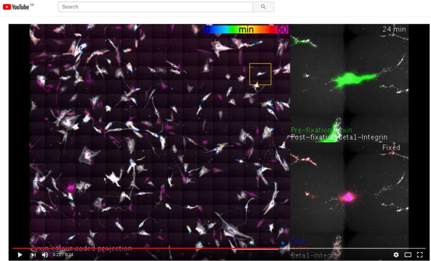
bioRxiv preprint first posted online May. 14, 2018; doi: http://dx.doi.org/10.1101/320416. The copyright holder for this
preprint (which was not peer-reviewed) is the author/funder. All rights reserved. No reuse allowed without permission.

Fig. S1. Assembled NanoJ-Fluidics system on a microscope. a) View of assembled pump array with syringes loaded on top of a
Nikon N-STORM microscope, an individual syringe pump unit sits inside the incubator and is kept at 37°C in the microscope incubator,
allowing the use of reagents equilibrated at the same temperature as the sample such as fixatives. b) Top view of the syringe pump
array. c) Zoom into the individual syringe pump unit inside incubator.

Almada & Pereira et al.   |   NanoJ-Fluidics                                                                 Supplementary Information   |   1
bioRxiv preprint first posted online May. 14, 2018; doi: http://dx.doi.org/10.1101/320416. The copyright holder for this
preprint (which was not peer-reviewed) is the author/funder. All rights reserved. No reuse allowed without permission.

Supplementary Note 1: Design and assembly

Fig. S2. NanoJ-Fluidics pump assembly. There are 4 steps to assemble a NanoJ-Fluidics system (a-d), and 2 steps to prepare an
experiment (e-f). a) Build the syringe pumps from LEGO bricks. b) Assemble the electronic controller. c) Prepare the motor cables for
wiring. d) Connect the pumps and power supply to the controller. e) Prepare the syringes. f) Prepare lid of the cell culture dish. g)
Thread syringes on the dish lid and mount syringes on LEGO pumps.

Design and capabilities. The NanoJ-Fluidics system is designed primarily for the sequential exchange of liquids in a glass-
bottom cell culture dish (or equivalent supports, such as ATTOfluor coverlslip holders for example). It consists of several parts:
LEGO based syringe pumps responsible for delivering reagents to the sample (Fig. 1a); electronics responsible for controlling
the pumps (Fig. 1b-d); a peristaltic pump responsible for removing reagents from the sample (Fig. 1d); fluid handling disposable
components (Fig. 1e-f). The syringe pumps are designed to accommodate syringes of any size, to be cost-effective and simple
to assemble. A syringe pump unit uses a simple gear and actuator system to translate fast motor motion to smooth and slower
linear motion, to enable consistent fluid flow. The result is that the flow-rate will depend on the gears, motor speed and the
syringe’s internal diameter. The pumps can be calibrated by measuring the flow-rate obtained at a given motor speed and syringe
volume. Given these reference values, the flow-rate for any other syringe diameter (Rd ) can be determined using Eq. S1:
                                                                       1        22
                                                                           d
                                                             Rd = Rr                                                                          (S1)
                                                                           dr

Where d is the syringe diameter, dr is the reference diameter and Rr is the reference rate. With the current design, a 1mm
BD Plastipak syringe with an internal diameter of 4.699mm was measured to obtain a nominal flow-rate of 2.3 ± 0.08 mL/s
at maximum motor speed. Based on this value, Table S1 summarizes the limits for different BD Plastipak syringe volumes

2   |   Supplementary Information                                                                      Almada & Pereira et al.   |   NanoJ-Fluidics
bioRxiv preprint first posted online May. 14, 2018; doi: http://dx.doi.org/10.1101/320416. The copyright holder for this
preprint (which was not peer-reviewed) is the author/funder. All rights reserved. No reuse allowed without permission.
calculated from Eq. S1. Table S1 also shows the maximum loading volume for each syringe given that this will be limited by
the length of the linear actuators on the pumps.
Nominal Volume (mL) Max. Volume (mL) Inner Diameter (mm) Max. Flow Rate (mL /s) Min. Flow Rate (mL /s)
1 0.75 4.699 2.3 0.6
2 2.5 8.7 7.9 2.0
5 4.8 11.989 15.0 3.7
10 6. 5 14.427 21.7 5.4
20 11 19.05 37.8 9.5
50 22 26.594 73.7 18.4
Table S1. Flow-rates obtained from Eq. S1 for several BD Plastipak syringes. Minimal flow rate is limited by the minimal rotation speed
achievable by the DC LEGO motors without stalling.
Both the syringe pump array (for media injection) and peristaltic pump (for media removal) are digitally run using an
Arduino UNO micro-controller and Adafruit Motorshield digital-to-analogue motor control. The Motorshield is an additional
electronic board that connects to the top of the Arduino controller, enabling it to run up to 4 LEGO motors. An Arduino can
be stacked with up to 32 Motorshields, limiting the controller to a maximum of 128 pumps, which should be sufficient for
most NanoJ-Fluidics applications. We provide custom open-source firmware that enables the Arduino-based electronics to be
programmatically controlled by a connected computer. We also provide a Java-based graphical user interface (GUI) for simple
control of the fluidics sequences (Fig. S2).
The software interface can be set to automatically perform a sequence of liquid injection and liquid removal steps (Sup. Note
Fig. S3) allowing an entire sample fixation or labelling protocol to be carried as shown in Fig. 2-3, 4.
Assembly. The steps to prepare a complete functioning NanoJ-Fluidics system (Fig. S2 and S1) consist of assembling the
pump array, controller, and wiring them together (Fig. S2a-d). Once the array and controller have been assembled, each
experiment only requires the lab-ware component to be prepared anew (Fig. S2 e-g). A full step-by-step guide for assembly
and preparation can be found in https://github.com/HenriquesLab/NanoJ-Fluidics/wiki.
NanoJ-Fluidics can also be used with more traditional microfluidics devices, such as Polydimethylsiloxane (PDMS) chips,
however we have focused on providing a series of protocols using off-the-shelf labware (e.g. glass-bottom cell culture dishes).
This approach makes the framework more accessible to laboratories that do not otherwise have the facilities to produce PDMS
devices. When using culture dishes the primary use of the system is limited to liquid exchange, this effectively reproduces
many laboratory protocols including cell fixation, immunolabeling, drug treatment, among others.
Fig. S3. User Interface. Screenshot of the NanoJ-Fluidics sequential control user interface.
Almada & Pereira et al. | NanoJ-Fluidics Supplementary Information | 3bioRxiv preprint first posted online May. 14, 2018; doi: http://dx.doi.org/10.1101/320416. The copyright holder for this preprint (which was not peer-reviewed) is the author/funder. All rights reserved. No reuse allowed without permission. Software interface and experiment automation. Control of the NanoJ-Fluidics pump array can be achieved in one of three ways: by using the provided Java-based GUI (Fig. S3); RS232 commands to the Arduino board; or by using the Application Programming Interface (API). A description of these is available at https: //github.com/HenriquesLab/NanoJ-Fluidics/wiki. The RS232 command scheme enables researchers with programming experience to run the controller directly in their own software packages. However, the GUI was designed to enable any user to directly run any number of Arduino controllers and pumps, as well as design a sequence of steps associated to an experimental protocol (Fig. S3). NanoJ-Fluidics is controlled by a set of Java classes described in the API. For example, these can be used to enable a protocol generated in the GUI to be programmatically started and manipulated by outside applications, such as a microscope control software such as Micro-Manager (1). This enables complex automated protocols, such as pre-screening in live imaging to try and capture specific events, such as mitosis (2, 3), before fixing and further analysis the sample (Fig. 4). Therefore, by using the NanoJ-Fluidics GUI and API, any combination of sample acquisition and liquid manipulation protocols can be performed in a completely automated manner. 4 | Supplementary Information Almada & Pereira et al. | NanoJ-Fluidics
bioRxiv preprint first posted online May. 14, 2018; doi: http://dx.doi.org/10.1101/320416. The copyright holder for this
preprint (which was not peer-reviewed) is the author/funder. All rights reserved. No reuse allowed without permission.

Supplementary Note 2: Resolution mapping

Fig. S4. Fourier Ring Correlation (FRC) resolution mapping for Fig. 2 and S1 using NanoJ-SQUIRREL. a) Individual live-cell
SRRF frames at different time-points pre-fixation (top-row), equivalent FRC map (middle-row) and overlay between SRRF frames and
the corresponding FRC map (lower-row); bottom values show average resolution calculated for the diffraction limited equivalent images.
The mean HILO resolutions for each image are shown beneath. b) Individual STORM rendering acquired post-fixation (top); equivalent
FRC map (middle); overlay between STORM frame and FRC map. Resolution maps calculated through NanoJ-SQUIRREL (4). All
scale bars are 10 µm.

Fig. S5. Fourier Ring Correlation (FRC) resolution mapping for Fig. 3 and S2 using NanoJ-SQUIRREL. Individual DNA-PAINT
super-resolution renderings (top-row), equivalent FRC map (middle-row) and overlay between SRRF frames and the corresponding
FRC map (lower-row). Resolution maps calculated through NanoJ-SQUIRREL. Black outlines in middle and bottom rows indicate the
cell shape mask used for calculation of mean and minimum resolutions across each channel.

To estimate the local resolution achieved in the super-resolution renderings associated to the main figures, we carried out
analysis using the Fourier Ring Correlation method (5), recently modified in (4) to generate a resolution map. Fig. S4 shows
that the best resolution achieved for live-cell imaging with SRRF is 175nm, and 43nm for fixed-cell imaging with STORM.
Fig. S5 shows that the best resolution achieved in the DNA-PAINT channels is ≥67nm, with the exception of the actin channel
which has an estimated best resolution of 97nm.

Almada & Pereira et al.   |   NanoJ-Fluidics                                                                 Supplementary Information   |   5
bioRxiv preprint first posted online May. 14, 2018; doi: http://dx.doi.org/10.1101/320416. The copyright holder for this
preprint (which was not peer-reviewed) is the author/funder. All rights reserved. No reuse allowed without permission.

Supplementary Note 3: Nanoscale morphological changes between pre- and post-fixation

Fig. S6. Analysis of changes in cell morphology and labelling pre- and post-fixation. a) Comparison of the distribution of GFP-
labelled Utrophin in the cell shown in Fig. 2 and Movie S1, imaged in HILO. The last timepoint pre-fixation (‘HILO GFP - live’) and
an image post-fixation of the same region (‘HILO GFP - fixed’) are shown. ‘Merge’ shows an overlay of the live (green) and fixed
(magenta) images. ‘Difference’ shows the result of subtracting the fixed image from the live image. b) As in (a), except using the
SRRF reconstructions of the data. The yellow arrows in ‘Merge’ show parts of the cell which moved during fixation as measured using
the elastic channel registration tool in NanoJ-SQUIRREL (4). c) Error maps generated between the (fixed) STORM reconstruction of
actin in this cell when using the live-cell GFP HILO image (left) or the fixed-cell GFP HILO image (right) as a reference. d) Error maps
generated between the SRRF reconstruction of GFP in the fixed cell when using the live-cell GFP HILO image (left) or the fixed-cell
GFP HILO image (right) as the reference. Scale bars = 10 µm.

To verify that there were no major alterations in cell morphology during fixation, we compared images of the same cell
immediately before and after fixation. Fig. S6a shows pre- and post-fixation images for HILO imaging of the cell shown
in Fig. 2, and Fig. S6b shows these changes for SRRF reconstructions of the same region. Overlaying both the SRRF and
HILO images pre- and post-fixation shows that the morphology of the cell remains largely constant during fixation. There is
movement of the bright filaments within the cell body and filopodia at the cell periphery on a sub-micron scale. This degree
of movement is comparable to or smaller than the frame-to-frame movement shown in Movie S1. There is also a loss of
fluorescence intensity during fixation in the top left portion of the region shown in Fig. S6.

6   |   Supplementary Information                                                                      Almada & Pereira et al.   |   NanoJ-Fluidics
You can also read