Applications of cell resealing to reconstitute microRNA loading to extracellular vesicles - Nature
←
→
Page content transcription
If your browser does not render page correctly, please read the page content below
www.nature.com/scientificreports
OPEN Applications of cell resealing
to reconstitute microRNA loading
to extracellular vesicles
Yuki Sonoda1, Fumi Kano2 & Masayuki Murata1,2*
MicroRNAs (miRNAs) are cargo carried by extracellular vesicles (EVs) and are associated with cell–cell
interactions. The response to the cellular environment, such as disease states, genetic/metabolic
changes, or differences in cell type, highly regulates cargo sorting to EVs. However, morphological
features during EV formation and secretion involving miRNA loading are unknown. This study
developed a new method of EV loading using cell resealing and reconstituted the elementary miRNA-
loading processes. Morphology, secretory response, and cellular uptake ability of EVs obtained from
intact and resealed HeLa cells were comparable. Exogenously added soluble factors were introduced
into multivesicular endosomes (MVEs) and their subsequent secretion to the extracellular region
occurred in resealed HeLa cells. In addition, miRNA transport to MVEs and miRNA encapsulation to
EVs followed a distinct pathway regulated by RNA-binding proteins, such as Argonaute and Y-box
binding protein 1, depending on miRNA types. Our cell-resealing system can analyze disease-specific
EVs derived from disease model cells, where pathological cytosol is introduced into cells. Thus, EV
formation in resealed cells can be used not only to create a reconstitution system to give mechanistic
insight into EV encapsulation but also for applications such as loading various molecules into EVs and
identifying disease-specific EV markers.
Extracellular vesicles (EVs), including microvesicles, apoptotic bodies, and exosomes, are released by a cell in an
environmentally dependent manner. Microvesicles and apoptotic bodies are generated by plasma membrane bud-
ding and detachment, while canonical exosomes are generated by more complicated and regulated processes1,2.
First, late endosomal membranes are invaginated to form multiple intraluminal vesicles (ILVs) inside multive-
sicular endosomes (MVEs). Second, MVEs fuse with the plasma membrane, releasing ILVs into the extracellular
region. These ILVs with a diameter of ~ 100 nm are referred to as “exosomes” in this study. EVs contain various
molecules, such as lipids, proteins, RNA, and DNA, derived from donor cells. After EVs are released from cells,
they are uptaken into recipient cells via direct membrane fusion with the plasma membrane or via endocytosis3,4.
The cargoes carried by EVs incorporated into recipient cells can change the biochemical environments of the
recipient cells by affecting their signal transduction pathways, metabolic pathways, and genetic modulation3,
causing tumor progression, metabolic disorders, neurological disorders, aging, and infections5. Various EV
functions depend on their production efficiencies, membranous components, and loaded cargoes. Therefore,
understanding the mechanisms underlying EV formation and secretion is crucial to using EVs for therapeutic
research and determining the pathogenesis of diseases via intercellular communications by EVs.
MicroRNAs (miRNAs), a type of EV cargo, inhibit a wide range of proteins and rearrange the cellular environ-
ment. After transcription and processing by Drosha form a precursor miRNA (pre-miRNA) in the nucleus, the
pre-miRNA is transported to the cytoplasm, where Dicer, an RNase III family enzyme, cleaves the loop part of
the pre-miRNA to form a double-stranded miRNA intermediate6,7. Argonaute (Ago), which has an RNase H-like
PIWI domain, binds to the double-stranded miRNA, forming the RNA-induced silencing complex (RISC), which
has an RNA-based silencing function8,9. Many studies have reported the molecular mechanisms underlying EV
formation, including specific miRNA in/from c ells10. Various molecules, such as Rab families, the endosomal
sorting complexes required for transport (ESCRT) machinery, RNA-binding proteins (RBPs), and the soluble
NSF attachment protein (SNAP) receptor (SNARE), are involved in EV formation, sorting, and secretion. The
component of the RISC and trans-activation-responsive RNA-binding protein (TARPB) that interacts with
Argonaute 2 (Ago2)11 is also involved in miRNA-loaded EV formation.
1
Department of Life Sciences, Graduate School of Arts and Sciences, The University of Tokyo, 3‑8‑1 Komaba,
Meguro‑ku, Tokyo 153‑8902, Japan. 2Cell Biology Center, Institute of Innovative Research, Tokyo Institute
of Technology, 4259 Nagatsuta, Midori‑ku, Yokohama, Kanagawa 226‑8503, Japan. *email: mmurata@
bio.c.u‑tokyo.ac.jp
Scientific Reports | (2021) 11:2900 | https://doi.org/10.1038/s41598-021-82452-5 1
Vol.:(0123456789)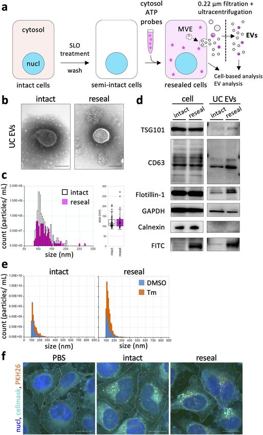
www.nature.com/scientificreports/
Figure 1. Morphological and biochemical characterization of EVs derived from resealed HeLa cells. (a) ▸
Reconstitution scheme of extracellular vesicle (EV) formation and secretion using HeLa cell resealing. Semi-
intact HeLa cells were prepared by permeabilizing HeLa cells with SLO treatment. Resealed cells were prepared,
as described in Methods, and were incubated under various conditions, and then culture media were collected.
The resulting EV fraction was obtained by ultracentrifugation and subjected to various experiments. (b)
Resealed HeLa cells were incubated in a 5% CO2 incubator for 120 min at 37 °C (post-incubation), and the
medium was changed with a fresh one. The cells were further incubated in a 5% C O2 incubator for 48 h at
37 °C, and EVs were prepared, as described in (a), and subjected to negative-stained electron microscopy (EM)
analysis. Scale bar = 100 nm. (c) Tunable resistive pulse sensing (TRPS) analysis was performed for EVs. Diluted
EVs normalized by the number of cells were loaded to an NP200 nanopore. The minimum number of events
recorded was 111 particles/measurement. The mean RMS noise was 4.4 ± 0.5 pA. The membrane stretch range
used was 47.97 nm, the voltage used was 0.68 V, and the current range used was 91.6–94.3 nA. (d) Western
blotting (WB) analysis was performed for whole cells and EVs obtained by ultracentrifugation (UC EVs). After
introducing cytosol containing 100 µg/mL of FITC-BSA, we obtained EVs and whole-cell lysates (− SLO as
“intact” and + SLO as “reseal”). Exogenously added FITC-BSA was observed only in the resealing sample. (e)
TRPS analysis was performed for EVs derived from tunicamycin (Tm)-treated or Tm-untreated cells. After
post-incubation for 120 min, resealed HeLa cells were incubated in a 5% C O2 incubator for 24 h at 37 °C in the
presence or absence of 5 µg/mL of Tm, and then EVs from each sample were prepared and analyzed by TRPS
analysis, as described in (c). The minimum number of events recorded was 792 particles/measurement. The
mean RMS noise was 4.7 ± 1.3 pA. The membrane stretch range used was 47.5 nm, the voltage used was 0.50 V,
and the current range used was 104.6–116.5 nA. (f) Fluorescence microscopy images of EVs endocytosed
by intact HeLa cells. EVs prepared from intact or resealed HeLa cells, as described in (b), were stained with
2 µM PKH26 dye. Fluorescence-labeled EVs from intact or resealed HeLa cells were incubated with intact
HeLa cells in a 5% CO2 incubator for 24 h at 37 °C. The cells were fixed and observed by confocal microscopy.
Fluorescence-labeled EVs from resealed HeLa cells were endocytosed by HeLa cells like EVs from intact HeLa
cells. White dotted lines outline the cell. Scale bar = 20 µm.
Studies have also investigated intracellular functions of the identified proteins and genes involved in EV
formation in living cells via knockout/knockdown of specific genes, expression of dominant active/negative
proteins, and utilization of modified m iRNA10,12,13. In addition, the cell-free reconstitution of cargo sorting to
isolated MVEs is especially interesting for determining the distinct sorting mechanisms for distinct EVs popula-
tions derived from cells with different phenotypes14–18. Recent advances in super-resolution microscopy allow us
to observe the detailed structure of MVEs and the dynamic behavior of DNA or EV-related proteins in MVEs
in living c ells13,19. Therefore, a mechanistic understanding of EV formation in/from cells has grown extensively.
We have been working on membrane traffic and organelle dynamics using semi-intact cells and evolution-
ary resealed cell coupling using quantitative image-based microscopic a nalysis20. Semi-intact cells have plasma
membranes that are permeabilized with the streptococcal toxin streptolysin O (SLO). SLO binds to cholesterol
in the plasma membrane and oligomerizes to form ~ 30-nm-diameter pores. SLO-mediated pores allow various
molecules, such as proteins, nucleotides, and membrane-impermeable small molecules, to enter cells. Therefore,
a semi-intact cell system enables the exchange of cytosol to modified cytosol that was prepared from other cells/
tissues, enabling us to reconstitute various intracellular phenomena, such as morphological changes in organelles
during mitosis, vesicular transport, and organelle-specific protein targeting21,22. As previously reported23, pores
are repaired in a Ca2+-dependent manner and SLOs are removed from cells through secretion microvesicles,
which make semi-intact cells intact again. These cells are called resealed cells. Resealed cells containing modified
cytosol are used as a cellular-type “test tube,” in which we can reconstitute modified cytosol-dependent intracel-
lular events and analyze the underlying mechanisms biochemically or morphologically (by using microscopic
analysis)24,25. In addition, introducing fluorescently labeled miRNAs into cells directly by cell resealing allows,
using confocal microscopy or super-resolution microscopy, the visualization and measurement of exogenously
added cytosol-dependent translocation, subcellular localization, co-localization with proteins, and dynamic
behaviors of miRNA quantitatively in resealed cells. Especially, various modifications of exogenously added cyto-
sol, such as immunodepletion of proteins, addition of dominant active/negative proteins, or addition of function-
blocking antibodies, allow us to investigate specific functions of proteins of interest in resealed cells. Therefore,
we tried to apply cell-resealing techniques to investigate the molecular mechanisms of EV formation in cells.
This study reproduced exogenously added cytosol-dependent EV formation using cell resealing. By using the
system coupled with quantitative image-based microscopic analysis, we analyzed the dynamic behavior, subcel-
lular localization, and co-localization of exogenously added, fluorescently labeled miRNA with CD63-positive
MVEs or ILVs quantitatively in resealed HeLa cells using confocal microscopy or super-resolution microscopy.
Finally, using morphometric analysis, we determined Ago-/Y-box binding protein 1 (YBX1)-mediated sorting/
recruiting mechanisms for EV formation, depending on the miRNA species in resealed HeLa cells.
Results
Morphological characterization of EVs derived from resealed HeLa cells. To reconstitute the EV
secretion in resealed HeLa cells, we first evaluated whether we could accomplish EV formation in and their
subsequent secretion from resealed HeLa cells. According to the resealing protocol described previously24,25,
we prepared resealed HeLa cells in which the cytosol was replaced with that prepared from murine lymphoma
L5178Y cells (protein concentration ~ 3 mg/mL), which has been successfully used for reconstitution of intracel-
lular membrane dynamics, such as cell cycle-dependent organelle dynamics or the membrane traffic between
Scientific Reports | (2021) 11:2900 | https://doi.org/10.1038/s41598-021-82452-5 2
Vol:.(1234567890)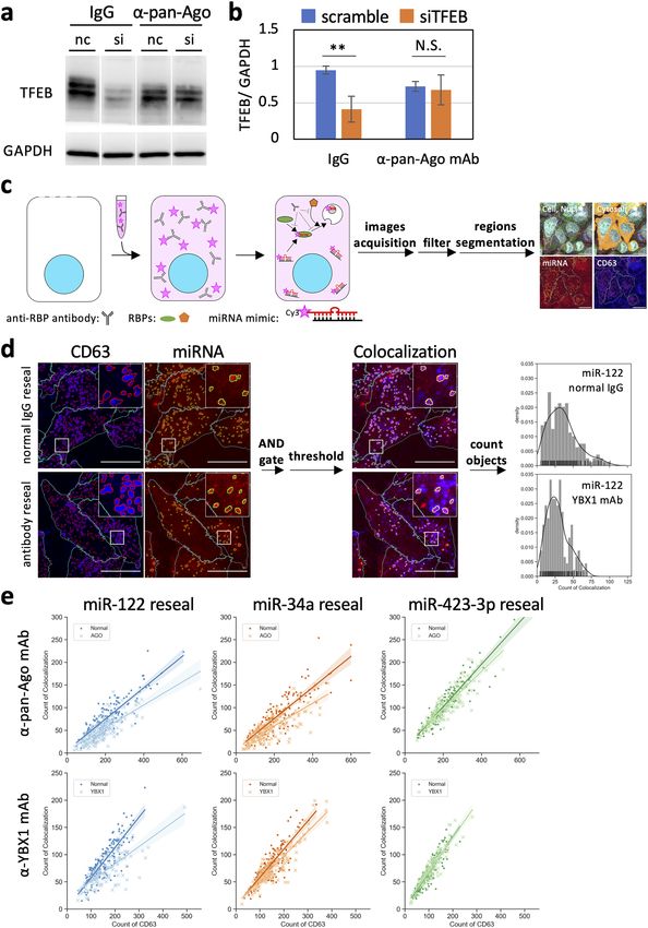
www.nature.com/scientificreports/

Scientific Reports |   (2021) 11:2900 |   https://doi.org/10.1038/s41598-021-82452-5        3

                                                                                       Vol.:(0123456789)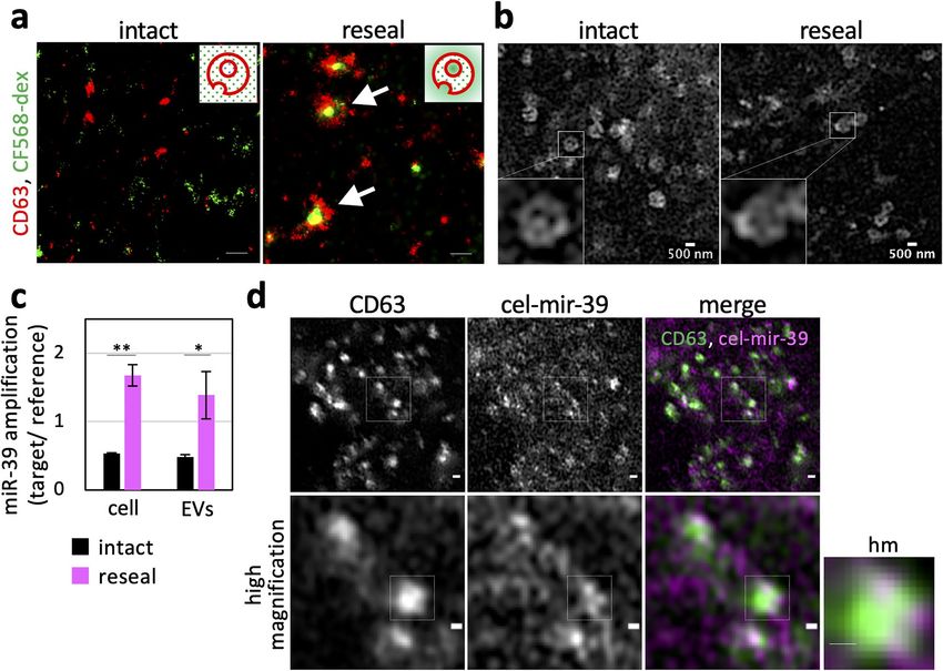
www.nature.com/scientificreports/
organelles in cells (Fig. 1a)21,22. The resealing efficiency, which was determined by flow cytometric and morpho-
metric analyses, was ~ 90% (Supplementary Fig. S1), as previously reported24,25.
SLO treatment for preparing semi-intact cells promotes ectocytosis and activates the production of plasma
membrane-derived EVs, and Ca2+ influx during cell resealing promotes lysosomal exocytosis23. These stress
responses potentially affect experimental results for elucidating the mechanisms underlying EV formation in
resealed cells. To estimate these stress responses of resealed HeLa cells, we first examined the activation of stress-
responsive kinases, such as p38 mitogen-activated protein kinase (p38 MAPK) and c-Jun N-terminal kinase
(JNK). After resealing, the resealed HeLa cells were incubated in a 5% C O2 incubator for 0, 30, 60, and 120 min
at 37 °C and then subjected to western blotting (WB) or immunofluorescence (IF) analysis using antibodies
against p38 MAPK, JNK, and their phosphorylated forms. We observed little or no activation of JNK during
resealing. In contrast, p38 MAPK was activated (phosphorylated) just after resealing but went back to the original
state within ~ 30 min (Supplementary Fig. S2. Similarly, IF analysis showed that the fluorescence signal of phos-
phorylated p38 MAPK in the nucleus (activated) decreased to the original state within 60 min (Supplementary
Fig. S3). These results indicated that at least 60 min of further incubation of resealed HeLa cells after resealing
(called post-incubation) is likely enough to restore the possible effects of the stress-induced by cell resealing on
EV formation in resealed HeLa cells.
Furthermore, we performed ultramicrostructural analysis of isolated EVs by negative-stained electron micros-
copy (EM). After 120 min post-incubation, we changed the medium of resealed HeLa cells with fresh medium
containing EV-depleted serum and further incubated the cells for 48 h. Incubation for 48 h was used to obtain
a sufficient amount of EVs from the medium of resealed HeLa cells for usual morphological or biochemi-
cal analysis. EVs in the cell medium were concentrated by differential ultracentrifugation (referred to as UC
EVs). Negative-stained EM showed that the UC EV morphology had no distinct structural difference between
intact (control) and resealed HeLa cells, and both were 100–200 nm or smaller in diameter (Fig. 1b). Next, we
obtained UC EVs as described above and measured the particle size distribution of the EVs by tunable resistive
pulse sensing (TRPS). The particle size for resealed cell-derived EVs was nearly 100 nm in diameter, which was
indistinguishable from EVs derived from intact HeLa cells under the same conditions (Fig. 1c). The measured
size of resealed cell-derived EVs was consistent with that previously reported but totally different from that of
membrane-derived apoptotic b odies3.
Biochemical characterization of EVs derived from resealed HeLa cells. Next, we compared pro-
tein compositions of UC EVs derived from resealed and intact HeLa cells by WB analysis. We prepared resealed
HeLa cells to which cytosol containing fluorescein isothiocyanate-bovine serum albumin (FITC-BSA) (protein
concentration 100 µg/mL) was introduced. After post-incubation, we discarded the medium and further incu-
bated resealed HeLa cells with fresh medium containing EV-depleted serum in a 5% CO2 incubator for 48 h
at 37 °C. Next, we collected the resealed HeLa cell and EV fraction samples, dissolved them in radioimmuno-
precipitation assay (RIPA) buffer, and subjected them to WB analysis. As a control, intact HeLa cells were pre-
incubated with FITC-BSA for 25 min, followed by incubation with medium for 120 min. After washing the cells,
they were further incubated them with fresh medium containing EV-depleted serum for 48 h, and intact HeLa
cell and EV fractions samples were prepared similar to resealed HeLa cells. As shown in Fig. 1d (right column,
intact and reseal), we detected standard EV marker proteins, such as TSG101, CD63, Flotillin-1, and glyceralde-
hyde 3-phosphate dehydrogenase (GAPDH). Especially, we detected the FITC-BSA band only in resealed HeLa
cells, indicating that FITC-BSA in exogenously added cytosol is, probably, diffusively and passively incorporated
into EVs. In contrast, we detected calnexin, a non-EV marker, only in the cell sample but not in the EV frac-
tion sample (Fig. 1d). We obtained similar results with regard to protein components of cell and EV fraction
samples using rat hepatoma H4IIEC3 resealed cells (Supplementary Fig. S4). Importantly, we detected no SLO
band in either the cell samples or the EV fraction, indicating that EV fractions prepared from the medium of
resealed HeLa cells do not contain microvesicles derived from the plasma membrane as SLO-induced blebbing
(Supplementary Fig. S5). Because we replaced the medium after 120 min of resealing, most SLOs on secreted
microvesicles in the medium were expected to be removed. Furthermore, some SLOs may have been retained on
the plasma membrane or endocytosed in resealed cells26; however, most were expected to undergo degradation
within 48 h as no SLO band was detected in the cell samples.
EV secretion into the culture medium is upregulated by oxidative stress27 or endoplasmic reticulum (ER)
stress28. The ER stress-induced increase in EV production was also found in tunicamycin (Tm)-treated resealed
cells (Fig. 1e and Supplementary Fig. S6). In addition, when EVs concentrated using Total Exosomes Isolation
reagent (Invitrogen kit) from cell culture (referred to as TEI EVs, the morphological and biochemical char-
acterization of which is described later) from intact and resealed HeLa cells were stained with PKH26, a lipid
membrane staining agent, and incubated with HeLa cells for 24 h, PKH26-labeled EVs from resealed HeLa cells
were incorporated into HeLa cells similar to those from intact HeLa cells (Fig. 1f). PKH26 could aggregate and
undergo endocytosis by c ells29; however, we could not detect any dots of PKH26 in phosphate-buffered saline
(PBS) without EVs. These results indicated that the secretory response and cellular uptake ability of EVs obtained
from intact and resealed HeLa cells are comparable.
Considering this biochemical data of EVs from resealed HeLa cells, together with the morphological informa-
tion (Fig. 1), we believe that EV formation successfully occurred in resealed HeLa cells using exogenously added
cytosol. In addition, we successfully introduced FITC-BSA into EVs (Fig. 1d).
Entry of exogenously added cytosolic components into EVs using cell resealing. We exam-
ined whether ILVs in resealed HeLa cells are morphologically filled with the exogenously added soluble cyto-
solic materials. Resealed HeLa cells introduced with CF568-dextran (CF568-dex) were incubated in a 5% CO2
Scientific Reports | (2021) 11:2900 | https://doi.org/10.1038/s41598-021-82452-5 4
Vol:.(1234567890)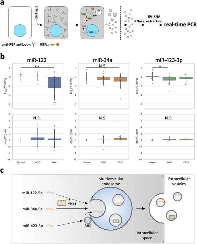
www.nature.com/scientificreports/
Figure 2. Encapsulation of exogenously added soluble cytosolic materials into ILVs and EVs in resealed
HeLa cells. (a) Super-resolution images showing encapsulated 10 kDa CF568-labeled dextran (CF568-dex) in
an intraluminal vesicle (ILV)-like structure. After introducing cytosol containing 100 µg/mL of CF568-dex,
intact and resealed HeLa cells were post-incubated for 120 min and subjected to immunofluorescence (IF)
analysis using anti-CD63 antibody (− SLO as “intact” and + SLO as “reseal”). The cells were observed by using
an N-STORM super-resolution system. Green and red represent CF568 and CD63 signals, respectively. Arrows
indicate CF568-dex incorporated in ILV-like structures. Inset: supposed models of ILV-like ultrastructure on
background noise signals (dots). Scale bar = 200 nm. (b) Super-resolution images showing similarity of the
ultrastructure of MVEs between intact and resealed HeLa cells. HeLa cells were incubated in DMEM without
phenol red and observed using LSM980 with Airyscan2 at 37 °C (live-cell imaging). Inset: similar MVE-like
ultrastructure observed in both intact and resealed HeLa cells. (c) Quantification of the amount of exogenously
added synthesized miRNA in whole cells and EVs. The bar plot shows the relative amount of cellular- and
EV-miRNA. After introducing cytosol containing a 1.0 nM synthesized cel-miR-39, intact and resealed HeLa
cells were post-incubated for 120 min and further incubated in a 5% C O2 incubator for 48 h at 37 °C. Total
cellular- and EV-RNA were extracted and miR-39-3p (guide strand) amount was measured by real-time PCR.
Fluorescent signal values of the target (FAM) were normalized by those of references (ROX). Data represent
results from three independent experiments (n = 3), expressed as the mean ± SD. *P < 0.05; **P < 0.01. (d)
Super-resolution images showing Cy3-labeled miR-39-3p in the MVE-like structure. After introducing cytosol
containing a 1.5 µM fluorescently labeled double-stranded miRNA mimic (Cy3 for cel-mir-39, guide strand;
Cy5 for cel-mir-39*, passenger strand), resealed HeLa cells were post-incubated for 120 min and subjected
to IF analysis using anti-CD63 antibody. The cells were observed using an LSM980 with Airyscan2. Arrows
indicate miRNA accumulated in MVEs. The right panels are a magnified image of the white boxes in the left
panel. hm: high magnification. Scale bar (upper images) = 500 nm. Scale bar (bottom images and cropped
images) = 200 nm.
incubator for 120 min at 37 °C and subjected to IF analysis using anti-CD63 antibody. CD63 is expressed in
various organelles (e.g., late endosomes, lysosomes, plasma membrane, other organelles), and it is particularly
enriched in MVEs or ILVs30. As a control, we incubated intact HeLa cells with CF568-dex for 25 min in advance.
ILVs immunostained with anti-CD63 antibodies in resealed HeLa cells were observed using super-resolution
microscopy. The green CF568 signals appeared encapsulated in red dotted CD63 in resealed HeLa cells, while
we observed little co-localization of CF568 spots with CD63 in intact HeLa cells (Fig. 2a). In our study, CF568-
positive dots were distinguished from those labeled with endocytosed CF568 in intact HeLa cells topologically.
Scientific Reports | (2021) 11:2900 | https://doi.org/10.1038/s41598-021-82452-5 5
Vol.:(0123456789)www.nature.com/scientificreports/
In addition, the size of each green CF568 spot was ~ 200 nm in diameter, which is virtually identical to ILVs
previously reported31. Super-resolution microscopy live imaging showed that the dynamics and morphology of
EGFP-CD63-labeled MVEs in intact and resealed HeLa cells were almost indistinguishable (Fig. 2b and Sup-
plementary Movies S1 and S2). These results indicated that the soluble cytoplasmic component in resealed HeLa
cells can be encapsulated in exosomes in resealed HeLa cells and that the morphological features of the ILVs of
MVEs in resealed HeLa cells are coincident with those previously reported13,19,31.
Encapsulation of exogenously added miRNA into ILVs and exosomes in resealed HeLa
cells. Subsequently, we examined the possible entry of exogenously added miRNA into ILVs and exosomes
from cytosol diffusively in resealed HeLa cells. To test this biochemically, we used cel-miR-39 (QIAGEN) as a
soluble and diffusive marker of miRNA. We prepared resealed HeLa cells using the cytosol containing cel-miR-39
and then obtained EVs from the medium of the resealed HeLa cells. It was reported that the combination of
polymer-based precipitation method (Total Exosomes Isolation reagents reagent) and miRNeasy Mini kit (QIA-
GEN) had high extraction efficiency and provided highly pure small RNA32. We cannot eliminate the possibility
that different subtypes of exosomes were obtained between the ultracentrifugation method and polymer-based
precipitation method. However, we could not detect obvious differences in morphologies between UC EVs and
TEI EVs via EM (Supplementary Fig. S7a). Moreover, we confirmed that TEI EVs also expressed known exoso-
mal marker proteins, similar to UC EVs (Supplementary Fig. S7b). The concentrated EVs in PBS were subjected
to RNase treatment to remove contaminated RNAs attached to the outside of EVs, and then RNA encapsulated
in the EVs (EVs-RNA) was extracted. The same amount of EVs-RNA was used for reverse transcription and real-
time PCR. We detected exogenously added cel-miR-39 in both resealed HeLa cells and EVs derived from the
resealed HeLa cells (Fig. 2c, reseal), indicating that the incorporation of miRNA introduced into exosomes and
their subsequent secretion to the extracellular region can occur in resealed HeLa cells. Concerning the origin of
extracellular RNAs, no consensus has been r eached6. Some researchers reported that most extracellular miRNAs
originate from non-vesicular Ago2-associated miRNAs (mainly from dead cells and apoptotic bodies) that are
Nase33,34. To reveal the origin of miRNA in TEI EVs from resealed cells, we examined the amount
resistant to R
of exogenously added miR-39 as follows. First, we treated the TEI EV fraction with RNase alone and quantified
the RNase-resistant miRNA by real-time PCR using the standard curve method (Supplementary Fig. S8). As a
result, exogenously added miR-39 content was reduced but substantially detected even in the presence of RNase,
indicating that this decreased amount of miRNA was likely to be extracellular miRNA free of proteins or EVs.
Furthermore, we found that the aforementioned RNase-resistant miRNA was poorly degraded by proteinase K
treatment in the presence of RNase, which can degrade protein-associated miRNA. We observed the complete
disappearance of RNase-resistant miRNA only when the membrane components were disrupted by sodium
dodecyl sulfate (SDS) in the presence of RNase. Together, these results suggested that RNase-resistant miRNA
was encapsulated in TEI EVs.
To examine the subcellular localization of exogenously added miRNA, we focused on the possible interac-
tion between Ago2 and miRNA, which are reported to form the RISC in the cytoplasm. First, we examined the
subcellular localization of Ago2, CD63, and Dcp1 by IF using corresponding antibodies. Ago2 and Dcp1a are
RNA-associated proteins, such as a marker of P-bodies that store RNA in the c ytoplasm35. Ago2 was localized
to either CD63 or Dcp1a extensively (CD63 and Ago2, localization efficiency; Pearson’s r = 0.474 ± 0.056; Dcp1a
and Ago2, Pearson’s r = 0.416 ± 0.024), but there was little co-localization between CD63 and Dcp1a (Pearson’s
r = 0.210 ± 0.032) (Supplementary Fig. S9a and S9b), indicating that Ago2 works as a recruiter of various types
of RNAs for late endosomes or P-bodies separately, as previously r eported35.
Ago2 unwinds short RNA duplexes, such as miRNA and short interfering RNA (siRNA), and stably associates
with single-stranded RNA. This single-stranded RNA is called the guide strand (i.e., mature miRNA), while the
opposite strand that is dissociated and undergoes degradation is called the passenger strand6,7. Next, we intro-
duced a double-stranded miRNA mimic (cel-miR-39) for which the 5′-end of the guide strand was labeled with
Cy3 and the 5′-end of the passenger strand with Cy5 into resealed HeLa cells, in which EGFP-CD63 or EGFP-
Dcp1a was transiently expressed, and we investigated the co-localization of fluorescent miRNA and EGFP-CD63/
EGFP-Dcp1a in fixed resealed HeLa cells 120 min after resealing using confocal microscopy. Both strands of the
miRNA introduced in resealed HeLa cells appeared to accumulate with CD63-positive but not Dcp1a-positive
structures (Supplementary Fig. S9c). Super-resolution microscopic analysis for close examination of the co-
localization of CD63 with the Cy3-labeled guide strand in resealed HeLa cells by IF analysis using anti-CD63
antibody showed that the guide strand appeared to accumulate in the vicinity of CD63-positive structures, form-
ing ILV-like structures (Fig. 2d). The CD63-positive structure was ~ 500 nm in diameter, and the accumulated
miRNA was ~ 200 nm in diameter. These results indicated that, at least, the Cy3-labeled guide strand introduced
into resealed HeLa cells is transported into CD63-positive MVEs via an innate cargo-sorting system.
Time‑dependent behavior of double‑stranded miRNA introduced into resealed HeLa
cells. Next, we investigated time-dependent changes of localization of the introduced fluorescently labeled
miRNA in resealed HeLa cells. HeLa cells were introduced with fluorescently labeled cel-miR-39, and the
resealed HeLa cells were incubated in a 5% CO2 incubator for 5, 30, and 120 min at 37 °C and then subjected
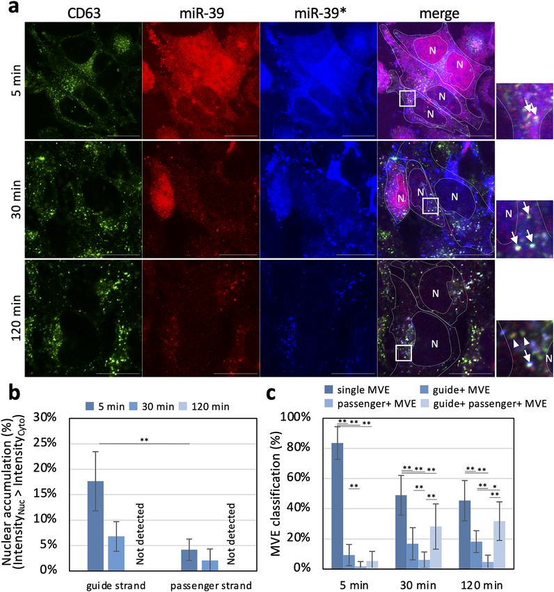
to IF analysis using anti-CD63 antibody. After 5 min incubation, both guide (Fig. 3a, miR-39) and passenger
(Fig. 3a, miR-39*) strands were distributed throughout the cells, but guide strands were more accumulated in
the nucleus compared to passenger strands. Fluorescence spots of either the guide or the passenger strand or
both of them began to emerge in the cytoplasm after 30 min incubation. After 120 min incubation, the guide
strand’s fluorescence signal was eliminated from the nucleus (Fig. 3a,b), while fluorescence spots of both guide
and passenger strands appeared in the cytoplasm (Fig. 3a,c). Quantitative image-based microscopic analysis
Scientific Reports | (2021) 11:2900 | https://doi.org/10.1038/s41598-021-82452-5 6
Vol:.(1234567890)www.nature.com/scientificreports/
Figure 3. Time-dependent localization changes of exogenously added fluorescent-labeled strands of miRNA
introduced into resealed HeLa cells. (a) Fluorescence microscopy images of the miRNA introduced in resealed
HeLa cells. After introducing cytosol containing a 1.5 µM fluorescently labeled miRNA mimic, resealed HeLa
cells were fixed at 5, 30, and 120 min after resealing and subjected to IF analysis using anti-CD63 antibody.
Fluorescence signals of guide (miR-39) strands, passenger (miR-39*) strands, and CD63-positive MVEs were
observed by confocal microscopy. Arrows indicate CD63-positive MVEs co-localized with both guide and
passenger strands. Arrowheads indicate CD63-positive MVEs co-localized with the guide strand but not with
the passenger strand. White dotted lines outline the cell and nucleus. Nuclear regions are indicated as “N”
inside the dotted line. Scale bar = 20 µm. (b) Bar plots show the population ratio of cells in which miRNAs
were accumulated in the nucleus. Data represent results from five frames (5 points and ~ 80 cells/sample; n = 5),
expressed as the mean ± SD. *P < 0.05; **P < 0.01. (c) Bar plots show the population ratio of MVEs classified
by the miRNA co-localization manner. For counting, we processed images using Laplacian of Gaussian (LoG)
filtering to remove noise. Observed MVEs were selected at random (20 points/cell) and classified by co-localized
miRNA bright spots. Data represent results from 14 cells (n = 14), expressed as the mean ± SD. *P < 0.05;
**P < 0.01. single, no co-localized; guide + , co-localized with guide strand, passenger + MVE, co-localized with
passenger strand.
Scientific Reports | (2021) 11:2900 | https://doi.org/10.1038/s41598-021-82452-5 7
Vol.:(0123456789)www.nature.com/scientificreports/
Scientific Reports | (2021) 11:2900 | https://doi.org/10.1038/s41598-021-82452-5 8
Vol:.(1234567890)www.nature.com/scientificreports/
◂Figure 4. Quantitative image-based microscopic analysis of exogenously added fluorescent-labeled miRNAs
loading to MVEs in resealed HeLa cells containing function-blocking antibodies. (a) Functional analysis of
exogenously added antibody in resealed cells by WB analysis. After introducing cytosol containing 10 µg/mL of
antibodies, HeLa cells were post-incubated for 45 min, transfected with the siRNA by lipofection, and further
incubated in a 5% C O2 incubator for 18 h at 37 °C. Next, the cells were lysed and subjected to WB analysis
using anti-TFEB and anti-GAPDH antibodies. Equivalent band intensities between scramble (nc) and siRNA
(si) in resealed HeLa cells were observed (α-pan-Ago). (b) Quantification of TFEB and GAPDH bands in
(a). The bar plot shows the relative TFEB amount in resealed HeLa cells. The TFEB amounts were compared,
normalized by GAPDH. Data represent results from three independent experiments (n = 3), expressed as
the mean ± SD. **P < 0.01. (c) Computer algorithm-defined segmentation scheme. After introducing cytosol
containing 1.5 µM fluorescently labeled miRNAs and 10 µg/mL of anti-Ago or anti-YBX1 antibodies, HeLa
cells were post-incubated for 120 min and subjected to IF analysis using anti-Ago2 and anti-CD63 antibodies.
Mouse anti-CD63 antibody was used to reseal HeLa cells containing rabbit anti-YBX1 antibody, and rabbit
anti-CD63 antibody was used to reseal HeLa cells containing mouse anti-pan Ago antibody. We obtained ~ 100
images (9 points × 9 Z-stack images) per sample and performed maximum-intensity projection prior to image
analysis. We detected the areas of nuclei, cells, miRNAs, and MVEs and performed LoG filtering, threshold
setting, and AND-gated image calculation for each image. Next, we counted the number of objects automatically
at the single-cell level. Scale bar in the right panel = 20 µm. (d) Detection scheme of CD63-positive MVEs,
miRNA dots, and their co-localization. Images were filtered using the AND gate, and co-localized objects were
enumerated automatically. The histogram shows the results of resealed HeLa cells containing miR-122 and anti-
YBX1 antibody [lower-middle panel in (e)]. Scale bar = 20 µm. (e) Scatter plots with linear regression models for
quantitative results obtained, as described in (c, d). Scatter plots with linear regression models show the number
of CD63-positive MVEs and co-localized objects. The 95% confidence intervals were drawn using translucent
bands around regression lines.
showed that CD63-positive dots are co-localized mainly with fluorescence spots of the guide or guide/passenger
strand of miRNA, not the passenger strand (Fig. 3c), which seemed reasonable because the passenger strand
is easily degraded in the cytoplasm after separating from the guide s trand6,7,36. As described earlier, < 120 min
after resealing, resealed HeLa cells might be under stress induced by resealing, so we could not exclude the pos-
sibility that the dynamic behavior of fluorescently labeled miRNAs in 5 or 30 min incubation samples showed
the behavior under stress. However, the transport of exogenously added miRNA to late endosomes (or CD63-
positive MVEs) within 120 min almost entirely in resealed HeLa cells.
The miRNA was strongly co-localized with CD63 (MVEs) and Lamp2 (lysosomes) (Supplementary Fig. S10,
arrow), but weakly co-localized with Rab7 (late endosomes) and Rab11 (recycling endosomes). A part of the
passenger strand was co-localized with CD63 and Lamp2, together with the guide strand. However, the passenger
strand was mainly co-localized with the organelle-nonspecific guide strand (Supplementary Fig. S10, arrowhead).
These morphological and biochemical results indicated that we could reconstitute miRNA transport to ILVs and
exosomes in resealed HeLa cells.
Inhibition of protein functions by introduced antibody in resealed HeLa cells. We focused on
go8,9 and YBX115,37,38, which are RBPs that interact with a large family of proteins and RNA species, including
A
miRNA, as representative cytosolic factors. We prepared the modified cytosol, which contained either anti-Ago
antibody or anti-YBX1 antibody, and examined its effect on the inhibition of miRNA transport to MVEs or EVs
in resealed HeLa cells by biochemical and morphological analyses. WB analysis using anti-pan-Ago, anti-Ago2
and, anti-YBX1 antibodies showed that YBX1 is present in HeLa cells and EVs, whereas pan-Ago and Ago2
were present only in HeLa cells but barely detected in EVs (Supplementary Fig. S11a). Immunoprecipitation and
immunodepletion experiments for HeLa cell lysates and L5178Y cytosol, respectively. The intensity of the upper
band (Ago; 90–100 kDa) in the antibody lane was clearly diminished. The lower band (Ago; nearly 70 kDa)
was a cross-reacting band (Radixin)39 that was not diminished. This suggests that the anti-pan-Ago antibody
recognized the full-length Ago in resealed HeLa cells. In the case of anti-YBX1 antibody, a decrease in YBX1
intensity was confirmed. These results showed that these antibodies significantly interacted with each antigen,
even in resealed HeLa cells (Supplementary Fig. S11b). Next, to examine the resealing efficiency and retention
rate of introduced immunoglobulin G (IgG)-type antibodies in resealed HeLa cells in which normal mouse IgG
or normal rabbit IgG was introduced by resealing, the resealed HeLa cells were incubated in a 5% C O2 incubator
for 12 h at 37 °C and then subjected to IF analysis using anti-mouse and anti-rabbit antibodies. We detected fluo-
rescence signals of both antibodies in the cytoplasmic region but not in the nuclear region in almost all resealed
HeLa cells (~ 90%), even after 12 h incubation (Supplementary Fig. S11c).
In addition, to confirm the inhibitory function of exogenously added antibody in cells, we examined the effect
of the introduced anti-pan-Ago antibody on the siRNA-mediated knockdown efficiency. Resealed HeLa cells
were incubated in a 5% C O2 incubator for 45 min at 37 °C and then transfected with the siRNA for silencing of
transcription factor EB (TFEB), a master transcriptional regulator of lysosomal biogenesis and autophagy, as a
representative sample. After incubation for 18 h, the cells were lysed and subjected to WB analysis. The knock-
down efficiency of control resealed HeLa cells, in which normal IgG was introduced, was ~ 50%, while that of
sample resealed HeLa cells, in which anti-pan-Ago antibody was introduced, was nearly 0, same as that of the
scrambled control (Fig. 4a,b). These results indicated that the anti-Ago and anti-YBX1 antibodies introduced
in resealed HeLa cells could recognize and interact with antigen proteins, inhibiting their functions, specifically
in resealed HeLa cells.
Scientific Reports | (2021) 11:2900 | https://doi.org/10.1038/s41598-021-82452-5 9
Vol.:(0123456789)www.nature.com/scientificreports/
Scientific Reports | (2021) 11:2900 | https://doi.org/10.1038/s41598-021-82452-5 10
Vol:.(1234567890)www.nature.com/scientificreports/
◂Figure 5. Quantitative analysis of miRNAs in isolated EVs derived from resealed HeLa cells, in which
function-blocking antibodies against RBPs were included. (a) Experimental scheme examining the effect of
function-blocking antibodies against RNA-binding proteins (RBPs) on loading of various endogenous miRNAs
in resealed HeLa cells. After introducing cytosol containing 10 µg/mL of anti-Ago or anti-YBX1 antibodies,
HeLa cells were incubated in a 5% C O2 incubator for 12 h at 37 °C. Total cellular- and EV-RNA were obtained,
as described in Fig. 2c, and the miR-122, miR-34a, and miR-423-3p amounts were measured by real-time PCR.
(b) Quantification of the amount of endogenously expressed miRNA in whole cells and EVs. Bar plots show
the relative cellular- and EV-miRNA amounts. Log2 fold-change (FC) values of each miRNA were calculated
using the Ct method, in which Ct was normalized to 18S rRNA and Ct to the control sample (normal).
When log2 FC values were greater than two times standard deviation (SD), we assumed them to be outliers
and excluded them. We selected TaqMan assays, which have the same sequences between humans and animals.
Therefore, TaqMan assays are presumed to detect them. EVs; miR-122, n = 10–13; miR-34a, n = 10–14; miR-
423-3p, n = 12–14. Cell; miR-122, n = 5–9; miR-34a, n = 9; miR-423-3p, n = 9–10. *P < 0.05; **P < 0.01. (c) Possible
model of loading mechanism of each miRNA to EVs. Considering all results concerning different efficiencies
for recruiting to MVEs among miRNAs (Fig. 4d) for miRNA loading to exosomes in (b), we schematically
proposed a possible model for loading of miRNA to exosomes. Both the loading and recruiting mechanisms
would depend on the miRNA species.
Quantitative analysis of subcellular localization of miR‑122, miR‑34a, and miR‑423‑3p in
resealed HeLa cells and the effect of antibodies against Ago and YBX1. After achieving the trans-
port and encapsulation of exogenously added miRNA into exosomes and EV secretion in resealed HeLa cells,
we examined the effect of anti-Ago and anti-YBX1 antibodies on the transport of miRNA mimics to CD63-
positive MVEs. We selected these miRNAs (miR-12240–43, miR-34a40,44,45, and miR-423-3p40,46,47) because they
are expressed in HeLa cells40,41,44,46. Furthermore, they interact with Ago proteins16,42,43,45,47, and studies have
investigated their sorting mechanism16,18,46. Three types of miRNAs, in which guide miRNA was conjugated
with Cy3, were introduced into HeLa cells in the presence of control IgG, anti-Ago antibody, or anti-YBX1
antibody by cell resealing. After 120 min, the cells were fixed and stained with anti-CD63 antibody. Fluores-
cent spots of exogenously added miRNA were heterogeneously distributed throughout the cytoplasm and we
saw many fluorescent spots that were positive only with CD63 or with both CD63 and miRNA, which made it
difficult to estimate the co-localization rate between miRNA and CD63 visually. Therefore, we performed auto-
matically quantitative image-based microscopic analysis. For single-cell analysis, HeLa cells were stained with
Hoechst 33,342 (for nucleus staining) and anti-Ago2 antibody (for cytosol staining) for segmentation of each
cell (Fig. 4c). Anti-Ago2 antibody was used as a marker of the cytoplasm because Ago2 was evenly distributed
throughout the cytoplasm. To quantify the co-localization of miRNA and CD63-positive MVEs in each cell,
miRNA- or CD63-positive dotted structures were segmented (Fig. 4d). To prevent secondary antibodies from
recognizing the antibodies incorporated into resealed HeLa cells, the host species of the primary antibodies were
different from the antibodies in resealed HeLa cells (Supplementary Fig. S12). The mean co-localization rates
of miR-122, miR-34a, and miR-423-3p, with CD63-positive MVEs, were 48.2% ± 14.6%, 48.0% ± 14.4%, and
60.8% ± 12.7%, respectively, in the presence of control IgG, indicating that miR-423-3p might be more actively
transferred to CD63-positive MVEs compared to other miRNAs. The co-localization of miR-122 and miR-34a
with CD63 appeared slightly repressed by the addition of anti-Ago antibody (Fig. 4e, α-pan-Ago antibody row).
Interestingly, anti-YBX1 antibody significantly decreased the co-localization of miR-122 with CD63-positive
MVEs (Fig. 4e, α-YBX1 antibody row). In contrast, the addition of these antibodies did not affect the subcellular
localization of miR-423-3p (Fig. 4e, miR-423-3p reseal column). Statistical analysis revealed that the co-localiza-
tion rates of miR-122 and miR-34a with CD63 were decreased by the addition of anti-Ago antibody, and the co-
localization of miR-122 with CD63 was also decreased by the addition of anti-YBX1 antibody (Supplementary
Fig. S13). Collectively, anti-Ago and anti-YBX1 antibodies displayed different effects on the localization of three
types of miRNA to CD63-positive late endosomes; especially, the localization of miR-122 to CD63 depended on
YBX1 in HeLa cells.
Type‑dependent encapsulation of miRNA into secreted EVs in resealed HeLa cells. Next,
we evaluated the encapsulation of endogenous miR-122, miR-34a, and miR-423-3p in secreted EVs. We pre-
pared resealed HeLa cells using cytosol containing control IgG, anti-Ago antibody, or anti-YBX1 antibody and
obtained EVs from the medium containing the EV-depleted serum of the resealed HeLa cells 12 h after reseal-
ing. Then the same amount of RNA in secreted EVs and the remained resealed HeLa cells were transcribed, and
the amount of endogenous miRNA was measured by real-time PCR (Fig. 5a). Anti-Ago or anti-YBX1 antibody
decreased the amount of miR-122 and miR-423-3p in secreted EVs, respectively (Fig. 5b). Although miR-122,
miR-34a, and miR-423-3p were expressed at lower levels than miRNAs, such as miR-21 and let-7a in HeLa
cells40, we could quantify with sufficient sensitivity the amounts of these miRNAs by real-time PCR in HeLa cells
and HeLa cell-derived EVs. Since the addition of these antibodies did not affect the amount of cellular miRNA,
miRNA degradation did not occur in their presence. In addition, to estimate the effect of resealing stress on
miRNA quantification, endogenous miRNAs in cells and EVs were prepared after post-incubation of resealed
HeLa cells, and miR-122 and miR-34a amounts were measured by real-time PCR. There was little difference in
Ct values between the intact and resealed conditions (Supplementary Fig. S14b).
Furthermore, we examined the amount of endogenously expressed miRNA (miR-122, miR-34a, and miR-
423-3p) following proteinase K or SDS treatment in the presence of RNase, as described previously. We found
that endogenously expressed miRNAs content was reduced but detectable, and these RNase-resistant miRNAs
Scientific Reports | (2021) 11:2900 | https://doi.org/10.1038/s41598-021-82452-5 11
Vol.:(0123456789)www.nature.com/scientificreports/
were poorly degraded by proteinase K but completely disrupted by SDS in the presence of RNase, as noted for
exogenously added miR-39, indicating that the detected extracellular miRNAs mainly originated from EVs.
The decrease in the amount of EV-loaded miR-122 by anti-YBX1 antibody seemed plausible because the anti-
body inhibited miR-122 transport to CD63-positive MVEs (Fig. 4e). In contrast, fluorescently labeled miR-423-3p
exhibited mostly normal accumulation with CD63-positive MVEs in the presence of anti-Ago antibody, while
anti-Ago antibody subtly decreased the amount of miR-423-3p in EVs. These results indicated that miR-423-3p
can be recruited to CD63-positive MVEs but cannot be loaded to ILVs as cargo. The loading process requires
RBPs, that of miR-122 depends on YBX1, and that of miR-423-3p depends on Ago. Overall, the transport of
miRNAs to late endosomes and their encapsulation to EVs followed a distinct pathway regulated by RBPs, such
as YBX1 and Ago, depending on miRNA types (Fig. 5c).
Discussion
One important aim of this study was to reconstruct EV formation in the cell-resealing system using exogenously
added cytosol. Via morphological and biochemical characterization of EVs derived from resealed cells, the
system will be useful for not only elucidating the regulatory mechanisms underlying EV formation in living
cells but also introducing various soluble biomaterials, such as proteins, miRNAs, and membrane-impermeable
biopharmaceuticals, into EVs. EV formation pathways depend on novel RBPs, such as Ago and YBX1.
Exogenously added materials, such as FITC-BSA or fluorescently labeled miRNA, can be incorporated into
EVs by cell resealing, and the incorporation appears to be via ILVs, which are concave membrane structures in
CD63-positive MVEs, in resealed HeLa cells. The miRNA in resealed cells can be actively recruited to MVEs or
ILVs, which depends on the miRNA type. In contrast, FITC-dex or FITC-BSA diffuses throughout the cells or
only the cytoplasm, respectively. Exclusion of FITC-BSA (66 kDa) from the nucleus is due to the size-dependent
barrier of nuclear pores, which prohibits free diffusion of > 40 kDa proteins across the nuclear membrane. The
even distribution of FITC-dex and FITC-BSA throughout the cytoplasm indicates the possibility of passive dif-
fusion of these molecules into EVs. However, endothelial cells secrete BSA-enriched EVs via transcytosis, which
might regulate the metabolic state of adipose t issue48. Therefore, we cannot exclude the possibility of selective
transport of FITC-BSA to EVs.
Previous studies reported that the majority of extracellular miRNA could be Ago2-associated instead of
EV-associated33,34. We found that exogenously added miR-39 and endogenously expressed miRNAs levels were
reduced but detectable in the presence of RNase, and they disappeared completely only when the membrane
components were disrupted by SDS. In addition, there was little Ago2 in the UC EV or TEI EV fraction. Together,
these results suggested that at least the RISC-derived miRNAs formed around Ago were not the main origin of
extracellular miRNAs in TEI EVs under our experimental conditions, and more importantly, the RNase-resistant
miRNAs were certainly encapsulated in TEI EVs. In the present study, we usually utilized the RNase-resistant
miRNAs as the EV-loaded miRNA in the resealed cell EVs. These results indicate that EVs from resealed cells
might become a powerful tool for drug delivery when we understand the mechanisms by which exogenously
added molecules of interest could be loaded to EVs by cell resealing.
Cell resealing coupled with quantitative image-based microscopic analysis is suitable for investigating the
biochemical requirements for miRNA-loaded EV formation in cells and for determining the mechanism underly-
ing this formation, because the analytical system allows us to dissect concerted biological reactions into several
elementary ones, biochemically and morphologically. One of the advantages of the cell-resealing system is to
investigate the biochemical requirements for reconstituted biological reactions by introducing modified cytosol.
For example, the addition of an antibody, which can inhibit protein functions in cells, into cytosol is the simplest
way to modify cytosol.
By introducing cytosol containing fluorescently labeled miRNA into resealed cells, we can detect the sub-
cellular localization, approximate amount, and aggregation states of miRNA in MVEs or ILVs in cells. Time-
dependent changes of subcellular localization or the states of introduced fluorescently labeled miRNA can also
be analyzed using IF analysis. In cells fixed 5 min after resealing, some of the cytoplasmic miRNA spots overlap
with MVEs, indicating that the translocation of some type of miRNA to MVEs is rapid. Especially, the whole
miRNA signal in cells weakens within 30–120 min, probably because of its d egradation49, whereas fluorescent
spots of miRNA in the cytoplasm become clearer. IF analysis shows that the bright spots of miRNA are mainly
co-localized with CD63-positive MVEs but not with Dcp1a-positive P-bodies. We cannot exclude the possibility
of some interference by endogenous miRNA; however, we believe that the influence of endogenous miRNAs on
the interaction between Ago or YBX1 and miRNA mimics is likely small because three miRNAs are endogenously
reviously40. Moreover, introducing excess amounts of miRNA
expressed at low levels in HeLa cells, as described p
mimic could cause a competitive inhibition of RISC f ormation36. It has been reported that the absolute amounts
of miRNAs are estimated to range 1 × 10²–1 × 10⁵ copies per cell (cpc)50,51. In our experiment, 1.5 µM fluores-
cently labeled miRNA mimic was introduced by resealing, and its levels were estimated to be nearly 2.4 × 10⁶
cpc, considering that the HeLa cell volume is 2.6 × 103 µm3, as reported p reviously52. Therefore, we assumed
that ≥ 10 times endogenous miRNA copies would have been introduced. Therefore, we speculate that the number
of miRNAs introduced was sufficient to form RISC in the resealed cells.
In contrast, miRNA introduced by microinjection accumulates partially in P-bodies and other unknown
bright spots in the cytoplasm36,53. Therefore, the mode of extranuclear efflux or cytoplasmic accumulation of
the miRNA introduced depends on the miRNA species, their modifications (e.g., single- or double-stranded),
or the methods of their introduction into cells (e.g., m icroinjection36, cationic polymer-based t ransfection54,55,
56 57,58
g ymnosis , reporter expression s ystem , or cell resealing). In addition, cel-miR-39, which is a synthetic
miRNA mimic, presumably does not have a target sequence in the human cells. This might result in no co-
localization of fluorescently labeled miR-39 with P-bodies.
Scientific Reports | (2021) 11:2900 | https://doi.org/10.1038/s41598-021-82452-5 12
Vol:.(1234567890)www.nature.com/scientificreports/
We suppose that fluorescently labeled miRNA may be incorporated a lot in cells by our cell-resealing system,
forming brighter fluorescence spots of miRNA. In addition, miRNA, which is unprotected by RBPs, can be
degraded by exoribonucleases such as 3′–5′ exoribonuclease 1 (Eri1) and small RNA-degrading nuclease (SDN)
families in the cytoplasm49. Therefore, the signal of unprotected miRNAs diffused in the cytoplasm could be easily
lost, enhancing miRNAs isolated from the cytoplasm by membranes or those protected by proteins. Overall, we
believe that cytoplasmic miRNA in resealed cells rapidly accumulates to CD63-positive MVEs.
IF analysis using fluorescently labeled miRNA shows that the Cy3-labeled guide strand introduced mainly
accumulates to CD63-positive MVEs in resealed cells. Double-stranded miRNA mimics might exist, which
accumulate to CD63-positive MVEs or nonmembranous structures in resealed cells. However, chemical modi-
fications, such as Cy3- or Cy5-labeling to miRNAs, have no effects on the intracellular behavior of modified
miRNAs35,36,56. Therefore, although the passenger strand is degraded selectively in the c ytoplasm6, exosomes
containing both guide and passenger strands could exist. This idea is supported by recent reports that analyzed
numerous passenger-strand miRNAs in exosomes of colon cancer cells59,60. Several questions, such as whether
guide and passenger strands would remain annealed in exosomes, need to be clarified in the future.
To examine the involvement of the protein in each elementary process in EV formation, including specific
miRNA in cells, our reconstitution system of miRNA transport to MVEs and EV secretion will allow easy and
direct comparison of the assay of the direct protein function in processes with RNAi-/CRISPRi-mediated knock-
down and CRISPR-mediated knockout methods. For the latter, complete depletion of the protein of interest
usually takes a few days at least and disturbs the endosomal trafficking pathway because of transfection reagents.
The direct effect of protein depletion leads to subsequent perturbation in various cellular processes, such as
transcription, translation, and signal transduction. Therefore, the phenomenon observed in the target protein-
depleted cells shows both direct and indirect involvement of the protein. Compared to these techniques, our
reconstitution system allows the examination of the direct protein function in reconstituted processes.
In our reconstitution system, anti-YBX1 antibody represses the transport of miR-122 to MVEs and also
decreases the amount of miR-122 in secreted EVs. There are several ways to inhibit miR-122-specific recruitment
to MVEs by anti-YBX1 antibody: direct perturbation of YBX1–miRNA binding, functional abnormality through
conformational changes by anti-YBX1 antibody binding, and perturbed interaction between YBX1 and other pro-
teins. The anti-YBX1 antibody we used (clone EP2708Y) recognizes the unstructured C-terminal domain (CTD)
of human YBX1 but not the RNA-interacting domain (cold-shock domain [CSD]) in the N-terminus, indicat-
ing that perturbation of the sequence-specific YBX1–miR-122 interaction through the CSD in the N-terminus
is not the major reason for this inhibition and also indicating the novel function of the CTD as the exosomal
miRNA sorting. In addition, the amount of miR-423-3p in secreted EVs significantly decreases by the addition
of anti-pan-Ago antibody, while the localization of miR-423-3p to CD63-positive MVEs is almost normal. These
findings indicate that a loading process of miRNA to EVs, whose molecular mechanism is distinct from that for
recruiting miRNAs to MVEs, exists. Therefore, the novel role of Ago in miRNA type-dependent loading can be
identified by dissecting miRNA encapsulation to EVs using our reconstitution system.
However, further studies are necessary to reveal the detailed mechanism of the observed differences between
the behaviors of these miRNAs. It is known that Ago recognizes miRNAs through its conserved PAZ domain
independently of miRNA sequences8,9, suggesting that the three miRNAs interact in the same manner; however,
different specificities and selectivities, depending on the combination of cell type and miRNA species, have been
reported. miR-122 interacts with and is sorted to exosomes with the La protein more efficiently than with Ago2
in MDA-MB-231 cells18. miR-34a increases Ago2 specificity via phosphorylation at the 5′-end in response to
DNA damage in A549 cells45. The efficiency of miR-423-3p binding to Ago2 is increased by neuronal apoptosis
ippocampus47. Furthermore, the requisite factors for miRNA encapsulation to EVs differ depending on
in the h
miRNA types, cell types, and cellular conditions, probably because of the difference in the selectivity and affinity
of each miRNA to respective RBPs or interactors, such as endogenously expressed R BPs10.
Our technique is widely applicable because most mammalian cells expressed cholesterol on their plasma
membrane, which is essential for SLO binding and/or SLO-mediated pore formation. Using our resealing cell
technique, we can introduce various types of cargoes, such as siRNA, membrane-impermeable proteins, peptides,
metabolites, and functional aptamers, into a variety of mammalian cells. Further experimentation, such as high-
efficient EV isolation methods61 or stoichiometric analysis62–64 coupled with our system, may be useful for future
studies. We believe that our reconstitution system using resealed cells would be beneficial for determining which
proteins are involved in the elementary processes of cargo encapsulation to EVs under specific cellular conditions.
One interesting application of our cell-resealing system is to analyze disease-specific EVs derived from disease
model cells where pathological cytosol is introduced into cells by cell resealing. For example, a diabetic hepatocyte
model was prepared by introducing cytosol prepared from the liver of leptin receptor-deficient type2 diabetic db/
db mice model. The hepatocytes reproduced the typical pathogenic phenotype, insulin resistance, that is, insulin
treatment did not repress glucogenesis-related gene e xpression25 and the abnormal enhancement of epidermal
growth factor receptor (EGFR) i nternalization24, which was also reported in the primary hepatocytes of diabetic
model rats65,66. Therefore, comparing the components or physicochemical properties of EVs derived from disease
model cells to healthy model cells would help identify various disease-specific markers in EVs with their cell-type
origins. This would be unique for our assays since body fluids include EVs from various tissues, making it chal-
lenging to determine the origins of EVs and the tissues in which these disease markers represent abnormalities.
In conclusion, our cell-resealing system for creating EVs from resealed cells is a novel and powerful tool
for EV basic and applied studies. EV formation in resealed cells can be used not only to create a reconstitution
system to give mechanistic insight into EV encapsulation but also for applications such as loading molecules of
interest into EVs and identifying disease-specific EV markers.
Scientific Reports | (2021) 11:2900 | https://doi.org/10.1038/s41598-021-82452-5 13
Vol.:(0123456789)You can also read