SLURP-1 CONTROLS GROWTH AND MIGRATION OF LUNG ADENOCARCINOMA CELLS, FORMING A COMPLEX WITH Α7-NACHR AND PDGFR/EGFR HETERODIMER - FRONTIERS
←
→
Page content transcription
If your browser does not render page correctly, please read the page content below
ORIGINAL RESEARCH
                                                                                                                                    published: 14 September 2021
                                                                                                                                    doi: 10.3389/fcell.2021.739391

                                            SLURP-1 Controls Growth and
                                            Migration of Lung Adenocarcinoma
                                            Cells, Forming a Complex With
                                            α7-nAChR and PDGFR/EGFR
                                            Heterodimer
                                            Maxim L. Bychkov 1† , Mikhail A. Shulepko 1† , Olga V. Shlepova 1,2 , Dmitrii S. Kulbatskii 1 ,
                                            Irina A. Chulina 3 , Alexander S. Paramonov 4 , Ludmila K. Baidakova 3 ,
                                            Viatcheslav N. Azev 3 , Sergey G. Koshelev 5 , Mikhail P. Kirpichnikov 1,6 ,
                                            Zakhar O. Shenkarev 2,4 and Ekaterina N. Lyukmanova 1,2,6*
                                            1
                                              Bioengineering Department, Shemyakin-Ovchinnikov Institute of Bioorganic Chemistry RAS, Moscow, Russia, 2 Phystech
                                            School of Biological and Medical Physics, Moscow Institute of Physics and Technology, Dolgoprudny, Russia, 3 Group
                                            of Peptide Chemistry, Branch of Shemyakin-Ovchinnikov Institute of Bioorganic Chemistry RAS, Pushchino, Russia,
                            Edited by:      4
                                              Department of Structural Biology, Shemyakin-Ovchinnikov Institute of Bioorganic Chemistry RAS, Moscow, Russia,
                      Anil K. Bamezai,      5
                                              Department of Molecular Neurobiology, Shemyakin-Ovchinnikov Institute of Bioorganic Chemistry RAS, Moscow, Russia,
    Villanova University, United States     6
                                              Biological Faculty, Lomonosov Moscow State University, Moscow, Russia
                      Reviewed by:
                         Takeshi Fujii,
Doshisha Women’s College of Liberal
                                            Secreted Ly6/uPAR-related protein 1 (SLURP-1) is a secreted Ly6/uPAR protein that
                          Arts, Japan       negatively modulates the nicotinic acetylcholine receptor of α7 type (α7-nAChR),
                      Patrizia Russo,
                                            participating in control of cancer cell growth. Previously we showed, that a recombinant
  Università Telematica San Raffaele,
                                  Italy     analogue of human SLURP-1 (rSLURP-1) diminishes the lung adenocarcinoma A549
                  *Correspondence:          cell proliferation and abolishes the nicotine-induced growth stimulation. Here, using
            Ekaterina N. Lyukmanova         multiplex immunoassay, we demonstrated a decrease in PTEN and mammalian target of
    ekaterina-lyukmanova@yandex.ru
 † These
                                            rapamycin (mTOR) kinase phosphorylation in A549 cells upon the rSLURP-1 treatment
           authors share first authorship
                                            pointing on down-regulation of the PI3K/AKT/mTOR signaling pathway. Decreased
                    Specialty section:      phosphorylation of the platelet-derived growth factor receptor type β (PDGFRβ) and
         This article was submitted to      arrest of the A549 cell cycle in the S and G2/M phases without apoptosis induction was
                              Signaling,
                a section of the journal    also observed. Using a scratch migration assay, inhibition of A549 cell migration under
  Frontiers in Cell and Developmental       the rSLURP-1 treatment was found. Affinity extraction demonstrated that rSLURP-1
                                Biology
                                            in A549 cells forms a complex not only with α7-nAChR, but also with PDGFRα and
            Received: 10 July 2021
        Accepted: 17 August 2021
                                            epidermal growth factor receptor (EGFR), which are known to be involved in regulation
     Published: 14 September 2021           of cancer cell growth and migration and are able to form a heterodimer. Knock-down
                              Citation:     of the genes encoding α7-nAChR, PDGFRα, and EGFR confirmed the involvement of
          Bychkov ML, Shulepko MA,          these receptors in the anti-migration effect of SLURP-1. Thus, SLURP-1 can target
          Shlepova OV, Kulbatskii DS,
           Chulina IA, Paramonov AS,        the α7-nAChR complexes with PDGFRα and EGFR in the membrane of epithelial cells.
              Baidakova LK, Azev VN,        Using chimeric proteins with grafted SLURP-1 loops we demonstrated that loop I is
       Koshelev SG, Kirpichnikov MP,
  Shenkarev ZO and Lyukmanova EN
                                            the principal active site responsible for the SLURP-1 interaction with α7-nAChR and
   (2021) SLURP-1 Controls Growth           its antiproliferative effect. Synthetic peptide mimicking the loop I cyclized by a disulfide
                and Migration of Lung       bond inhibited ACh-evoked current at α7-nAChR, as well as A549 cell proliferation and
     Adenocarcinoma Cells, Forming
           a Complex With α7-nAChR          migration. This synthetic peptide represents a promising prototype of new antitumor
     and PDGFR/EGFR Heterodimer.            drug with the properties close to that of the native SLURP-1 protein.
      Front. Cell Dev. Biol. 9:739391.
      doi: 10.3389/fcell.2021.739391        Keywords: lung cancer, intracellular signaling, Ly6/uPAR, nicotinic acetylcholine receptor, SLURP-1, A549, Lynx1

Frontiers in Cell and Developmental Biology | www.frontiersin.org                1                                     September 2021 | Volume 9 | Article 739391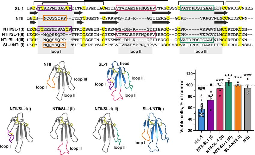
Bychkov et al.                                                                                SLURP-1 Forms Complex With α7-nAChR/EGFR/PDGFR

INTRODUCTION                                                              Carlisle et al., 2007; Chernyavsky et al., 2009; Davis et al.,
                                                                          2009; Mucchietto et al., 2018). In keratinocytes, the α7-nAChR
Nicotinic acetylcholine receptors (nAChRs) are ligand-gated ion           activation upregulates some transcriptional factors e.g., signal
channels that are activated by acetylcholine (ACh) and are                transducer and activator of transcription 3 (STAT3), GATA-
involved in regulation of many vital processes in the central             binding factor 2 (GATA2) (Arredondo et al., 2007b), and
and peripheral nervous system, including synaptic transmission            NFκB (Chernyavsky et al., 2010); so, the dysregulation of α7-
and plasticity, neuronal plasticity, memory, cognition, addictive         nAChR can also lead to loss of control over gene expression
behavior, pain transmission and muscle contraction (Feduccia              and drive the progression of epithelial cancers. Activation of
et al., 2012; Koukouli and Maskos, 2015; Zoli et al., 2018).              the PI3K/AKT/mTOR pathway by α7-nAChR can inhibit the
Recently, however, a lot of data on the expression of nAChRs              cell senescence and apoptosis resulting in the chemotherapy
in non-neuronal cells, and on their participation in regulation           resistance (Dasgupta et al., 2006; Junhui et al., 2009). In addition,
of epithelial cell homeostasis and the immune system were                 the α7-nAChR activation can promote angiogenesis (Brown
published (Wessler and Kirkpatrick, 2009; Kulbatskii et al., 2018;        et al., 2012) and inhibit immunosurveillance (Qiu et al., 2004).
Zoli et al., 2018). Nicotinic acetylcholine receptor of α7 type           On the other hand, the activation of α3-containing nAChR by
(α7-nAChR) is one of the most widespread type of nAChRs                   nicotine mediates remodeling of the extracellular environment
expressed in epithelial and immune cells (Zoli et al., 2018).             (Arredondo et al., 2003), creating a microenvironment favorable
The non-neuronal α7-nAChR signaling is implicated in the                  for migration and invasion. Thus, nAChRs and particularly
regulation of inflammation (de Jonge and Ulloa, 2007), terminal           the α7 receptors are involved in all stages of epithelial cancer
epithelial homeostasis (Grando, 2006), vascularization (Heeschen          progression; and inhibition of α7-nAChRs may be a promising
et al., 2002), and even in cytokine secretion during COVID-               strategy for lung cancer therapy.
19 infection (Kashyap et al., 2020) and regulation of expression             There are several endogenous three-finger proteins belonging
of ACE2,–the SARS-CoV-2 entry receptor (Leung et al., 2020;               to the lymphocyte antigen 6/urokinase plasminogen activator
Russo et al., 2020).                                                      receptor (Ly6/uPAR) family that negatively modulate nAChRs
   There are also strong evidences of α7-nAChR involvement                without complete inhibition and have the promising properties
in oncogenic transformation and tumor progression                         for cancer therapy. One of such proteins, the human secreted
upon activation by nicotine (Grando, 2014; Schaal and                     Ly6/uPAR-related protein 1 (SLURP-1), was initially described
Chellappan, 2014). Nicotine and tobacco nitrosamines                      as a paracrine regulator of keratinocyte homeostasis. Point
[4-(methylnitrosamino)-1-(3-pyridyl)-1-butanone (NNK) and                 mutations in the SLURP1 gene lead to the development of skin
N-nitrosonornicotine (NNN)] contained in tobacco smoke                    disease, palmoplantar keratoderma Mal de Meleda (Arredondo
cause oncogenic transformation of normal cells (Arredondo                 et al., 2005; Perez and Khachemoune, 2016). SLURP-1 has
et al., 2006a) and drive proliferation, migration, and invasion of        a rather flexible spatial structure (Paramonov et al., 2020),
breast, pancreatic, and lung cancer cells (Dasgupta et al., 2009;         and site-directed mutagenesis suggested the possibility of its
Gankhuyag et al., 2017; Sarlak et al., 2020). Notably, nicotine           simultaneous interaction with several target receptors, by means
and nitrosamines demonstrate greater affinity to the α7 receptor          of three elongated and conformationally mobile loops, and
than endogenous ACh (Grando, 2014). In lung cancer cells the              a β-structural core (“head”) of the protein (Shulepko et al.,
pro-oncogenic effect of nicotine can be further enhanced by               2021). SLURP-1 interacts with α7-nAChRs (Chernyavsky et al.,
upregulation of the α7-nAChR expression in response to the                2015; Lyukmanova et al., 2016a), induces keratinocyte apoptosis
nAChR activation by nicotine (Schuller, 2012). The nicotine               (Arredondo et al., 2005), and protects the oral keratinocytes
signaling in lung cancer cells can also be enhanced by mitogenic          from oncogenic transformation by tobacco-derived nitrosamines
receptor tyrosine kinases (RTKs) activation due to formation              (Arredondo et al., 2007a; Kalantari-Dehaghi et al., 2012). SLURP-
of heterocomplexes between α7-nAChRs and some RTKs, such                  1 expression is down-regulated in primary and metastatic
as epidermal growth factor receptor (EGFR) (Chernyavsky                   melanomas compared with normal cells (Bergqvist et al., 2018;
et al., 2015). Along with the cell membrane, α7-nAChRs are                Arousse et al., 2019), moreover the elevated level of SLURP-1 in
also located on the mitochondrial membrane, where they can                plasma correlates with a better survival prognosis for pancreatic
inhibit a mitochondrial membrane pore formation and apoptosis             cancer patients (Throm et al., 2018). Thus, SLURP-1 can be
induction (Gergalova et al., 2012; Kalashnyk et al., 2012).               considered a prototype antitumor drug, but its effect on cancer
   Activation of cell-surface α7-nAChR can lead to two main               and normal cells, its targets and active centers should be studied
types of response: ionotropic, associated with the Ca2+ influx            in details.
into the cytosol; and metabotropic, which activates mitogenic                Previously we have shown that recombinant analogue of
intracellular signaling pathways without opening of the nAChR             human SLURP-1 (rSLURP-1) selectively inhibits ACh-evoked
channel (Grando, 2014). The protein kinase C (PKC), mitogen-              currents through α7-nAChR (Lyukmanova et al., 2016a) and
activated protein kinase (MEK)/extracellular signal-regulated             suppresses the growth of different carcinoma cells (Lyukmanova
kinase (ERK), and phosphoinositide 3-kinase (PI3K)/protein                et al., 2014, 2018; Shulepko et al., 2020a). The recombinant
kinase B (AKT)/mammalian target of rapamycin (mTOR)                       protein also suppresses the nicotine-induced lung cancer cell
pathways are the main intracellular mediators of the α7-nAChR             proliferation via interaction with α7-nAChR (Shulepko et al.,
signaling responsible for growth, migration, and invasion of              2020b). The PI3K/AKT/mTOR and inositol 1,4,5-trisphosphate
lung cancer cells (Tsurutani et al., 2005; Arredondo et al., 2006b;       (IP3) pathways are probably involved in the antiproliferative

Frontiers in Cell and Developmental Biology | www.frontiersin.org     2                                  September 2021 | Volume 9 | Article 739391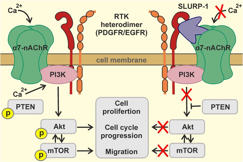
Bychkov et al.                                                                               SLURP-1 Forms Complex With α7-nAChR/EGFR/PDGFR

activity of rSLURP-1 in lung adenocarcinoma A549 cells                   complete medium supplemented with Pen-Strep (10,000 U/mL
(Shulepko et al., 2020b). In the present study, we further               penicillin, 10,000 U/mL streptomycin). Primary fibroblast cells
investigated the rSLURP-1 effects in A549 cells, determined the          were grown at 37◦ C and 5% CO2 . All cells were subcultured at
intracellular pathways involved in its action, revealed its new          least twice a week.
non-cholinergic molecular targets, and identified the primary               To study an influence of the peptides or proteins on the A549
active site responsible for the SLURP-1 antitumor activity.              or primary fibroblasts cell growth, the cells were seeded in 96-
                                                                         well cell culture plates in the corresponding complete medium
                                                                         (0.5 × 10⁴ cells/well) and grown for 24 h. Thereafter rSLURP-
MATERIALS AND METHODS                                                    1 from 250 µM DMSO stock or peptides from 10 mM DMSO
                                                                         stock solutions were diluted in the cell medium and added
Materials                                                                to the cells for further incubation during 48 h. The maximal
Genes of the chimeric proteins NTII/SL-1(I), NTII/SL-1(II),              DMSO concentration did not exceed 0.5%. The added DMSO
NTII/SL-1(III), and SL-1/NTII(I) were obtained by site-                  did not influence the cell growth as was established in the
directed mutagenesis on the basis of pET22b/STII/NTII                    additional experiments.
and pET22b/SLURP-1 expression plasmids, respectively                        To analyze cell viability, we used water soluble tetrazolium salt
(Lyukmanova et al., 2007; Shulepko et al., 2013). rSLURP-1               1 (WST-1) colorimetric test as described earlier (Lyukmanova
and chimeric protein SL-1/NTII(I) were isolated and refolded             et al., 2016a). Briefly, WST-1 (Santa Cruz, Dallas, TX,
from Escherichia coli inclusion bodies as described previously           United States) and 1-m-PMS (1-methoxy-5-methylphenazinium
(Shulepko et al., 2013). Chimeric proteins NTII/SL-1(I), NTII/SL-        methyl sulfate, Santa Cruz) were added to the cells in
1(II), and NTII/SL-1(III) were produced in E. coli according to          concentrations of 0.25 mM and 5 µM, respectively, for 1 h, and
the protocols described in Lyukmanova et al. (2007). The purity          formation of colored product was measured at 450 nm with
and homogeneity of the protein preparations was confirmed by             background subtraction at 655 nm on microplate reader. The
HPLC, MALDI-MS, and SDS-PAGE. Disulfide bond formation                   data were normalized to averaged read-out from the control wells
was confirmed in the reaction with Ellman’s reagent (Sigma-              containing cells without added compounds.
Aldrich, United States). The correct folding of the recombinant
proteins was confirmed by 1D 1 H NMR spectroscopy.
                                                                         Knock-Down of CHRNA7, EGFR, and
Protein Phosphorylation Analysis                                         PDGFRA Genes in A549 Cells
Phosphorylation of cellular signaling proteins was analyzed using        To block expression of native α7-nAChR, EGFR, and platelet-
the Bio-Plex magnetic beads assay with Bio-Plex Pro cell signaling       derived growth factor receptor type α (PDGFRα), A549 cells
reagent kit (Bio-Rad). Cells were incubated for 48 h with                were transfected with siRNA (Supplementary Table 1, Synthol,
1 µM rSLURP-1 added from 100% DMSO stock (protein stock                  Russia). Cells were seeded in T25 cell culture flasks (1 × 10⁵ cells
concentration 250 µM) and lysed using provided buffer. Analysis          per well) and grown for 24 h. Then four different siRNA
was performed on Bio-Plex 200 machine (Bio-Rad) according to             were mixed (1 µg per well), the mixture was diluted in the
the manufacturer instructions using the Bio-Plex Manager 6.2             100 µl transfection buffer (Pan-Biotech, Germany), incubated
software (Bio-Rad).                                                      for 5 min and mixed with 15 µl of pre-diluted PanFect A-plus
                                                                         transfection reagent (Pan-Biotech, Germany). The final mixture
Cell Cultivation and Viability Assay                                     was incubated for 30 min and added to A549 cells. The cells were
Human lung adenocarcinoma A549 cells (ATCC, United States)               incubated in CO2 -incubator during 4 h and the cell media was
were grown (37◦ C, 5% CO2 ) in a DME medium with phenol red              replaced by the fresh one. After 48 h incubation, the cells were
(PanEco, Russia), 10% fetal calf serum (Thermo Fisher Scientific,        detached by Versene solution and divided into two parts. The first
United States) and 2 mM L-glutamine (PanEco), abbreviated                part was incubated with TRITC-labeled α-Bgtx (Sigma-Aldrich,
below as the A549 complete medium. Mouse primary lung                    T0195) for detection of expression of functional α7-nAChRs on
fibroblasts were isolated from the lung of mouse embryos (the            the cell membrane. For EGFR and PDGFR detection, cells were
study was approved by IBCH RAS IACUC, protocol #312 from 21              incubated with primary antibodies (Abs) (sc-373746, Santa Cruz,
December 2020) according to the previously described procedure           for EGFR detection and ABIN5611263, Antibodies online, for
(Edelman and Redente, 2018). Briefly, mouse embryos were                 PDGFRα) and with secondary TRITC-conjugated Abs (615-025-
isolated from female mice after cervical dislocation. The lung           214, Jackson Immunoresearch). Expression of surface receptors
was removed from the thoracic cavity, placed into a 100 mm               was analyzed by flow cytometry. The second part of the cells was
tissue culture petri dish, and cut into small pieces using a razor       seeded in 96-well culture plates (50 × 104 cells per well) and
blade or surgical scissors. Then, 1 mL of StemPro Accutase Cell          wound-healing assay was performed as described below.
Dissociation Reagent (Thermo Fisher Scientific, United States)
was added to the chopped lung and incubated at 37◦ C for 30 min.         Wound Healing (Scratch) Assay
Then, the tissue was washed twice in HBSS and incubated at               The in vitro wound healing (scratch) assay was performed as
37◦ C for 20 min in 0.5 mL of 0.25% Trypsin-EDTA. After                  described elsewhere (Varankar and Bapat, 2018) with some
incubation the cells were centrifuged for 5 min at 1000 × g and          changes. In brief, A549 cells and primary fibroblasts were seeded
resuspended in fibroblast complete medium which was the A549             in 96-well cell culture plates in the corresponding complete

Frontiers in Cell and Developmental Biology | www.frontiersin.org    3                                  September 2021 | Volume 9 | Article 739391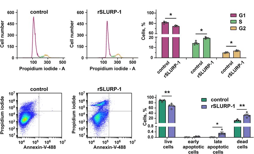
Bychkov et al.                                                                              SLURP-1 Forms Complex With α7-nAChR/EGFR/PDGFR

medium (5 × 10⁴ cells/well) and grown for 24 h. Then the media          non-reducing PAGE loading buffer for detection of EGFR and in
from the wells was changed to serum-free media to minimize cell         reducing PAGE buffer for detection of α7-nAChR and PDGFRα.
proliferation. After 8 h the wells were scratched with a sterile        Western blotting was used to detect α7-nAChR (primary Abs
10 µl pipette tip. Then, the cells were washed with PBS and             ABIN5611363, Antibodies Online, 1:1000 and secondary Abs
treated with rSLURP-1 or peptide for 48 h. Pictures were analyzed       111-035-003, Jackson Immunoresearch, 1:5000), EGFR (primary
after 0 and 24 h at 20× magnification at CloneSelect Imager             Abs sc-120, Santa Cruz, 1:1000 and secondary Abs 715-035-150,
(Molecular Devices, United States). The center of the plate was         Jackson Immunoresearch, 1:5000) and PDGFRα (primary Abs
marked as a central reference point to ensure recording of the          ABIN5611263, Antibodies Online, 1:1000 and secondary Abs
same area during the time course. Digital images were taken, and        715-035-150, Jackson Immunoresearch, 1:5000).
the scratch area was quantified using ImageJ (NIH, United States)
and MS Excel software by measurement % of scratch surface,              Real Time PCR for miRNA Detection
occupied by migrating cells. In each experiment, the duplicate          Total mRNA from the cultured cells was extracted by the Aurum
measurements have been averaged.                                        Total RNA Mini Kit (Bio-Rad) according to the manufacturer’s
                                                                        instructions. Total cDNA was synthesized using the Mint
Cell Cycle Arrest in A549 Cells                                         revertase (Evrogen, Russia) with miRNA-specific stem-loop
Cells were seeded in T25 Cell Culture Flask (5 × 10⁵ cells/flask)       primer. After that, real-time PCR was performed with the primers
and incubated with 1 µM rSLURP-1 for 48 h. Then the cells               described in the Supplementary Table 1, and ready-to-use qPCR
were detached from the flasks by trypsin, washed with EBSS, and         mix with the SYBR Green I fluorescent dye (Evrogen). Negative
fixed in ice-cold 70% ethanol for 4 h. After fixation, the cells        controls contained all the components of the PCR mixture but
were washed twice by EBSS, and DNA was extracted by 5 min               with cDNA replaced by mRNA gave no signal. All PCR reactions
incubation with the DNA extraction buffer (200 mM Na2 HPO4              were performed using Roche Light cycler 96 real-time detection
with 0.004% Triton X-100, pH 7.8). Then the cells were washed           thermal cycler (Roche, Switzerland). Data was analyzed by the
by EBSS, resuspended in the DNA staining solution (EBSS,                1Ct method (Livak and Schmittgen, 2001) using Light-Cycler
50 mg/ml propidium iodide, 0.2 mg/ml DNase free RNAse),                 96 SW1.01 software (Roche). The expression level of the genes
and analyzed by Cytoflex flow cytometer (Beckman Coulter,               was normalized to the expression level of the housekeeping
United States). Data were analyzed by ModFit LT software (Verity        non-coding RNA U6.
House, United States).

Study of Apoptosis in A549 Cells                                        Design and Chemical Synthesis of
To investigate apoptosis in A549 cells, we used Annexin V for           Peptides Mimicking the SLURP-1 Loops
detection of phosphatidylserine externalization,–one of the early       The      amide     forms     of     the   peptides     (“loop   I”:
apoptosis markers. Briefly, A549 cells were seeded on a 35-mm           VKAYTCKEPXTSASCRTITRA, X stands for norleucine
Petri dish (1 × 10⁵ cells/dish) and incubated with 1 µM rSLURP-         (methionine       bioisosteric     replacement);     “loop    III”:
1 for 48 h. After incubation, the cells were detached by the            GCVARDPDSIGAAHLIFCG) were obtained by chemical
Versene solution and washed in annexin-binding buffer (V13246,          synthesis using the Fmoc/TBTU methodology (Chan and White,
Thermo Fisher Scientific). Then, the cells were incubated with          2000), starting with aminomethyl polystyrene acylated with
Annexin V conjugated to Alexa 488 (A13201 Thermo Fisher                 Rink amide-linker at an initial loading of 0.4 mmol/g. Standard
Scientific) for 20 min, washed by annexin-binding buffer, and           protocols were followed during stepwise chain elongations,
were analyzed on Cytoflex flow cytometer (Beckman Coulter,              except that N-terminal amino-acids were introduced using the
United States). Data were analyzed by CytExpert 2.4 software            corresponding Nα -Boc derivatives of valine and glycine. After
(Beckman-Coulter, United States).                                       completing the chain elongation, the protecting groups and the
                                                                        linear peptides were removed from the support using a mixture
Affinity Purification and Western Blotting                              of TFA-DCM-TIPS-H2 O-anisole (95: 3.5: 0.5: 0.5: 0.5: 0.5, 30 ml)
Recombinant analogue of human SLURP-1 (1 mg/ml) was                     in an inert atmosphere. The reaction mixtures were filtered from
coupled to NHS-activated Sepharose 4 Fast Flow resin (Cat#              the polymer, evaporated in vacuo at a temperature not exceeding
17-0906-01, GE Healthcare) according to the manufacturer’s              40◦ C to a quarter of the initial volume and slowly added to cold
manual. The empty resin blocked by 500 mM ethanolamine was              diethyl ether. The precipitates were then filtered off on a glass
used as a negative control. The membrane fraction of A549 cells         filter, washed with diethyl ether and dried in a vacuum desiccator
(5 × 10⁷ cells per sample) was solubilized in 2% Triton X-100           over potassium hydroxide pillets and paraffin overnight.
(Cat# A4975, Panreac), diluted 10 times with TBS buffer [100 mM             The disulfide bridges were formed under slightly basic
Tris (141940.0914, Panreac), 150 mM NaCl (141659, Panreac),             conditions (200 ml of 0.1 M ammonium bicarbonate solution
pH 8.0], and incubated with the resin for 1 h in TBS. After that,       per 70–100 mg of peptide sample). The disappearance of
unspecific bound proteins were sequentially washed out from             sulfhydryl groups was assessed by the quantitative Ellman’s test
the resin with five volumes of TBS, five volumes of TBS + 1 M           (Andreu et al., 1994). After 3–4 days, the reaction mixtures
NaCl + 0.5% Triton X-100, and five volumes TBS + 0.5%                   were evaporated in vacuum to a volume of 50–60 ml and
Triton X-100. The specifically bound proteins were eluted by five       lyophilized. After that, first crude HPLC purifications were
volumes of 200 mM Glycine (131340, Panreac) (pH 2.6) into               achieved using Vaydac C18 (250 × 21.4) column with isocratic

Frontiers in Cell and Developmental Biology | www.frontiersin.org   4                                 September 2021 | Volume 9 | Article 739391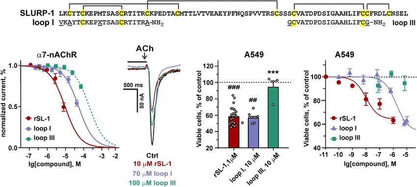
Bychkov et al.                                                                                 SLURP-1 Forms Complex With α7-nAChR/EGFR/PDGFR

elution using aqueous acetonitrile (21% acetonitrile/0.1% TFA               concentration at half-maximal efficacy, [protein] is the protein
in water for “loop I” and 24% for “loop III,” 10 ml/min). The               concentration. Analysis was performed using the GraphPad
fractions collected were analyzed by mass-spectrometry and the              Prism 6.0 software.
appropriate fractions were lyophilized and additionally purified
by HPLC using a Jupiter C4 chromatographic column (4.6 × 250,
Phenomenex, acetonitrile gradient 20–25% in 0.1% TFA at a flow              RESULTS
rate of 0.2 ml/min). The homogeneity and purity (>95%) of
peptides were confirmed by HPLC, MALDI-MS, and 1 H-NMR                      rSLURP-1 Reduces PTEN, mTOR, and
spectroscopy (Supplementary Figure 3).
                                                                            PDGFRβ Phosphorylation in A549 Cells
                                                                            Recently, we have shown that nicotine stimulates growth
Electrophysiological Recordings From                                        of lung carcinoma A549 cells and down-regulates the
X. laevis Oocytes                                                           expression of phosphatase and tensin homolog deleted on
For expression of human α7-nAChRs in Xenopus oocytes, the                   chromosome 10 (PTEN), while rSLURP-1 abolishes these
linearized plasmid was transcribed using the T7 mMessage-                   negative effects of nicotine (Shulepko et al., 2020b). At
mMachine transcription kit (Thermo Fisher Scientific, Carlsbad,             the same time, rSLURP-1 by itself inhibits growth of A549
CA, United States). The harvesting of stage V–VI oocytes                    cells, and this effect depends on α7-nAChR, EGFR, and
from anesthetized female Xenopus laevis frog was previously                 β-adrenergic receptor (Shulepko et al., 2020b). α7-nAChR
described (Peigneur et al., 2019). Oocytes were injected with               and EGFR are both enhance the cancer cell proliferation via
42 nL of cRNA at a concentration of 1 ng/nL using a micro-                  activation of the PI3K/AKT/mTOR signaling pathway, and
injector (WPI, United States). The oocytes were incubated in                PTEN is a major negative regulator of this pathway (Misiura
a solution containing (in mM): 96 NaCl, 2 KCl, 1.8 CaCl2 , 2                et al., 2020; Witayateeraporn et al., 2020; Cochard et al.,
MgCl2 , and 5 HEPES, (pH 7.4), supplemented with 50 mg/L                    2021). Therefore, we decided to study the effect of rSLURP-
gentamycin sulfate.                                                         1 on the phosphorylation level of the certain components
    Two-electrode voltage-clamp recordings were performed at                of this intracellular signaling pathway (AKT, PTEN, and
room temperature (18–22◦ C) using a Geneclamp 500 amplifier                 mTOR) in A549 cells.
(Molecular Devices , Downingtown, PA, United States)
                           R
                                                                               The Bio-Plex magnetic immunoassay revealed that rSLURP-1
controlled by a pClamp data acquisition system (Axon                        significantly inhibited PTEN phosphorylation at the S380 site,
Instruments , Union City, CA, United States). Whole-cell
                 R
                                                                            resulting in PTEN activation (Scully et al., 2014; Figure 1).
currents from oocytes were recorded 1–4 days after injection.               In addition, we found that the rSLURP-1 application
Bath solution composition was (in mM): 96 NaCl, 2 KCl,                      significantly reduced the mTOR phosphorylation (activator
1.8 CaCl2 , 2 MgCl2 , and 5 HEPES (pH 7.4). Voltage and                     of the PI3K/AKT pathway) in A549 cells, thereby reducing
current electrodes were filled with 3 M KCl. Resistances of                 its activity. Interestingly, we also observed a decrease in
both electrodes were kept between 0.7 and 1.5 M. During                    PDGFRβ phosphorylation at the Y751 site upon incubation
recordings, the oocytes were voltage-clamped at a holding                   with rSLURP-1 (Figure 1), which may lead to decrease in
potential of −70 mV and continuously superfused with                        phosphorylation of PI3K (Kazlauskas and Cooper, 1990)
solutions. α7-nAChRs currents were evoked by 100 ms pulses                  and AKT (Zhang et al., 2015). Indeed, we found weak
of 100 µM ACh at 2 mL/min with 1–2 min washout periods                      insignificant decrease of the AKT (S473) phosphorylation
between applications. Data were sampled at a frequency of                   (Figure 1). The observed simultaneous PTEN activation and
100 Hz and low-pass filtered at 20 Hz by using a four-pole                  mTOR/PDGFRβ inactivation revealed down-regulation of
Bessel filter. Peak current amplitude was measured prior to and             the PI3K/AKT/mTOR signaling pathway in A549 cells upon
following the incubation with rSLURP-1 and peptides. Data                   incubation with rSLURP-1.
were analyzed using pClamp Clampfit 10.0 (Molecular Devices ,       R

Downingtown, PA, United States) and Origin 7.5 software                     rSLURP-1 Induces Cell Cycle Arrest but
(Originlab , Northampton, MA, United States).
             R

                                                                            Not Apoptosis in A549 Cells
                                                                            Activation of the PI3K/AKT/mTOR signaling pathway is
Statistical Analysis and Curve Fitting                                      implicated in hyperproliferation and resistance to apoptosis
Data are presented as mean ± SEM. Sample numbers (n)                        (Mercurio et al., 2021). Since rSLURP-1 down-regulates this
are indicated in the figure legends. Statistical analysis was               pathway, we studied its influence on cell cycle progression
done using two-tailed t-test, one sample two-tailed t-test, or              and apoptosis in A549 cells. Flow cytometry analysis revealed
one-way ANOVA followed by a Dunnett’s post hoc test as                      that rSLURP-1 induced significant reduction of A549 cell
indicated in the figure legends. Differences in the groups                  number in the G1 cell cycle phase from ∼81 to ∼74%
were considered statistically significant at p < 0.05. To                   with simultaneous increase of cell nuclei in the S and
assess the concentration-response relationships, normalized data            G2/M phases from ∼14 to ∼19% and from ∼5 to ∼7%,
points were fitted with the standard slope inhibition equation:             respectively (Figures 2A,B). The Annexin V phosphatidylserine
y(%) = 100% × (1–1/[1 + EC50 /[protein]], where y(%) is the                 externalization assay revealed significant increase in the
amplitude of the protein-induced effect, EC50 is the protein                number of non-apoptotically dead A549 cells, but did not

Frontiers in Cell and Developmental Biology | www.frontiersin.org       5                                September 2021 | Volume 9 | Article 739391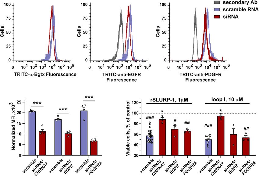
Bychkov et al. SLURP-1 Forms Complex With α7-nAChR/EGFR/PDGFR
FIGURE 1 | Effect of rSLURP-1 on phosphorylation of the Akt/mTOR and MAP/ERK pathways participants in A549 cells. A549 cells were incubated for 48 h with
1 µM of rSLURP-1; and PTEN (S380), AKT (S473), mTOR (S2448), PDGFRβ (Y751), Src (Y416), ERK (T202/Y204; T185/Y187), and p38 (T180/Y182)
phosphorylation was detected by Bio-Plex magnetic beads assay. Data are presented as mean ± SEM, n = 4; * (p < 0.05) and ** (p < 0.01) indicate the significant
difference by a two-tailed t-test.
reveal presence of apoptotic cells (Figures 2C,D). Thus, (Figures 3I,J). Thus, rSLURP-1 showed selective anti-migration
incubation of A549 cells with rSLURP-1 results in the activity toward cancer cells.
cell cycle arrest in the S and G2/M phases and induces We previously hypothesized that rSLURP-1 in A549 cells
non-apoptotic cell death. controls PTEN expression by acting on STAT3 (Shulepko
et al., 2020b). There are other molecules that are involved in
the regulation of gene expression,–small non-coding miRNAs
rSLURP-1 Inhibits Migration of A549 (Catalanotto et al., 2016), and some of them controls genes
Lung Cancer Cells but Not of Normal related to migration (Xu et al., 2012; Othman and Nagoor, 2014;
Lung Fibroblasts Smolarz and Widlak, 2021). Here, we tested the effect of rSLURP-
Recombinant analogue of human SLURP-1 decreases the 1 on expression of these miRNA. No significant effect was found
phosphorylation level of PDGFRβ (Y751) which could be (Supplementary Figure 1). Therefore, the influence of rSLURP-1
associated with the receptor inhibition, while PDGFRβ activation on cell migration is not associated with the changes in expression
leads to actin reorganization and cell migration (Heldin et al., of regulatory miRNAs.
1998). Another protein, involved in the action of rSLURP-
1, –mTOR, is also implicated in the control of cell motility,
cytoskeleton assembly, and epithelial-mesenchymal transition Loop I Is the Primary Site of SLURP-1
(Zhou and Huang, 2011). In line with this, SLURP-1 has Responsible for Antiproliferative Activity
been shown to inhibit migration of pancreatic cancer cells Lymphocyte antigen 6/urokinase plasminogen activator receptor
(Throm et al., 2018). proteins have conservative spatial organization and contain the
Therefore, we decided to test whether rSLURP-1 can inhibit β-structural core stabilized by invariant disulfide bonds (“head”)
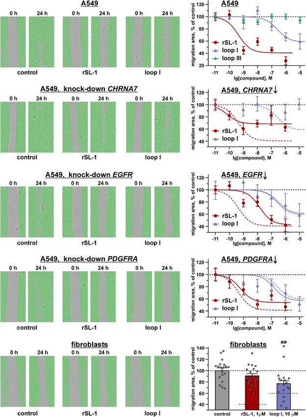
migration of A549 cells. The scratch assay showed that rSLURP-1 and three elongated loops (“fingers”) protruding into solvent.
significantly reduced migration of A549 cells up to ∼40% of the Ly6/uPAR proteins usually interact with their targets by these
control with EC50 in nanomolar range (∼0.4 nM) (Figures 3A,B loops (Vasilyeva et al., 2017). To identify the active site of
and Table 1). At the same time, rSLURP-1 at concentrations up SLURP-1, we constructed three chimeric proteins containing
to 1 µM did not affect the migration of primary lung fibroblasts grafted loops (I, II, or III) of SLURP-1 on a scaffold of short
Frontiers in Cell and Developmental Biology | www.frontiersin.org 6 September 2021 | Volume 9 | Article 739391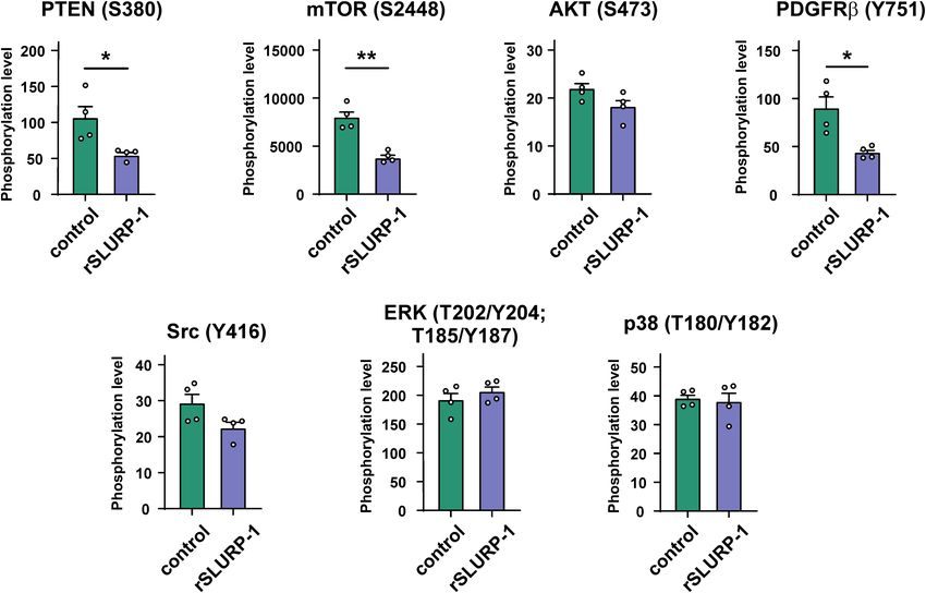
Bychkov et al.                                                                                                 SLURP-1 Forms Complex With α7-nAChR/EGFR/PDGFR

  FIGURE 2 | Influence of rSLURP-1 on cell cycle progression and apoptosis in A549 cells. (A) Representative nuclei population distributions of cells after 48 h
  incubation in absence (control) or presence of 1 µM rSLURP-1. (B) Percentage of cells in each cell cycle phase determined by ModFitLT software. Data are
  presented as mean ± SEM, n = 4; * (p < 0.05) indicates the significant difference from the control by two-tailed t-test. (C) Representative pictures of
  phosphatidylserine externalization analysis upon the rSLURP-1 treatment of A549 cells by flow cytometry with Annexin V-488 and Propidium iodide. The cells were
  incubated with 1 µM rSLURP-1 or without it (control) during 48 h. (D) Percentage of cells with externalized phosphatidylserine and bound propidium iodide. The data
  are presented as % of live, early apoptotic, late apoptotic and dead cells ± SEM (n = 4). * (p < 0.05) and ** (p < 0.01) indicate the significant difference from the
  control by two-tailed t-test.

three-finger neurotoxin II from cobra Naja oxiana (NTII)                               I region is the primary site of the SLURP-1 molecule responsible
(Figures 4A,B). The chimeric proteins were named NTII/SL-                              for antiproliferative activity in A549 cells.
1(I), NTII/SL-1(II), and NTII/SL-1(III). NTII was chosen as a
scaffold, because it shares the conserved three-finger structure,
but does not interact with α7-nAChRs (Lyukmanova et al.,
2007) and does not inhibit the proliferation of A549 cells
                                                                                       Synthetic Peptide Mimicking the Loop I
(Bychkov et al., 2019). Preservation of the overall three-finger                       of SLURP-1 Inhibits α7-nAChRs
spatial structure by NTII/SL-1 chimeras was confirmed by                               Expressed in X. laevis Oocytes
1 H NMR spectroscopy (Supplementary Figure 2). We have
                                                                                       To further study the role of the loop I region in the SLURP-1
previously shown that rSLURP-1 inhibits A549 cell growth in                            activity, we synthesized two peptide mimetics (Figure 5A).
a concentration-dependent manner and the maximum effect                                The first peptide (VKAYTCKEPXTSASCRTITRA-NH2 , 21
(∼60% of viable cells relative to the control) was observed at a                       residues, X stands for norleucine) mimics the structure
concentration of 1 µM after 48 h incubation (Shulepko et al.,                          of loop I of the SLURP-1 molecule. The second peptide
2020b). Here, we examined the antiproliferative activity of the                        (GCVATDPDSIGAAHLIFCG-NH2 , 19 residues) has the
chimeras in comparison with rSLURP-1 in A549 cells using a                             sequence of loop III and was used as a negative control.
single concentration of 1 µM. The chimeras NTII/SL-1(II) and                           Note, that the NTII/SL-1(III) chimera did not demonstrate
NTII/SL-1(III) and NTII itself were found to be completely                             antiproliferative activity in A549 cells (Figure 4C). Here and
inactive, while the chimera NTII/SL-1(I) containing the first                          after the peptides are called as “loop I” and “loop III,” respectively
loop of rSLURP-1 demonstrated antiproliferative activity slightly                      (Figure 5A). Initially, we tested the activity of peptides in
weaker than that of rSLURP-1 (inhibition effect up to ∼70% of                          comparison with rSLURP-1 against α7-nAChRs expressed in
viable cells relative to the control, Figure 4C). To further confirm                   X. laevis oocytes. In line with the previous findings (Lyukmanova
the importance of the loop I region for rSLURP-1 activity, we                          et al., 2016a), rSLURP-1 and loop I demonstrated reversible
designed an inverse chimera containing grafted loop I of NTII                          concentration-dependent inhibition of α7-nAChRs with IC50
on a scaffold of rSLURP-1. As expected, the SL-1/NTII(I) chimera                       values of 12 ± 1 and 74 ± 4 µM, respectively. At the same time,
did not suppress growth of A549 cells (Figure 4C). Thus, the loop                      loop III was much less active (IC50 > 250 µM, Figure 5B).

Frontiers in Cell and Developmental Biology | www.frontiersin.org                  7                                        September 2021 | Volume 9 | Article 739391
Bychkov et al. SLURP-1 Forms Complex With α7-nAChR/EGFR/PDGFR FIGURE 3 | Influence of rSLURP-1 and the “loop I” and “loop III” peptides on migration of A549 cells (A–H) and primary mouse lung fibroblasts (I,J). The data obtained for knockdown of the CHRNA7, EGFR, and PDGFRA genes are shown on the panels (C,D), (E,F), and (G,H), respectively. (A,C,E,G,I) Representative pictures of scratch assay, obtained on CloneSelect Imager after 24 h incubation of the cells with 1 µM of rSLURP-1 or 10 µM of the “loop I” peptide. (B,D,F,H,J) Effect of different rSLURP-1 and the “loop I” and “loop III” peptides concentrations on the migration of the cells. Data are presented as a mean scratch surface, occupied by migrating cells (% normalized to control), ±SEM, n = 7–21; The parameters describing the dose-response curves (EC50 , A1 ) and results of statistical comparisons are given in Table 1. Control level (100%) and position of the migration inhibition curves for rSLURP-1 and the “loop I” peptide in A549 cells without knock-down are shown by dashed lines. (J) Control level (100%) and the migration inhibition effects for 1 µM of rSLURP-1 and 10 µM of the “loop I” peptide in A549 cells are shown by dashed lines. ## (p < 0.01) indicates significant difference from control group by one-way ANOVA/Dunnett’s test. Frontiers in Cell and Developmental Biology | www.frontiersin.org 8 September 2021 | Volume 9 | Article 739391
Bychkov et al.                                                                                                   SLURP-1 Forms Complex With α7-nAChR/EGFR/PDGFR

TABLE 1 | Parameters descripting the effect of CHRNA7, EGFR, and PDGFRA knock-down on the anti-migration activity of rSLURP-1 and synthetic peptide “loop I” in
A549 cellsa .

Gene knockdown                                                   rSLURP-1                                                                Peptide “loop I”

                                               A1 , %                                EC50 , nM                               A1 , %                            EC50 , nM

Control                                      40.8 ± 8.7                               0.4 ± 0.2                            58.7 ± 7.2                        226 ± 85 (***)
α7-nAChR                                    68.5 ± 4.9 (#)                          0.07 ± 0.04                               n.d.                                n.d.
EGFR                                         42.6 ± 8.7                           14.1 ± 4.7 (###)                         60.3 ± 8.6                          291 ± 133
PDGFR                                      54.0 ± 4.9 (##)                            0.6 ± 0.2                            56.7 ± 9.9                           155 ± 78
a Dataare presented as mean ± SEM, n = 7–21.      ∗∗∗  (p < 0.001) indicates the significant difference between parameters describing rSLURP-1 and “loop I” curves by a
two-tailed t-test. # (p < 0.05), ## (p < 0.01), and ### (p < 0.001) indicate the significant difference from parameters describing the control curve by a two-tailed t-test.

  FIGURE 4 | Design and study of antiproliferative activity of chimeric NTII/SL-1 and SL-1/NTII proteins. (A) Amino acid sequence alignment of SLURP-1, NTII, and
  chimeras. Cys residues are shown by yellow. Disulfide bonds are shown by brackets. The β-strands are designated by arrows. Sequences of the grafted loops are
  highlighted by color frames. (B) Schemes depicting the chimeric NTII/SL-1 and SL-1/NTII proteins. Color coding is the same as on the panel (A). (C) Antiproliferative
  activity of the chimeric proteins in A549 cells according to WST-1 test. Cells were incubated during 48 h with 1 µM of the proteins. Data are presented as % of
  control (untreated cells, dashed line) ± SEM (n = 4–22). # (p < 0.05) and ### (p < 0.001) indicate significant difference from the control by one-sample t-test.
  * (p < 0.05) and *** (p < 0.001) indicate significant difference from the “rSLURP-1” group by one-way ANOVA/Dunnett’s test.

The “Loop I” Peptide Suppresses Growth                                                      Water soluble tetrazolium salt 1 test revealed similar
and Migration of A549 Cells but Not of                                                   significant decrease of A549 cell viability upon treatment with
                                                                                         10 µM of “loop I” peptide or 1 µM of rSLURP-1 (Figure 5D).
Normal Fibroblasts                                                                       Comparison of the dose-response curves revealed EC50 of
We have previously shown that rSLURP-1 suppresses the growth                             1.8 ± 0.3 µM and 10.1 ± 2.6 nM for “loop I” and rSLURP-
of various carcinomas, but demonstrates much weaker activity on                          1, respectively (Figure 5E). Observed maximal effects were
the normal immortalized human cells, including keratinocytes                             comparable (45.8 ± 5.1% and 59.0 ± 2.7% relative to the control
Het-1A and lung fibroblasts WI-38 (Lyukmanova et al., 2016a,                             for the “loop I” peptide and rSLURP-1, respectively), although
2018; Shulepko et al., 2020b). Here, the antiproliferative and                           the larger concentration of the “loop I” peptide was required
anti-migration activities of the “loop I” and “loop III” peptides                        (Figures 5D,E). The similar situation was observed for loop I
were studied in comparison with rSLURP-1 in A549 cells or                                in the scratch migration assay (Figures 3A,B). EC50 of the anti-
normal primary lung fibroblasts.                                                         migration activity was increased from 0.4 nM (rSLURP-1) to

Frontiers in Cell and Developmental Biology | www.frontiersin.org                    9                                        September 2021 | Volume 9 | Article 739391
Bychkov et al. SLURP-1 Forms Complex With α7-nAChR/EGFR/PDGFR
FIGURE 5 | Design and activity of peptides, mimicking the SLURP-1 loops I and III. (A) Amino acid sequence alignment of rSLURP-1 and the synthetic peptides
“loop I” and “loop III.” Cys residues are shown by yellow. Disulfide bonds are shown by brackets. (B) The dose-response curves for the inhibition of ACh-evoked
currents at α7-nAChRs expressed in X. laevis oocytes by rSLURP-1 and synthetic peptides. The data are normalized to the peak amplitude of current recorded
without compounds application (100%) and presented as mean ± SEM (n = 4–6 oocytes for each compound). The standard slope (nH = 1) inhibition equation was
fitted to normalized data. (C) Representative responses to 100 ms pulses of 100 µM ACh recorded in absence and presence of rSLURP-1, “loop I” and “loop III”
peptides. The period of oocyte pre-incubation with compounds is shown by horizontal bars above current traces (not in the timescale), the application of ACh is
shown by arrow. (D,E) Antiproliferative activity of rSLURP-1, and “loop I” and “loop III” peptides in A549 cells upon 48 h incubation according to WST-1 test. Data
are presented as % of control (untreated cells, dashed line) ± SEM (n = 4–22). ## (p < 0.01) and ### (p < 0.001) indicate significant difference from the control by
one-sample t-test. *** (p < 0.001) indicates significant difference from the “rSLURP-1” group by one-way ANOVA/Dunnett’s test.
∼230 nM (“loop I” peptide, Table 1), while the maximal effects mutagenesis data, this mutation inactivates the rSLURP-1 protein
were comparable (∼41 and 59%, respectively). Both the rSLURP- and prevents the interaction with α7-nAChR (Shulepko et al.,
1 protein and “loop I” peptide demonstrated significantly 2021). As expected, the K29A rSLURP-1 mutant did not extract
diminished activity on normal fibroblasts in comparison with the α7-nAChR, while EGFR and PDGFRα did (Figure 6). Thus,
cancer cells (Figures 3I,J). No effect of “loop III” on the A549 cell loop I of SLURP-1 is responsible for the interaction with α7-
viability and migration was observed up to the 10 µM peptide nAChR, while some other parts of the molecule are involved in
concentration (Figures 3B, 5D,E). the interaction with EGFR and PDGFRα .
rSLURP-1 Forms Complexes With
rSLURP-1 and “Loop I” Antiproliferative
α7-nAChR and Receptor Tyrosine
Activity in A549 Cells Is Mediated Only by
Kinases EGFR and PDGFRα
We recently proposed that some of the effects of rSLURP-1 in
α7-nAChR, While in the Regulation of
keratinocytes may be mediated by interaction with a second, Migration α7-nAChR, EGFR, and
different from α7-nAChR, molecular target (Shulepko et al., PDGFRα Are Involved
2021). This second target may be some receptor of the RTK To determine the role of α7-nAChR, EGFR, and PDGFRα in the
family, for example, EGFR forming a complex with α7-nAChR rSLURP-1 effects, we knocked down the corresponding receptors
(Chernyavsky et al., 2015). On the other hand, the observed genes, and studied the effect of rSLURP-1 and “loop I” on the
inhibition of PDGFRβ phosphorylation by rSLURP-1 (Figure 1) proliferation and migration of A549 cells (Figures 7A–D). In
suggests an interaction of rSLURP-1 with PDGFRs. line with the previous findings (Shulepko et al., 2020b), the
In attempt to uncover this second target, we performed affinity antiproliferative activity of rSLURP-1 was almost completely
purification of proteins from a membrane fraction of A549 cells abolished by the CHRNA7 knockdown, while knockdown of the
that bind rSLURP-1. The NHS-Sepharose with coupled rSLURP- EGFR and PDGFRA genes did not affect the viability of A549 cells
1 was used for affinity extraction; and resulting Western-blots in the presence of rSLURP-1 (Figure 7E). Similarly, to rSLURP-1,
were stained by antibodies to α7-nAChR, EGFR, and PDGFRα. the antiproliferative activity of the “loop I” peptide was inhibited
Surprisingly, we found that rSLURP-1 extracts all three receptors only by the CHRNA7 knockdown (Figure 7E).
from the membrane fraction (Figure 6). To test the specificity The knock-down of the CHRNA7 gene significantly
of the affinity extraction procedure, we used rSLURP-1 K29A diminished the maximal anti-migration effects of the rSLURP-1
mutant with a substitution in the loop I. According to the protein and completely abolished the activity of the “loop
Frontiers in Cell and Developmental Biology | www.frontiersin.org 10 September 2021 | Volume 9 | Article 739391Bychkov et al. SLURP-1 Forms Complex With α7-nAChR/EGFR/PDGFR
FIGURE 6 | Analysis of rSLURP-1 targets in A549 cells. NHS-Sepharose coupled with rSLURP-1 or rSLURP-1[K29A] and rSLURP-1[D81A] mutants was incubated
with a membrane fraction of A549 cells and extracted proteins were analyzed by Western blotting using antibodies against α7-nAChR (A), EGFR (B), and PDGFRα
(C). For detection of EGFR non-reducing SDS-PAGE was used. Lines: “Input”–the membrane fraction of A549 cells used for analysis; “empty”–proteins extracted
from the membrane fraction by empty resin without rSLURP-1 or its mutant; “rSL-1”, “rSL-1[K29A]” or “rSL-1[D81A]”–proteins extracted from the membrane fraction
by resin coupled with rSLURP-1 or its mutants. Bands corresponding to the α7-nAChR, EGFR, and PDGFRα receptors are shown by arrows.
I” peptide (Figures 3C,D and Table 1). Block of the EGFR the loop C of the primary receptor’s subunit. The loop C covers
expression did not influence the maximal amplitude of the the orthosteric ligand binding site of the nicotinic receptor,
anti-migration effect, while the EC50 value for rSLURP-1 became and its movement is coupled with the receptor activation (Law
more than order of magnitude higher (increase from 0.4 to et al., 2005; Gulsevin, 2020). Loop II and loop III of SLURP-
14 nM, Figures 3E,F and Table 1). At the same time no effect on 1 were supposed to form additional contacts with the primary
the activity of the “loop I” peptide was observed (Figures 3E,F). and complementary receptor subunits (Shulepko et al., 2021).
This indicates that EGFR is involved in the anti-migration effect The data obtained here revealed the primary role of loop I
of rSLURP-1, but not of the “loop I” peptide. Similarly, the of the SLURP-1 molecule for the interaction with α7-nAChR
knock-down of the PDGFRA gene did not affect the migration of (Figures 4, 5). Accordingly, the K29A mutation in loop I resulted
A549 cells in the presence of the “loop I” peptide (Figures 3G,H), in a decrease in the affinity of rSLURP-1 toward α7-nAChR
while significant reduction in the maximal effect amplitude, but in A549 cells (Figure 6A) as it has been demonstrated in
not in EC50 value, was observed for rSLURP-1 (Figures 3G,H keratinocytes (Shulepko et al., 2021). The reduced activity of the
and Table 1). Taking together, the data obtained show that the “loop I” peptide, as compared to the whole rSLURP-1 molecule,
anti-migration activity of the “loop I” peptide depends only on confirms the multiple point mode of the α7-nAChR/SLURP-1
the α7 nicotinic receptor, while the activity of rSLURP-1 depends interaction proposed earlier (Shulepko et al., 2021). In line with
on the α7-nAChR and RTKs (EGFR and PDGFRα ). this, we observed the weak inhibitory activity of the “loop III”
peptide at α7-nAChRs expressed in oocytes and reduced affinity
of the mutant with substitution D81A in loop III toward α7-
DISCUSSION nAChR (Figures 5C, 6A). This indicates that loop III also plays
some role in the interaction with the receptor.
Activation of α7-nAChRs by nicotine results in increase of Notably, the mode of the α7-nAChR/SLURP-1 interaction
viability, proliferation, and motility of epithelial cells (Lupacchini differs from that found for other Ly6/uPAR proteins and
et al., 2020). Using the lung adenocarcinoma A549 cells, for their targets. For example, the region of loop II was found
the first time we showed that rSLURP-1 significantly reduced to be important for interaction of mambalgins with the acid
not only proliferation of cancer cells, but also their migration sensitive ion channel ASIC1 (Salinas et al., 2021), for fasciculins
(Figure 3),–the process that underlies tumor invasion and with acetylcholinesterase (Falkenstein and Pena, 1997), for α-
metastasis. The opposite effect of rSLURP-1 to that of nicotine cobratoxin with the GABAA receptor (Kudryavtsev et al., 2015),
is in good agreement with its negative modulation of the α7 as well as for human neuromodulator Ly6/neurotoxin 1 (Lynx1)
receptor (Lyukmanova et al., 2016a). Notably, very weak rSLURP- with α7-nAChR (Lyukmanova et al., 2013). Loop II frequently
1 effect was detected in normal cells (primary mouse fibroblasts), contains almost all the determinants necessary for efficient
pointing on existence of so called “pharmacological window,” interaction of snake α-neurotoxins with the nicotinic receptors
similar to that found for antiproliferative effect on normal (Lyukmanova et al., 2007, 2016b; Rahman et al., 2020). Similarly
keratinocytes (Lyukmanova et al., 2018). to the α7-nAChR/SLURP-1 complex, the α-neurotoxins mainly
Recently, we proposed the mutagenesis-based model of the interact with the loop C of the nicotinic receptors.
α7-nAChR/SLURP-1 complex, according which loops I, II, Early reports have described SLURP-1 as a selective ligand
and III of SLURP-1 form multiple contacts with the receptor of α7-nAChR (Lyukmanova et al., 2016a). However, recently we
(Shulepko et al., 2021). Loop I was proposed to interact with have proposed that, except α7-nAChR, SLURP-1 can also interact
Frontiers in Cell and Developmental Biology | www.frontiersin.org 11 September 2021 | Volume 9 | Article 739391Bychkov et al. SLURP-1 Forms Complex With α7-nAChR/EGFR/PDGFR FIGURE 7 | Influence of CHRNA7, EGFR, and PDGFRA knock-down on the rSLURP-1 and “loop I” peptide antiproliferative activity in A549 cells. (A–C) Representative histograms of the cell distribution after transfection with scramble RNA (purple) or siRNA to CHRNA7, EGFR, or PDGFRA (red) according to the intensity of TRITC-labeled α-Bgtx (A), TRITC-labeled antibody against EGFR (B), and TRITC-labeled antibody against PDGFRα (C). (D) Median fluorescence intensities for TRITC-labeled α-Bgtx, TRITC-labeled antibody against EGFR, or TRITC-labeled antibody against PDGFRα in cells transfected with scramble RNA and in cells with the blocked α7-nAChR, EGFR, or PDGFRα expression (n = 4). ∗∗∗ (p < 0.001) indicates the significant difference between the groups by two-tailed t-test. (E) Influence of 1 µM rSLURP-1 or 10 µM “loop I” on proliferation of cells transfected with scramble RNA and in cells with blocked α7-nAChR, EGFR, or PDGFRα expression upon 48 h incubation. Data presented as % of control ± SEM (n = 3–25). # (p < 0.05), ## (p < 0.01), and ### (p < 0.001) indicate significant difference from control (untreated cells, dashed line) by one-sample t-test. ∗ (p < 0.05) indicates significant difference from the cells transfected with scramble RNA by one-way ANOVA/Dunnett’s hoc test. with other receptors, probably from the RTK family (Shulepko In line with this assumption, rSLURP-1 extracted not only et al., 2021). It has been suggested, that the site responsible α7-nAChR, but also EGFR and PDGFRα from the A549 cell for the interaction with RTK(s) is located in the “head” region membrane fraction (Figures 6B,C). The involvement of these of the SLURP-1 molecule. The hypothesis about the SLURP- receptors in the anti-migration effect of SLURP-1 was confirmed 1/RTK interaction was also supported by dependence of the by knock-down experiments (Figures 3E–H). Knock-down of SLURP-1-induced inhibition of A549 cell growth simultaneously the EGFR or PDGFRA genes diminished EC50 or maximal on α7-nAChR and EGFR (Shulepko et al., 2020b). Moreover, amplitude of the rSLURP-1 effect, respectively. Thus, anti- SLURP-1-induced pro-inflammatory cytokine secretion by mast migration activity of SLURP-1 in A549 cells depends on all cells was found to be not connected with α7-nAChR, that also three receptors: α7-nAChR, EGFR, and PDGFRα (Figure 3). points on the existence of other targets of SLURP-1 (Ertle et al., Interestingly, the effect of knock-down on the antiproliferative 2021). Here, we also found some evidences to support this activity in A549 cells was different (Figure 7E). In this case, the hypothesis. One of them is given by the migration scratch assay CHRNA7 knock-down almost completely abolished the rSLURP- in A549 cells, where the knock-down of CHRNA7 gene does 1 antiproliferative effect, while knock-down of the EGFR or not completely abolish the anti-migration effect of rSLURP-1 PDGFRA genes did not influence the maximal amplitude of the (Figures 3C,D). effect. This finding illustrates the difference in the molecular The data mentioned above imply the presence of alternative mechanisms that control proliferation and migration of A549 mechanisms of the antiproliferative and anti-migration activity cells. Interestingly, the CHRNA7 knock-down led to complete of SLURP-1 mediated by other (non α7-nAChR) receptor(s). cancelation of the antiproliferative and anti-migration effect Frontiers in Cell and Developmental Biology | www.frontiersin.org 12 September 2021 | Volume 9 | Article 739391
Bychkov et al.                                                                                        SLURP-1 Forms Complex With α7-nAChR/EGFR/PDGFR

  FIGURE 8 | Scheme illustrating the mechanism of SLURP-1 action in A549 cells.

of the “loop I” peptide (Figures 3, 5E) indicating that α7-                        Figure 1), indicating that the effects of SLURP-1 in A549 cells
nAChR is the only target of this peptide. Apparently, rSLURP-                      are probably not mediated by these pathways. This is in a
1 interacts with EGFR and PDGFRα not by loop I, but                                good agreement with the data of the inhibitory analysis showed
by other parts of the molecule as it was proposed earlier                          that these pathways are not involved in the SLURP-1 action
(Shulepko et al., 2021).                                                           (Shulepko et al., 2020b).
   Participation of two different RTKs in the SLURP-1 activity                        Phosphoinositide 3-kinase is one the key players of the
may look quite surprising. Two possibilities can be considered                     PI3K/AKT/mTOR signaling pathway (Porta et al., 2014). It was
to explain it: simultaneous formation of two types of the                          shown previously, that α7-nAChR can form a complex with
complexes, –α7-nAChR/EGFR and α7-nAChR/PDGFR, both                                 PI3K in lung cancer cells (Chernyavsky et al., 2015). From
of which can bind SLURP-1, or formation of a complex                               the other hand, the direct interaction of PI3K with EGFR and
between α7-nAChR and the EGFR/PDGFR heterodimer.                                   PDGFR was reported (Valius et al., 1993; Klinghoffer et al.,
Previously, the direct interaction of α7-nAChR with EGFR                           1996; Kharbanda et al., 2020). In addition, we also found
(Chernyavsky et al., 2015) and the possible formation                              the weak insignificant decrease in phosphorylation of non-
of functional heterodimeric complexes EGFR/PDGFR                                   receptor tyrosine kinase Src at position Y416 under action
(Saito et al., 2001; Perrone et al., 2009; Chakravarty et al.,                     of SLURP-1 (Figure 1), corresponding to inactivation of this
2017) were reported. Thus, regardless of choice of the                             kinase (Irtegun et al., 2013). The Src kinase can be directly
correct model, we have shown here for the first time that                          activated by various RTKs (including EGFR and PDGFR)
complexes of RTK and α7-nAChR can interact with a                                  (Takikita-Suzuki et al., 2003; Furcht et al., 2015) and, in turn,
common modulator.                                                                  induces activation of the PI3K kinase and STAT3 transcription
   The PI3K/AKT/mTOR pathway is known to regulate growth,                          factor (Byers et al., 2009; Beadnell et al., 2018). In line with
migration, and drug resistance in A549 cells (Wang et al.,                         that, the involvement of STAT3 in the action of SLURP-1
2020; Morelli et al., 2021), Recently we have proposed that                        on A549 cells was previously shown by inhibitory analysis
rSLURP-1 regulates this pathway by increasing expression                           (Shulepko et al., 2020b).
of PTEN, –the negative regulator of PI3K (Shulepko et al.,                            Induction of the cell cycle arrest in the S and G2/M phases
2020b). In line with this hypothesis, here we observed that                        in A549 cells upon incubation with rSLURP-1 (Figures 2B,C) is
SLURP-1 induces significant inhibition of mTOR (S2448) and                         consistent with the fact that activation of the PI3K/AKT/mTOR
PTEN (S380) phosphorylation (Figure 1), which corresponds                          signaling pathway is required to overcome G2/M cell cycle
to inactivation of mTOR and activation of PTEN (Scully et al.,                     checkpoint (Kandel et al., 2002). Indeed, partial deficiency
2014). The observed activation of PTEN on a functional level                       in the mTOR activity is sufficient to block cells from G2/M
can explain the reduction of A549 cell growth and migration                        checkpoint recovery (Hsieh et al., 2018). Thus, inhibition of the
by rSLURP-1. Interestingly, we did not observe the changes                         PI3K/AKT/mTOR pathway components, including mTOR, by
in phosphorylation of the key kinases from other intracellular                     rSLURP-1 may attenuate cell cycle progression and lead to the
pathways (ERK and p38 mitogen-activated protein kinase,                            accumulation of cells in the G2/M phase.

Frontiers in Cell and Developmental Biology | www.frontiersin.org             13                                September 2021 | Volume 9 | Article 739391
You can also read