Clinically-approved CFTR modulators rescue Nrf2 dysfunction in cystic fibrosis airway epithelia - JCI
←
→
Page content transcription
If your browser does not render page correctly, please read the page content below
Clinically-approved CFTR modulators rescue Nrf2 dysfunction in
cystic fibrosis airway epithelia
Dana C. Borcherding, … , Scott M. Plafker, Assem G. Ziady
J Clin Invest. 2019. https://doi.org/10.1172/JCI96273.

 Research   In-Press Preview Pulmonology

Cystic Fibrosis (CF) is a multi-organ progressive genetic disease caused by loss of functional cystic fibrosis
transmembrane conductance regulator (CFTR) channel. Previously, we identified a significant dysfunction in CF cells and
model mice of the transcription factor nuclear-factor-E2-related factor-2 (Nrf2), a major regulator of redox balance and
inflammatory signaling. Here we report that approved F508del CFTR correctors VX809/VX661 recover diminished Nrf2
function and colocalization with CFTR in CF human primary bronchial epithelia by proximity ligation assay,
immunoprecipitation, and immunofluorescence, concordant with CFTR correction. F508del CFTR correctors induced Nrf2
nuclear translocation, Nrf2-dependent luciferase activity, and transcriptional activation of target genes. Rescue of Nrf2
function by VX809/VX661 was dependent on significant correction of F508del and was blocked by inhibition of corrected
channel function, or high-level shRNA knockdown of CFTR or F508del-CFTR. Mechanistically, F508del-CFTR
modulation restored Nrf2 phosphorylation and its interaction with the coactivator CBP. Our findings demonstrate that
sufficient modulation of F508del CFTR function corrects Nrf2 dysfunction in CF.

Find the latest version:
https://jci.me/96273/pdf
Clinically-approved CFTR modulators rescue Nrf2 dysfunction in cystic fibrosis

                                                 airway epithelia

Dana C. Borcherding1#, Matthew E. Siefert1#, Songbai Lin1,3, John Brewington1,2, Hesham Sadek4,

John P. Clancy1,2, Scott M. Plafker5, & Assem G. Ziady1,2*
1
    Division of Pulmonary Medicine, Cincinnati Children’s Hospital Medical Center, Cincinnati, Ohio, USA.
2
    Department of Pediatrics, University of Cincinnati College of Medicine, Cincinnati, Ohio, USA.
3
    Department of Medicine, Division of Digestive Diseases, Emory University, Atlanta, Georgia, USA.
4
    Department of Internal Medicine, University of Texas Southwestern Medical Center, Dallas, Texas, USA.
5
    Aging and Metabolism Research Program, Oklahoma Medical Research Foundation, Oklahoma City, Oklahoma,

USA.

# Authors contributed equally to the work.

* Correspondence should be addressed to Assem G. Ziady, Division of Pulmonary Medicine, Cincinnati Children’s

Hospital Medical Center, 3333 Burnet Avenue, MLC 2021, Cincinnati, Ohio, 45229, USA. Phone: 513-803-9094;

E-mail: assem.ziady@cchmc.org.

Summary
We report a direct relationship between CFTR and Nrf2 and that restoration of F508del function by approved
therapies corrects Nrf2 dysregulation in CF airway epithelia.

Competing Financial Interests
The authors have declared that no conflict of interests exist.

                                                           6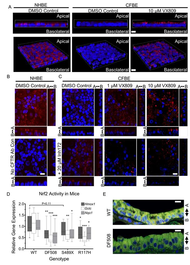
Abstract

Cystic Fibrosis (CF) is a multi-organ progressive genetic disease caused by loss of functional cystic

fibrosis transmembrane conductance regulator (CFTR) channel. Previously, we identified a significant

dysfunction in CF cells and model mice of the transcription factor nuclear-factor-E2-related factor-2

(Nrf2), a major regulator of redox balance and inflammatory signaling. Here we report that approved

F508del CFTR correctors VX809/VX661 recover diminished Nrf2 function and colocalization with

CFTR in CF human primary bronchial epithelia by proximity ligation assay, immunoprecipitation, and

immunofluorescence, concordant with CFTR correction. F508del CFTR correctors induced Nrf2 nuclear

translocation, Nrf2-dependent luciferase activity, and transcriptional activation of target genes. Rescue of

Nrf2 function by VX809/VX661 was dependent on significant correction of F508del and was blocked by

inhibition of corrected channel function, or high-level shRNA knockdown of CFTR or F508del-CFTR.

Mechanistically, F508del-CFTR modulation restored Nrf2 phosphorylation and its interaction with the

coactivator CBP. Our findings demonstrate that sufficient modulation of F508del CFTR function corrects

Nrf2 dysfunction in CF.

                                                     7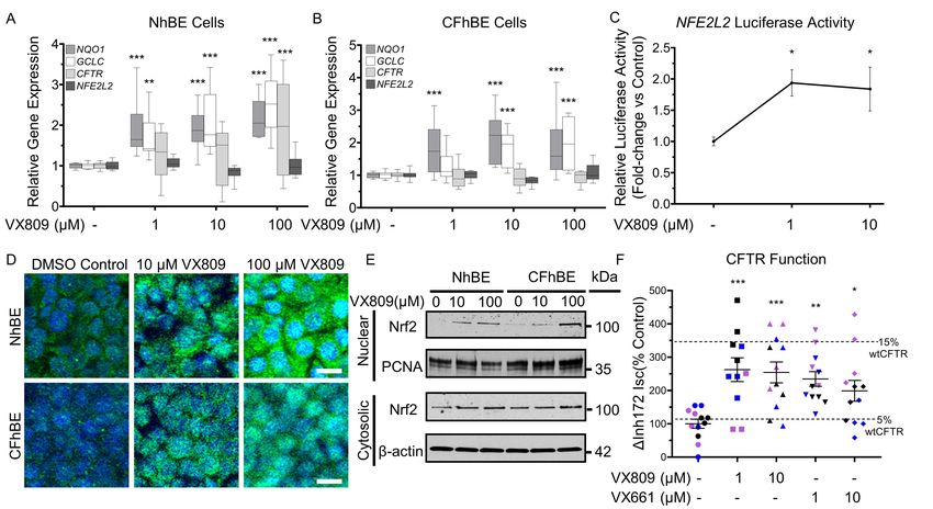
Introduction

       Cystic Fibrosis (CF) is an autosomal recessive disease characterized by progressive loss of lung

function, pancreatic exocrine dysfunction, and gastrointestinal complications, which culminate in

dramatically shortened life expectancy. CF patients have a loss of functional cystic fibrosis

transmembrane conductance regulator (CFTR) channel, an important Cl -/HCO3- channel in epithelial cells

(1). The most common mutation in CFTR is the F508del variant, which is a Class II mutation, resulting

in defective protein folding (2). F508del-CFTR is retained in the endoplasmic reticulum (ER) and not

transported to the cell membrane. Furthermore, F508del exhibits reduced gating and stability at the

plasma membrane. CF airway epithelia exhibit many secondary defects, including abnormalities in

pathways involved in inflammatory responses (3-5).          However, no direct link between CFTR and

regulators of inflammation, such as Nrf2 (4), has yet been reported.

       Transforming CF care, several small molecule compounds that target CFTR dysfunctions have

been developed(6). VX809 (Lumacaftor™) is a “corrector” that is reported to bind to and partially

correct the folding of F508del-CFTR, increasing its delivery to the cell membrane (7). Alone, VX809

significantly improves sweat chloride levels (a measure of CFTR function in sweat glands), but does not

elicit significant changes in nasal epithelial CFTR function or in lung function (8). In CF patients

homozygous for the F508del mutation, combination therapy with VX809 and the CFTR potentiator

VX770 (Ivacaftor™) significantly improves lung function, and reduces the number of events leading to

hospitalization by 39- 61% (9).           VX661 (Tezacaftor™, a corrector with similar efficacy as

Lumacaftor™), when combined with VX770, reduced pulmonary exacerbations by 35% with an

improved safety profile in a Phase 3 clinical trial (10).

       Secondary defects in CF are important contributors to the pathophysiology of the disease.

Previously, we discovered the dysfunction of Nuclear-factor-E2-related factor 2 (Nrf2) in CF airway

epithelia (11;12). Nrf2 is a transcription factor that regulates redox balance, and its dysfunction is

associated with numerous disease states, including acute lung injury (13), emphysema (14), chronic
                                                       8
obstructive pulmonary disease (COPD) (15;16), pulmonary fibrosis (17), and asthma (18). The primary
regulator of Nrf2 is Kelch-like erythroid cell-derived protein with cap’n’collar homology-associated
protein 1 (Keap-1), which sequesters Nrf2 in the cytoplasm (19;20). Keap1 is a substrate adaptor of a
Cullin-3 E3 ligase complex that poly-ubiquitinates Nrf2 and thereby targets it to the 26S proteasome for
degradation. As a consequence, Nrf2 levels and half-life are suppressed under basal conditions (21;22).
Reactive oxygen species (and xenobiotics) oxidize Keap1 cysteine residues and induce a conformational
change that dissociates Keap1 and Nrf2 from the E3 ligase complex resulting in Nrf2 stabilization (23-
25). Stabilized Nrf2 then translocates into the nucleus and binds to antioxidant response elements (AREs)
present in the promoters of its cognate target genes, stimulating the transcription of numerous antioxidant
and detoxifying genes (26;27).
Previously, we discovered that CF epithelia have reduced nuclear Nrf2 protein expression and
activity, and corresponding increases in steady state oxidants and enhanced inflammatory signaling (11).
We found that inhibition of CFTR (with CFTRinh-172) decreases Nrf2 activity, and increases H2O2 levels
in non-CF cells (11). Conversely, Nrf2 knockdown significantly decreases CFTR expression (28). We
showed that the cAMP competitor Rp-cAMPS, which decreases pCREB, stimulates Nrf2 activity (29).
Mechanistically, we demonstrated that in CF cells increased pCREB binding to CREB Binding Protein
(CBP) decreased its interaction with and diminished Nrf2 activation (29). Concomitantly, we found that
an increased pCREB-CBP in CF cells promoted the CBP-mediated activation of NF-κB, implicating Nrf2
dysfunction in inflammatory signaling in CF. In vivo, we found that activation of Nrf2 with the
triterpenoid CDDO in CF mice significantly reduced inflammatory responses to Pseudomonas
aeruginosa-derived LPS or flagellum (30).
To further investigate the relationship between CFTR and Nrf2, we evaluated whether VX809 or
VX661 impact Nrf2 dysregulation. We discovered that (1) VX809/VX661 activate Nrf2; (2) Nrf2 and
CFTR colocalize to within 40 nm and coimmunoprecipitate; (3) Nrf2-CFTR interaction is decreased in
CF; (4) VX809/VX661 correction of Nrf2 activity and colocalization with CFTR is dependent on CFTR
9function at the cell membrane; and (5) mechanistically, CFTR correctors rescue Nrf2 interaction with its
coactivator CBP. In addition to serving as tools to study F508del correction and Nrf2 biology, our studies
of both VX809 and VX661 are clinically relevant as both drugs are prescribed regularly to CF patients
with two copies of the F508del CFTR variant. Nrf2 activity and colocalization with CFTR were further
confirmed in established murine models of CF. These findings represent the first evidence of a direct
interface between CFTR and Nrf2. Given the association of Nrf2 with the resolution of inflammation
(11;12;30-39), our studies suggest that sufficient correction of CFTR function has the potential to
modulate inflammatory signaling in CF.
10Results
VX809 increases Nrf2 nuclear translocation and activity. To determine the effects of CFTR modulation
on Nrf2, primary non-CF and CF human bronchial epithelial (NhBE and CFhBE) cells were incubated
with a range of doses of VX809 (1-100 µM) for 48 hrs. The CFhBE cells used in this and subsequent
experiments were homozygous for the F508del-CFTR variant, the most common genotype in CF (2).
VX809 induces the expression of Nrf2-regulated genes, including NQO1 (NAD(P)H quinone
dehydrogenase 1), and GCLC (glutamate-cysteine ligase catalytic subunit) (Figure 1, A and B).
Furthermore, VX809 increases Nrf2-dependent luciferase activity in CFhBE cells, indicating the
compound promotes Nrf2 activation (Figure 1C). We next investigated whether VX809 also regulates
CFTR gene expression and found that after incubation with 1-100 µM VX809 for 48 hrs, CFTR gene
expression significantly increases in primary NhBE cells, but does not change in CFhBE cells (Figure 1,
A and B). Consistent with increased Nrf2 function, we found that VX809 treatment enhances the nuclear
localization of Nrf2 by immunofluorescence (IF) (Figure 1D).
VX809 treatment also increases Nrf2 cytosolic and nuclear protein levels (Figure 1, D and E), but
not Nrf2 gene (NFE2L2) expression (Figure 1, A and B), in both CF and non-CF cells. In agreement
with this, VX809 dose-dependently increases Nrf2 protein levels in the cytosolic and nuclear fractions of
CFhBE cells by up to 2-fold and almost 30-fold, respectively, by Western blot analysis (Figure 1E).
Nrf2 fluorescence in the nucleus significantly increases after incubation with VX809 for 48 hr in both
NhBE and CFhBE cells (Figure 1D), which agrees with the increase in nuclear Nrf2 observed by Western
blotting (Figure 1E). A MTT cell viability assay confirmed that VX809 (10-100 µM) is not cytotoxic
(Figure S1). Both the IF and Western blot data are consistent with the stimulation of Nrf2 activity
following VX809 treatment. These data demonstrate that VX809, especially at higher doses, is able to
correct Nrf2 dysfunction in CF by inducing translocation of Nrf2 to the nucleus and increasing Nrf2
transcriptional activity. While 100 µM VX809 induces more nuclear Nrf2 accumulation than lower doses
11(Figure 1, D and E), we used the lowest effective doses of 1-10 µM (Figure 1, A, B, and C) in the
remaining studies for physiological and clinical relevance.
Prolonged incubation with corrective modulators improves F508del-CFTR function. Ussing chamber
studies were performed to confirm that incubation with CFTR corrector compounds improves stimulated
F508del CFTR function in primary CFhBE cells. In the absence of corrector compounds, cells from five
F508del-CFTR homozygous donors exhibited stimulated (ΔcAMP+VX770) CFTR currents at 6.2% of
those from four NhBE donors (Figure S2, A and B). Pre-incubation with 1-10 µM VX809 for 48 hours
increases F508del CFTR function three- to four-fold, with a similar trend of improvement observed with
VX661 pre-incubation (Figure S2, A and B). In cells pre-treated with 1µM VX809, stimulated F508del
CFTR function reaches 17.3% of NhBE donors, similar to previously published studies (40). In cells also
pre-treated with VX770, alone or in combination with corrector, very little stimulated F508del CFTR
current is observed due to baseline CFTR activation in the presence of VX770. For these groups,
however, a statistically significant increase in inhibited CFTR function (ΔInh172) is noted in cells
pretreated with VX770 combined with 1-10µM VX809, with a trend towards an increase in current in
cells pretreated with VX770 and 1µM VX661 (Figure S2, C and D). Across all studies of stimulated
CFTR function, no corrector-induced difference is consistently noted in other electrophysiologic
measures, including baseline resistance, current, amiloride-inhibited current, low-chloride induced
current, or ATP-stimulated currents (Figure S3).
To mirror the non-stimulated conditions in our co-localization studies, we also examined the
corrector-induced change in Inh172-inhibited, unstimulated CFTR current (i.e., in absence of cAMP or
VX770). Unstimulated F508del CFTR function in three CFhBE donors is 4.1% of NhBE donors in the
absence of corrector compounds (Figure 1F and S4). Similar to our stimulated studies, this is rescued to
>10% of NhBE function following 48 hour pre-incubation with 1-10µM VX809 or VX661 (Figure 1F
and S4). These results demonstrate that both corrector drugs alone increase baseline F508del-CFTR
12function without stimulation by cAMP activators or VX770. As with studies of stimulated CFTR
function, there is no consistent corrector-induced differences in baseline resistance or baseline, amiloride-
sensitive, low chloride-induced, DIDS-sensitive, or ATP-stimulated currents (Figure S5).
Prolonged incubation with VX809 or VX661 increases mature F508del-CFTR. Primary CFhBE vehicle
control cells have a weak or undetectable mature band C, compared to a readily detectable band C in non-
CF NhBE cells (Figure S6, A and B). However, treatment for 48 hr with 1-10 µM VX809 or VX661
produced a detectable band C for F508del CFTR, indicating an increase in glycosylated mature protein
with corrector drugs (Figure S6, A and B). These data are consistent with correction of F508del-CFTR
trafficking, increased CFTR function at the plasma membrane (by Ussing chamber assay (Figure 1F)),
and increased Nrf2 activity following treatment with CFTR correctors (Figure 1, A-C).
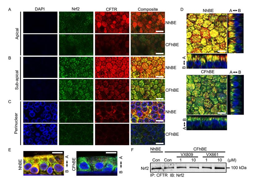
Colocalization of Nrf2 and F508del CFTR is decreased in primary human CF airway epithelia. To
explore the mechanism by which VX809 activates Nrf2, we examined the localization of Nrf2 with
respect to F508del CFTR, since VX809/VX661 change F508del CFTR localization. Dual IF with
antibodies against Nrf2 (green) and CFTR (red) reveals that the two proteins colocalize (yellow) in
primary NhBE cells (Figure 2, A-C). To focus on membrane and cytoplasmic staining, cells were
minimally permeabilized. As expected, IF for F508del CFTR protein is significantly lower in primary
CFhBE cells compared with NhBE cells, and is mainly limited to the peri-nuclear to sub-apical planes
(Figure 2, A-C and Figure S7). Importantly, the colocalization of Nrf2 with CFTR is significantly
diminished in CF cells compared to non-CF cells (Figure 2, A-E and Figure S7). Colocalization
(yellow) in both the primary NhBE and CFhBE cells appears highest in the peri-nuclear to sub-apical
plane, although colocalization is observed from the basolateral plane to below the apical plane in the
NhBE cells (Figure 2, A-E and Figure S8A).
13To directly test if CFTR and Nrf2 interact, we conducted immunoprecipitations (IP) from whole
cell lysates with an antibody against CFTR, followed by immunoblotting with an anti-Nrf2 antibody.
Consistent with our IF studies, Nrf2 is pulled down with CFTR, and the association between CFTR and
Nrf2 in CFhBE cells is decreased compared to that in NhBE cells, while 48 hr treatment with 1 or 10 µM
VX809 or VX661 increases this association (Figure 2F and Figure S8, B and C). We also examined the
interaction between CFTR and Nrf2 by proximity ligation assay (PLA), in which a red fluorescent signal
indicates that the two proteins are < 40 nm (~3 times the length of the average 100 kDa protein) apart. In
wild-type NhBE cells, Nrf2 and CFTR colocalize to within 40 nm, as indicated by readily detectable red
PLA signals (Figure 3, A and B). Conversely, CFhBE cells display a very weak PLA signal using
antibodies for Nrf2 and CFTR, suggesting little interaction between these two proteins in the CF
condition (Figure 3, A and C). The negative control (mouse IgG used instead of primary anti-CFTR
antibody), did not produce a red fluorescent signal (Figure 3B). The PLA data corroborate our IF
(Figure 2, A-C) and IP findings regarding CFTR and Nrf2 interactions (Figure 2F), and together, the
results reveal a reduced association between F508del CFTR and Nrf2 in CF versus non-CF cells. Close
Nrf2-CFTR association from the peri-nuclear compartment through the sub-apical membrane suggests
that the interaction may occur early in the proteins’ life cycles and continues while they traffic to the
membrane. In CFhBE cells, treatment with VX809 significantly increases PLA signal (Figure 3, A and
C). Inhibition of CFTR function with 20 µM CFTRinh-172 blocks the VX809-stimulated Nrf2-F508del
CFTR interaction, as assessed by diminished red fluorescent PLA signal, compared to cells treated with
VX809 alone (Figure 3C).
Nrf2 target gene expression and Nrf2-CFTR colocalization are decreased in CF mouse models. To
confirm the physiological relevance of cross talk between Nrf2 and CFTR, we examined Nrf2 function
and interaction with CFTR in CF mutant mouse lungs. In F508del-CFTR (DF508) and knockout S489X-
CFTR mice, expression of Nrf2 target genes Hmox1, Nqo1, and Gclc is decreased to ~ 40-75% of
14expression levels measured in wild-type (WT) mice (Figure 3D). Reduced Nrf2 function is also evident
in mice with the less severe R117H-CFTR Class IV variant, where R117H-CFTR expression is ~10% of
CFTR expression in wild-type mice (personal communication, Dr. Craig Hodges, Ph.D.). Furthermore,
IF imaging for Nrf2 (green) and CFTR (red) demonstrate significant colocalization (yellow) in airway
epithelial cells of WT mice versus reduced levels in DF508 mice (Figure 3E and Figure S9). Further
studies of the mechanism of activation of Nrf2 by VX809/VX661, were conducted in ALI cultures of
primary human cells.
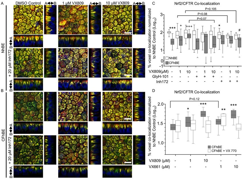
VX809 and VX661 rescue F508del CFTR and Nrf2 interaction in CF cells. VX809 affects the cellular
localization of F508del-CFTR, and activates Nrf2, possibly by promoting the interaction of the
transcription factor with CFTR (Figure 3). To test this hypothesis, we investigated whether VX809
modulates the association of Nrf2 and CFTR in primary CF cells using immunofluorescence. Consistent
with the PLA data, IF reveals F508del CFTR and Nrf2 colocalization (yellow) with 1 or 10 µM VX809 in
CFhBE cells, to levels similar to those observed in control NhBE cells, following 72 hr of drug exposure
(Figure 4, B and C, and Figure S10). Quantitation revealed that CFhBE control cells have < 40% of the
Nrf2-CFTR colocalization observed in NhBE control cells (Figure 4 C). VX809 (1-10 µM) treatment
significantly and dose-dependently increases Nrf2-F508del CFTR colocalization in CFhBE cells, and at
the 10 µM dose VX809 increases colocalization to ~67% of NhBE control levels (Figure 4C). VX809
treament increases F508del CFTR levels (shown in red, Figure 4B). Incubation with 1-10 µM VX661 for
48 hr had a similar corrective effect on Nrf2-CFTR colocalization in CFhBE cells (Figure 4D). Addition
of the potentiator VX770 to either VX809 or VX661 did not significantly increase colocalization over
CFhBE vehicle control and was similar or lower than either corrector alone (Figure 4D), consistent with
previous reports that VX770 reduces VX809 correction of F508del CFTR function in vitro (41). None of
the treatments fully corrected Nrf2-CFTR colocalization.
15Prolonged inhibition of CFTR decreases Nrf2 and CFTR interaction. To investigate the role of CFTR
function in regulating Nrf2-CFTR interaction, we inhibited the channel following treatment with
correctors. Colocalization of Nrf2 with CFTR is decreased in NhBE cells after incubation with CFTR inh-
172, suggesting that functional CFTR modulates interaction between CFTR and Nrf2 in wild-type cells
(Figure 4, A and C). Consistent with PLA studies shown in Figure 3C, treatment of CFhBE cells with
10 µM VX809 combined with 20 µM CFTRinh-172 trends to (p=0.07) to reduce Nrf2-CFTR
colocalization versus treatment with 10 µM VX809 alone (Figure 4, B and C), and incubation with 1-10
µM VX809 and 20 µM CFTRinh-172 does not significantly increase Nrf2-CFTR colocalization over that
of the inhibitor alone. In order to confirm that inhibitor effect is not due to competition with VX809, we
used a second inhibitor of CFTR, GlyH-101. While the thiazolidinone CFTR inh-172 interacts with the
intracellular Arg347 residue of CFTR, GlyH-101 is a glycine hydrazide analogue that blocks the
extracellular region of CFTR’s pore (37;38). Treatment of CFhBE cells with 10 µM VX809 + 20 µM
GlyH-101 significantly reduces Nrf2-CFTR colocalization versus treatment with 10 M VX809 alone
(Figure 4C). VX809 fails to significantly increase Nrf2-CFTR colocalization in CFhBE cells treated
with GlyH-101 over GlyH-101 alone or vehicle control. We also investigated whether VX809 altered
Keap1 protein expression, the primary regulator of Nrf2 degradation and activation. We found no impact
of VX809 treatment, suggesting that the effects of VX809 are not mediated through changes in total
Keap1 levels (Figure S6C). These data suggest that CFTR function, even during trafficking, is required
for Nrf2 colocalization throughout the cell.
Acute manipulation of CFTR function modulates Nrf2-CFTR colocalization. Based on the eveidence
above, we further investigated acute activation of CFTR and it’s role in Nrf2 interactions. In NhBE cells,
acute treatment with CFTRinh-172 significantly reduces colocalization, while forskolin trends to increase
it over vehicle control cells, and does so significantly combination with VX809 (Figure S11A). In
conjuction with our other CFTR-inhibitor studies, results with acute forskolin treatment support the
16notion that Nrf2-CFTR colocalization is CFTR function dependent. In CFhBE cells acute treatment with
CFTRinh-172 arrogated VX809 induced Nrf2/CFTR colocalization, similar to the effects of prolonged
inhibition, further indicating a rapid sensitivity of colocalization to CFTR function (Figure S11B). Acute
treatment with amiloride, which inhibits ENaC, trends to increase Nrf2 colocalization with CFTR. In
addition to corrector, both amiloride and DIDS, a non-CFTR chloride channel inhibitor, result in VX809-
induced Nrf2-CFTR colocalization that significantly differ from CF vehicle control (Figure S11B).
Knockdown of F508del expression in CFhBE cells blocks VX809 activation of Nrf2. To further examine
the CFTR-dependence of VX809 activation of Nrf2 we used shRNA lentivirus to knockdown F508del
gene expression in CFhBE cells. When CFTR gene expression is reduced by >50% compared to
scrambled control (Figure 5A), VX809-induced increase in HMOX1, NQO1, and GCLC mRNA levels
(Figure 5, B-D) is significantly inhibited. Importantly, knockdown combined with VX809 does not
change Nrf2 gene expression (Figure S12), supporting the notion that CFTR-dependent VX809 activation
of Nrf2 occurs at the protein level.
Correction of F508del-CFTR with VX661 stimulates Nrf2 transcriptional activity. Similar to VX809,
treatment of primary CFhBE cells with the newer corrector drug VX661 for 48 hr also increases
expression of the Nrf2 target genes HMOX1 and NQO1 (Figure 5, E and F). The combination of VX661
and the potentiator VX770 also stimulated expression of both HMOX1 and NQO1 significantly compared
to DMSO control, but not over the same dose of VX661 alone except for NQO1 at the 1 M dose
(Figure 5, E and F). Potentiation by VX770 of VX809/VX661-corrected F508del-CFTR can further
increase Nrf2 activated gene expression over the effects of corrector alone, but fails to do so consistently.
Nevertheless, these data further support the importance of CFTR functional correction rather than
localization for Nrf2 activation.
17Activation of Nrf2 by VX809/VX661 is dependent on CFTR function. To examine whether CFTR function
was necessary for Nrf2 transcriptional activation, we co-treated primary differentiated cultures with
VX809/VX661 and CFTRinh-172. After 48-72 hr, 20 µM CFTRinh-172 blocked the VX809 or VX661
induced increase in gene expression of GCLC, HMOX1 and NQO1 in CFhBE cells, with a partial
blockade in HMOX1 expression by VX809 at 48 hr (Figure 6 and 7A). Taken together with shRNA
knockdown experiments (Figure 5), inhibitor studies indicate that VX809 correction of Nrf2 is CFTR
function dependent. Importantly, CDDO-Me, a triterpenoid activator of Nrf2 that is not dependent on
CFTR function, served as positive control and increased GCLC, HMOX1, and NQO1 mRNA levels after
72 hr (Figure 6, A-C). Treatment with 20 µM CFTRinh-172 did not reverse the Nrf2 stimulatory effect of
CDDO-Me (Figure 6, A-C), indicating that the inhibitory action of CFTRinh-172 is mediated through
CFTR and not by direct action on Nrf2.
Co-incubation with 20 µM GlyH-101 blocks the VX661-induced stimulation of Nrf2 target gene
expression, including HMOX1 (at 1 M VX661) and NQO1, in CFhBE cells after 72 hours (Figure 7, B
and C), similar to the block of the increase in Nrf2-CFTR colocalization observed in imaging studies
(Figure 4C). The similar inhibitory effects of CFTRinh-172 and GlyH-101 on corrector-induced Nrf2
activity and Nrf2-CFTR colocalization further support the notion that corrector effects are CFTR function
dependent.
Conversely, activation of CFTR stimulated Nrf2 activity. Incubation of NhBE cells with forskolin
for 6 hr stimulates HMOX1 or GCLC gene expression over 2- and 5-fold, respectively, indicating that
activation of wild-type CFTR function increases Nrf2 transcriptional activity (Figure S13A). In CFhBE
cells, 6 hr treatment with CFTRinh172 blocks HMOX1 and GCLC gene expression stimulated by 10 µM
VX809 48 hr pre-/co-treatments, while 6 hr co-treatments with amiloride (ENaC inhibitor) and DIDS
(non-CFTR chloride channel inhibitor) did not significantly inhibit VX809-induced Nrf2 activity (Figure
S13B).
18Partial CFTR knockdown in NhBE cells does not inhibit Nrf2 activity induced by VX809. Infection of
NhBE cells with CFTR shRNA lentivirus decreases CFTR gene expression by ~60% compared to
scrambled control (Figure 8A). Partial knockdown experiments allow us to compare the effect of
decreased CFTR function and Nrf2-CFTR colocalization, and are complementary to the studies using the
inhibitors of CFTR function, CFTRinh-172 and GlyH-101. Partial knockdown of CFTR in NhBE cells
does not decrease the basal gene expression of Nrf2 target genes (HMOX1, NQO1, and GCLC), but does
block VX809-mediated activation of Nrf2, although high doses of corrector trend toward Nrf2 activation
(Figure 8, B-D). These data support the importance of CFTR function over localization (given the partial
knockdown) VX809-mediated activation of Nrf2.
CFTR modulation stimulates Nrf2 phosphorylation and increased interaction with CBP. To explore the
mechanism by which correction of F508del CFTR function increases Nrf2 activity, we
immunoprecipitated Nrf2, followed by immunoblotting with antibody against phosphoserine to assess
serine phosphorylation of Nrf2 in CFhBE vs. NhBE cells (Figure 9A). Nrf2 serine phosphorylation is
required for its translocation to the nucleus (42;43). Incubation of CFhBE cells with 1-10 µM VX809 or
VX661 significantly increased Nrf2 phosphoserine levels (Figure 9A), consistent with increases in Nrf2
nuclear localization and activity observed in our other studies CFTR modulation studies (Figures 1, 6, 7,
and 8). Furthermore, previous studies demonstrated that lack of CFTR function resulted in an increase in
phospho-CREB which increasingly bound CBP and decreased CBP-Nrf2 interaction in primary CF
epithelial cells and F508del mutant mice (12). CBP, a transcriptional coactivator, binds and maximally
activate Nrf2 (44;45). In previous studies, artificial restoration of Nrf2-CBP interaction corrected a large
portion of the Nrf2 dysfunction observed in CF (12). We hypothesized that correction of F508del CFTR
function with VX809/661 would also correct Nrf2-CBP interaction. To test this, we immunoprecipitated
CBP, and probed for Nrf2 in CF and non CF primary epithelia treated for 48 hr with VX809 or VX661.
We found that, in a dose dependent manner, VX661 significantly (while VX809 trended) increased CBP
19interaction with Nrf2 (Figure 9B and C) 1.5-3 fold. This increase of Nrf2 interaction with CBP was
CFTR-function dependent as co-incubation with CFTR inh-172 abrogated any increase in Nrf2-CBP
association over that observed with DMSO alone (Figure 9, B and C). No significant changes were
observed in complementary studies with NhBE cells (Figure 9, B and D). These data are consistent with
the increase in Nrf2 activation observed with F508del CFTR modulation and demonstrate a link between
CFTR function and Nrf2 activation.
20Discussion
The primary novel findings of this study are that the F508del-CFTR correctors VX809 and VX661
reverse the dysregulation of Nrf2 activity in primary human CF epithelial cells, and that this rescue is
CFTR function dependent. Previously, we found that dysfunction of Nrf2 significantly contributes to the
dysregulation of inflammatory signaling in CF airway epithelia (11;12;30). This is consistent with a large
body of evidence that demonstrates dysregulation of Nrf2 inflammatory signaling in the airway for a
variety of diseases, including asthma and COPD (46-53). The crucial role of Nrf2 in regulating
inflammation and the present findings that CFTR interacts with and regulates Nrf2 suggest a link between
CFTR and inflammatory signaling. The implication of a direct relationship between CFTR and Nrf2 is
that clinical modulation of F508del CFTR has the potential to also correct Nrf2 abnormalities and
mitigate inflammatory signaling in CF patients. As inflammation continues to be a major cause of lung
function decline in patients receiving F508del-CFTR corrector therapy, the implications of this study are
clinically relevant. Complete normalization of Nrf2 activity, which may not be necessary for significant
modulation of inflammation in patients, was only achieved at high doses of corrector exposure where 100
µM VX809 elevated Nrf2 nuclear localization to non-CF levels. However, 100 µM is approximately 7-
fold higher than the maximum serum concentrations of correctors that can be attained in vivo in patients
(achievable serum concentrations are 1-14 µM) (8). Nevertheless, at these clinically relevant doses of
VX809 and VX661 (54), we found that while Nrf2 activity was not completely normalized, it was
significantly stimulated, supporting the notion that F508del CFTR correction can be achieved clinically
may positively impact Nrf2 activation. The development of more effective modulators of CFTR function
holds promise for complete correction of Nrf2, and associated regulation of inflammatory signaling
(11;12;30).
Throughout our studies we found that the abilities of VX809 and VX661 to activate Nrf2 are
CFTR dependent. To explore the mechanism by which VX809/VX661 correct Nrf2 function in CF
epithelial cells we dissected the impact of the modulators’ effects on CFTR processing versus CFTR
21function. Others have shown that VX809 increases CFTR levels at the plasma membrane by binding
directly to the first nucleotide binding domain (NBD1) and stabilizing its interaction with the membrane-
spanning domain-1 (MSD1) in F508del-CFTR, allowing for better folding (55;56). In examining whether
changes in the processing of CFTR contributed to Nrf2 correction, we found that CFTR and Nrf2
colocalize in perinuclear compartments and the subapical membrane space (Figure 2) to within 40 nm
(Figure 3, A-C) by PLA. Multiple immunofluorescence studies (totaling n = 360 confocal z-stacks from
multiple CF and non-CF subjects, corrected for multiple comparisons) show that the colocalization of
Nrf2 and CFTR is significantly decreased in CF compared to non-CF airway epithelial cells (Figures 2
and 4). Moreover PLA (Figure 3) and CFTR immunoprecipitation experiments (Figure 2F) also
demonstrate that the close association between Nrf2 and CFTR is diminished in CF vs. non-CF cells. Our
present studies do not distinguish between direct interaction or one that involves intermediary proteins.
Nrf2 and/or Keap1 have not been shown to bind to any PDZ interactors of CFTR. The interaction is
unlikely to occur at the C-terminal PDZ motif of CFTR, as our pulldown studies employed the 24-1 anti-
CFTR antibody which recognizes CFTR’s C-terminus. We also found that Nrf2 interaction with CFTR is
physiologically relevant in vivo, where it is significantly higher in the lung airway epithelia of wild-type
mice versus the airways of F508del-CFTR mice (Figure 3E and S9). The decreased levels of mutant
CFTR in F508del homozygous cells (63) does not fully account for the significant decrease in the fraction
of F508del-CFTR and Nrf2 that colocalize in CFhBE compared to NhBE cells.
Our exploration of the functional consequence of VX809/VX661 treatment revealed that Nrf2
activation by CFTR modulators is CFTR function-dependent (Figures 6-8). Even the Nrf2-CFTR
interaction is modulated, at least in part, by CFTR function, and is reduced by CFTR inhibition in NhBE
cells (Figures 3, 4, S11, and S13). Furthermore, ~50% knockdown of F508del CFTR expression by
shRNA or blockade of corrected F508del CFTR function by CFTR inh172 or GlyH-101 inhibits Nrf2
activity in CFhBE cells stimulated with VX809/VX661 (Figures 5-7). The use of two inhibitors of CFTR
that bind at different sites confirmed that the loss of VX809/VX661 effect is not due to the blocking of
22modulator binding sites on CFTR. Conversely, activation of CFTR function increases both corrector-
mediated Nrf2 activition and Nrf2-CFTR colocalization in NhBE cells with wild-type CFTR (Figures
S11A and S13A), and partial CFTR knockdown in NhBE cells does not completely block 10 M
corrector activation of Nrf2 (Figure 8). Taken together our data of CFTR expression or functional
inhibition indicate that modulator effects on Nrf2 activity are mediated through CFTR activity, and not
necessarily by altering the interaction with CFTR or the levels of CFTR. This dependence on CFTR
function is consistent with our previous studies that showed that the lack of CFTR function reduces Nrf2
activity, leading to increased steady state intracellular hydrogen peroxide and inflammatory cytokine
production (11;12).
Mechanistically, our data indicate that one link that exists between CFTR function and Nrf2 in
non-CF cells is restored in CF cells by CFTR modulation. Nrf2 interaction with its coactivator CBP in
the nucleus is necessary for maximal transcriptional activity (44;45;57). In the context of CF, lack of
CFTR function results in a feedback response that increases levels of cAMP and phosphorylated CREB
(58;59). Increases in pCREB bind CBP and sequester it from interaction with Nrf2, reducing Nrf2
activity (57;60). Previously, we reported that in CF cell lines, primary cells, and animals Nrf2 exhibits a
reduced interaction with CBP compared with non-CF controls, and that artificial reversal of reduced Nrf2-
CBP interaction corrects Nrf2 dysfunction (12). In the present studies, we examined the three major steps
in Nrf2 activation: inhibition by Keap1 (Figure S6C), serine phosphorylation (Figure 9A), and
interaction with CBP (Figure 9B, C, and D). The data show that F508del CFTR modulation-mediated
activation of Nrf2 was only coupled to increased serine phosphorylation and increased interaction with
CBP in a CFTR-function dependent fashion. The implication of our studies are that VX809/VX661
normalize CFTR colocalization with Nrf2, but rescue of Nrf2 activity in CF is due to modulation of
CFTR function, which relieves feedback responses to CFTR dysfunction, allowing for sufficient levels of
CBP to interact with Nrf2.
23With present clinical doses and modulators, our studies suggest that it would be necessary to
combine VX809 or VX661 and VX770 with a Nrf2-acitvating drug to attain sufficient Nrf2 rescue to
significantly impact Nrf2-regulated inflammatory pathways in F508del homozygotes. The combination
of VX809 and VX770 moderately improves lung function in CF patients homozygous for the F508del
mutation, which is not observed with VX809 therapy alone (9). Addition of VX770 to VX809/VX661
did improve Nrf2-CFTR colocalization (Figures 4D) and Nrf2 activity (Figure 5E, and F), but rarely did
so over either VX809 or VX661 alone. VX770 in combination with VX809/VX661 does not completely
restore CFTR function in patients (61), however, new generation modulators that more efficiently correct
CFTR expression, localization, and function may fully correct Nrf2 dysfunction and downstream
regulation of inflammatory signaling.
Our studies demonstrate a significant correction of a secondary defect directly linked to
inflammation in CF primary cells following correction of F508del-CFTR with VX809. Although partial
restoration of Nrf2 is achieved at clinical concentrations of VX809 and VX661, full correction requires
supratherapeutic clinical doses. Nevertheless, the important implication of our findings is that full clinical
correction of CFTR dysfunction would correct Nrf2 regulation, and influence inflammatory signaling. In
the absence of full mutant CFTR correction, additional treatment with Nrf2 activators may be necessary to
significantly modulate Nrf2 function to influence inflammation in CF patients. VX809/VX661 activation
of Nrf2 is dependent on the correction of CFTR function, but also involves the correction of Nrf2
colocalization with CFTR. Given the role of Nrf2 in deactivating inflammatory signaling (39;47;50), our
data suggest a link between CFTR and inflammatory signaling in CF epithelial cells.
24Methods
Cell culture. Primary non-cystic fibrosis (CF) or CF human bronchial epithelial cells (NhBE or CFhBE,
respectively) were purchased from ChanTest/Chales River Laboratories (Wilmington, MA), received
from the Pulmonary Medicine CF RDP Translational Core at Cincinnati Children’s Hospital Medical
Center, or provided by Dr. Scott H. Randell (Marsico Lung Institute, Tissue Procurement and Cell Culture
Core, The University of North Carolina at Chapel Hill, USA, UNC IRB#03-1396), and prepared as
described previously (62). Cells were cultured at air-liquid interface (ALI) on semipermeable filters, as
described previously (29;63), in media containing Ultroser™ G (Pall, Port Washington, NY), following
the Vertex Pharmaceuticals, Inc. formula (64). The cells were differentiated, forming tight junctions and
cilia. The non-CF cells from 6 donors had the following codes: DD007K, DD005K, DD021K, DD029J,
DD032L, DD053K. The CF cells from 12 donors had the following codes: KK002L, KK003K,
KK003M, KK004i, KK006F, KK006G, KK011i, KK012B, KK013F, KK022M, KK024N, KK027H. The
primary cells used were all de-indentified and their use was approved under CCHMC IRB #2014-8600.
RNA isolation and quantitative real-time PCR (qPCR). Differentiated NhBE and CFhBE cells were
treated with the indicated doses of VX809, VX661, VX770 (Selleck Chemicals, Houston, TX), CDDO
methyl ester (Cayman Chemical, Ann Arbor, MI), CFTR inh-172 and/or GlyH-101 (Tocris, Minneapolis,
MN) for 48 or 72 hr. Alternatively, differentiated NhBE or CFhBE cells were incubated on the
basolateral side with DMSO control or VX809 (10 µM) for 48 hr, then co-incubated acutely for 6 hr with
apical forskolin (Tocris, Minneapolis, MN), amiloride (Sigma, St. Louis, MO), DIDS (Sigma), or
CFTRinh-172. Negative controls were treated with DMSO. Total RNA from cells was isolated using
Aurum Total RNA Mini Kit (Bio-Rad, Hercules, CA), and reverse-transcribed in a 20µl reaction
containing random primers and iScript Reverse Transcriptase (Bio-Rad). Real-time PCR was performed
with a StepOnePlus instrument using Power SYBR Green PCR Master Mix (Applied Biosystems,
Carlsbad, CA). Primer pairs used for human samples were: HMOX1 (Fwd 5′-
25CTTCTTCACCTTCCCCAACA-3′; Rev 5′-GCTCTGGTCCTTGGTGTCAT-3′), GCLC (Fwd 5′-
CCTCCAGTTCCTGCACATCT-3′; Rev 5′-GGGTAGGATGGTTTGGGTT-3′), NQO1 (Fwd 5′-
CAAATCCTGGAAGGATGGAA-3′; Rev 5′-GGTTGTCAGTTGGGATGGAC-3′), CFTR (Fwd 5′-
TTGGATGACCTTCTGCCTCT-3′; Rev 5′-CTCCTGCCTTCAGATTCCAG-3′), NFE2L2 (NRF2) (Fwd
5′-GAGAGCCCAGTCTTCATTGC-3′; Rev 5′-TGCTCAATGTCCTGTTGCAT-3′), 18S rRNA (Fwd 5′-
GTGGAGCGATTTGTCTGGTT-3′; Rev 5′-CGCTGAGCCAGTCAGTGTAG-3′). Primer pairs used for
mouse lung samples were: Hmox1 (Fwd 5′-GCCGAGAATGCTGAGTTCATG-3′; Rev 5′-
TGGTACAAGGAAGCCATCACC-3′), Gclc (Fwd 5′-CTGCACATCTACCACGCAGT-3′; Rev 5′-
TTCATGATCGAAGGACACCA-3′), Nqo1 (Fwd 5′-CGCCTGAGCCCAGATATTGT-3′; Rev 5′-
GCACTCTCTCAAACCAGCCT-3′), 18S rRNA (Fwd 5′-GTAACCCGTTGAACCCCATT-3′; Rev 5′-
CCATCCAATCGGTAGTAGCG-3′). All samples were run in duplicate or triplicate. The relative fold
increase of specific RNA was calculated by the comparative cycle of threshold detection method and
values were normalized to 18S rRNA. Fold changes in gene expression were calculated using the 2 -ddCt
method after normalization to 18S rRNA. Data was collected from 3-4 independent experiments.
Subcellular protein fractionation. NhBE and CFhBE were treated with 0, 0.1, 1, 10 or 100 µM VX809 for
24 hr. Nuclear extracts were prepared as described previously (65). Briefly, NhBE and CFhBEs were
harvested in ice-cold PBS and centrifuged at 4000 x g for 3 min at 4 °C. The pellet was resuspended in
buffer containing 10 mM HEPES (pH 7.0), 1.5 mM MgCl 2, and 10 mM KCl on ice for 10 min, vortexed,
centrifuged at 4000 x g, and the cytoplasmic protein in the supernatant was collected. Pellets were
resuspended in buffer containing 20 mM HEPES (pH 7.9), 420 mM NaCl, 1.5 mM MgCl 2, 0.2 mM
EDTA, and 25% glycerol and incubated on ice for 20 min. Samples were centrifuged at 16,800 x g and
the nuclear fraction in the supernatant was collected. Protein concentration was determined by Bradford
assay (Bio-Rad, Hercules, CA). All buffers contained protease inhibitor cocktail (Roche, Nutley, NJ) and
1 mM Na3VO4, 20 mM NaF, and 1 mM Na4P2O7. All extracts were stored at -80 °C until analysis.
26Western blot analysis. The anti-CFTR antibody (mouse clone #570) was received from the CFTR
antibody distrubtion program at the University of North Carolina, the anti-phosphoserine antibody was
purchased from Invitrogen (Carlsbad, CA), and the anti-Nrf2 antibody was generated and validated
previously (12;66;67). Differentiated NhBE and CFhBE cells grown on filters were treated with the
indicated doses of VX809 or VX661 for 48 hr. Equal amounts of protein samples (20-30 μg) were
separated on 7% SDS-PAGE gels and transferred to nitrocellulose membranes. Membranes were blocked
in 5% milk, incubated with primary antibodies overnight at 4°C, then washed and incubated with
horseradish peroxidase-conjugated secondary antibodies for 1 hr. Products were exposed to the
SuperSignal ECL detection system (Pierce, Rockford, IL) and photographed. β-actin and PCNA served as
loading controls. Experiments were repeated at least 3 times.
Co-immunoprecipitation. Differentiated NhBE and CFhBE cells grown on ALI were incubated with the
indicated doses of VX809 or VX661 for 48 hr. Lysis of cells, and all precipitation procedures with the
lysates, were conducted at 4 °C. Protein concentration was measured using an aliquot of each lysate, and
lysates were diluted at 4 °C to equal concentrations with cold lysis buffer. Lysates (with equal
concentrations of protein) were pre-cleared with uncoated Dynabeads, then mixed with anti-CFTR
antibody (mouse clone #24-1, R&D systems, Minneapolis, MN), anti-Nrf2 antibody, or anti-CBP
antibody (cat# sc-1211, Santa Cruz, Dallas, TX) conjugated to Dynabeads (5g antibody/15L beads),
and incubated overnight at 4 °C. Following at least 3 washes in cold buffer (25 mM Tris, pH 7.5, 150 mM
NaCl, Roche Complete Protease Inhibitor, 1% Triton X-100), protein was eluted at 37 °C in 50 μL SDS-
PAGE sample loading buffer, subjected to gel electrophoresis on 7.5% gels, and analyzed by Western blot
analysis for Nrf2 or CFTR as described above.
Immunofluorescence. Differentiated NhBE and CFhBE cells grown on filters were treated with the
indicated doses of VX809, VX661, VX770, and/or CFTR inh-172 for 48-72 hr. Alternatively, NhBE or
27CFhBE cells on ALI were treated on the basolateral side with DMSO control or VX809 (1, or 10 µM) for
48 hr, and then co-treated on the apical side for the last 2 hours with forskolin, amiloride, DIDS, or
CFTRinh-172. Cells on filters were fixed in 4% paraformaldehyde (PFA) for 30 min. Cells were
permeabilized with 0.1% Triton X-100 for 10 min, blocked with 2.5% horse serum (Vector, Burlington,
CA) for 1 hr, then incubated with anti-Nrf2 and/or anti-CFTR antibodies overnight at 4 °C. For the Nrf2
nuclear localization experiments, cells were fixed in cold methanol for 15 min and permeabilized with
0.2% Triton X-100 for 20 min. Mouse lungs were fixed in 4% PFA, paraffin-embedded, and sectioned
onto slides. Mouse sections were deparaffinized, rehydrated, permeabilized with 0.1% Triton X-100 for
10 min, and antigen retrieval was done by microwave heating (1 min full power, then 10 min 10% power)
in sodium citrate buffer, followed by blocking. Samples were then incubated with species-matched Alexa
Fluor®-conjugated secondary antibodies (Alexa Fluor® 594 goat anti-mouse IgG, or Alexa Fluor® 488
goat anti-rabbit IgG, Life Technologies, Carlsbad, CA) and nuclei were counterstained with 4’,-6-
diamidino-2-phenylindole (DAPI). Filters were cut and mounted on slides. Specimens were visualized
using a Nikon A1R Laser Scanning Confocal Microscope (Melville, NY) and representative images were
analyzed under the same conditions for each experiment, using NIS Elements Advanced Research
software (Nikon) or Imaris software (Bitplane, Concord, MA). Colocalization (shown in yellow) for Nrf2
and CFTR was quantified as percent of colocalized voxels (voxels that were positive for both red and
green channels above the threshold set for each channel) in the sample volume for each sample in Imaris
software. All samples in each experiment were set to the same thresholds for each channel, and the
thresholds set to include 40% of the data in each channel for the NhBE controls for each experiment.
Data are normalized to NhBE control for each of 4-5 independent experiments.
Luciferase reporter assay. Primary CFhBE cells were grown in 48-well plates until cells were 60–80%
confluent. Transient transfection assays were performed using Lipofectamine LTX reagent (Life
Technologies, Carlsbad, CA) with 1 µg of a firefly luciferase plasmid (pNrf2-fluc) under the control of
28the Nrf2-dependent promoter for glutathione-S-transferase 1 (GST1) and/or 100 ng of Renilla luciferase plasmid (Promega, Madison, WI) as an internal control for transfection efficiency. Six hours after transfection, cells were supplemented with complete media. One day following transfection, cells were incubated with the indicated doses of VX809 for 48 hr. Cells were lysed in Passive Lysis Buffer (Promega, Madison, MI) and examined for firefly and/or Renilla luciferase activity using a SpectraMax L 1-channel luminometer (Molecular Devices, San Jose, CA). Protein concentration was determined by BCA protein assay. Firefly luciferase activity was normalized to protein levels or Renilla luciferase activity. Data are expressed as fold-change vs. vehicle control in 4 independent experiments. CFTR Electrophysiology Assays. Ion transport, including CFTR function, was quantified as short-circuit current (ISC) as previously described (68) with minor modifications. Mature primary NhBE and CFhBE cells grown on 0.33cm2 semipermeable filters, were mounted in Ussing chambers (Physiologic Instruments, San Diego, CA), and equilibrated in symmetric Ringer’s buffer. Cells were placed under voltage clamp conditions to measure baseline ISC and resistance, then transitioned to a 6mM Cl- apical buffer (replacing sodium chloride with sodium gluconate) to create a basolateral-to-apical Cl - gradient. Culture quality was good with an average transepithelial resistance of 546.8µΩ*cm 2 (n=127, SEM=35.6); seven cultures (5.5%) were excluded due to transepithelial resistance measures
into Prism software (GraphPad, San Diego, CA). To allow for comparisons between donors, all I SC

values were normalized to the experimental DMSO control and are reported as percent of internal control.

Baseline CFTR Electrophysiology Assays. Unstimulated CFTR function was assessed using a modified

version of the above stimulated protocol. Mature primary cells were mounted in Ussing chambers in a

basolateral-to-apical Cl- gradient and sodium transport was inhibited with amiloride. Alternate Cl -

transporters were then blocked with 4,4’-Diisothiocyano-2,2’-stilbenedisulfonic acid (DIDS, 100µM) in

the apical compartment. CFTR function was then inhibited with CFTR inh-172 (10µM) in the apical

compartment; this CFTR inhibition was quantified as proxy for unstimulated CFTR function. For each

step, the ISC tracing reached plateau before moving to the next treatment.       Average transepithelial

resistance was 703.3µΩ*cm2 (n=59, SEM=45.9); one culture (1.7%) was excluded due to transepithelial

resistance measures
hairpin RNA (shRNA) lentiviral vectors, targeted against human CFTR (MISSION ® shRNA Lentiviral

Transduction Particles SHCLNV-NM_000492; Sigma Aldrich), in order to knockdown CFTR gene

expression. Scrambled control lentiviral vector (SH02; Sigma Aldrich) served as negative control. After

screening, plasmid TRCN0000082965 provided the best knockdown, as confirmed by qPCR. Cells were

infected with CFTR shRNA or scrambled control lentivirus for 4 days, and then incubated with the

indicated doses of VX809 for 48 hr before harvesting for RNA. Each of 4 independent experiments

contained 3-8 replicates.

Animals. Lungs from CF mouse models with either the F508del Cftr mutation (Cftr tm1kth) (69), the R117H

Cftr mutation (Cftrtm2Mrc) (70), or the S489X Cftr mutation (Cftrtm1Unc) (71) were received from the Case

Western Reserve University Cystic Fibrosis Mouse Models Core (Cleveland, OH).             The mice are

backcrossed for 10 generations on the C57Bl/6J strain to make the mutations congenic. All animals were

cared for according to a Case Western Reserve University approved protocol and Institutional Animal

Care and Use Committee guidelines. Animals were housed in standard polysulfone microisolator cages in

ventilated units with corncob bedding. Mice were given ad libitum access to chow (Harlan Teklad 7960,

Harlan Teklad Global Diets, Madison, WI) and sterile water. All animals were maintained on a 12 hr

light, 12 hr dark schedule at a mean ambient temperature of 22°C.

Statistical analysis. Data were analyzed by 1- or 2-way ANOVA with Dunnett’s multiple comparison

testing to measure significant differences. Values of P ≤ 0.05 are considered statistically significant.

Data are presented by box plots ranging from the 25th to 75th percentile with lines marking group medians

and whiskers spanning the 5th to 95th percentile. Data presented by scatter plot are shown including a

mean line ± standard error of the mean. The number of subjects tested is denoted by the N number. The

number of experiments of each condition are indicated in each figure legend, and each tested condition

was replicated 2-4 times per experiment.
                                                   31
Study approval. The studies with de-identified primary human bronchial epithelial cells are considered
exempt (IRB #2014-8600) by the Institutional Review Board at Cincinnati Children’s Hospital Medical
Center (CCHMC). Studies with animals are approved by CCHMC’s institutional animal care and use
committee under IACUC protocol #2017-0095.
32Author Contributions
A.G.Z. supervised the project. D.C.B., A.G.Z., and J.B. wrote the manuscript. A.G.Z., S.L., D.C.B., and
M.E.S. contributed to the conceptualization of the hypothesis and project studies presented in the paper.
A.G.Z., D.C.B., M.E.S., S.L., and J.B. designed experiments and interpreted results. M.E.S., D.C.B.,
S.L., and J.B. performed and analyzed experimental data. S.M.P., H.S., and J.P.C. provided unique
reagents, primary cells, and aided with experimental design and interpretation of results.
33Acknowledgements
The authors thank the CCHMC CF RDP Translational Studies Core for providing primary airway
epithelial cells. Provision of primary cells to our sources was by Dr. Scott Randell, Ph.D. at the University
of North Carolina and was supported by grants from the Cystic Fibrosis Foundation (BOUCHE15R0 and
NIH (DK065988). The authors also thank the CCHMC Confocal Imaging Core (CIC), directed by Dr.
Matthew Kofron, for their help with confocal imaging techniques and analysis. The authors also thank
the Case Western Reserve University Cystic Fibrosis Mouse Models Core, for providing CF mouse
tissues. Ms. Gail M. Macke, HTL (ASCP), provided assistance with histology. Dr. Rhonda Szczesniak,
Ph.D. at CCHMC aided with statistical analyses. Drs. Changsuk Moon, Ph.D. and Anjaparavanda P.
Naren, Ph.D. at CCHMC provided technical advice on the proximity ligation assay. Technical edits to the
manuscript and fact checking were provided by Dr. Katye M. Fichter, Ph.D. of FichterScript, LLC. This
work was supported by the NHLBI (1R01HL109362-01 (Ziady)).
34Reference List
1. Riordan,J.R., Rommens,J.M., Kerem,B., Alon,N., Rozmahel,R., Grzelczak,Z., Zielenski,J.,
Lok,S., Plavsic,N., Chou,J.L. et al 1989. Identification of the cystic fibrosis gene: cloning and
characterization of complementary DNA. Science 245:1066-1073.
2. Drumm,M.L., Ziady,A.G., and Davis,P.B. 2012. Genetic variation and clinical heterogeneity in
cystic fibrosis. Annu. Rev. Pathol. 7:267-282.
3. Muhlebach,M.S., Stewart,P.W., Leigh,M.W., and Noah,T.L. 1999. Quantitation of inflammatory
responses to bacteria in young cystic fibrosis and control patients. Am. J. Respir. Crit Care Med.
160:186-191.
4. Cantin,A.M., Hartl,D., Konstan,M.W., and Chmiel,J.F. 2015. Inflammation in cystic fibrosis lung
disease: Pathogenesis and therapy. J. Cyst. Fibros. 14:419-430.
5. Wagener,J.S., Kahn,T.Z., Copenhaver,S.C., and Accurso,F.J. 1997. Early inflammation and the
development of pulmonary disease in cystic fibrosis. Pediatr Pulmonol Suppl 16:267-268.
6. Cutting,G.R. 2015. Cystic fibrosis genetics: from molecular understanding to clinical application.
Nat. Rev. Genet. 16:45-56.
7. Sinha,C., Zhang,W., Moon,C.S., Actis,M., Yarlagadda,S., Arora,K., Woodroofe,K., Clancy,J.P.,
Lin,S., Ziady,A.G. et al 2015. Capturing the Direct Binding of CFTR Correctors to CFTR by
Using Click Chemistry. Chembiochem.
8. Clancy,J.P., Rowe,S.M., Accurso,F.J., Aitken,M.L., Amin,R.S., Ashlock,M.A., Ballmann,M.,
Boyle,M.P., Bronsveld,I., Campbell,P.W. et al 2012. Results of a phase IIa study of VX-809, an
investigational CFTR corrector compound, in subjects with cystic fibrosis homozygous for the
F508del-CFTR mutation. Thorax 67:12-18.
9. Wainwright,C.E., Elborn,J.S., Ramsey,B.W., Marigowda,G., Huang,X., Cipolli,M., Colombo,C.,
Davies,J.C., De,B.K., Flume,P.A. et al 2015. Lumacaftor-Ivacaftor in Patients with Cystic Fibrosis
Homozygous for Phe508del CFTR. N. Engl. J. Med. 373:220-231.
10. Taylor-Cousar,J.L., Munck,A., McKone,E.F., van der Ent,C.K., Moeller,A., Simard,C.,
Wang,L.T., Ingenito,E.P., McKee,C., Lu,Y. et al 2017. Tezacaftor-Ivacaftor in Patients with
Cystic Fibrosis Homozygous for Phe508del. N. Engl. J. Med. 377:2013-2023.
11. Chen,J., Kinter,M., Shank,S., Cotton,C., Kelley,T.J., and Ziady,A.G. 2008. Dysfunction of Nrf-2
in CF epithelia leads to excess intracellular H2O2 and inflammatory cytokine production. PLoS.
One. 3:e3367.
12. Ziady,A.G., Sokolow,A., Shank,S., Corey,D., Myers,R., Plafker,S., and Kelley,T.J. 2012.
Interaction with CREB binding protein modulates the activities of Nrf2 and NF-kappaB in cystic
fibrosis airway epithelial cells. Am. J. Physiol Lung Cell Mol. Physiol 302:L1221-L1231.
35You can also read