Arabidopsis FHY3 and HY5 Positively Mediate Induction of COP1 Transcription in Response to Photomorphogenic - Plant Cell
←
→
Page content transcription
If your browser does not render page correctly, please read the page content below
This article is a Plant Cell Advance Online Publication. The date of its first appearance online is the official date of publication. The article has been
edited and the authors have corrected proofs, but minor changes could be made before the final version is published. Posting this version online
reduces the time to publication by several weeks.

Arabidopsis FHY3 and HY5 Positively Mediate Induction
of COP1 Transcription in Response to Photomorphogenic
UV-B Light                 C W OA

Xi Huang,a,b,c,1 Xinhao Ouyang,b,c,d,1 Panyu Yang,b,e On Sun Lau,c,2 Gang Li,c,3 Jigang Li,b,c Haodong Chen,b
and Xing Wang Dengb,c,f,4
a Collegeof Life Sciences, Beijing Normal University, Beijing 100875, China
b Peking-Yale Joint Center for Plant Molecular Genetics and Agro-Biotechnology, National Laboratory of Protein Engineering and
Plant Genetic Engineering, College of Life Sciences, Peking University, Beijing 100871, China
c Department of Molecular, Cellular, and Developmental Biology, Yale University, New Haven, Connecticut 06520-8104
d Rice Research Institute, Sichuan Agricultural University, Chengdu, Sichuan 611130, China
e Department of Botany, College of Life Sciences, Hunan Normal University, Changsha 410081, China
f Shenzhen Institute of Crop Molecular Design, Shenzhen 518107, China

As sessile organisms, higher plants have evolved the capacity to sense and interpret diverse light signals to modulate their
development. In Arabidopsis thaliana, low-intensity and long-wavelength UV-B light is perceived as an informational signal to
mediate UV-B–induced photomorphogenesis. Here, we report that the multifunctional E3 ubiquitin ligase, CONSTITUTIVE
PHOTOMORPHOGENESIS1 (COP1), a known key player in UV-B photomorphogenic responses, is also a UV-B–inducible
gene. Two transcription factors, FAR-RED ELONGATED HYPOCOTYL3 (FHY3) and ELONGATED HYPOCOTYL5 (HY5), directly
bind to distinct regulatory elements within the COP1 promoter, which are essential for the induction of the COP1 gene
mediated by photomorphogenic UV-B signaling. Absence of FHY3 results in impaired UV-B–induced hypocotyl growth and
reduced tolerance against damaging UV-B. Thus, FHY3 positively regulates UV-B–induced photomorphogenesis by directly
activating COP1 transcription, while HY5 promotes COP1 expression via a positive feedback loop. Furthermore, FHY3 and
HY5 physically interact with each other, and this interaction is diminished by UV-B. Together, our findings reveal that COP1
gene expression in response to photomorphogenic UV-B is controlled by a combinatorial regulation of FHY3 and HY5, and
this UV-B–specific working mode of FHY3 and HY5 is distinct from that in far-red light and circadian conditions.

INTRODUCTION                                                                   reactive oxygen species, and inhibition of photosynthesis (Brosché
                                                                               et al., 2002; Frohnmeyer and Staiger, 2003). By contrast, low-
Though a minor component of sunlight, UV-B light (280 to 315                   fluence and long-wavelength UV-B acts as a positive signal that
nm) that reaches the earth’s surface exerts strong influences on                promotes plant photomorphogenic development, which is char-
plant growth and development. Plants have evolved the capacity                 acterized by hypocotyl growth inhibition (Kim et al., 1998), flavonoid
to sense UV-B and perceive it not only as a damaging stimulus                  accumulation (Christie and Jenkins, 1996), and acclimation to UV-B
but also as an informational signal. In general, there are two broad           stress (Kliebenstein et al., 2002). Such acclimation renders plants
categories of plant responses to UV-B, nonspecific and specific                  a certain level of tolerance against harmful UV-B (Frohnmeyer and
pathways, featuring UV-B–induced damage and photomorpho-                       Staiger, 2003; Ulm and Nagy, 2005; Hectors et al., 2007; Jenkins,
genic responses, respectively. In response to high-fluence and                  2009).
short-wavelength UV-B light, plants experience stress-related                     Molecularly, photomorphogenic UV-B is effective in triggering
physiological processes, including DNA damage, generation of                   differential gene expression in Arabidopsis thaliana. Based on ge-
1 These
                                                                               netic and transcriptional analyses, several positive regulators in-
         authors contributed equally to this work.
2 Current address: Department of Biology, Stanford University, Stanford,
                                                                               volved in UV-B–specific responses have been isolated. They
CA 94305.                                                                      include the bZIP transcription factor ELONGATED HYPOCOTYL5
3 Current address: Shandong Agricultural University, Taian, Shandong           (HY5) (Ulm et al., 2004), the UV RESISTANCE LOCUS8 (UVR8)
271018, China.                                                                 (Brown et al., 2005), and the multifunctional E3 ubiquitin ligase
4 Address correspondence to deng@pku.edu.cn.
                                                                               CONSTITUTIVE PHOTOMORPHOGENESIS1 (COP1) (Oravecz
The author responsible for distribution of materials integral to the findings
presented in this article in accordance with the policy described in the       et al., 2006). Mutants for each of these three genes suffer from
Instructions for Authors (www.plantcell.org) is: Xing Wang Deng (deng@         decreased activation of UV-B–inducible genes, leading to reduced
pku.edu.cn).                                                                   inhibition of hypocotyl elongation, impaired anthocyanin accumu-
C
  Some figures in this article are displayed in color online but in black and   lation, and defective acclimation under UV-B (Brown et al., 2005;
white in the print edition.
W
  Online version contains Web-only data.
                                                                               Oravecz et al., 2006; Favory et al., 2009).
OA
   Open Access articles can be viewed online without a subscription.              HY5, an extensively studied bZIP transcription factor, func-
www.plantcell.org/cgi/doi/10.1105/tpc.112.103994                               tions downstream of multiple photoreceptors and plays key

The Plant Cell Preview, www.aspb.org ã 2012 American Society of Plant Biologists. All rights reserved.                                            1 of 17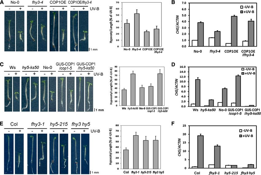
2 of 17    The Plant Cell

roles in promoting photomorphogenesis under diverse light             RESULTS
conditions (Oyama et al., 1997; Ang et al., 1998; Osterlund et al.,
2000; Ulm et al., 2004). HY5 targets a large number of light-         COP1 Expression Is Induced by Photomorphogenic UV-B
responsive genes in vivo by directly binding to the ACGT-
containing elements (ACEs) of their promoters (Oyama et al.,          In response to low-fluence and long-wavelength UV-B, a set of
1997; Ang et al., 1998; Lee et al., 2007; Zhang et al., 2011). It     genes was found to be activated in a temporal manner (Oravecz
also mediates crosstalk between light and hormone signaling,          et al., 2006). To determine how COP1 is regulated by photomor-
such as abscisic acid, gibberellins, and auxins (Oyama et al.,        phogenic UV-B, we examined the expression pattern of COP1 in
1997; Cluis et al., 2004; Lau and Deng, 2010), and integrates         4-d-old seedlings grown under no UV-B light (2UV-B) and then
light and stress responses, such as low temperature (Catalá           transferred to UV-B light (+UV-B). The accumulation of COP1
et al., 2011). In the UV-B–specific signaling pathway, HY5 serves      transcripts rises within 1 h and reaches a peak of eightfold in-
as a hub that is required for the accumulation of transcripts of      duction after 3 h of exposure to +UV-B and then decreases (Figure
a subset of UV-B–responsive genes. HY5’s own UV-B–induced             1A). However, when using 4-d-old seedlings grown in darkness
expression largely depends on UVR8 and COP1 (Ulm et al.,              and then transferred to far-red, red, or blue light conditions, we
2004; Oravecz et al., 2006; Favory et al., 2009). However, the        found the expression of COP1 was only slightly affected. These
specific cis-element that is responsible for mediating low-fluence      results suggest that, compared with monochromatic far-red and
and long-wavelength UV-B–responsive gene expression remains           visible light signals, photomorphogenic UV-B preferably induces
undefined.                                                             COP1 expression. Besides, COP1 protein abundance continues to
   Recently, the molecular mechanism for UVR8-mediated UV-B           increase over 12 h of photomorphogenic UV-B irradiation (Figure
perception was structurally described (Christie et al., 2012; Wu      1B). These results show that photomorphogenic UV-B induces
et al., 2012). Unlike the other photoreceptors, UVR8 possesses        COP1 expression at both mRNA and protein levels.
an internal chromophore shaped by the tryptophan (Trp) resi-
                                                                      FHY3 Binds to the COP1 Promoter via an FHY3
dues Trp-233 and Trp-285. Without UV-B, UVR8 forms a sym-
                                                                      Binding Site Motif in Vitro and in Vivo
metric homodimer that is stabilized by intra- and intermolecular
interactions, principally through the Arg residues Arg-286 and        The induction of COP1 by photomorphogenic UV-B prompted
Arg-338 and surrounding Trp residues. Exposure to UV-B leads          us to explore transcription factors involved in the regulation of
to a conformational switch from dimeric to monomeric UVR8
within seconds. The monomeric UVR8 then triggers down-
stream signaling pathways (Rizzini et al., 2011; Wu et al., 2011;
Christie et al., 2012; Wu et al., 2012). Upon UV-B irradiation,
UVR8 rapidly accumulates in the nucleus and interacts with
COP1 (Kaiserli and Jenkins, 2007). Interestingly, there appears
to be residual nuclear UVR8, which constitutively associates
with chromatin regions of several UV-B–activated genes, in-
cluding HY5, regardless of the presence or absence of UV-B
(Brown et al., 2005; Cloix and Jenkins, 2008).
   In far-red and visible light–induced photomorphogenesis, which
will be designated as traditional photomorphogenesis hereafter,
COP1 is a central repressor that targets photomorphogenesis-
promoting transcription factors, including HY5 for 26S proteasome–
mediated degradation. The function of COP1 is modulated primarily
via nucleocytoplasmic translocation and interaction with regulatory
factors (Yi and Deng, 2005). By contrast, in UV-B–induced photo-
morphogenesis, COP1 is a positive regulator for HY5, triggering
downstream UV-B–specific responses via an unknown mechanism
(Favory et al., 2009).
   Here, we show that COP1 gene expression is induced by
photomorphogenic UV-B. FAR-RED ELONGATED HYPOCOTYL3
(FHY3) and HY5 activate COP1 expression by respectively tar-
                                                                      Figure 1. COP1 Is Induced by UV-B at the mRNA and Protein Levels.
geting their distinct motifs in the COP1 promoter region, con-
tributing to UV-B–induced photomorphogenesis and tolerance            (A) Changes in COP1 transcript levels in 4-d-old wild-type (Col) seed-
against damaging UV-B. The genetic and molecular evidence             lings transferred from darkness (Dk) to far-red (FR), red (R), or blue (B)
                                                                      light conditions or from 2UV-B to +UV-B and harvested at indicated time
presented in this study demonstrates that in the UV-B–specific
                                                                      points. The transcript level at 0 h was set as 1. Error bars represent
responses, FHY3 is a positive regulator upstream of COP1,
                                                                      standard deviation (SD) of three biological replicates.
while HY5 forms a positive feedback loop on COP1. Our findings         (B) Changes in COP1 protein levels in 4-d-old wild-type (Col) seedlings
uncover the elaborate control of photomorphogenic UV-B on             transferred from 2UV-B to +UV-B and harvested at indicated time
COP1 by dual transcriptional regulation to ensure efficient early      points. Anti-RPN6 was used as a loading control.
signaling.                                                            [See online article for color version of this figure.]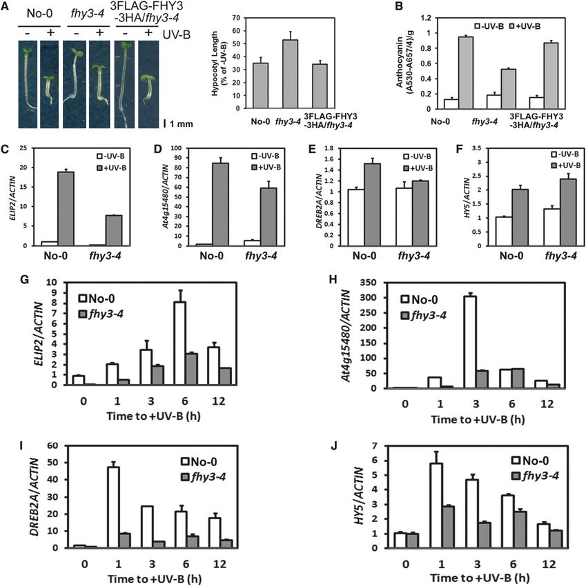
FHY3 and HY5 Activate COP1 under UV-B 3 of 17
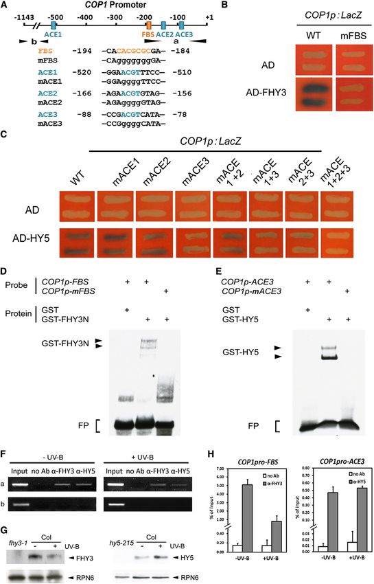
COP1 expression. Examination of the COP1 promoter identified enrich the ACE3-containing “a” fragment, but not the control “b”
a putative FHY3 binding site (FBS; Lin et al., 2007) ;190 bp up- fragment (Figure 2F). ChIP-qPCR assays confirmed that the
stream of the COP1 start codon (Figure 2A). A 1143-bp fragment COP1 promoter can be immunoprecipitated by antibodies
of the COP1 promoter, which was reported to be sufficient for against HY5 under both 2UV-B and +UV-B (Figures 2G and 2H).
COP1 promoter activity (Kang et al., 2009) was cloned and used These data illustrate that through the ACE3 site, HY5 is also able
for yeast one-hybrid assays. We found that the transcriptional to bind to the COP1 promoter regardless of photomorphogenic
activation domain fused FHY3 (AD-FHY3), but not the AD control, UV-B treatment.
induced LacZ reporter gene expression driven by the COP1 pro-
moter (Figures 2A and 2B). Site-directed mutagenesis of this pu-
FHY3 and HY5 Are Capable of Mediating COP1
tative FBS abolished AD-FHY3 binding to the COP1 promoter.
Activation in Response to UV-B
Subsequently, an electrophoretic mobility shift assay (EMSA) was
performed to confirm the direct binding of FHY3 to the FBS on the Since FHY3 and HY5 exhibit direct binding to the COP1 promoter
COP1 promoter. The first 200 amino acids of FHY3, C-terminally via FBS and ACE3, respectively, these two transcription factors
fused to glutathione S-transferase, (GST-FHY3N) was previously might contribute to the regulation of COP1 expression. To test
shown to harbor DNA binding activity (Lin et al., 2007; Li et al., this hypothesis, we first took advantage of the transgenic line
2010). In our assay, addition of GST-FHY3N, but not GST, resulted FHY3p:FHY3-GR/fhy3-4 expressing an FHY3-glucocorticoid re-
in slower migration of the FBS probe, indicating that FHY3 is able ceptor (GR) fusion protein driven by the FHY3 native promoter
to bind to the COP1 promoter. When assayed with a probe with (Lin et al., 2007). The fusion protein will translocate from the cy-
the FBS site mutated, GST-FHY3N was not able to bind to the toplasm into the nucleus after treatment with dexamethasone
COP1 promoter fragment (Figure 2D). (DEX) (Lloyd et al., 1994). The seedlings were grown under 2UV-B
To investigate whether FHY3 protein associates with the COP1 for 4 d and then treated with DEX before transferred to 2UV-B
promoter in vivo, a chromatin immunoprecipitation (ChIP) assay and +UV-B, respectively. The expression of COP1 is induced
was performed followed by regular PCR and quantitative PCR within 2 h under +UV-B but not 2UV-B (Figure 3A). Neverthe-
(qPCR) analyses. In 4-d-old 2UV-B– and +UV-B–grown seed- less, FHY1, an identified target of FHY3 in far-red light signaling
lings, the “a” fragment, which contains the FBS motif of the COP1 (Lin et al., 2007), is induced under 2UV-B instead of +UV-B (see
promoter, was found to be enriched by antibodies against native Supplemental Figure 3A online), consistent with no change in
or transgenic FHY3. However, the “b” fragment, which is further the FHY1 mRNA level in wild-type seedlings grown under 2UV-
upstream of the COP1 promoter, was used as a negative control B and then transferred to +UV-B (see Supplemental Figure 3B
and was not enriched (Figure 2F; see Supplemental Figure 1A online).
online). The reduced enrichment in +UV-B–grown seedlings can Then, a dual luciferase assay was conducted in Nicotiana
probably be ascribed to the lower protein level of FHY3 under benthamiana to check the effects of FHY3 and HY5 on COP1
+UV-B (Figures 2G and 2H; see Supplemental Figures 1B and 1C transcription. Recombinant reporter constructs were generated to
online). Taken together, these results indicate that FHY3 protein allow the wild-type or mutated COP1 promoter to drive expression
directly binds to the COP1 promoter through the FBS motif under of the firefly luciferase (LUC) reporter gene (Figure 3B). Under
both 2UV-B and +UV-B. photomorphogenic UV-B, the overexpression of FHY3 leads to
a 3.7-fold increase in the activity of the LUC reporter under the
HY5 Binds to the COP1 Promoter via an ACE COP1 promoter (Figure 3C). By contrast, it had no effect when
Element in Vitro and in Vivo either UV-B irradiation was absent (Figure 3C) or the FBS motif in
the COP1 promoter was disrupted (Figure 3D). ACE3 mutation still
In addition to the FBS motif, three putative ACEs, typical HY5 led to the induction of LUC expression by FHY3 (Figure 3D). In
binding sites, are present in the COP1 promoter region. Yeast one- a parallel analysis, HY5 also activated the COP1 promoter-driven
hybrid assays were again used to identify the ACE(s) that is im- LUC expression in a UV-B–dependent manner, though with re-
portant for HY5 binding. AD-HY5 was able to bind the wild-type duced effectiveness compared with FHY3 (Figure 3C). With the
COP1 promoter, whereas mutation in ACE3 resulted in little affinity mutated ACE3 construct, HY5 was no longer able to promote LUC
of AD-HY5 to the COP1 promoter, demonstrating that ACE3 is the expression (Figure 3D). The FBS mutation exerted no influence on
dominant, if not the only, site for HY5 binding (Figure 2C). The the induction of LUC expression by HY5 (Figure 3D). Altogether,
ACE3 is located ;80 bp upstream of the COP1 start codon and these results demonstrate that upon UV-B irradiation, FHY3 and
100 bp downstream of the FBS motif (Figure 2A). HY5-HOMOLOG HY5 can promote transcription of COP1 in plant cells.
(HYH) overlaps in function with HY5 in the stimulation of some Furthermore, to get more insight how FHY3 and HY5 activate
gene expression by UV-B (Brown and Jenkins, 2008), but no ob- COP1, the dual luciferase assay was perform in Arabidopsis. Un-
vious signal for HYH binding to the COP1 promoter was detected der photomorphogenic UV-B, in wild-type plants, the transiently
(see Supplemental Figure 2 online). In an EMSA test, the wild-type overexpressed FHY3 and HY5 led to 2.6- and 2.0-fold increases,
but not the mutated ACE3 probe was bound by the recombinant respectively, in the activity of the LUC reporter under the COP1
GST-HY5, as evidenced by the presence of bands with retarded promoter. However, little reporter activity was detected when
mobility (Figure 2E). uvr8-6 plants were infiltrated (Figure 3E), indicating that the loss of
Furthermore, ChIP-PCR analysis validated the in vivo associa- UVR8 function impinges on FHY3 and HY5 in COP1 activation.
tion of HY5 protein with the COP1 promoter in 2UV-B– and +UV- Together with the fact that UVR8 is a UV-B receptor (Rizzini et al.,
B–grown wild-type seedlings. Anti-HY5 antibodies were found to 2011; Wu et al., 2011; Christie et al., 2012; Wu et al., 2012), we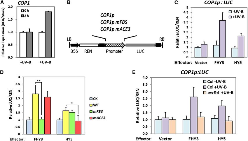
Figure 2. FHY3 and HY5 Bind to the COP1 Promoter in Vitro and in Vivo.
(A) Diagram of fragments of the COP1 promoter. The adenine of the translational start codon (ATG) is designated as position +1. Orange and blue
blocks indicate putative FBS and ACEs, respectively. Arrows indicate the fragments amplified in ChIP-PCR assays. The wild-type and mutated sites of
the COP1 promoter subfragments are shown in uppercase and lowercase letters, respectively.
FHY3 and HY5 Activate COP1 under UV-B            5 of 17

conclude that the activation of COP1 by FHY3 and HY5 is de-                then transferred to +UV-B for various time periods. The temporal
pendent on UVR8 initiated UV-B signaling.                                  induction of all these genes by photomorphogenic UV-B was
                                                                           much weaker in fhy3-4 than in No-0 (Figures 4G to 4J). These
FHY3 Is Critical for UV-B–Induced Photomorphogenesis                       results demonstrate that FHY3 positively contributes to the tran-
at the Seedling Stage                                                      scriptional response to photomorphogenic UV-B, particularly at
                                                                           an early stage.
As shown above, FHY3 is able to bind to the COP1 promoter                     Furthermore, introduction of a 35S promoter–driven 3FLAG-
and activate its transcription under UV-B. COP1 is a positive              FHY3-3HA construct in fhy3-4 (see Supplemental Figure 5C
regulator of UV-B–induced photomorphogenesis. Therefore, it is             online) rescues the defects in its hypocotyl growth (Figure 4A)
possible that FHY3 may be critical in this pathway.                        and anthocyanin accumulation (Figure 4B), supporting that FHY3
   As inhibition of hypocotyl elongation is a characteristic response      is involved in UV-B–induced photomorphogenesis at the seed-
to photomorphogenic UV-B (Favory et al., 2009; Gardner et al.,             ling stage.
2009), the relative hypocotyl length, a percentage defined as the              FAR-RED IMPAIRED RESPONSE1 (FAR1) is homologous to
hypocotyl length under +UV-B relative to the length under 2UV-B,           FHY3 and shares functional redundancy with FHY3 in phyto-
was measured using 4-d-old seedlings grown in typical photo-               chrome A (phyA) signaling, circadian clock, chloroplast deve-
morphogenic UV-B experimental conditions (see Supplemental                 lopment, and chlorophyll biosynthesis (Wang and Deng, 2002;
Figure 4 online). Consistent with earlier reports (Oravecz et al.,         Hiltbrunner et al., 2005; Allen et al., 2006; Lin et al., 2007; Genoud
2006; Favory et al., 2009; Gardner et al., 2009), a UV-B photore-          et al., 2008; Li et al., 2010; Li et al., 2011; Ouyang et al., 2011;
ceptor mutant uvr8-6 and a key promoter mutant cop1-4 showed               Tang et al., 2012). Thus, we are interested if FAR1 plays a role in
much longer relative hypocotyl length than the wild type, but such         UV-B–induced photomorphogenesis as well. Surprisingly, far1
a defect was not found in the other photoreceptor mutants phyAB,           mutants showed no apparent difference from wild-type seedlings
phyABDE, and cry1 cyr2 or the DNA repair mutant uvr1-1. These              in UV-B–promoted hypocotyl growth (see Supplemental Figure 6
observations confirmed that the condition used is specific to                online). In addition, the fhy3 far1 double mutants displayed the
UV-B–induced photomorphogenesis instead of UV-B stress.                    same extent of hypocotyl growth in the presence of UV-B as that
   Interestingly, we found that fhy3-4 mutants displayed a dramat-         of fhy3 single mutant (see Supplemental Figure 6 online). These
ically reduced ability to inhibit hypocotyl elongation with UV-B ir-       results suggest that FAR1 is not essential in photomorphogenic
radiation compared with wild-type Nossen-0 (No-0) (Figure 4A).             UV-B response.
The relative hypocotyl length in No-0 seedlings was around 35%
but was 53% in fhy3-4 mutants. This defect was also observed in            A T-DNA Insertion hy5 Null Mutant Exhibits Impaired
four additional fhy3 mutant alleles, fhy3-1, fhy3-3, fhy3-5, and           UV-B–Induced Photomorphogenesis
fhy3-10 (see Supplemental Figures 5A and 5B online). Photomor-
phogenic UV-B accelerates biosynthesis of sunscreen flavonoids              Previous studies have reported reduced UV-B–mediated gene
(Oravecz et al., 2006). Quantitatively, we found that there was less       activation in hy5-1 and hy5-215 mutants and impaired UV-B tol-
anthocyanin content in fhy3-4 than in No-0 (Figures 4B).                   erance in hy5-1, suggesting that HY5 is a positive regulator in
   Next, we examined the expression of several marker genes                mediating gene expression and protective survival under UV-B
responsive to photomorphogenic UV-B in 4-d-old seedlings                   (Ulm et al., 2004; Oravecz et al., 2006; Gruber et al., 2010; Stracke
grown under 2UV-B and +UV-B. We found that the photo-                      et al., 2010). Here, we found that hy5-ks50, a T-DNA insertion hy5
morphogenic UV-B–mediated transcript accumulation of EARLY                 null mutant, is also impaired in UV-B-induced photomorphogen-
LIGHT-INDUCIBLE PROTEIN2 (ELIP2) (Figure 4C), At4g15480                    esis. The relative hypocotyl length of the mutant seedlings was
(Figure 4D), and DEHYDRATION-RESPONSIVE ELEMENT                            around 74% (see Supplemental Figure 7A online), and there was
BINDING PROTEIN2A (DREB2A) (Figure 4E) was diminished in                   much less anthocyanin content in hy5-ks50 than that in Wassi-
fhy3-4, but HY5 (Figure 4F) accumulated slightly more tran-                lewskija (Ws) (see Supplemental Figure 7B online). These defects
scripts. Next, we continued to analyze the temporal expression             were restored through the overexpression of 35S:HY5 (see
of these genes using 4-d-old seedlings grown under 2UV-B and               Supplemental Figure 7 online). This indicates that the hy5-ks50

Figure 2. (continued).

(B) and (C) FHY3 (B) and HY5 (C) bind to the COP1 promoter in yeast one-hybrid assays. Empty vector expressing the AD domain alone is the negative
control. WT, the wild type.
(D) and (E) GST-FHY3N (D) and GST-HY5 (E) specifically bind to COP1p-FBS and COP1p-ACE3 probes respectively in EMSA assays. FP, free probes.
(F) Representative results of ChIP-PCR assays. Chromatin fragments prepared from 4-d-old wild-type (Col) seedlings grown under 2UV-B and +UV-B
were immunoprecipitated by the polyclonal anti-FHY3 and anti-HY5 antibodies, respectively. Input, PCR reactions using the samples before immu-
noprecipitation. Ab, antibody. a and b correspond to the DNA fragments shown in (A).
(G) FHY3 and HY5 protein levels of 4-d-old wild-type (Col) seedlings grown under 2UV-B and +UV-B. Anti-RPN6 was used as a loading control.
(H) qPCR analyses of the FBS-containing fragment (COP1pro-FBS) and the ACE3-containing fragment (COP1pro-ACE3) in the COP1 promoter in anti-
FHY3 and anti-HY5 ChIP assays, respectively, using 4-d-old wild-type (Col) seedlings grown under 2UV-B and +UV-B. The ChIP values were nor-
malized to their respective DNA inputs. Error bars represent SD of three biological replicates.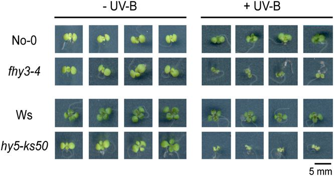
6 of 17     The Plant Cell

Figure 3. FHY3 and HY5 Target COP1 to UV-B–Dependent Transcriptional Activation.

(A) The transcript levels of COP1 in 4-d-old FHY3p:FHY3-GR/fhy3-4 seedlings grown under 2UV-B and then treated with 10 mM DEX before being
transferred to 2UV-B and +UV-B. Relative expression levels were normalized to the Mock (equal volume of ethanol) treatment. Error bars represent SD
of three biological replicates.
(B) Structure of the dual-luciferase reporter construct in which the firefly LUC reporter gene is driven by the wild type, FBS-mutated (mFBS), or ACE-
mutated (mACE3) COP1 promoter. The REN luciferase reporter gene is controlled by the constitutive 35S promoter. The T-DNA left border and right
border are indicated as LB and RB, respectively.
(C) The relative LUC activity normalized to the REN activity (LUC/REN) in tobacco leave cells transiently transformed with the indicated effectors and
wild-type reporter construct COP1p:LUC under 2UV-B and +UV-B. Empty effector vectors were included as negative controls.
(D) The LUC/REN values in tobacco leave cells transiently transformed with the indicated effectors and wild-type (WT) or mutated reporter constructs
under +UV-B. Empty reporter vectors were included as negative controls. *P < 0.05 and **P < 0.01 by Student’s t test. CK, empty reporter vector. Error
bars represent SD of four biological replicates.
(E) The relative LUC activity normalized to the REN activity (LUC/REN) in wild-type (Col) and mutant (uvr8-6) Arabidopsis leave cells transiently
transformed with the indicated effectors and wild-type reporter construct COP1p:LUC under 2UV-B and +UV-B. Empty effector vectors were included
as negative controls.

null mutant, similar to the other hy5 alleles reported, is defective         with shrunken and pale leaves (Figure 5). The hypersensitivity to
in UV-B–induced photomorphogenesis and thus was used for                     UV-B stress in fhy3-4 and hy5-ks50 mutants might be ascribed
further investigation in our analysis.                                       to their failure to properly respond to photomorphogenic UV-B,
                                                                             further demonstrating the involvement of FHY3 and HY5 in
Both fhy3 and hy5 Mutants Are Hypersensitive to                              UV-B–induced photomorphogenesis and acclimation.
Damaging UV-B
                                                                             Absence of FHY3 or HY5 Results in Abnormal
Earlier studies reported that UV-B–induced photomorphogenesis                Accumulation of COP1 Transcript and
is a fundamental event for plants to acquire tolerance against               Protein under Photomorphogenic UV-B
damaging UV-B (Favory et al., 2009). Based on the observations of
abnormal UV-B–induced photomorphogenesis in our fhy3 and hy5                 To further explore the roles of FHY3 and HY5 in UV-B–specific
mutants, we attempted to investigate if they suffer from UV-B–               signaling, especially in regulating COP1 expression in vivo,
induced damage. Wild-type seedlings were apparently resistant                temporal expression of UV-B responsive genes was analyzed
to the UV-B stress. By contrast, the leaves of fhy3-4 mutants                using 4-d-old seedlings grown under -UV-B and then transferred
shriveled, and hy5-ks50 mutants displayed even more severe defects           to +UV-B for various time periods.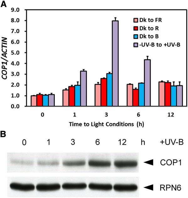
FHY3 and HY5 Activate COP1 under UV-B              7 of 17

Figure 4. FHY3 Is Involved in UV-B–Induced Photomorphogenesis at the Seedling Stage.
(A) Phenotypes of 4-d-old wild-type (No-0), fhy3-4, and 3FLAG-FHY3-3HA/fhy3-4 seedlings grown under 2UV-B and +UV-B. Quantitative analysis of
hypocotyl length is presented as percentage of 2UV-B. Error bars represent SD of three biological replicates. Bar = 1 mm.
(B) Quantitative analysis of anthocyanin accumulation of the seedlings in (A). Error bars represent SD of three biological replicates.
(C) to (F) The transcript levels of UV-B–induced marker genes ELIP2 (C), At4g15480 (D), DREB2A (E), and HY5 (F) in 4-d-old wild-type (No-0) and fhy3-
4 seedlings grown under 2UV-B to +UV-B. The transcript level in No-0 under 2UV-B was set as 1. Error bars represent SD of three biological replicates.
(G) to (J) The transcript levels of UV-B–induced marker genes ELIP2 (G), At4g15480 (H), DREB2A (I), and HY5 (J) in 4-d-old wild-type (No-0) and fhy3-4
seedlings transferred from 2UV-B to +UV-B and harvested at indicated time points. The transcript level at 0 h in No-0 was set as 1. Error bars represent
SD of three biological replicates.
[See online article for color version of this figure.]
8 of 17 The Plant Cell
CHALCONE SYNTHASE (CHS), which encodes a key enzyme 8 online). Under photomorphogenic UV-B, GUS-COP1/cop1-5
at the first committed step in anthocyanin biosynthesis, is another exhibited comparable hypocotyl growth to No-0. When crossed
UV-B–induced marker gene (Oravecz et al., 2006). In No-0, CHS with GUS-COP1/cop1-5, hy5-ks50 showed no recovery of hypo-
was rapidly activated with a peak of 64-fold induction after 3 h of cotyl shortening (Figure 7C). Besides, in response to photomor-
UV-B treatment and starts to fall afterwards. In fhy3-4, the CHS phogenic UV-B, GUS-COP1/cop1-5 displayed stronger induction
transcript rose with a diminished and retarded peak of 39-fold of CHS than No-0, but GUS-COP1/hy5-ks50 failed to induce CHS
increase after 6 h of UV-B treatment (Figure 6A). COP1 transcript like hy5-ks50 (Figure 7D). In the absence of HY5, COP1 over-
peaked with 6.3- and 3.9-fold increases in No-0 and fhy3-4, re- expression was not sufficient to regain the regular response to
spectively, after 6 h of UV-B treatment (Figure 6B). At the protein photomorphogenic UV-B, suggesting epistasis of hy5-ks50 mu-
level, compared with No-0, less COP1 protein accumulated in tation over COP1 overexpression. According to the phenotypes
fhy3-4 over 12 h of photomorphogenic UV-B irradiation (Figure of COP1OE/fhy3-4 and GUS-COP1/hy5-ks50, we deduce that the
6C). In parallel assays, the UV-B–induced transcription of CHS main function of HY5 is downstream of FHY3 and COP1.
was found to be almost completely lost in hy5-ks50 (Figure 6D). Next, we crossed fhy3-1 with hy5-215, which was reported to
COP1 transcript peaked with 6.7- and 3.6-fold increase in Ws and have a reduced inhibition of hypocotyl elongation under +UV-B
hy5-ks50, respectively (Figure 6E). The increase in COP1 protein (Jiang et al., 2012). We observed that fhy3-1 hy5-215 double
mediated by photomorphogenic UV-B was largely abolished in mutant seedlings closely resembled the hy5-215 single mutants
hy5-ks50 (Figure 6F). These results indicate that both FHY3 and in hypocotyl growth upon UV-B treatment (Figure 7E). Consis-
HY5 are required for the full activation of COP1 and COP1 protein tently, fhy3-1 hy5-215 mimicked hy5-215 in its photomorpho-
accumulation mediated by photomorphogenic UV-B. genic UV-B–mediated CHS induction (Figure 8F). These results
also agree with the notion that HY5 acts as a main downstream
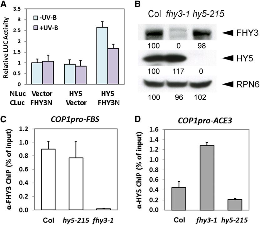
COP1 and HY5 Genetically Interact with FHY3 in Mediating transcription factor of COP1 in the central pathway in UV-B–
UV-B Photomorphogenic Responses mediated photomorphogenesis. It is likely that participating in
COP1 gene activation is one of the regulatory roles of HY5.
In order to further define the genetic role of FHY3 in the photo-
morphogenic UV-B signaling pathway, we examined the possible Photomorphogenic UV-B Affects the Functional
genetic interaction between COP1, FHY3, and HY5. The seedlings Interaction between FHY3 and HY5
of a previously described COP1 overexpression line, COP1OE (see
Supplemental Figure 8 online), displayed a stronger inhibition of It was previously noted that the antagonistic roles of FHY3 and
hypocotyl elongation compared with the wild-type, confirming HY5 in phyA signaling and their cooperative functions in the cir-
COP1’s positive role in UV-B signaling (Figure 7A). When in- cadian clock both rely on their physical interaction (Li et al., 2010;
troduced into fhy3-4, the COP1 overexpression completely Li et al., 2011). It is therefore of interest to examine whether FHY3
suppressed the reduced inhibition of hypocotyl elongation found and HY5 functionally interact in UV-B responses. We first con-
in fhy3-4, to a level that is similar to COP1OE (Figure 7A). ducted firefly luciferase complementation imaging (LCI) assays
Quantitative analysis of the CHS transcripts confirmed that (Chen et al., 2008) by transiently overexpressing CLuc and NLuc
COP1OE/fhy3-4 phenocopies COP1OE (Figure 7B). Therefore, fusion proteins in N. benthamiana leaf cells. Obvious LUC activity
we conclude that COP1 acts downstream of FHY3. was observed when CLuc-FHY3N and HY5-NLuc were coex-
GUS-COP1/cop1-5 was previously found to rescue the con- pressed under 2UV-B, whereas much less was observed under
stitutive photomorphogenesis of cop1-5 in darkness by over- +UV-B (Figure 8A). This result suggests that UV-B attenuates the
expressing COP1 (von Arnim et al., 1997; see Supplemental Figure association between FHY3 and HY5 in plant cells.
In addition, the presence of both the FBS motif and the ACE
element in the COP1 promoter also hints at a possible functional
interaction between FHY3 and HY5. Thus, we examined whether
the loss of HY5 or FHY3 affected the other with regard to their
protein abundance and their affinity to the COP1 promoter. In
hy5-215, the protein level of FHY3 is comparable to that of the
wild type (Figure 8B). ChIP of FHY3 also precipitated the FBS
motif–containing promoter fragment of COP1 at a similar effi-
ciency with the wild type (Figure 8C). However, in fhy3-1, more
HY5 proteins accumulate (Figure 8B), and antibodies against HY5
enriched twofold more DNA fragments containing the ACE3 in the
fhy3-1 mutant than that in the wild type (Figure 8D). These ob-
servations reveal that FHY3 or HY5 are each able to associate
with the COP1 promoter in the absence of the other.
Figure 5. FHY3 and HY5 Are Required for Tolerance to Damaging UV-B.
Wild-type (No-0 and Ws), fhy3-4, and hy5-ks50 seedlings were grown DISCUSSION
under white light supplemented with photomorphogenic UV-B for 12 d
and then were irradiated without (2UV-B) or with (+UV-B) damaging The E3 ubiquitin ligase COP1 is a well-known repressor in tradi-
broadband UV-B for 12 h. Bar = 5 mm. tional photomorphogenesis, primarily via mediating the degradationFHY3 and HY5 Activate COP1 under UV-B 9 of 17 Figure 6. FHY3 and HY5 Are Required for Accumulation of COP1 Transcripts and Proteins under Photomorphogenic UV-B. (A) and (B) Changes in CHS (A) and COP1 (B) transcript levels in 4-d-old wild-type (No-0) and fhy3-4 seedlings transferred from 2UV-B to +UV-B and harvested at indicated time points. The transcript level at 0 h in No-0 was set as 1. Error bars represent SD of three biological replicates. (C) COP1 protein levels in 4-d-old wild-type (No-0) and fhy3-4 seedlings transferred from 2UV-B to +UV-B and harvested at indicated time points. Anti- RPN6 was used as a loading control. (D) and (E) CHS (D) and COP1 (E) transcript levels in 4-d-old wild-type (Ws) and hy5-ks50 seedlings transferred from 2UV-B to +UV-B and harvested at the indicated time points. The transcript level at 0 h in Ws was set as 1. Error bars represent SD of three biological replicates. (F) COP1 protein levels in 4-d-old wild-type (Ws) and hy5-ks50 seedlings transferred from 2UV-B to +UV-B and harvested at indicated time points. Anti- RPN6 was used as a loading control. of photomorphogenesis-promoting transcription factors (Yi and Thus, this work uncovers a functional mode of FHY3 and HY5 Deng, 2005). By contrast, COP1 plays a positive role in UV-B– beyond phyA signaling and circadian regulation. induced photomorphogenesis, but how photomorphogenic UV-B regulates COP1 is unknown. Our analyses reveal that COP1 is Photomorphogenic UV-B Regulates COP1 Expression a UV-B–inducible gene, whose full activation requires tran- via FHY3 and HY5 Binding LREs scription factors FHY3 and HY5. Furthermore, FHY3 and HY5 are engaged in UV-B–induced photomorphogenesis and accli- During the last decades, the reprogramming of the plant tran- mation and show a genetic interaction with COP1 (Figure 9). scriptome by light has been extensively examined. Transcriptional
10 of 17     The Plant Cell

Figure 7. COP1, FHY3, and HY5 Genetically Interact with Each Other under Photomorphogenic UV-B.

(A) and (B) Phenotypes (A) and CHS transcript levels (B) of 4-d-old wild-type (No-0), fhy3-4, COP1OE, and COP1OE/fhy3-4 seedlings grown under
2UV-B and +UV-B.
(C) and (D) Phenotypes (C) and the CHS transcript levels (D) of 4-d-old wild-type (Ws and No-0), hy5-ks50, GUS-COP1/cop1-5, and GUS-COP1/hy5-
ks50 seedlings grown under 2UV-B and +UV-B.
(E) and (F) Phenotypes (E) and the CHS transcript levels (F) of 4-d-old wild-type (Col), fhy3-1, hy5-215, and fhy3-1 hy5-215 seedlings grown under
2UV-B and +UV-B. Quantitative analysis of hypocotyl length is presented as percentage of 2UV-B. The transcript level in the wild type under -UV-B
was set as 1. Error bars represent SD of three biological replicates. Bars = 1 mm.
[See online article for color version of this figure.]

reprogramming requires light-responsive cis-elements (LREs),               motif and the ACE element in the COP1 promoter serve as LREs
which are commonly present in the promoter regions of light-               to positively mediate COP1 expression in this transcriptional
regulated genes. Combinations of LREs confer distinct tran-                response to photomorphogenic UV-B (Figures 2 and 3).
scriptional responses to various light signals and further depict             As a multifunctional E3 ligase, COP1 is subjected to changes in
a sophisticated light regulated transcriptional network (Puente            activity in response to diverse stimuli. For example, the crypto-
et al., 1996; Jiao et al., 2007). Such transcriptional regulation is       chromes rapidly inactivate COP1 through their direct association
also coupled with UV-B–specific signaling. Photomorphogenic                 (Wang et al., 2001; Yang et al., 2001), four functional redundant
UV-B promotes differential gene expression (Ulm et al., 2004;              SUPPRESSOR OF PHYA (SPA) proteins act as regulatory sub-
Brown et al., 2005; Oravecz et al., 2006; Favory et al., 2009),            units of COP1-SPA heterogeneous complexes (Saijo et al., 2003;
histone modification, and chromatin remodeling (Casati et al.,              Zhu et al., 2008), and at a relatively slower rate, light inhibits its E3
2008). In Arabidopsis, although the UVBox, a promoter motif, is            ligase activity partially by excluding it from the nucleus (von Arnim
required for the induction of ARABIDOPSIS THALIANA NAC                     and Deng, 1994). Here, we establish how COP1 is controlled at
DOMAIN PROTEIN13 by shorter-wavelength and high-intensity                  the transcriptional level in Arabidopsis. Specifically, the accurate
UV-B, this element is not regulated by longer-wavelength UV-B              composition of the LREs for FHY3 and HY5 in the COP1 promoter
(Safrany et al., 2008). In our study, COP1 was induced upon                (Figure 2) defines its ability to positively respond to the stimulus of
treatment with photomorphogenic UV-B (Figure 1). The FBS                   photomorphogenic UV-B (Figure 3).
FHY3 and HY5 Activate COP1 under UV-B              11 of 17

Figure 8. Photomorphogenic UV-B Affects the Functional Interaction between FHY3 and HY5.

(A) The interaction between FHY3 and HY5 under 2UV-B and +UV-B in LCI assays. Error bars represent SD of four biological replicates.
(B) FHY3 and HY5 protein levels in 4-d-old wild-type (Col), fhy3-1, and hy5-215 seedlings grown under +UV-B. Anti-RPN6 was used as a loading
control. Band intensities of FHY3, HY5, and RPN6 in Col seedlings were set to 100. Relative band intensities were then calculated and are indicated by
numbers below blots.
(C) and (D) qPCR analysis of the FBS-containing fragment (COP1pro-FBS) in the COP1 promoter in anti-FHY3 ChIP assays (C) and the ACE3-
containing fragment (COP1pro-ACE3) in the COP1 promoter in anti-HY5 ChIP assays (D) using 4-d-old wild-type (Col), hy5-215, and fhy3-1 seedlings
grown under +UV-B. The ChIP values were normalized to their respective DNA inputs. Error bars represent SD of three biological replicates.
[See online article for color version of this figure.]

FHY3 Is Associated with UV-B–Induced                                         COP1 expression. Also, it was found that photomorphogenic
Photomorphogenesis                                                           UV-B entrains the plant clock. Without UV-B pulses, amplified
                                                                             circadian rhythms are eliminated in clock genes like CIRCADIAN
UV-B–induced photomorphogenesis was first observed in Arabi-                  CLOCK ASSOCIATED1, GIGANTEA, and EARLY FLOWERING4
dopsis in the late 1990s and expanded light control of plant de-             (ELF4) (Fehér et al., 2011). Among these genes, ELF4 loses its
velopment beyond the traditional photomorphogenesis by far-red               rhythmic expression in fhy3 mutants in visible light and it needs
and visible light wavelengths (Kim et al., 1998). To date, this re-          FHY3 for transcriptional activation (Li et al., 2011). Thus, FHY3
sponse has been shown in several plant species, such as maize                might be a link between UV-B–induced photomorphogenesis and
(Zea mays) (Casati and Walbot, 2003; Casati et al., 2008), cu-               the circadian clock. Interestingly, FAR1 does not seem to function
cumber (Cucumis sativus) (Shinkle et al., 2010), and moss (Phys-             in early photomorphogenic UV-B responses (see Supplemental
comitrella patens) (Wolf et al., 2011). In Arabidopsis, besides HY5,         Figure 6 online). Based on previous promoter-swapping experi-
UVR8, and COP1, there are a growing number of factors found                  ments, FHY3 may undertake a divergent role from FAR1 through
to be involved in the responses to photomorphogenic UV-B, in-                protein subfunctionalization (Lin et al., 2008).
cluding two potential Damaged DNA Binding Protein1 (DDB1)
binding WD40 proteins, REPRESSOR OF UV-B PHOTOMOR-                           The Expression and Activity of FHY3 Is Regulated by
PHOGENESIS1 (RUP1) and RUP2. They are UVR8-interacting                       Photomorphogenic UV-B
proteins and act downstream of UVR8 and COP1 in a negative
feedback loop (Gruber et al., 2010).                                         Time-course microarray-based expression profiling of light-
   Recently, the identification of genome-wide binding sites of               responsive genes has documented the light regulation of
FHY3 profoundly extended our understanding of the diversity of               transcription factors (Tepperman et al., 2001, 2004; Jiao et al.,
FHY3 function, especially in environmental adaptation (Ouyang                2003). For example, HY5 protein accumulates under various light
et al., 2011; Stirnberg et al., 2012; Tang et al., 2012). In response to     conditions (Oyama et al., 1997; Hardtke et al., 2000; Osterlund
photomorphogenic UV-B, FHY3 functions primarily via activating               et al., 2000). FHY3 is repressed by far-red light (Lin et al., 2007) but
12 of 17 The Plant Cell
Figure 2; see Supplemental Figure 1 online). However, our earlier
microarray data did not detect altered COP1 mRNA levels in re-
sponse to FHY3 translocation into the nucleus in far-red light or
dark conditions (Ouyang et al., 2011). The transcriptional activa-
tion activity of FHY3 toward the COP1 promoter is strictly induced
by the UV-B signaling (Figures 3C and 3E).
COP1 and HY5 Form a Positive Feedback Loop in
UV-B–Induced Photomorphogenesis
As one of the core transcription factors in light control of plant
development, HY5 is induced by light and its protein accumulation
is promoted by multiple photoreceptors under distinct wavelengths
of light (Oyama et al., 1997; Hardtke et al., 2000; Osterlund et al.,
2000). The SPA-COP1 complexes are substrate receptors for
CULLIN4-DDB1 E3 ligases, which ubiquitinate HY5 for selective
degradation by the 26S proteasome (Osterlund et al., 2000;
Saijo et al., 2003; Chen et al., 2006, 2010; Zhu et al., 2008).
In UV-B–induced photomorphogenesis, HY5 acts downstream
of UVR8 and COP1 (Ulm et al., 2004; Oravecz et al., 2006; Favory
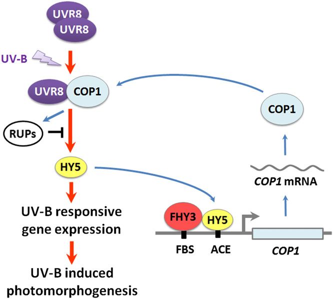
et al., 2009). Meanwhile, HY5 directly associates with the COP1
Figure 9. A Model for the Functional Interaction of COP1, FHY3, and promoter via the ACE element independent of UV-B irradiation
HY5 in UV-B–Induced Photomorphogenesis.
(Figure 2) and induces COP1 expression specifically dependent
Under photomorphogenic UV-B, UVR8 monomerizes and interacts with on UV-B signaling (Figures 3C and 3E). Thus, COP1 and HY5
COP1, triggering downstream signaling, including HY5-regulated gene form a positive feedback loop in UV-B photomorphogenic
expression, which is repressed by RUPs. COP1 is a UV-B–inducible responses.
gene. FHY3 and HY5 bind to the FBS motif and the ACE element in the Upon UV-B treatment, three-fourths of the early UV-B–activated
COP1 promoter, respectively, and activate COP1 transcription. This tran-
genes lose induction in cop1-4, but more than a half remain un-
scriptional modulation ensures more active UVR8-COP1-HY5 core path-
changed in hy5-1 (Oravecz et al., 2006), suggesting there is a group
way. Red arrows, positive regulation in the core pathway; blue arrows,
positive regulation in UV-B–mediated gene induction; black bar, negative of COP1-dependent genes that are regulated independent of
regulation. HY5. Besides, there may no longer be a COP1-mediated deg-
radation of HY5 under UV-B (Favory et al., 2009). In this case,
the mutual transcriptional regulation between COP1 and HY5
is activated by photomorphogenic UV-B (see Supplemental might stand as one of their interaction modes. Still, the positive
Figures 3B and 9A online). This differential regulation shows that feedback is important, together with FHY3-induced COP1 ex-
FHY3 can be specifically controlled by distinct wavelengths of pression, to produce adequate early signal transducers to maintain
light. Meanwhile, FHY1, a target of FHY3 under far-red light, UVR8-COP1–mediated signaling. Downstream of UVR8-COP1,
shows no transcriptional response to UV-B (see Supplemental RUP1 and RUP2 rapidly accumulate their transcripts and pro-
Figure 3 online). Such distinct transcriptional behaviors of FHY3 teins and result in a negative feedback loop to repress UV-B–
and FHY1 indicate a fine-tuning of light signals that differentiates induced gene expression. Meanwhile, RUP1 and RUP2 are likely
the far-red light pathway from the UV-B pathway. Additionally, to sequester UVR8 from COP1. In these two ways, RUP1 and
though FHY3 mRNA level is rapidly upregulated by photomor- RUP2 prevent an exaggerated UV-B photomorphogenesis re-
phogenic UV-B, its protein level is eventually downregulated sponse and further balance UV-B acclimation and plant deve-
(Figure 2G; see Supplemental Figures 1B, 9B, and 9C online). lopment (Gruber et al., 2010; Figure 9). A recent study on another
This observation indicates that FHY3 might be regulated by negative regulator, SALT TOLERANCE, that might antagonize
photomorphogenic UV-B in a posttranscriptional manner, which HY5 transcriptional activity (Jiang et al., 2012) also supports
awaits further investigation. coordination of positive and negative feedback processes as
FHY3 contains an N-terminal C2H2 zinc finger domain, a cen- crucial for appropriate UV-B photomorphogenic responses in
tral putative core transposase domain, and a C-terminal SWIM planta.
zinc finger domain (named after SWI2/SNF and MuDR trans-
posases) (Makarova et al., 2002). The C2H2 zinc finger domain The Combinatorial Functional Interaction of the FHY3-HY5
mediates its DNA binding activity, and the transposase catalytic Pair Is Distinct in UV-B–Specific Response from That in
domain and SWIM motif are important for its transcriptional ac- Far-Red Light Signaling and Circadian Regulation
tivation activity (Lin et al., 2008). FHY3 shows high affinity to the
COP1 promoter (Figure 2), whose FBS-containing region is on the Two contrasting working models between FHY3 and HY5 were
list of global FBSs (Ouyang et al., 2011). A series of ChIP assays hypothesized in recent years. In far-red light responses, FHY3
collectively suggests that FHY3 binds to the COP1 promoter in and FAR1 directly activate FHY1 and FHL, which produce chap-
far-red, dark, 2UV-B, and +UV-B conditions (Ouyang et al., 2011; erone proteins to facilitate the phyA-FHY1/FHL nuclear importFHY3 and HY5 Activate COP1 under UV-B 13 of 17
complex for downstream signaling, while they are down- METHODS
regulated by phyA in a negative feedback loop (Lin and Wang,
2004; Hiltbrunner et al., 2005; Genoud et al., 2008). HY5 acts Plant Materials and Growth Conditions
antagonistically to FHY3 and suppresses FHY1/FHL expression
The wild-type Arabidopsis thaliana used in this study was of the Columbia
by interfering with FHY3/FAR1 binding to the FHY1/FHL pro-
(Col), Ws, No-0, and Landsberg erecta ecotypes. Some of the mutants and
moters (Li et al., 2010). However, FHY3 and HY5 work in con- transgenic lines used in this study were described previously: fhy3-1 and
cert in circadian expression of ELF4. FHY3 is tied to circadian far1-2 (Whitelam et al., 1993; Kang et al., 2009), fhy3-3, fhy3-4, fhy3-5,
regulation in the daytime phase, relied on its role in gating red and fhy3-10 (Wang and Deng, 2002), fhy3-4 far1-2 and FHY3p:FHY3-GR/
light signaling to the circadian clock and in maintaining the fhy3-4 (Lin et al., 2007), 3FLAG-FHY3-3HA/fhy3-4 (Li et al., 2011), hy5-
rhythmic expression of ELF4. FHY3/FAR1 and HY5 directly 215, and hy5-ks50 (Oyama et al., 1997), 35S-HY5/hy5-ks50 (Hardtke et al.,
activate ELF4 expression during the day (Allen et al., 2006; Li 2000), COP1OE, and cop1-4 (McNellis et al., 1994), GUS-COP1/cop1-5
et al., 2011). (von Arnim et al., 1997), phyAB (Reed et al., 1994), phyABDE (Franklin
In UV-B photomorphogenic responses, FHY3 and HY5 share et al., 2003), cry1 cry2 (Mao et al., 2005), uvr8-6 (Favory et al., 2009), and
uvh1-1 (Liu et al., 2001).
some similarity in their temporal mRNA accumulation upon
The Arabidopsis seeds were surface-sterilized and sown on solid 1%
UV-B treatment (see Supplemental Figure 9 online), their mutant
Murashige and Skoog medium supplemented with 1% Suc for bio-
phenotypes and their effects on COP1 and CHS expression. Our chemical assays or with 0.3% Suc for phenotypic analysis and cold
data clearly defined a set of two cis-elements, the FBS motif and treated at 4°C for 4 d. Then, for photomorphogenic UV-B treatment,
the ACE element, that act in the FHY3- and HY5-mediated seedlings were grown under continuous white light (3 mmol$m22$s21,
transcriptional modulation of COP1 in response to UV-B. In the measured by LI-250 Light Meter; LI-COR Biosciences) supplemented with
COP1 promoter, there is probably little steric hindrance for FHY3 Philips TL20W/01RS narrowband UV-B tubes (1.5 mmol$m22$s21,
and HY5 binding since the cis-elements bound by these two measured by a TN-340 UV-B light meter) under a 350-nm cutoff (half-
proteins are ;100 bp away (Figure 2A). This distance is even maximal transmission at 350 nm) filter ZUL0350 (2UV-B; Asahi Spectra)
greater than that in the ELF4 promoter (30 bp). Additionally, or a 300-nm cutoff (half-maximal transmission at 300 nm) filter ZUL0300
(+UV-B; Asahi Spectra). For damaging UV-B treatment, seedlings
photomorphogenic UV-B diminishes the physical contact of
were grown under continuous white light (30 mmol$m 22 $s 21 )
FHY3 and HY5 (Figure 8A), possibly due to the reduced FHY3
supplemented with narrowband UV-B (1.5 mmol$m22$s21) for 12 d and
protein abundance (Figure 2G; see Supplemental Figures 1B, then were irradiated by Philips TL40W/12RS broadband UV-B tubes
9B, and 9C online). Either FHY3 or HY5 alone can maintain a low (20 mmol$m22$s21) with ZUL0350 (2UV-B) or ZUL0300 (+UV-B) filters for
level of UV-B–mediated COP1 upregulation (Figures 6B and 6E), 12 h. The UV-B light conditions are essentially identical to those reported
to show affinity to the COP1 promoter (Figures 8C and 8D), and recently (Oravecz et al., 2006; Favory et al., 2009). For monochromatic
to accomplish limited photomorphogenic responses to UV-B light treatment, cold-treated seeds were grown in darkness for 4 d
when the other one is absent (Figure 4; see Supplemental Figure and then transferred to far-red light (0.5 mmol$m 22$s 21), red light
7 online). These data suggest that FHY3 and HY5 work in a (10 mmol$m22$s21), or blue light (4 mmol$m22$s21).
noncompetitive manner. FHY3 acts as a positive regulator of
UV-B–induced photomorphogenesis through directly activating Site-Directed Mutagenesis and Plasmid Construction
COP1 transcription, while HY5 forms a positive feedback on For yeast one-hybrid assays, to generate the COP1p:LacZ reporter
COP1. Such dual transcriptional regulation is required for the full construct, a reported COP1 promoter region (Kang et al., 2009) was
activation of UV-B–induced COP1 gene expression. To some amplified and cloned into the EcoRI-XhoI site of pLacZi2m (Lin et al.,
extent, HY5 might compensate for fhy3 mutation by elevating its 2007). LacZ reporter genes driven by mutant COP1 promoters were
own mRNA and protein levels (Figures 4F and 8B). Compared generated using the COP1p:LacZ reporter plasmid as the template and
with far-red light and circadian conditions, photomorphogenic the QuikChange site-directed mutagenesis kit (Stratagene). The AD-
UV-B endows the same set of signaling intermediates, FHY3 FHY3, AD-HY5, and AD-HYH constructs were described previously
and HY5, a distinct working mode. (Wang and Deng, 2002; Li et al., 2010). For EMSA assays, the GST-HY5
and GST-FHY3N constructs were described previously (Ang et al., 1998;
Taken together, our data support a refined UV-B signaling
Lin et al., 2007). For LCI assays, NLuc, CLuc, HY5- NLuc, and CLuc-
model (Figure 9). In UV-B photomorphogenic responses, a core
FHY3N were described previously (Li et al., 2010). For dual-luciferase
UV-B signaling pathway is established by UVR8-COP1-HY5. assays, to generate the COP1p:LUC reporter construct, the reported
Upon UV-B treatment, UVR8 undergoes a rapid conformational COP1 promoter region (Kang et al., 2009) was again amplified and cloned
switch from homodimer to monomer, which enables its in- into the HindIII-XhoI site of the pGreenII 0800-LUC (Hellens et al., 2005).
teraction with COP1. The UVR8-COP1 complex triggers down- LUC reporter genes driven by mutant COP1 promoters were generated
stream signaling events, including gene expression regulated using the COP1p:LUC reporter plasmid as the template and the Quik-
by HY5, which is repressed by a negative feedback formed by Change site-directed mutagenesis kit (Stratagene). The 35S:FHY3 and
RUPs. In UV-B regulation of gene expression, COP1 is one of 35S:HY5 constructs were described previously (Li et al., 2010).
the UV-B–inducible genes. FHY3 and HY5, whose physical in- All the primers are listed in Supplemental Table 1 online, and all the
constructs were confirmed by sequencing analyses.
teraction is altered by UV-B, directly bind to the FBS motif and
the ACE element in the COP1 promoter respectively and acti-
vate the transcription of COP1. This combinatorial transcrip- Immunoblot Analysis
tional modulation helps produce abundant COP1 and thus For anti-FHY3 immunoblots, Arabidopsis seedlings were ground to a fine
ensures a more active UVR8-COP1-HY5 UV-B core signaling powder and total proteins were eluted in 23 SDS loading buffer. For all
pathway. other immunoblots, Arabidopsis seedlings were homogenized in a protein14 of 17 The Plant Cell
extraction buffer containing 50 mM Tris, pH 7.5, 150 mM NaCl, 1 mM 2 cm in diameter) were excised, ground in liquid nitrogen, and homog-
EDTA, 10 mM NaF, 25 mM b-glycerophosphate, 2 mM Na3VO4, 10 mM enized in 100 mL of the passive lysis buffer. Eight microliters of this crude
NaF, 10% glycerol, 0.1% Tween 20, 1 mM DTT, 1 mM phenyl- extract was mixed with 40 mL of luciferase assay buffer, and the firefly LUC
methylsulfonyl fluoride, and 13complete protease inhibitor cocktail activity was measured using a GLOMAX 20/20 luminometer (Promega).
(Roche). Forty microliters of Stop and Glow Buffer was then added to the reaction,
Primary antibodies used in this study were anti-flag (Sigma-Aldrich), and the Renillia luciferase (REN) activity was measured. Four biological
anti-HY5 (Osterlund et al., 2000), anti-FHY3 (Saijo et al., 2008), anti-COP1, replicates were measured for each sample.
and anti-RPN6 (Chen et al., 2006) antibodies.
Anthocyanin and Hypocotyl Measurement
Quantitative Real-Time PCR Anthocyanins were extracted and quantified as previously described (Noh
Total RNA was extracted from Arabidopsis seedlings using the RNeasy and Spalding, 1998). Briefly, Arabidopsis seedlings were harvested and
plant mini kit (Qiagen). Reverse transcription was performed using the placed into extraction solution (18% 1-propanol and 1% HCl) and boiled
SuperScript II first-strand cDNA synthesis system (Invitrogen) according for 3 min. Then, the mixture was left in darkness for at least 3 h at room
to the manufacturer’s instructions. Real-time qPCR analysis was per- temperature. After a brief centrifugation to pellet the tissue debris, the
formed using Power SYBR Green PCR Master Mix (Applied Biosystems) supernatant was removed and diluted with the extraction solution. The
with a Bio-Rad CFX96 real-time PCR detection system. Each experiment anthocyanin content was presented as A535 2 2(A650) g21 fresh weight.
was repeated with three independent samples, and RT-PCR reactions For each line under each condition (2UV-B or +UV-B), hypocotyl
were performed in three technical replicates for each sample. The primers length was analyzed using three biological replicates. In each replicate, at
used for quantitative RT-PCR are listed in Supplemental Table 1 online. least 30 Arabidopsis seedlings were measured. The relative hypocotyl
length was presented as the percentage of the hypocotyl length under
+UV-B with respect to that under 2UV-B (percentage of 2UV-B).
Yeast One-Hybrid Assays
Plasmids of AD fusions were cotransformed with the LacZ reporter genes LCI Assays
driven by wild-type and mutant COP1 promoters into the yeast strain
Transient LCI assays in N. benthamiana were performed as described
EGY48. Transformants were grown on proper dropout plates containing
previously (Chen et al., 2008). Briefly, Agrobacterium tumefaciens con-
5-bromo-4-chloro-3-indolyl-b-D-galactopyranoside for blue color de-
taining the indicated constructs were infiltrated into N. benthamiana leaves.
velopment. Yeast transformation was conducted as described in the
After infiltration, plants were grown under 2UV-B and +UV-B for 3 d, and
Yeast Protocols Handbook (Clontech).
luciferase signals were then viewed in an IVIS Spectrum imaging system
(Caliper LifeSciences) and quantified with the Living Image 4.0 software.
ChIP
Accession Numbers
Arabidopsis seedlings grown under 2UV-B and +UV-B for 4 d were used
for ChIP assays following the procedure described previously (Lee et al., Sequence data from this article can be found in the Arabidopsis Genome
2007). Briefly, 5 g of seedlings were first cross-linked with 1% formal- Initiative or GenBank/EMBL databases under the following accession
dehyde under vacuum, and then the samples were ground to powder in numbers: COP1 (At2g32950), ELIP2 (At4g14690), At4g15480, DREB2A
liquid nitrogen. The chromatin complexes were isolated and sonicated (At5g05410), CHS (At5g13930), FHY3 (At3g22170), HY5 (At5g11260),
and then incubated with anti-H3 (Millipore), anti-flag (Sigma-Aldrich), anti- HYH (At3g17609), and FHY1 (At2g37678).
FHY3, and anti-HY5 antibodies. The precipitated DNA was recovered and
analyzed by PCR methods using the primers in Supplemental Table 1 Supplemental Data
online. Real-time PCR was performed as described above.
The following materials are available in the online version of this article.
EMSA Supplemental Figure 1. 3FLAG-FHY3-3HA Associates with the COP1
Promoter under 2UV-B and +UV-B.
EMSAs were performed using the biotin-labeled probes and the Lightshift
Supplemental Figure 2. HYH Does Not Bind to the COP1 Promoter.
Chemiluminescent EMSA kit (Pierce) according to the manufacturer’s
instructions. Briefly, 0.5 mg of GST or GST fusion proteins were incubated Supplemental Figure 3. FHY1 Expression Is Not Induced by Photo-
together with biotin-labeled probes in 20-mL reaction mixtures containing morphogenic UV-B.
10 mM Tris-HCl, 150 mM KCl, 1 mM DTT, 50 ng/mL poly (dI-dC), 2.5%
Supplemental Figure 4. Relative Hypocotyl Length of Wild-Type and
glycerol, 0.05% Nonidet P-40, 100 mM ZnCl2, and 0.5 mg/mL BSA for
Various Mutant Seedlings.
20 min at room temperature and separated on 6% native polyacrylamide
gels in Tris-Gly buffer. The labeled probes were detected according to the Supplemental Figure 5. Additional fhy3 Alleles Are Defective in UV-B–
instructions provided with the EMSA kit. Sequences of the comple- Induced Hypocotyl Growth.
mentary oligonucleotides used to generate the biotin-labeled probes are Supplemental Figure 6. FAR1 Is Barely Involved in UV-B–Induced
shown in Supplemental Table 1 online. Photomorphogenesis.
Supplemental Figure 7. UV-B–Induced Photomorphogenesis Is
Transient Transcription Dual-Luciferase Assay
Impaired in hy5-ks50.
Transient dual-luciferase assays in Nicotiana benthamiana and Arabi- Supplemental Figure 8. COP1 Is Overexpressed in COP1OE and
dopsis were performed as described previously (Hellens et al., 2005). After GUS-COP1/cop1-5.
infiltration, plants were left under 2UV-B or +UV-B for 3 d, and then leaf
Supplemental Figure 9. COP1, HY5, and FHY3 Show Rapid Expres-
samples were collected. Firefly luciferase and Renillia luciferase were
sion Responses to Photomorphogenic UV-B.
assayed using the dual luciferase assay reagents (Promega) and were
performed as previously described (Liu et al., 2008). Briefly, leaf discs (1 to Supplemental Table 1. Summary of Primers Used in This Study.You can also read