Centromere deletion in Cryptococcus deuterogattii leads to neocentromere formation and chromosome fusions - eLife
←
→
Page content transcription
If your browser does not render page correctly, please read the page content below
RESEARCH ARTICLE

                                   Centromere deletion in Cryptococcus
                                   deuterogattii leads to neocentromere
                                   formation and chromosome fusions
                                   Klaas Schotanus, Joseph Heitman*

                                   Department of Molecular Genetics and Microbiology, Duke University Medical
                                   Center, Durham, United States

                                   Abstract The human fungal pathogen Cryptococcus deuterogattii is RNAi-deficient and lacks
                                   active transposons in its genome. C. deuterogattii has regional centromeres that contain only
                                   transposon relics. To investigate the impact of centromere loss on the C. deuterogattii genome,
                                   either centromere 9 or 10 was deleted. Deletion of either centromere resulted in neocentromere
                                   formation and interestingly, the genes covered by these neocentromeres maintained wild-type
                                   expression levels. In contrast to cen9D mutants, cen10D mutant strains exhibited growth defects
                                   and were aneuploid for chromosome 10. At an elevated growth temperature (37˚C), the cen10D
                                   chromosome was found to have undergone fusion with another native chromosome in some
                                   isolates and this fusion restored wild-type growth. Following chromosomal fusion, the
                                   neocentromere was inactivated, and the native centromere of the fused chromosome served as the
                                   active centromere. The neocentromere formation and chromosomal fusion events observed in this
                                   study in C. deuterogattii may be similar to events that triggered genomic changes within the
                                   Cryptococcus/Kwoniella species complex and may contribute to speciation throughout the
                                   eukaryotic domain.

*For correspondence:
heitm001@duke.edu
                                   Introduction
                                   Eukaryotic organisms have linear chromosomes with specialized regions: telomeres that cap the
Competing interests: The           ends, origins of replication, and centromeres that are critical for chromosome segregation. During
authors declare that no
                                   cell division, the centromere binds to a specialized protein complex known as the kinetochore
competing interests exist.
                                   (Cheeseman, 2014). Most centromeres are regional, sequence-independent, and defined by the
Funding: See page 22               replacement of the canonical histone H3 by the histone homolog CENP-A (CenH3 or Cse4)
Received: 14 February 2020         (Henikoff and Furuyama, 2010). In humans, centromeres contain higher-order a-satellite DNA
Accepted: 16 April 2020            arrays that span 0.1 to 4.8 Mb (McNulty and Sullivan, 2018), which is in contrast to most fungal cen-
Published: 20 April 2020           tromeres, which contain transposable elements and repetitive sequences (Friedman and Freitag,
                                   2017). Fungal regional centromeres range from the small centromeres of Candida albicans, (the
Reviewing editor: Wolf-Dietrich
                                   CENP-A enriched regions range from 2 to 3.3 kb and are located in 4 to 18 kb open-reading frame
Heyer, University of California,
Davis, United States
                                   ORF-free regions), to the large regional centromeres described in Neurospora crassa, (which range
                                   from 174 to 287 kb and consist mainly of truncated transposable elements) (Sanyal et al., 2004;
   Copyright Schotanus and         Smith et al., 2011). Similar to mice, some fungi have pericentric regions (Guenatri et al., 2004). The
Heitman. This article is
                                   most prominent examples are the centromeres of Schizosaccharomyces pombe, which have a
distributed under the terms of
                                   CENP-A-enriched region comprised of a central core flanked by heterochromatic pericentric regions
the Creative Commons
Attribution License, which         divided into outer and inner repeats (Ishii et al., 2008; Rhind et al., 2011). Budding yeast have
permits unrestricted use and       sequence-dependent centromeres, which are short and have a conserved organization with three
redistribution provided that the   centromere DNA elements (consensus DNA elements (CDEs) I-III) (Kobayashi et al., 2015). How-
original author and source are     ever, the budding yeast Naumovozyma castellii has unique consensus DNA elements that differ
credited.                          from those of other budding yeast species (Kobayashi et al., 2015).

Schotanus and Heitman. eLife 2020;9:e56026. DOI: https://doi.org/10.7554/eLife.56026                                              1 of 26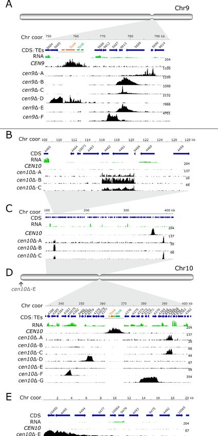
Research article                                                                             Chromosomes and Gene Expression

                                      Infrequently, centromeres can be spontaneously inactivated, resulting in neocentromere forma-
                                  tion (i.e., evolutionary new centromeres) (Ventura et al., 2007). Neocentromere formation can occur
                                  either while the native centromeric sequence is still present on the chromosome or when the native
                                  centromere has been mutated or deleted (e.g., from chromosomal rearrangements or g irradiation
                                  damage Burrack and Berman, 2012; Tolomeo et al., 2017; Ventura et al., 2007). In addition, sev-
                                  eral studies have described neocentromere formation after deletion of native centromeres by molec-
                                  ular genetic engineering in fungi, chickens, and Drosophila (Alkan et al., 2007; Ishii et al., 2008;
                                  Ketel et al., 2009; Shang et al., 2013). In some organisms, the formation of neocentromeres can be
                                  deleterious, leading to disease, cancer, or infertility (Burrack and Berman, 2012; Garsed et al.,
                                  2014; Nergadze et al., 2018; Scott and Sullivan, 2014; Warburton, 2004). For example, human
                                  neocentromeres are often identified in liposarcomas (Garsed et al., 2014). However, neocentromere
                                  formation also can be beneficial, leading to speciation (Ventura et al., 2007).
                                      Fungal neocentromeres are well described in the diploid yeast C. albicans and the haploid fission
                                  yeast S. pombe (Ishii et al., 2008; Ketel et al., 2009; Thakur and Sanyal, 2013). Deletion of C. albi-
                                  cans native centromere 5 or 7 has been shown to induce neocentromere formation and does not
                                  result in chromosome loss (Ketel et al., 2009; Thakur and Sanyal, 2013). In these cases, neocentro-
                                  meres conferred chromosomal stability similar to the native centromere (Ketel et al., 2009;
                                  Mishra et al., 2007). Deletion of a native centromere in S. pombe led to either neocentromere for-
                                  mation or chromosome fusion (Ishii et al., 2008; Ohno et al., 2016). S. pombe neocentromeres
                                  formed in telomere-proximal regions near heterochromatin, and neocentromere organization fea-
                                  tured a CENP-A-enriched core domain and heterochromatin at the subtelomeric (distal) side. Inter-
                                  estingly, neocentromere formation occurred at the same regions in both wild-type and
                                  heterochromatin-deficient strains, suggesting that heterochromatin is dispensable for neocentro-
                                  mere formation in S. pombe, although the rate of survival by chromosome fusion was significantly
                                  increased in heterochromatin-deficient mutants (Ishii et al., 2008). Deletion of kinetochore proteins
                                  (mhf1D and mhf2D) led to a shift of CENP-A binding, resulting in a CENP-A-enriched region directly
                                  adjacent to the native centromere (Lu and He, 2019).
                                      In some cases, neocentromeres span genes that are silenced, such as the neocentromeres in C.
                                  albicans. However the mechanisms that mediate silencing of neocentromeric genes are unknown in
                                  C. albicans, as proteins that are necessary for heterochromatin formation and gene silencing in other
                                  species ( HP1, Clr4, and DNA methyltransferase) are absent in C. albicans (Ketel et al., 2009). Neo-
                                  centromeres of S. pombe can also span genes. These genes are upregulated during nitrogen starva-
                                  tion and expressed at low levels during stationary growth in wild-type cells, but are silenced under
                                  all conditions tested when spanned by neocentromeres. In addition to neocentromeric genes, genes
                                  located within native centromeres have been identified in other fungi as well as rice and chicken
                                  (Nagaki et al., 2004; Schotanus et al., 2015; Shang et al., 2013).
                                      Recently, the centromeres of the human pathogenic fungus Cryptococcus deuterogattii were
                                  characterized and compared to those of the closely related species Cryptococcus neoformans (cen-
                                  tromeres ranging from 27 to 64 kb), revealing dramatically smaller centromeres in C. deuterogattii
                                  (ranging from 8.7 to 21 kb) (Janbon et al., 2014; Yadav et al., 2018). C. deuterogattii is responsible
                                  for an ongoing cryptococcosis outbreak in the Pacific Northwest regions of Canada and the United
                                  States (Fraser et al., 2005). In contrast to the sister species C. neoformans, C. deuterogattii com-
                                  monly infects immunocompetent patients (Fraser et al., 2005). C. deuterogattii is a haploid basidio-
                                  mycetous fungus with 14 chromosomes (D’Souza et al., 2011; Farrer et al., 2015; Yadav et al.,
                                  2018). The dramatic reduction in centromere size in C. deuterogattii may be attributable to loss of
                                  the RNAi pathway (Farrer et al., 2015; Yadav et al., 2018). The centromeres of C. deuterogattii
                                  consist of truncated transposable elements, and active transposable elements are missing through-
                                  out the genome (Yadav et al., 2018). This is in stark contrast to C. neoformans, which has active
                                  transposable elements in centromeric regions (Dumesic et al., 2015; Janbon et al., 2014;
                                  Yadav et al., 2018).
                                      Neocentromeres are frequently formed near genomic repeats, yet C. deuterogattii lacks active
                                  transposons that might seed neocentromere formation. Thus, C. deuterogattii is a unique organism
                                  in which to study centromere structure and function. To elucidate centromeric organization, the
                                  native centromeres of chromosomes 10 and 9 were deleted, leading to characterization of the first
                                  neocentromeres in the Basidiomycota phylum of the fungal kingdom.

Schotanus and Heitman. eLife 2020;9:e56026. DOI: https://doi.org/10.7554/eLife.56026                                             2 of 26
Research article Chromosomes and Gene Expression
Results
Deletion of centromere 9 and 10 results in neocentromere formation
To determine if neocentromere formation occurs in the C. deuterogattii reference strain R265, either
centromere 9 or 10 was deleted. Centromere 9 (CEN9) was deleted by CRISPR-Cas9-mediated
transformation. Two guide RNAs flanking the centromere were used and CEN9 was replaced with a
NAT dominant drug-resistance gene by homologous recombination. Biolistic transformation was
used to replace centromere 10 (CEN10) with either the NAT or NEO dominant drug-resistance gene
via homologous recombination. Viable transformants with the correct integration and deletion were
obtained and confirmed by 5’ junction, 3’ junction, loss of deleted regions, and spanning PCRs as
well as Southern blot analysis for cen10D (Figure 1—figure supplement 1, Figure 1—figure supple-
ment 2). Multiple independent cen9D and cen10D deletion mutants (cen9D-A to -F and cen10D-A to
-G) were obtained from independent transformations. Pulsed-field gel electrophoresis (PFGE) con-
firmed that cenD mutants had a wild-type karyotype and that chromosome 9 and 10 remained linear,
because a circular chromosome would not have entered the gel (Figure 1—figure supplement 3).
The formation of neocentromeres on chromosome 10 in C. deuterogattii was infrequent. A total
of 99 independent biolistic transformations resulted in only seven confirmed cen10D mutants (7/21
total candidate transformants, 33% homologous integration), suggesting that CEN10 deletion is
lethal in most circumstances. In comparison, deletion of nonessential genes by homologous recombi-
nation in the C. deuterogattii R265 strain typically results in ~100 colonies with a high success rate
(~80–90% homologous integration). We estimate that the likelihood of deleting a centromere and
recovering a viable colony is at least 1000-fold lower than would be expected from the deletion of a
non-essential gene. The deletion of CEN9 was more efficient as this was mediated by CRISPR-Cas9
cleavage with two guide RNAs and a repair allele.
Chromatin immunoprecipitation of mCherry-CENP-A followed by high-throughput sequencing
(ChIP-seq) for six cen9D (-A to -F) and seven cen10D mutants (-A to -G) was performed (Figure 1).
Prior to the ChIP-seq experiment, all of the centromere deletion mutants were streak purified from
single colonies. The sequence reads were mapped to a complete whole-genome assembly, followed
by the normalization of the reads by subtraction of the input from the ChIPed sample (Yadav et al.,
2018). To quantify the ChIP-seq data, the CENP-A-enriched regions were compared with the centro-
meres previously identified based on CENP-C enrichment. Both the CENP-A- and CENP-C-enriched
peaks were congruent for all of the native centromeres (Yadav et al., 2018). This analysis identified
13 of the 14 native centromeres (CEN1-8, CEN11-14 and depending on the centromere mutant
either CEN9 or CEN10), indicating that, as expected, the native centromere of chromosome 9 or 10
was missing in all of the cen9D and cen10D deletion mutants respectively (Figure 1). Instead, neo-
centromeres were observed.
Except for the neocentromere of isolate cen10D-E, the neocentromeres formed in close proximity
to the native centromere (CEN9 and CEN10). Almost all neocentromeres were shorter than the
native centromere, with the exception of cen10D-G which was larger than native centromere 10
(Table 1).
In three of the independent cen9D mutants (cen9D-B, -C and -E), neocentromeres formed at the
same chromosomal location (Figure 1A). Interestingly, two independent cen10D mutants (cen10D-A
and cen10D-C) contained two CENP-A-enriched regions on chromosome 10, with a primary peak
and a smaller secondary peak with reduced levels of CENP-A (1.3- to 1.75-fold lower) compared to
the primary CENP-A peak (Figure 1C). The chromosomal location of the secondary peak was similar
to the neocentromere of cen10D-B (which had only one neocentromere) (Figure 1B).
The two CENP-A-enriched regions suggest four possible models: 1) aneuploidy in which cells har-
bor two chromosomes, 2) a dicentric chromosome with two neocentromeres (neodicentric), 3) insta-
bility between two different neocentromere states (neocentromere switching), 4) or only one CENP-
A-enriched region functions as a centromere and the second CENP-A-enriched region is not bound
by the kinetochore (Figure 1).
The neocentromeres were located in unique, nonrepetitive sequences and were not flanked by
repetitive regions. The GC content of neocentromeres is similar to the overall GC content of chro-
mosome 9 and 10, whereas the native centromere has a lower GC content (Table 1). Comparing the
reference genome with de novo genome assemblies of cen10D-A, cen10D-B, and cen10D-E con-
firmed that transposable elements did not enter these genomic regions during neocentromere
Schotanus and Heitman. eLife 2020;9:e56026. DOI: https://doi.org/10.7554/eLife.56026 3 of 26Research article Chromosomes and Gene Expression Figure 1. Centromere deletion leads to neocentromere formation. For each panel, the chromosome coordinates are indicated. Genes (CDS) are shown in blue arrows and the truncated transposable elements, located in the native centromere (CEN9 or CEN10), are colored according to their class (Tcn4 in orange and Tcn6 in green). Previously generated RNA-sequencing obtained from wild-type cells was re-mapped and shown in green. In each panel, the wild-type CENP-A content is shown. In the wild type, CENP-A is only enriched at the native centromeres. For each cenD mutant, the Figure 1 continued on next page Schotanus and Heitman. eLife 2020;9:e56026. DOI: https://doi.org/10.7554/eLife.56026 4 of 26
Research article                                                                                    Chromosomes and Gene Expression

Figure 1 continued
neocentromeric region is shown by enrichment of CENP-A and the fold enrichment is indicated on the right of each ChIP-seq track. (A) Schematic full
overview of chromosome 9, the indentation represents the native centromere 9 position. The light grey area points to the zoomed-in chromosomal
region shown with the detailed view of the native centromere (CEN9) and the location of the cen9D mutant neocentromeres. Neocentromeres of
cen9D-B, cen9D-C and cen9D-E formed at the same chromosomal location. The dark gray line, below the transposable elements, indicates the deleted
region in the cen9D mutants (B) Detailed view of the neocentromere of cen10D-B and the secondary CENP-A peak of cen10D-A and cen10D-C. (C)
Overview of the chromosomal 10 region spanning 100 to 410 kb. cen10D-A and cen10D-C have two regions enriched with CENP-A (primary and
secondary). (D) Schematic full overview of the full chromosome 10, the indentation represents the chromosomal location of the native centromere
(CEN10). The light grey areas point to the zoomed-in chromosomal regions shown in panel C and below. The neocentromere of cen10D-E is indicated
with an arrow. Lower panel, detailed view of the native centromere (CEN10) and the neocentromeres formed in cen10D-A, cen10D-C, cen10D-D,
cen10D-F and cen10D-G mutants. The dark gray line, below the transposable elements, indicates the deleted region in the cen10D mutants (E) Detailed
view of the telocentric neocentromere of cen10D-E.
The online version of this article includes the following figure supplement(s) for figure 1:
Figure supplement 1. Confirmation of centromere 9 and 10 deletion by PCR.
Figure supplement 2. Centromere 10 is deleted in cen10D isolates.
Figure supplement 3. cen9D and cen10D mutants have a wild-type karyotype.
Figure supplement 4. ChIP-qPCR with additional kinetochore proteins.
Figure supplement 5. cen10D mutants have elongated cell morphology.

                                    formation (Supplementary file 1). Instead of spanning repeats and transposable elements like the
                                    native centromeres, neocentromeres span genes.
                                        All of the neocentromeres of chromosome 9 formed in a region within 26 kb of the chromosomal
                                    location of the native centromere 9 (Figure 1A). Interestingly, the neocentromeres of the indepen-
                                    dent cen9D-B, cen9D-C, and cen9D-E mutants all formed at the same chromosomal location and had
                                    the same length (4.41 kb); these neocentromeres spanned three genes. One gene was completely
                                    covered by CENP-A and this gene encodes a transglycosylase SLT domain-containing protein. The
                                    two other genes (a gene encoding a xylosylphosphotransferase and a gene encoding glutamate syn-
                                    thase (NADPH/NADH)) were partially covered with CENP-A. Mutant cen9D-A had a 3.87 kb long
                                    neocentromere located 26 kb 3’ to the native centromere and spanned two genes. The first gene
                                    was completely spanned by CENP-A and encodes an ESCRT-II complex subunit (Vps25) protein. The
                                    second gene was only partially covered and encodes an iron regulator protein. The neocentromere
                                    of cen9D-D was located directly to the left of the native centromere and was 4.37 kb in length. This
                                    neocentromere spanned two genes, coding for a hypothetical protein (92% covered by CENP-A)
                                    and a gene encoding for Derlin-2/3 that was completely covered by CENP-A. Lastly, the neocentro-
                                    mere of mutant cen9D-F was 3.83 kb in length and spanned one gene (encoding a xylosylphospho-
                                    transferase, XPT1), which was completely covered by CENP-A. This neocentromere was located 12
                                    kb away (3’) from the native centromere 9.
                                        Like the neocentromeres of cen9D mutants, the neocentromeres of cen10D mutants also spanned
                                    genes and interestingly, the kinetochore protein CENP-C was located inside the neocentromere of
                                    cen10D-B and in the secondary peak of cen10D-A and -C (Table 1, Figure 1B). The neocentromere
                                    in cen10D-B spanned 4.46 kb, was located 242 kb away from the 3’ region of the native CEN10, and
                                    was located 115 kb from the telomere (Figure 1B). In addition to the gene encoding CENP-C, the
                                    CENP-A-enriched region spanned a hypothetical protein (Table 1). The primary CENP-A-enriched
                                    region of cen10D-A and cen10D-C spanned a gene encoding a serine/threonine-protein phosphatase
                                    2A activator 2 (RRD2) and a hypothetical protein (Figure 1D). This neocentromere spanned 2.85 kb
                                    and was located closer to the native CEN10 (21 kb from the native centromere) than the neocentro-
                                    mere of cen10D-B and the secondary CENP-A peak of cen10D-A and cen10D-C. The neocentromere
                                    of cen10D-D was the smallest neocentromere (2.5 kb) and partially (88.4%) spanned a gene encoding
                                    a Ser/Thr protein kinase and formed 7.4 kb from the location of the native CEN10 (Figure 1D). The
                                    neocentromere of cen10D-E spanned two hypothetical proteins, was 4.38 kb in length and was
                                    located directly adjacent to the right telomere (Figure 1E). Mutant cen10D-F had a neocentromere
                                    of 2.64 kb, which spanned one hypothetical gene completely and two genes (hypothetical and a
                                    hexokinase (HXK1)) partially; the neocentromere formed at a chromosomal location 20 kb 5’ of the
                                    native centromere (Figure 1D). The neocentromere of cen10D-G was the largest neocentromere
                                    with a CENP-A-enriched region of 7.97 kb, and was in fact larger than the native CEN10. This

Schotanus and Heitman. eLife 2020;9:e56026. DOI: https://doi.org/10.7554/eLife.56026                                                        5 of 26
Research article Chromosomes and Gene Expression
Table 1. Genes located inside neocentromeres.
The chromosomal locations, sizes, and GC content (%) for the native centromere and cenD mutants are shown. For the neocentro-
meres, gene ID, predicted function, and the amount of CENP-A coverage are indicated.
Size
compared
to native
Size centromere GC Genes spanned % covered by Exons inside
Chr coor (bp) (kb) (%) % by neocentromere Gene ID Neocentromere neocentromere
Native Chr9:755,771– 6.84 - 43.6 - - - -
centromere 762,621
9
cen9D-A Chr9:785,352– 3.87 56.6 46.1 Escrt-II complex CNBG_5690 100
789,247 subunit (VPS25)
Iron regulator 1 CNBG_9614 14.6 Last exon
cen9D-B Chr9:775,164– 4.41 64.5 46.6 Xylosylphosphotransferase CNBG_5687 6.9
780,756 (XPT1)
Transglycosylase SLT CNBG_9613 100
domain-containing protein
Glutamate synthase CNBG_5689 33.7
(NADPH/NADH)
cen9D-C Chr9:775,164– 4.41 64.5 46.6 Xylosylphosphotransferase CNBG_5687 6.9
780,756 (XPT1)
Transglycosylase SLT CNBG_9613 100
domain-containing protein
Glutamate synthase CNBG_5689 33.7
(NADPH/NADH)
cen9D-D Chr9:750,902– 4.37 63.9 41.9 Hypothetical protein CNBG_5684 92.8
755,294
Derlin-2/3 CNBG_5685 100
cen9D-E Chr9:775,164– 5.56 81.3 50 Xylosylphosphotransferase CNBG_5687 6.9
780,756 (XPT1)
Transglycosylase SLT CNBG_9613 100 Last exon
domain-containing protein
Glutamate synthase CNBG_5689 33.7 Last exon
(NADPH/NADH)
cen9D-F Chr9:771,614– 3.83 56.0 51.5 Xylosylphosphotransferase CNBG_5687 100
775,469 (XPT1)
Native Chr10:362,876– 6.77 - 42.6 - - - -
centromere 369,657
10
cen10D-A Chr10:115,954– 4.46 65.9 46.9 CENPC/MIF2 CNBG_4461 88.3 1, 2, 3, 4 (only
120,422 5th is outside)
Hypothetical protein CNBG_4462 100
Chr10:391,090– 2.85 42.1 48.9 Serine/threonine-protein CNBG_9459 10.6 Last exon (5th)
393,946 phosphatase 2A
activator 2(RRD2)
Hypothetical protein CNBG_4366 100
Hypothetical protein CNBG_4365 23.4 Last exon (3th)
cen10D-B Chr10:115,954– 4.46 65.9 46.9 CENPC/MIF2 CNBG_4461 88.3 1, 2, 3, 4 (only
120,422 5th
is outside)
Hypothetical protein CNBG_4462 100
Table 1 continued on next page
Schotanus and Heitman. eLife 2020;9:e56026. DOI: https://doi.org/10.7554/eLife.56026 6 of 26Research article Chromosomes and Gene Expression
Table 1 continued
Size
compared
to native
Size centromere GC Genes spanned % covered by Exons inside
Chr coor (bp) (kb) (%) % by neocentromere Gene ID Neocentromere neocentromere
cen10D-C Chr10:115,954– 4.46 65.9 46.9 CENPC/MIF2 CNBG_4461 88.3 1, 2, 3, 4 (only
120,422 5th
is outside)
Hypothetical protein CNBG_4462 100
Chr10:391,090– 2.85 42.1 48.9 Serine/threonine-protein CNBG_9459 10.6 Last exon (5th)
393,946 phosphatase 2A
activator 2(RRD2)
Hypothetical protein CNBG_4366 100
Hypothetical protein CNBG_4365 23.4 Last exon (3th)
cen10D-D Chr10:352,648– 2.51 37.1 48 Ser/Thr protein kinase CNBG_4379 88.4
355,154
cen10D-E Chr10:1–4,385 4.38 64.7 53.2 Hypothetical protein CNBG_10450 100
Hypothetical protein CNBG_4495 100
cen10D-F Chr10:342,517– 2.64 39.0 45.5 Hypothetical protein CNBG_4383 18.6 Last two exons
345,159
Hypothetical protein CNBG_10075 100
Hexokinase (HXK1) CNBG_4382 15.3 Last three exons
cen10D-G Chr10:378,389– 7.97 117.7 46.5 High osmolarity CNBG_4373 100
386,366 signaling protein (SHO1)
Hypothetical protein CNBG_4372 100
Hypothetical protein CNBG_4371 100
Hypothetical protein CNBG_4370 100
neocentromere spanned four genes, including a gene coding for a high osmolarity protein (Sho1)
and three genes coding for hypothetical proteins (Figure 1D).
To test if the kinetochore was binding to the CENP-A-enriched regions of chromosomes 9 and
10, and to validate if the neocentromeres were fully functional as centromeres, two additional kinet-
ochore proteins were epitope-tagged with GFP. cen9D mutants were transformed with an overlap
PCR product expressing CENPC-GFP. As the neocentromeres of three cen10D mutants spanned the
gene encoding CENP-C, all cen10D mutants were transformed with an overlap PCR product,
expressing Mis12-GFP. In addition to the cen9D and cen10D mutants, the wild type was transformed
with constructs expressing Mis12-GFP and CENP-C-GFP, and these served as controls. ChIP-qPCRs
for cen9D mutants, cen10D mutants, and wild-type strains with Mis12-GFP or CENP-C-GFP were per-
formed (Figure 1—figure supplement 4). Because Mis12 is an outer kinetochore protein, the form-
aldehyde cross-linking was extended to 45 min (15 min was used for CENP-A and CENP-C) for this
protein. For all qPCR analyses, the native centromere 6 CEN6) was used as an internal control and
for each neocentromere specific primer pairs were designed. For cen9D and cen10D mutants a simi-
lar level of Mis12 or CENP-C enrichment at the neocentromeres and (CEN6) was observed. This sug-
gested that the CENP-A-enriched regions of chromosome 9 of the cen9D mutants and chromosome
10 of cen10D mutants identified by ChIP-seq were functional centromeres and indeed neocentro-
meres (Figure 1—figure supplement 4).
Previously generated RNA sequence data were remapped to the C. deuterogattii reference strain
R265 and analyzed to determine if the regions where neocentromeres formed in the cenD mutants
were transcribed in the wild type (Figure 1; Supplementary file 2; Schneider et al., 2012). In the
wild-type strain, all genes spanned by neocentromeres in the cenD mutants were expressed (Fig-
ure 1). However, the expression levels of the neocentromeric genes were lower than their neighbor-
ing genes. For example, the expression level of the gene (CNBG_5686) flanking native centromere 9
was three times higher than the genes spanned by neocentromeres in cen9D mutants (Figure 1A).
Schotanus and Heitman. eLife 2020;9:e56026. DOI: https://doi.org/10.7554/eLife.56026 7 of 26Research article Chromosomes and Gene Expression
The same trend was observed in the cen10D mutants. Here, the expression level of the gene
(CNBG_4365) 3’ flanking the neocentromere (primary CENP-A peak) of cen10D-A and cen10D-C was
more than six times higher than the genes spanned by the neocentromere (Figure 1D). Also, the
neocentromere of cen10D-D is flanked by genes whose expression was two times higher than the
genes spanned by the neocentromere (Figure 1D). This suggests that neocentromeres are formed
in chromosomal regions with lower gene expression in C. deuterogattii. The majority of the genes
(24/28) flanking native centromeres are transcribed in the direction towards the native centromere.
All of the neocentromeres observed span one or more genes and most of the flanking genes are
transcribed in the direction away from the neocentromere.
The expression levels of the neocentromeric genes in cen9D and cen10D mutants were assayed
by qPCR (Figure 2). The neocentromeric genes of chromosome 9 were normalized to actin. To com-
pensate for the ploidy levels of chromosome 10 in cen10D mutants, a housekeeping gene located
on chromosome 10 was used to normalize the expression of genes spanned by neocentromeres
located on chromosome 10. The expression levels of the CENP-A-associated neocentromeric genes
were all found to be similar to the wild-type strain (Figure 2).
Figure 2. Expression of neocentromeric genes. Expression of the neocentromeric genes was assessed by qPCR for all cenD mutants and expression is
shown as Log2DDCt. For cen10D-A, cen10D-B and cen10D-C, two genes were selected from each neocentromeric region, all other cenD mutants are
represented by one gene spanned by CENP-A. cen10D-B has only one CENP-A-enriched region, and in this case, the genes located within primary
peak of cen10D-A and cen10D-C served as controls. The qPCRs of cen10D mutants are normalized with a housekeeping gene located on chromosome
10. The qPCRs of cen9D mutants are normalized with actin. Error bars show standard deviation.
Schotanus and Heitman. eLife 2020;9:e56026. DOI: https://doi.org/10.7554/eLife.56026 8 of 26Research article Chromosomes and Gene Expression
Neocentromere formation can reduce fitness
cen10D mutants were noted to grow more slowly than wild type. To investigate this, the growth of
cen10D and wild-type strains was measured during the course of a 22-hour cell growth experiment
(Figure 3A). The majority of cen10D mutants exhibited slower growth rates compared to the wild-
type parental strain R265. Six of seven cen10D mutants exhibited significant fitness defects com-
pared to the wild-type strain, with doubling times ranging from 101 to 111 min compared to 81 min
for the wild type (Figure 3B). In contrast, one mutant, which has a telocentric neocentromere
(cen10D-E), grew similarly to the wild type and had a similar doubling time (84 min for the mutant vs
81 min for the wild-type strain). Compared to the wild type, cen10D mutants with increased doubling
times produced smaller colonies during growth on non-selective media (Figure 1—figure supple-
ment 5C).
To compare fitness, a competition assay was performed with 1:1 mixtures of wild-type and cen9D
or cen10D mutants grown in liquid YPD medium (Figure 3C). With no growth defect, the expecta-
tion was that the wild-type strain and centromere deletion mutants would grow at the same growth
rate, resulting in a 1:1 ratio. In fact, fewer cen10D cells were found in the population after growth in
competition with the wild-type strain, and this observation is consistent with the slower doubling
time of cen10D mutants resulting in reduced fitness compared to wild type (Figure 3). Compared to
Figure 3. cen10D mutant strains have reduced fitness compared to the wild-type strain. (A) Six out of seven cen10D mutants had a longer doubling
time and slower growth than the wild-type strain. In contrast cen10D-E grows similarly to the wild type. Error bars show standard deviation. (B) Doubling
times and fold change compared to wild type are shown. (C) Competition assays with the wild type and cen9D and cen10D mutant strains. Mixed
cultures (1:1) were grown overnight and plated with and without selection agents. After four days, colonies were counted and the percentage of cenD
mutants (black) and wild type (grey) in each culture was plotted. As a control (C) a wild-type strain with a NAT marker was mixed with the wild type.
The online version of this article includes the following figure supplement(s) for figure 3:
Figure supplement 1. cen10D mutants with chromosomal fusion have a wild-type growth rate.
Schotanus and Heitman. eLife 2020;9:e56026. DOI: https://doi.org/10.7554/eLife.56026 9 of 26Research article Chromosomes and Gene Expression
the wild-type cells, there were fewer cen9D mutant cells in the population. However, the number
was closer to a 1:1 ratio (Figure 3C). The ratio of the cen9D mutants in the population was similar to
the ratio of the cen10D-E mutant, which had a wild-type growth rate. Due to this observation, we
hypothesize that the growth rate of the cen9D mutants is similar to wild type.
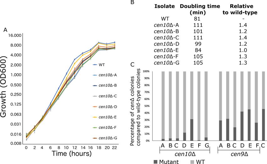
cen10D isolates are aneuploid
Because deletion of a centromere could lead to defects in chromosome segregation, cenD mutants
were assessed for aneuploidy (Figure 4). Overall, cen10D mutants exhibited a mixture of large and
small colony sizes during growth on YPD medium at 37˚C, while cen9D mutants exhibited a uniform,
wild-type like, colony size (Figure 1—figure supplement 5).
Aneuploidy in C. neoformans often leads to a similar mixed colony size phenotype as that
observed in the cen10D mutants (Sun et al., 2014). To exacerbate the aneuploidy-associated slow
growth phenotype, four cen10D mutants were grown at elevated temperature (37˚C), causing these
isolates to produce smaller, growth-impaired and larger, growth-improved colonies (Figure 3—fig-
ure supplement 1). Three small and two large colonies were selected from each isolate and whole-
genome analysis was performed based on Illumina sequencing. Sequences were mapped to the ref-
erence R265 genome, revealing that the small colonies were indeed aneuploid (Figure 4A). The
small colonies of cen10D-B and cen10D-C had ploidy levels for chromosome 10 in the range of 1.25-
to 1.36-fold higher compared to the other 13 chromosomes, which suggested that only a proportion
of the cells (25% to 36%), were aneuploid (Figure 4B). The remainder of the genome was euploid.
Chromosome 10 of the small colonies derived from isolate cen10D-A and cen10D-E exhibited ploidy
levels ranging from 1.1- to 1.14-fold, reflecting less aneuploidy. Importantly, for all of the large colo-
nies derived from isolates cen10D-A, cen10D-B, cen10D-C, and cen10D-E the fold coverage of chro-
mosome 10 was restored to the wild-type euploid level (1.0 fold compared to wild type). The ploidy
levels of chromosome 10 were 1-fold for all of the large colonies compared to wild type, indicating
that the ploidy level of chromosome 10 of the large colonies was restored to euploid.
cen10D chromosome is rescued by chromosome fusion
Based on whole-genome sequencing and PFGE analysis, fusion of cen10D chromosome 10 to other
chromosomes was a common event in the large colonies (Figure 5, Figure 6). Whole-genome
sequence analysis revealed that sequences corresponding to the 3’ subtelomeric region of chromo-
some 10 (including one gene) were absent in the sequences obtained from all of the large colonies
analyzed (Figure 4—figure supplement 1A). In addition, the large colonies of cen10D-A were miss-
ing sequences for two genes in the 5’ subtelomeric region of chromosome 4 (Figure 4—figure sup-
plement 1B). Large colonies of cen10D-B were missing 18.5 kb at the 5’ subtelomere of
chromosome 7 (including eight genes) (Figure 4—figure supplement 1C). The large colonies of
cen10D-E lacked a small part of one gene in the 3’ subtelomeric region of chromosome 1. In total, of
the 14 subtelomeric genes that were lost in these three chromosome-fusion isolates, ten encoded
hypothetical proteins and four encoded proteins with predicted functions. BlastN analysis in the de
novo genome assemblies of the large colonies confirmed that the subtelomeric regions were not
located on minichromosomes or inserted in other chromosomes. Seven genes have homologs in C.
neoformans and are present in C. neoformans deletion libraries (Liu et al.,
2008; Supplementary file 3). This observation suggested that either subtelomeric deletions
occurred, or that chromosomal fusions led to the loss of subtelomeric regions. Notably, sequences
from the small colonies spanned the entire genome with no evidence of these subtelomeric dele-
tions (Figure 4—figure supplement 1F).
We hypothesized that the subtelomeric gene loss was due to chromosomal fusion and tested this
hypothesis with de novo genome assemblies and PFGE (Figure 5 and Figure 6). Based on de novo
genome assemblies for the large colonies of cen10D-A, cen10D-B, and cen10D-E, chromosome 10
fused with chromosome 4, 7, or 1, respectively (Figure 5 and Supplementary file 3). In the large col-
ony of cen10D-A (cen10D-A-L1), the fusion occurred between chromosome 10 and chromosome 4
(Figure 5A). Chromosomal fusion led to the loss of the CNBG_10211 gene (on chromosome 4), and
the fusion junction was within the CNBG_6174 gene of chromosome 4. For cen10D-B-L1, the chro-
mosomal fusion occurred between chromosomes 10 and 7 (Figure 5B). Seven genes of chromosome
7 were lost in the fused chromosome. The chromosome fusion junction was intergenic on
Schotanus and Heitman. eLife 2020;9:e56026. DOI: https://doi.org/10.7554/eLife.56026 10 of 26Research article Chromosomes and Gene Expression Figure 4. cen10D mutants are aneuploid. The whole genomes of small and large colonies derived from four cen10D mutants were sequenced and read coverage (corresponding to ploidy levels) was plotted. Small colonies of cen10D mutants were partially aneuploid for chromosome 10, while the large colonies are euploid. (A) Genome-wide read depth coverage for small and large colonies. On the right, the fold coverage for the highest ploidy level is indicated for each sample. For example, chromosome 10 of cen10D-B-S1 had an aneuploidy level of 1.35-fold compared to the wild-type strain. Chromosome 4 had a small region with increased read depth due to the ribosomal rDNA gene cluster and was excluded from the analysis. Chromosome 8 of cen10D-E was duplicated. In addition, cen10D-E-S3 had an additional duplicated region of 162 kb of chromosome 5 that spans the sequence of native centromere 5. (B) Detailed view of read depth of chromosome 10. As in panel A, read depth is indicated on the right. The native centromeric location is shown by a black square. Due to the deletion of centromere 10, the location of the native centromere lacks sequence reads for each sample. The online version of this article includes the following figure supplement(s) for figure 4: Figure 4 continued on next page Schotanus and Heitman. eLife 2020;9:e56026. DOI: https://doi.org/10.7554/eLife.56026 11 of 26
Research article                                                                                    Chromosomes and Gene Expression

Figure 4 continued
Figure supplement 1. Deletion within subtelomeric regions in chromosome fusion isolates.

                                   chromosome 7. cen10D-E-L1 was due to a chromosomal fusion between chromosomes 10 and 1
                                   (Figure 5C). The fusion was intragenic for both chromosomes. The fusion point occurred in
                                   CNBG_6141 on chromosome 10 and CNBG_10308 on chromosome 1.
                                      Because all of the large cen10D colonies had chromosome 10 fusions, we examined the fusion
                                   location on chromosome 10 in detail. The fusions occurred 1.7, 0.3, and 3.6 kb from the chromo-
                                   some 10 gene CNBG_6142, respectively (Figure 5—figure supplement 1). The fusion occurred in
                                   unique DNA sequences and was not flanked by repetitive regions. The overlapping region between
                                   chromosome 10 and the fused chromosome was at most 6 bp, suggesting that these fusions

Figure 5. cen10D mutants undergo chromosome fusion leading to improved fitness at 37˚C. Chromosomal fusions were studied in detail for three
cen10D mutants restored to wild-type growth levels at 37˚C (large colonies). After chromosome fusion, the fused chromosomes of cen10D-A-L and
cen10D-B-L lost the gene CNBG_6141, which is located in the 3’ subtelomeric region of chromosome 10. Genes present in the fused chromosome are
depicted in green, and genes lost after chromosome fusion are indicated in red. Gray highlights indicate regions present in both the parental and
fused chromosomes. Each fusion occurred in a unique nonrepetitive region. (A) cen10D-A-L1, the fusion occurred between chromosome 10 and
chromosome 4. (B) In cen10D-B-L1, chromosomal fusion occurred between chromosomes 10 and 7. (C) cen10D-E-L1 chromosomal fusion occurred
between chromosomes 10 and 1.
The online version of this article includes the following figure supplement(s) for figure 5:
Figure supplement 1. Chromosome fusion in large cen10D colonies.

Schotanus and Heitman. eLife 2020;9:e56026. DOI: https://doi.org/10.7554/eLife.56026                                                      12 of 26
Research article Chromosomes and Gene Expression
Figure 6. Chromosome fusion results in neocentromere inactivation and karyotype reduction. (A) Neocentromeres are inactive after chromosomal
fusion. For each neocentromere two qPCR primer pairs located in genes spanned by the neocentromere in cen10D-A and cen10D-B mutant were used
in a ChIP-qPCR experiment. Analyzed is the CENP-A enrichment of 1) a cen10D mutant, 2) a large colony derived from the cen10D mutant, and 3) the
wild-type strain. Centromere 6 (CEN6) was included as a positive control, and actin was included as a negative control. Data are shown for cen10D-A,
cen10D-A-L1, cen10D-B, cen10D-B-L2, and wild type. For cen10D-A and cen10D-A-L1 mutants, the chromosomal regions investigated are indicated
according to the primary and secondary CENP-A peaks of the cen10D-A mutant. The cen10D-B mutant has only one CENP-A-enriched region which co-
localized with the secondary CENP-A peak of cen10D-A and this region is labeled with neocen in cen10D-B and cen10D-B-L1. Error bars show standard
deviation. (B) PFGE analysis shows that the band corresponding to chromosome 10 was lost in the large colonies and instead larger bands appear due
to the fusion of chromosome 10 with other chromosomes. cen10D deletion mutants and small colonies derived from 37˚C show a wild-type karyotype.
Chromosome 10 of the large colonies was fused to chromosome 13, 10, or 1, respectively. Due to limitations of PFGE conditions, the chromosome 10–
chromosome 1 fusion did not separate from chromosomes 2, 3, and 4. The positions of the fused chromosomes are indicated with arrows.
occurred via microhomology-mediated end joining (MMEJ) (also known as alternative nonhomolo-
gous end-joining [Alt-NHEJ]).
Chromosome fusion may result in loss of the neocentromeres, and the kinetochore may bind to
the native centromere of the fused chromosome and function as the active centromere. This hypoth-
esis was tested by performing ChIP-qPCR for CENP-A binding (Figure 6A). For each neocentromere
(either of the cen10D-A or cen10D-B mutant), CENP-A enrichment was tested with four primer sets
located in the neocentromere. The CENP-A enrichment for these four locations was tested in 1) the
initial cen10D mutant, 2) a large colony derived from a specific cen10D mutant and 3) wild type
(Figure 6A). As expected, the ChIP-qPCR analysis showed CENP-A enrichment for the neocentro-
meres of the initial cen10D-A and cen10D-B mutants. The neocentromeric regions of cen10D-A and
cen10D-B were not enriched with CENP-A in the wild-type strain, showing there was no occupancy
by CENP-A prior to neocentromere formation. For all analyzed cen10D chromosome 10 fusion iso-
lates, the neocentromeres were not CENP-A-associated, and were similar to the wild-type back-
ground levels. Therefore, the neocentromeres were no longer active in the chromosome fusion
strains (Figure 6A). This suggests that the native centromere of the fusion partner of chromosome
10 (i.e., chromosome 1, 4, or 7) was the active centromere of the Chr10-Chr1, Chr10-Chr4, and
Chr10-Chr7 fusions.
In addition to cen10D-A, -B, and -E mutants, whole-genome sequencing was performed for two
large colonies of cen10D-C. Although it was not possible to identify the chromosome fusion based
on whole-genome sequencing data for either of the large colonies of the cen10D-C mutant, PFGE
analysis showed that cen10D-C-L1 had a fusion between chromosomes 10 and 13 (Figure 6b).
cen10D-C-L2 had read coverage of 1.99-fold for a region of ~200 kb of chromosome 10 (Figure 4B).
The rest of chromosome 10 was euploid, suggesting that the ~200 kb region was duplicated and
was either a single chromosome or fused to another chromosome in this isolate. PFGE analysis
Schotanus and Heitman. eLife 2020;9:e56026. DOI: https://doi.org/10.7554/eLife.56026 13 of 26Research article Chromosomes and Gene Expression
suggested that this fragment was duplicated on chromosome 10, resulting in a larger chromosome
(Figure 6B). In contrast to the other fused chromosomes, this chromosomal fragment did not fuse to
a chromosome with a native centromere, and the fact that the mutant still exhibited a fitness defect
was consistent with this interpretation. The larger chromosome was euploid, suggesting that the
unstable neocentromere(s), rather than causing aneuploidy, resulted in a fitness cost in this isolate.
Discussion
Composition of neocentromeres in C. deuterogattii
The native centromeres of C. deuterogattii are found in repetitive regions and are flanked by, but
do not contain, protein-encoding genes (Yadav et al., 2018). By contrast, neocentromeres of C.
deuterogattii span genes, lack repetitive elements, and like the native centromeres, are flanked by
genes. In general (with one exception), the neocentromeres of C. deuterogattii are significantly
shorter than the native centromeres, whereas most neocentromeres in other species have similar
lengths as the native centromeres.
Native centromeres of S. pombe have a central core that is enriched with CENP-A and flanked by
repetitive pericentric regions (Ishii et al., 2008). While neocentromere formation in S. pombe favors
repeats in the pericentric regions, neocentromere formation is possible without the repetitive peri-
centric regions (Ishii et al., 2008). The majority of the neocentromeres in C. albicans and chickens
are formed close to native centromeres due to seeding of CENP-A that is located near the native
centromere (the so-called CENP-A cloud) (Ketel et al., 2009; Shang et al., 2013). Our results and
the earlier reports discussed, suggest that the chromosomal location of the native centromere is the
main determinant of neocentromere formation. One exception was the neocentromere of cen10D-E,
which directly flanked the left telomere. Interestingly, this was the only cen10D mutant that had a
growth rate similar to wild type.
Several C. deuterogattii neocentromeres formed in the same location; however, there is no
apparent consensus between the different regions occupied by different neocentromeres. A similar
trend has been observed in neocentromere formation in C. albicans (Ketel et al., 2009). Evolution-
ary new centromeres (ECNs) in the largest crucifer tribe Arabideae originated several times inde-
pendently and are located in the same chromosomal location (Mandáková et al., 2020). Our results
suggest that neocentromeres form by mechanisms that do not rely on nearby transposable ele-
ments/repeats to initiate de novo centromere assembly.
Neocentromeric genes are expressed
Neocentromeres induced in several species can span genes, resulting in silencing or reduced gene
expression. For example, all genes within five independent neocentromeres in C. albicans that
spanned nine genes were suppressed (Burrack et al., 2016). In S. pombe, neocentromeres span
genes that are only expressed in response to nitrogen starvation in the wild-type strain, and neocen-
tromere formation silences these genes during nitrogen starvation (Ishii et al., 2008). The native
centromere 8 of rice contains an approximately 750 kb CENP-A-enriched region with four genes
that are expressed in both leaf and root tissues of three closely related species (Fan et al., 2011;
Nagaki et al., 2004). Neocentromeres of rice span genes that are expressed at similar levels as in
the wild type (Zhang et al., 2013). Chicken neocentromeres have been induced on chromosome Z
or 5 (Shang et al., 2013). Chromosome Z neocentromeres span eight genes, but in wild-type cells
only MAMDC2 is expressed during normal growth. The other seven genes were either not expressed
at any detectable level in all tested developmental stages or were only expressed during early
embryonic stages (Shang et al., 2013). When a neocentromere formed, expression of the
MAMDC2-encoding gene was reduced 20- to 100-fold. Chromosome 5 of chickens is diploid, and
neocentromeres on this chromosome span genes that are expressed. The hypothesis behind this
phenomenon is that one allele functions as a centromere, while the other allele codes for the genes.
Because the cen10D mutants of C. deuterogattii were aneuploid, the expression of genes
spanned by chromosome 10 neocentromeres was normalized to expression levels of a housekeeping
gene located on chromosome 10. The expression of genes enriched for CENP-A chromatin is similar
to that of wild type, and if the allelic hypothesis, like in chickens, were valid, the expectation would
be a 60% reduction in expression levels. The genes spanned by neocentromeres of cen9D mutants
Schotanus and Heitman. eLife 2020;9:e56026. DOI: https://doi.org/10.7554/eLife.56026 14 of 26Research article Chromosomes and Gene Expression
are also expressed at wild-type levels. As the cen9D mutants have uniform, wild-type colony sizes,
the ploidy levels of these mutants were not tested and we hypothesize that these mutants are hap-
loid/euploid.
Genes contained in regions in which C. deuterogattii neocentromeres formed in cenD mutants
were actively expressed in the wild-type strain, and this is similar to human neocentromeres that can
form in regions with or without gene expression (Alonso et al., 2010; Marshall et al., 2008). How-
ever, we have identified chromosomal regions that lack gene expression on chromosomes 9 and 10,
although these regions were not close to the native centromere.
Of the C. deuterogattii genes spanned by the neocentromere region, one encodes the kineto-
chore component CENP-C. Several independent biolistic transformations were performed to delete
the gene encoding CENP-C, but all attempts were unsuccessful. This suggests that CENPC is an
essential gene. In fission yeast, deletion of the gene encoding the CENP-C homolog Cnp3 was lethal
at 36˚C, but mutants were still viable at 30˚C (Suma et al., 2018). However, CENP-A was mislocal-
ized in the cnp3D mutants. Another gene partially located inside a C. deuterogattii neocentromere
encodes the serine/threonine-protein phosphatase 2A activator 2 (RRD2). The RRD2 homolog is not
essential in S. cerevisiae (Higgs and Peterson, 2005).
Compared with other haploid fungi, the neocentromeric genes of C. deuterogattii are similar to
the native centromeric genes of the haploid plant pathogenic fungus Zymoseptoria tritici. Z. tritici
has short regional centromeres with an average size of 10.3 kb, and 18 out of 21 native centromeres
have a total of 39 expressed genes (Schotanus et al., 2015).
cen10D mutants with two CENP-A-enriched regions
The appearance of two CENP-A-enriched regions of C. deuterogattii cen10D mutants could be
explained in a few ways. First, neocentromere formation could lead to a dicentric chromosome 10 in
which the centromeres may differ in functional capacity. Dicentric chromosomes are not by definition
unstable, for example the dominant-negative mutation of the mammalian telomere protein TRF2
results in chromosome fusions, leading to the formation of dicentric chromosomes (Stimpson et al.,
2010). The formation of dicentric chromosomes occurred in 97% of the fused mammalian chromo-
somes, which were stable for at least 180 cell divisions (Stimpson et al., 2010). Several microscopic
studies showed that chromosomes with two regions of centromere-protein enrichment are stable
(Higgins et al., 2005; Stimpson et al., 2012; Stimpson et al., 2010; Sullivan and Willard, 1998).
This suggests that a dineocentric chromosome 10 could be stable in the population. Second, the
two CENP-A-enriched peaks could be the result of a mixed population and either due to an unstable
primary neocentromere and/or aneuploidy. The primary neocentromere could be associated with
the majority of the cells, whereas the secondary CENP-A peak would be only found in a small num-
ber of cells (and the primary neocentromere is lost in these isolates). This is reflected by lower
CENP-A enrichment for the secondary peak, and the hypothesis of putative dicentrics is due to a
mixture of alleles in the population. Third, the neocentromeres could be unstable, which could lead
to the formation of two CENP-A-enriched regions with centromere function switching between the
regions. However, our data would argue against this latter model. Prior to the ChIP-seq analysis of
the cen10D mutants, colonies were isolated by streak purification (eight times), suggesting that the
presence of two distinct CENP-A peaks occurs continuously.
cen10D mutants are partially aneuploid
Neocentromere formation in chickens results in a low number of aneuploid cells (Shang et al.,
2013). Based on whole-genome sequencing of a population of cells, the C. deuterogattii cen10D iso-
lates are partially aneuploid for chromosome 10. For fully aneuploid isolates, the coverage of Illu-
mina reads is expected to be 2-fold; the cen10D isolates with two CENP-A peaks showed
aneuploidy levels up to 1.28-fold or were even euploid. This suggests that, like the chicken neocen-
tromeric isolates, only a small number of cells in a population of C. deuterogattii cen10D isolates are
aneuploid.
cen10D mutants have reduced fitness
In C. albicans, deletion of centromere 5 results in neocentromere formation, and these isolates have
fitness similar to the wild-type strain (Ketel et al., 2009). Similar results were reported for
Schotanus and Heitman. eLife 2020;9:e56026. DOI: https://doi.org/10.7554/eLife.56026 15 of 26Research article Chromosomes and Gene Expression
neocentromeres in chicken and S. pombe, in which strains with neocentromeres or chromosome
fusion have a growth rate similar to the wild-type strain (Ishii et al., 2008; Shang et al., 2013).
If centromere deletions occurred in nature, we hypothesize that the wild type would outcompete
all of the cenD isolates. The virulence of the cenD mutants was not assayed. Based on reduced fitness
of the cenD mutants, we hypothesize that pathogenicity of the cenD mutants would be lower than
the wild type. However, when chromosome fusion occurs the growth rate is restored to a near wild-
type level and we hypothesize that the isolates with 13 chromosomes could have virulence similar to
the wild type. Several genes were lost due to the fusion events in the cenD mutants; to our knowl-
edge these lost genes have not been associated with pathogenicity of C. deuterogattii.
Neocentromere stains exhibit impaired growth and chromosome fusion
restores wild-type growth at elevated temperatures
Deletion of a centromere in S. pombe leads to either neocentromere formation or chromosome
fusion due to a noncanonical homologous recombination pathway (Ishii et al., 2008; Ohno et al.,
2016). This is in contrast to neocentromere formation in C. deuterogattii, which results in 100% neo-
centromere formation. Based on PFGE analysis, the karyotype of the cenD isolates is wild type at 30˚
C, but chromosome fusion can occur at 37˚C within the cen10D mutants and lead to improved
growth at 30˚C.
The location of the cen10D neocentromere had no apparent influence on the ability to undergo
chromosome fusion as we have shown with a telocentric neocentromere, a dicentric mutant, and a
neocentromere located 118 kb away from the telomere.
The fused chromosomes have no or only short homology at the breakpoints that is insufficient for
homologous recombination, suggesting that the chromosome fusions arise via MMEJ. Future experi-
ments to test this hypothesis could involve deleting genes involved in the MMEJ pathway, such as
CDC9 and DNL4 (Sinha et al., 2016).
A prominent chromosome fusion occurred during the speciation of humans. Compared to other
great apes, humans have a reduced karyotype, which is due to the fusion of two ancestral chromo-
somes that resulted in chromosome 2 in modern humans, Denisovans, and Neanderthals
(Miga, 2017). Human chromosome2 still harbors signatures of telomeric repeats at the fusion point
(interstitial telomeric sequences [ITS]), suggesting that this chromosome is derived from a telomere-
telomere fusion. By synteny analysis, the inactive centromere of chimpanzee chromosome 2b can be
identified on human chromosome 2, and there are relics of a satellite DNA at this now extinct cen-
tromere (Miga, 2017). Moreover, a dominant-negative mutation of the human telomeric protein
TRF2 leads to telomere-telomere fusions, mainly between acrocentric chromosomes
(Stimpson et al., 2010; van Steensel et al., 1998). In the fungal species Malassezia, chromosome
breakage followed by chromosome fusion has led to speciation (Sankaranarayanan et al., 2020).
The short regional centromeres (3–5 kb) are fragile and this led most likely to chromosome reduc-
tion. By contrast in C. deuterogattii, the chromosomes involved in chromosomal fusion of the
cen10D mutants were all metacentric, and fusion occurred in non-telomeric sequences.
Another example of telomeric fusions is the presence of ITS regions in several genomes. In bud-
ding yeast, the experimental introduction of an ITS into an intron of the URA3 gene resulted in four
classes of chromosome rearrangements, including: 1) inversion, 2) gene conversion, 3) mini-chromo-
some formation due to deletion or duplication, and 4) mini-chromosome formation due to transloca-
tion (Aksenova et al., 2013). Based on our de novo genome assemblies of the C. deuterogattii
large-colony cen10D mutants, chromosome fusions occurred with no signs of chromosome rear-
rangements. Thus, these chromosome fusions did not produce ITS regions, which would otherwise
destabilize the genome.
Conclusions
Our work shows that, like in other model systems, neocentromeres can be induced in C. deuterogat-
tii. However, C. deuterogattii neocentromeres have several unique characteristics, such as spanning
genes whose expression is unaffected by centromere assembly. In some instances, deletion of
CEN10 led to chromosome fusion, resulting in enhanced fitness and leading to inactivation of the
neocentromere. Presumably, deletion of other centromeres could be carried out, leading to a C.
Schotanus and Heitman. eLife 2020;9:e56026. DOI: https://doi.org/10.7554/eLife.56026 16 of 26Research article Chromosomes and Gene Expression
deuterogattii strain with only one or a few chromosomes, as was recently reported in S. cerevisiae
(Luo et al., 2018; Shao et al., 2018).
Materials and methods
Key Resources Table Template and Guidelines.
Key resources table
Reagent type Additional
(species) or resource Designation Source or reference Identifiers information
Genetic reagent R265 This study R265 expressing
Cryptococcus mCherry-CENPA
deuterogattii
Genetic reagent cen104-A This study R265 centromere 10
Cryptococcus deletion mutant with
deuterogattii expressing mCherry-CENPA
Genetic reagent cen104-B This study R265 centromere 10
Cryptococcus deletion mutant with
deuterogattii expressing mCherry-CENPA
Genetic reagent cen104-C This study R265 centromere 10
Cryptococcus deletion mutant with
deuterogattii expressing mCherry-CENPA
Genetic reagent cen104-D This study R265 centromere 10
Cryptococcus deletion mutant with
deuterogattii expressing mCherry-CENPA
Genetic reagent cen104-E This study R265 centromere 10 deletion
Cryptococcus mutant with expressing
deuterogattii mCherry-CENPA
Genetic reagent cen104-F This study R265 centromere 10
Cryptococcus deletion mutant with
deuterogattii expressing mCherry-CENPA
Genetic reagent cen104-G This study R265 centromere 10
Cryptococcus deletion mutant with
deuterogattii expressing mCherry-CENPA
Genetic reagent cen104-A-S1 This study Small colony derived
Cryptococcus from R265 centromere 10A
deuterogattii deletion mutant with
expressing mCherry-CENPA
Genetic reagent cen104-A-L1 This study Large colony derived from
Cryptococcus R265 centromere 10A deletion
deuterogattii mutant with expressing
mCherry-CENPA
Genetic reagent cen104-B-S1 This study Small colony derived
Cryptococcus from R265 centromere 10B
deuterogattii deletion mutant with
expressing mCherry-CENPA
Genetic reagent cen104-B-S2 This paper Small colony derived
Cryptococcus from R265 centromere 10B
deuterogattii deletion mutant with
expressing mCherry-CENPA
Genetic reagent cen104-B-S3 This study Small colony derived
Cryptococcus from R265 centromere
deuterogattii 10B deletion mutant with
expressing mCherry-CENPA
Genetic reagent cen104-B-L1 This study Large colony derived
Cryptococcus from R265 centromere
deuterogattii 10B deletion mutant with
expressing mCherry-CENPA
Continued on next page
Schotanus and Heitman. eLife 2020;9:e56026. DOI: https://doi.org/10.7554/eLife.56026 17 of 26You can also read