Dreaming with hippocampal damage - eLife
←
→
Page content transcription
If your browser does not render page correctly, please read the page content below
SHORT REPORT

                                    Dreaming with hippocampal damage
                                    Goffredina Spanò1, Gloria Pizzamiglio1, Cornelia McCormick2, Ian A Clark1,
                                    Sara De Felice1, Thomas D Miller3, Jamie O Edgin4, Clive R Rosenthal5,
                                    Eleanor A Maguire1*
                                    1
                                      Wellcome Centre for Human Neuroimaging, UCL Queen Square Institute of
                                    Neurology, University College London, London, United Kingdom; 2Department of
                                    Neurodegenerative Diseases and Geriatric Psychiatry, University Hospital Bonn,
                                    Bonn, Germany; 3Department of Neurology, Royal Free Hospital, London, United
                                    Kingdom; 4Department of Psychology, University of Arizona, Tucson, United States;
                                    5
                                      Nuffield Department of Clinical Neurosciences, University of Oxford, Oxford,
                                    United Kingdom

                                    Abstract The hippocampus is linked with both sleep and memory, but there is debate about
                                    whether a salient aspect of sleep – dreaming – requires its input. To address this question, we
                                    investigated if human patients with focal bilateral hippocampal damage and amnesia engaged in
                                    dreaming. We employed a provoked awakening protocol where participants were woken up at
                                    various points throughout the night, including during non-rapid eye movement and rapid eye
                                    movement sleep, to report their thoughts in that moment. Despite being roused a similar number
                                    of times, dream frequency was reduced in the patients compared to control participants, and the
                                    few dreams they reported were less episodic-like in nature and lacked content. These results
                                    suggest that hippocampal integrity may be necessary for typical dreaming to occur, and aligns
                                    dreaming with other hippocampal-dependent processes such as episodic memory that are central
                                    to supporting our mental life.

*For correspondence:
                                    Introduction
e.maguire@ucl.ac.uk                 Dreaming has intrigued humans for thousands of years, being variously interpreted as having pre-
                                    monitory, religious, psychoanalytic, or mnemonic significance. Defined as an internally-generated
Competing interests: The
                                    subjective mental experience during the sleep state (Cipolli et al., 2017), dreaming can be present
authors declare that no
                                    at initial sleep onset (hypnagogic sleep; Horikawa et al., 2013; Stickgold et al., 2000), during non-
competing interests exist.
                                    rapid eye movement (NREM) sleep (Antrobus et al., 1995; Foulkes, 1962; Nielsen, 2000;
Funding: See page 11                Siclari et al., 2017; Wamsley, 2013; Wamsley et al., 2007), and rapid eye movement (REM) sleep
Received: 20 February 2020          (Hobson et al., 2000; Maquet et al., 2000; Maquet et al., 2005).
Accepted: 05 May 2020                   Although dreams are not a precise replay of our memories (Fosse et al., 2003; Stickgold et al.,
Published: 08 June 2020             2001a), it has been proposed that dreaming may be associated with memory consolidation pro-
                                    cesses (Payne, 2010; Wamsley, 2014; Wamsley et al., 2010; Wamsley and Stickgold, 2019).
Reviewing editor: Muireann
Irish, University of Sydney,
                                    Indeed, in rodents and humans, patterns of brain activity exhibited during a learning experience
Australia                           were found to be subsequently expressed during sleep (Peigneux et al., 2004; Wilson and
                                    McNaughton, 1994).
   Copyright Spanò et al. This
                                        Bilateral damage to a brain structure called the hippocampus is known to adversely affect mem-
article is distributed under the
                                    ory processing (Miller et al., 2017; Miller et al., 2020; Scoville and Milner, 1957; Spiers et al.,
terms of the Creative Commons
Attribution License, which          2001) and daydreaming (McCormick et al., 2018). While sleep dreaming has been examined previ-
permits unrestricted use and        ously in patients with hippocampal lesions, results are mixed, with some studies reporting repetitious
redistribution provided that the    and stereotyped dreams (Torda, 1969a; Torda, 1969b; Stickgold et al., 2000), whereas others
original author and source are      claim that hippocampal damage has no effect on dreaming (Solms, 1997; Solms, 2013). Possible
credited.                           reasons for these disparate findings include differences in the sleep stages sampled, the inclusion in

Spanò et al. eLife 2020;9:e56211. DOI: https://doi.org/10.7554/eLife.56211                                                        1 of 15
Short report Neuroscience
eLife digest Dreaming has intrigued humans for thousands of years, but why we dream still
remains somewhat of a mystery. Although dreams are not a precise replay of our memories, one
idea is that dreaming helps people process past experiences as they sleep. If this is true, then part
of the brain called the hippocampus that is important for memory should also be necessary for
dreaming.
Damage to the hippocampus can cause a condition called amnesia that prevents people from
forming new memories and remembering past experiences. However, studies examining dreaming
in people with amnesia have produced mixed results: some found that damage to the hippocampus
had no effect on dreams, while others found it caused people to have repetitive dreams that lacked
detail. One reason for these inconsistencies is that some studies asked participants about their
dreams the next morning by which time most people, particularly those with amnesia, have
forgotten if they dreamed.
To overcome this limitation, Spanò et al. asked participants about their dreams immediately after
being woken up at various points during the night. The experiment was carried out with four people
who had damage to both the left and right hippocampus and ten healthy volunteers. Spanò et al.
found that the people with hippocampal damage reported fewer dreams and the dreams they had
were much less detailed.
These findings suggest that a healthy hippocampus is necessary for both memory and dreaming,
reinforcing the link between the two. Hippocampal damage is associated with a number of diseases,
including dementia. If these diseases cause patients to dream less, this may worsen the memory
difficulties associated with these conditions.
several studies of patients with psychiatric conditions and below-average IQ and, in other instances,
patients were interviewed at a point temporally remote from any dreaming that might have
occurred, presenting a challenge for these patients who usually suffer from amnesia.
It remains uncertain, therefore, whether hippocampal integrity is necessary for dreaming to occur.
If, as we predicted, bilateral hippocampal damage degrades dreaming, this would reinforce the link
between dreaming and hippocampal-dependent processes such as memory, potentially moving us
closer to an understanding of why we dream. By contrast, if hippocampal-damaged patients who
have a reduced ability to experience complex, imagery-rich, spatio-temporal mental events during
wake are nevertheless able to have such experiences during sleep, this would need to be explicitly
accounted for within theories of hippocampal function.
Here, we sought to mitigate the issues affecting previous studies while investigating the fre-
quency, features and content of dreaming in rare patients with selective bilateral hippocampal dam-
age and matched control participants. To do this we employed a provoked awakening protocol
(Nguyen et al., 2013; Siclari et al., 2013; Wamsley et al., 2016). This widely-used approach
involved waking participants up at various times during their night’s sleep to report their thoughts in
that moment. In this way we examined sleep mentation in a direct and immediate manner.
Results
We assessed four patients (all right-handed males; mean age 58.25 years, SD ±20.82) with selective
bilateral hippocampal lesions and a specific episodic memory deficit (Table 1, Material and methods,
Supplementary file 1, Spanò et al., 2020). Patients were matched to ten healthy control partici-
pants (all right-handed males; mean age 59.2 years, SD ±15.89) based on a number of demographic
factors including age, gender, body mass index, non-verbal IQ, and a range of sleep quality meas-
ures (Table 1; Material and methods, Supplementary file 1, Supplementary file 2). We conducted
in-home sleep recording using portable polysomnography (PSG) on two consecutive nights – one
habituation night to allow for familiarization with the PSG equipment and one experimental night for
the collection of dream reports. The purpose of the PSG recording was to ensure that we awakened
participants during both NREM and REM sleep, and in a similar manner for the patient and control
groups. During the dream sampling night, participants were woken up after a period of 3 min from
Spanò et al. eLife 2020;9:e56211. DOI: https://doi.org/10.7554/eLife.56211 2 of 15Short report Neuroscience
Table 1. Demographic characteristics.
Age Chronicity LHPC volume RHPC volume LHPC % volume loss relative to RHPC % volume loss relative to
Group (years) (years) (mm3) (mm3) CTLa CTLa WASI
CTL 59.20 n.a. 3173.18a (338.89) 3285.91a (300.81) n.a. n.a. 14.50
(15.89) (2.37)
HPC1 61 6 2506 2803 21.03% 14.70% 12
HPC2 72 8 1736 1698 45.29% 48.32% 10
HPC3 72 11 2607 2755 17.84% 16.16% 12
HPC4 28 11 2819 2804 11.16% 14.67% 14
All patients (HPC1-4) and control participants (CTL) were right-handed males. Mean and standard deviation in parentheses are shown for control partici-
pants and individual data for the four patients. aThe control group consisted of eleven participants (mean age 55.64 years ± 16.47). LHPC = left hippocam-
pus; RHPC = right hippocampus; n.a. = not applicable; WASI = Wechsler Abbreviated Scale of Intelligence (Wechsler, 1999) Matrix Reasoning subtest
scaled score. See Supplementary file 1 and Supplementary file 2 for additional neuropsychological and sleep quality data of the participants.
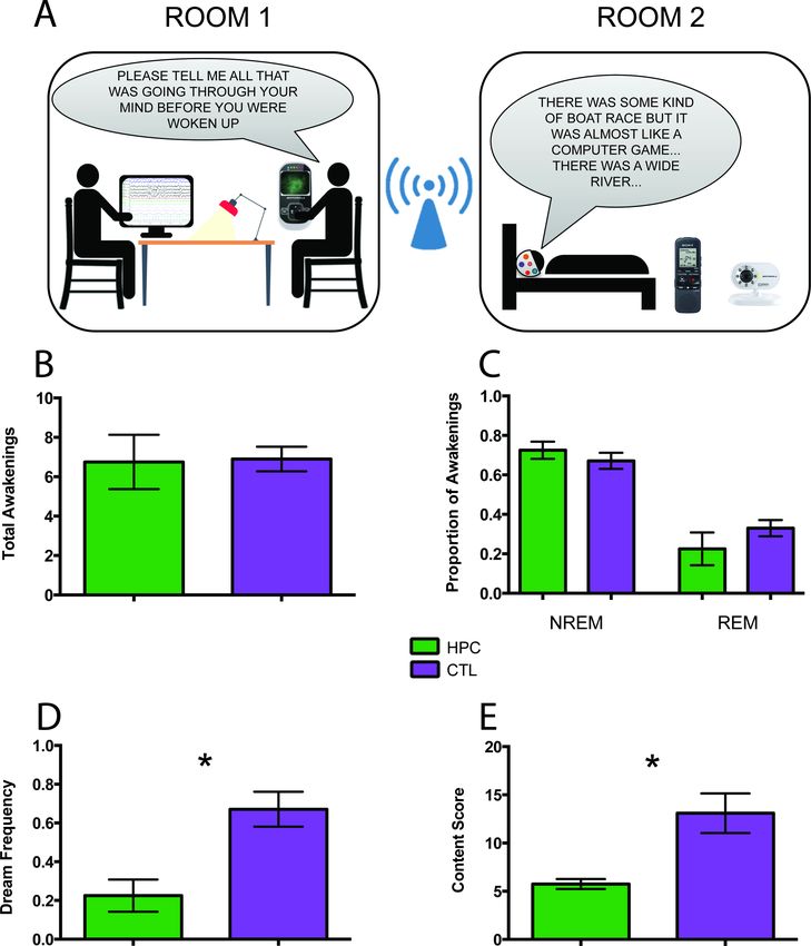
the onset of either NREM or REM sleep at various times throughout the night (Materials and meth-
ods, Figure 1A).
Table 2 shows the group summary data and the results of the between-group statistical analyses
for each of the measures that are described below. While, for the sake of economy, we present the
findings in terms of these group comparisons, given the small sample of these rare patients, caution
should be exercised in interpreting the results. We, therefore, include the individual patient data in
Tables 1 and 2 permitting the patients to be considered also as a series of case studies.
The number of awakenings was not different between patients and control participants
(Figure 1B). Furthermore, there were no significant group differences in the proportion of awaken-
ings from NREM and REM sleep (Figure 1C). After an awakening, participants were instructed via a
two-way intercom to describe everything that was going through their mind before they were woken
up in that moment. They were occasionally probed (e.g. Can you tell me more about that?) to obtain
further information (Materials and methods). The amount of probing did not differ between groups.
Dream reports were subsequently transcribed for further analyses (Materials and methods;
Figure 2).
Although the two groups were woken up a similar number of times, they differed in terms of
dream frequency, with patients reporting significantly fewer dreams compared to control partici-
pants (Figure 1D). There were no group differences in terms of the proportion of dream recall dur-
ing NREM and REM sleep. Of note, one patient did not report any dreams at all, but instead
typically stated ‘I can’t picture it’ (Figure 2).
Perhaps the patients reported fewer dreams than control participants simply because they forgot
any dreams they may have had. Three different types of awakening were evident: when participants
reported a dream, when they stated they did not dream at all (no dream), and when they dreamt
but could not recall the content (this is known as a blank dream). Although there was a significant
difference between patients and controls for dreams (see above) and for no dreams, they did not
differ in terms of the proportion of blank dreams, which was low for both groups. This suggests that
patients could distinguish between situations when they did not dream and those when they dreamt
but could not remember. However, no validated objective measure of dreaming exists, and this
should be borne in mind when interpreting participants’ subjective reports. It is notable that in previ-
ous studies, these particular patients could retain information over several minutes, including report-
ing on their daydreaming (e.g. McCormick et al., 2016; McCormick et al., 2018), which speaks
against a rapid decay of sleep mentation as an explanation for their reduced dream frequency.
The patients had so few dream reports that comparisons with the control participants should be
treated with caution. Nevertheless, we wondered whether any differences in features and content
were evident between the groups for the few dreams the patients reported. Because one patient
had no dreams whatsoever, he was not included in these analyses.
We first examined the number of informative words (see Materials and methods; Stickgold et al.,
2001b) used in the dream narratives and, while the patients used fewer such words, overall there
was no significant difference between the groups. Nevertheless, adjudicating between the possibility
of patients having a generic problem with expressing themselves verbally versus merely having little
Spanò et al. eLife 2020;9:e56211. DOI: https://doi.org/10.7554/eLife.56211 3 of 15Short report Neuroscience Figure 1. Experimental set-up and key findings. (A) Two researchers were located in Room one which was adjacent to Room two where the participant slept. The participant was woken up at various times during their night’s sleep to report their thoughts in that moment. PSG recordings informed the decisions about when to awaken the participant to ensure sampling during non-rapid eye movement (NREM) and rapid eye movement (REM) sleep. We used a Bluetooth intercom system equipped with a camera for continuous visual monitoring and communication with the participant. (B) The number of Figure 1 continued on next page Spanò et al. eLife 2020;9:e56211. DOI: https://doi.org/10.7554/eLife.56211 4 of 15
Short report                                                                                                                  Neuroscience

Figure 1 continued
total awakenings was not different between the patients (HPC) and control (CTL) participants. (C) There were also no significant group differences in the
proportion of awakenings from NREM and REM sleep. (D) In contrast, the patients reported significantly fewer dreams than the control participants,
expressed here as the total number of dreams divided by the total number of awakenings (+ / - 1 SEM; p=0.028). (E) The few dreams the patients had
were significantly less rich in content compared to those of the control participants (n = 3 patients, as one patient had no dreams at all and was not
included in this analysis; + / - 1 SEM; p=0.018). For other measures see Table 2.

                                     to describe because the few dreams they had were so impoverished, is challenging. The same issue
                                     pertains for assessments during wake. A number of studies addressed this concern during tasks
                                     involving the imagination of scenes or future scenarios, counterfactual thinking, describing the pres-
                                     ent and pictures of scenes. Bilateral hippocampal damage does not affect narrative construction or
                                     verbal descriptive ability (e.g. Race et al., 2011; Race et al., 2013; Mullally et al., 2012;
                                     Mullally and Maguire, 2014; Miller et al., 2020). Considering specifically the patients in the current

Table 2. Dream characteristics.

                                              HPC               CTL
                                                                                                                 HPC1        HPC2       HPC3       HPC4
                                              M (SD)            M (SD)            U        ES        P-Value
General analysesa
Number of awakenings                          6.75 (2.75)       6.90 (1.97)       18.5     0.11      0.829       10.00       4.00       5.00       8.00
Proportion of awakenings during NREM          0.73 (0.09)       0.67 (0.13)       14.0     0.47      0.383       0.80        0.75       0.60       0.75
Proportion of awakenings during REM           0.23 (0.17)       0.33 (0.13)       13.0     0.55      0.309       0.00c       0.25       0.40       0.25
Number of probes per awakening                3.82 (1.75)       4.22 (1.13)       17.0     0.23      0.671       5.80        3.50       1.60       4.38
Dream frequency                               0.23 (0.17)       0.67 (0.28)       4.5      1.45      0.028       0.40        0.25       0.00       0.25
Proportion of dreams during NREM              0.38 (0.48)       0.52 (0.20)       14.5     0.43      0.415       1.00        0.00       0.00       0.50
Proportion of dreams during REM               0.38 (0.48)       0.48 (0.20)       16.0     0.31      0.555       0.00        1.00       0.00       0.50
Proportion of no dreams                       0.65 (0.31)       0.21 (0.24)       4.0      1.52      0.022       0.60        0.75       1.00       0.25
Proportion of blank dreams                    0.13 (0.25)       0.12 (0.15)       15.5     0.35      0.496       0.00        0.00       0.00       0.50
                                 b
Overall qualitative attributes
Number of informative words                   43.17 (16.06)     95.55 (55.20)     7.0      0.81      0.176       55.50       25.00      .          49.00
Complexity                                    2.67 (0.58)       3.32 (0.70)       5.5      1.00      0.098       3.00        2.00       .          3.00
Vividness                                     3.10 (0.79)       3.88 (1.08)       7.5      0.75      0.203       2.80        4.00       .          2.50
Bizarreness                                   1.58 (1.01)       2.27 (0.95)       8.0      0.70      0.232       2.75        1.00       .          1.00
Emotional valence                             2.75 (0.25)       2.81 (0.22)       13.0     0.19      0.720       2.75        3.00       .          2.50
Proportion of self-references                 0.84 (0.29)       0.90 (0.19)       15.0     0.00      1.000       1.00        1.00       .          0.50
                             b
Content characterization
Internal (episodic) details                   4.08 (1.47)       9.13 (3.56)       2.0      1.54      0.028       5.75        3.00       .          3.50
External (semantic/other) details             0.17 (0.29)       1.07 (1.60)       8.5      0.64      0.258       0.00        0.00       .          0.50
Content score                                 5.75 (0.90)       13.10 (6.49)      1.0      1.74      0.018       6.75        5.00       .          5.50

M = mean; SD = standard deviation; ES = effect size; HPC = hippocampal-damaged patients; CTL = control participants; NREM = non-rapid eye move-
ment sleep; REM = rapid eye movement sleep; HPC1 4 = each individual hippocampal-damaged patient. P-values relate to between-group non-paramet-
ric Mann-Whitney U tests with significant differences depicted in bold. aAll patients included; means are per awakening. bHCP3, who had no dream
reports at all, was excluded; means are per dream report. cFor HCP1, during 20% of his awakenings towards the end of the night, the EEG cap stopped
functioning and so designation to NREM or REM sleep was not possible. Hence, it could be that this zero score for REM awakenings is an underestimate,
given that REM is more common in the latter part of the night. Note that his dream reports from these awakenings were still included in the dream qualita-
tive attributes and content analyses. See Table 2—source data 1 file for the data underpinning this table.
The online version of this article includes the following source data for Table 2:
Source data 1. This file contains the individual participant data for every dream-related measure that is summarised in Table 2.

Spanò et al. eLife 2020;9:e56211. DOI: https://doi.org/10.7554/eLife.56211                                                                       5 of 15
Short report Neuroscience
Figure 2. Example dream reports. Experimenter probing is shown in italics. HPC1 4 = the four hippocampal-damaged patients; CTL = an example
control participant.
study, they too had no difficulty performing verbal fluency tests or other tasks where total word
count was measured (Supplementary file 1). Given these general and specific findings, it is unlikely
that the patients’ performance was driven by an underlying expressive verbal problem.
Next we assessed overall qualitative features of dream reports using experimenter ratings (Mate-
rials and methods; De Gennaro et al., 2011; Oudiette et al., 2012). There were no significant
group differences in terms of general complexity, vividness, bizarreness, emotional valence (there
were no nightmares), and proportion of self-references. In order to probe the dream reports in more
depth, we used two scoring methods that are often employed for examining complex mental events
(Materials and methods). The first involved a scoring regime typically used for autobiographical
memories, the Autobiographical Interview (Levine et al., 2002). This allowed us to measure the
amount of episodic (internal) and non-episodic (external) details. The patients included significantly
fewer internal details in their dream narratives relative to controls, whereas there was no significant
group difference in terms of external details. A second method, usually employed for scoring the
Spanò et al. eLife 2020;9:e56211. DOI: https://doi.org/10.7554/eLife.56211 6 of 15Short report Neuroscience
content of imagined scenes (Hassabis et al., 2007), showed that the dream reports of patients were
significantly less rich in content compared to those of the control participants (Figure 1E).
Discussion
By studying rare patients with selective bilateral hippocampal damage we found that dream fre-
quency was reduced compared to control participants, and the few dreams they had were less epi-
sodic-like in nature and lacked content. This accords with previous studies that reported stereotyped
dreaming in patients with brain damage that extended beyond the hippocampi (Torda, 1969a;
Torda, 1969b; Stickgold et al., 2000), and echoes the effects of hippocampal damage on imagina-
tion (Hassabis et al., 2007), episodic memory (Miller et al., 2017; Miller et al., 2020; Spiers et al.,
2001) and daydreaming (McCormick et al., 2018). Given our patients’ circumscribed lesions, these
results suggest that hippocampal integrity may be necessary for typical dreaming to occur. There
are several possible explanations for degraded dreaming in the patients, which we consider in turn.
Despite the tendency to confine dreaming to the sleep state, studies have shown that there is
often continuity between waking life experiences and dreaming (Andrillon et al., 2015; Fosse et al.,
2003; Horikawa et al., 2013; Schredl and Hofmann, 2003). Given that the patients had difficulty
retaining information over longer time scales while awake, then perhaps during subsequent sleep
there was little material to process. Hence it could be that the capacity to dream was intact, but
underused, in the patients.
Alternatively, the core capacity for dreaming might have been compromised, with this in turn
affecting memory processing. Dreaming has been linked with the consolidation of information into
long-term memory (Payne, 2010; Wamsley, 2013; Wamsley, 2014; Wamsley et al., 2010;
Wamsley and Stickgold, 2019), but it is unknown whether dreaming plays a functional role in this
process (Wamsley, 2014). If it does, then patients with hippocampal damage may lack this dream-
related mechanism for facilitating memory processing, and this could contribute to their amnesia.
Another aspect of sleep that has been associated with episodic memory consolidation is slow-
wave sleep (SWS), a stage within NREM sleep (Rasch and Born, 2013). We have previously shown
that the hippocampal-damaged patients tested here had significantly reduced SWS and slow wave
activity, whereas the time spent in other sleep stages was comparable to that of control participants
(Spanò et al., 2020). It is unlikely that the patients’ degraded dreaming was caused solely by
decreased SWS. This is because we sampled dreaming across both NREM and REM sleep, and of
the dreams sampled during NREM sleep in the control participants, just 5% (SD 11.3) were during
SWS. Overall, however, reduced slow wave activity along with sub-optimal dreaming – if dreaming
plays a functional role – may constitute a twofold blow that adversely affected the proper function-
ing of episodic memory in these patients.
A different perspective on the current results involves looking beyond memory. The hippocampus
has been implicated in a range of other cognitive functions including thinking about the future
(Addis et al., 2007; Hassabis et al., 2007; Kurczek et al., 2015), spatial navigation (Maguire et al.,
2006; O’Keefe and Nadel, 1978), daydreaming (Karapanagiotidis et al., 2017; McCormick et al.,
2018), aspects of decision making (Mullally and Maguire, 2014; McCormick et al., 2016), and
visual perception (Lee et al., 2005; McCormick et al., 2017; Mullally et al., 2012). Patients with
bilateral hippocampal damage are also impaired at mentally constructing scene imagery
(Hassabis et al., 2007), and scene imagery features prominently across cognition (Clark et al.,
2019; Clark et al., 2020). Consequently, it has been proposed that one role of the hippocampus
could be to facilitate the scene construction process (Hassabis and Maguire, 2007; Maguire and
Mullally, 2013). This may explain why dreaming is degraded in the context of hippocampal pathol-
ogy, as it too typically involves (and possibly requires) scene imagery.
In summary, while the functional role of dreaming is as yet unknown, we conclude from our results
that hippocampal integrity may be a prerequisite for typical dreaming to occur, and that dreaming
seems to align with an array of other cognitive functions that are hippocampal-dependent and which
play crucial roles in supporting our everyday mental life.
Spanò et al. eLife 2020;9:e56211. DOI: https://doi.org/10.7554/eLife.56211 7 of 15Short report Neuroscience
Materials and methods
Participants
For all patients, hippocampal lesions resulted from leucine-rich glycine-inactivate-1 antibody-com-
plex limbic encephalitis (LGI1-antibody-complex LE; Miller et al., 2017; Miller et al., 2020). This
study was conducted a median of 9.5 years after hippocampal damage occurred (mean 9 years ± SD
2.45). Patients (HPC) and the dream control participants (CTL) were closely matched on a number of
demographic factors: gender (all males), age (Table 1; MWU = 19.00, p=0.89, Cohen’s d = 0.08),
body mass index (HPC mean 27.68 ± 2.51; CTL 25.79 ± 2.41; MWU = 14.00, p=0.40, Cohen’s
d = 0.47) and general cognitive ability assessed with the Matrix Reasoning subtest of the Wechsler
Abbreviated Scale of Intelligence (WASI; Wechsler, 1999; Table 1; MWU = 7.00, p=0.06, Cohen’s
d = 1.13). Participants had no history of psychiatric disorders (e.g., depression, anxiety). Each partici-
pant gave written informed consent for participation in the study, for data analysis and for publica-
tion of the study results. ‘Materials and methods’ were approved by the University College London
Research Ethics Committee.
The patients entered the sleep study having already been characterized, relative to matched
healthy control participants, in terms of their lesion selectivity and neuropsychological profile as part
of previous research studies. Full details of that characterization process are available in
McCormick et al., 2016, McCormick et al., 2017, McCormick et al., 2018 and Spanò et al., 2020.
In summary, manual (blinded) segmentation of the hippocampi from T2-weighted high resolution
structural MRI scans (0.5 0.5 0.5 mm voxels) showed that our patients had substantial volume
loss relative to controls in the left (MWU = 2.00, p=0.009, Cohen’s d = 1.83) and right (MWU = 3.00,
p=0.013, Cohen’s d = 1.67) hippocampus (Table 1). Expert neuroradiological examination confirmed
there was no damage outside of the hippocampi. In addition, automated whole brain voxel-based
morphometry showed there were no volume differences between patients and controls anywhere
else in the brain. Supplementary file 1 provides the neuropsychological profile (summary data and
statistical analyses) of the patients across a range of cognitive tests, and indicates the selective
nature of their memory loss.
Since all patients included in the current study had suffered from LGI1-antibody-complex LE, our
findings might potentially not generalize to other forms of hippocampal amnesia. However, it is
important to note that other aetiologies that lead to hippocampal-mediated amnesia such as viral
encephalitis, hypoxic brain injury secondary to drug overdose, or toxic shock syndrome are associ-
ated with circumscribed hippocampal lesions, but frequently also involve anatomical damage else-
where (Heinz and Rollnik, 2015; Raschilas et al., 2002). In addition, these aetiologies lead to co-
morbidities and broader cognitive impairment (Heinz and Rollnik, 2015; Hokkanen and Launes,
2007; McGrath et al., 1997; Peskine et al., 2010; Thakur et al., 2013; Rosene et al., 1982), which
were absent from the clinical and neuropsychological profile of the patients reported here. There-
fore, the selection of such a rare group of patients with circumscribed hippocampal lesions allowed
us to pinpoint the direct role of the hippocampus in dreaming without the interference of potential
confounds associated with heterogeneity in aetiology.
Other features associated with LGI1-antibody-complex LE in its initial presentation – such as focal
seizures and hyponatraemia related to hypothalamic damage – are also unlikely to explain the
effects we observed. Our patients were seizure-free when they were discharged after initial admis-
sion, they were not prescribed antiepileptic medication, and none of the patients had seizure recur-
rence following initial treatment. Thus, unlike in temporal lobe epilepsy, which is associated with
ongoing seizures and hippocampal sclerosis (Kapur and Prevett, 2003), our patients enabled us to
study effects on dreaming that were not coincidental with, and sequelae of, seizure activity. More-
over, patients were not undergoing treatment for hyponatraemia, which is consistent with published
evidence that persistent hyponatraemia is not a characteristic feature of LGI1-antibody-complex LE
(Bastiaansen et al., 2017). Therefore, the findings in the current study are unlikely to stem from the
above-mentioned potential issues.
As dreaming might be influenced by sleep quality (Schredl, 2009), we confirmed that patients
did not differ significantly from control participants on subjective measures of the general quality
and pattern of sleep, as well as on objective measures of sleep-related breathing disorders, and
sleep-wake patterns across one week (see Supplementary file 2). Specifically, participants com-
pleted standardized questionnaires assessing habitual sleep habits over the last month (The
Spanò et al. eLife 2020;9:e56211. DOI: https://doi.org/10.7554/eLife.56211 8 of 15Short report Neuroscience
Pittsburgh Sleep Quality Index; Buysse et al., 1989), level of daytime sleepiness (The Epworth
Sleepiness Scale; Johns, 1991), and chronotype – whether someone is a ‘morning’ or an ‘evening’
type of person (The Morningness-Eveningness Questionnaire; Horne and Ostberg, 1976). More-
over, we assessed the severity of obstructive sleep apnoea with the WatchPAT-200 (Itamar Medical
Ltd., Caesarea, Israel), a wrist-worn device that measures the Peripheral Arterial Tone (PAT) signal
by means of a plethysmographic based finger-mounted probe. Signals were automatically analysed
with the zzzPAT software (version 4.4.64 .p, Itamar Medical Ltd., Caesarea, Israel) to identify respira-
tory events and sleep states. The outcome measure employed in this study was the PAT apnoea-
hypopnea index (AHI), which provides the number of apnoea and hypopnea events per hour during
the night.
In order to assess sleep-wake patterns, participants wore an Actiwatch 2 (Phillips Respironics
Mini-Mitter) for seven consecutive days and nights on their non-dominant wrist. Light and activity
data were collected in 30 s epochs and analyzed using the Philips Actiware 6.0.2 software package
(Respironics Actiware 6.0.2). Data were scored based on available guidelines (Chow et al., 2016),
with a medium sensitivity (40 activity cpm), with sleep onset occurring after an immobility period of
10 min, and rise time following an increase in activity level and in light level above 1.0 mW/cm2. Vari-
ables of interest were sleep efficiency (in percent), total sleep time (in minutes), sleep fragmentation
index (an index of restlessness), night-to-night variability for sleep duration (Lemola et al., 2013),
average bedtime and mean sleep midpoint (clock time halfway between bedtime and rise time).
Procedure
Participants slept in the same room on both nights. Two sleep researchers were located in a sepa-
rate, nearby, room one of whom performed sleep staging in real time during online visualization of
noise-reduced EEG recordings. An independent, registered sleep technologist, blind to participant
group membership and the study aims, later off-line scored the PSG recordings to verify the sleep
staging, in line with the revised American Academy of Sleep Medicine manual (Berry et al., 2015).
For all participants, awakenings occurred after sleep onset, throughout the night. We aimed to
assess dreams from both NREM and REM sleep, and we therefore staggered awakenings at intervals
that allowed for entry into these stages, or approximately between 30–90 min intervals. For exam-
ple, a participant may have been woken at 30 min, returned to sleep and was woken again at 90
min. A maximum of 10 awakenings were scheduled per night. Awakenings were not collected at
pre-ordained points during the night (for example, after the third REM period for all participants),
rather they were based on participants’ specific sleep architecture in order to maximize the number
of reports collected. Every participant had a different sleep onset and duration, and so the awaken-
ings were not scheduled at precisely the same clock time across participants. However, as shown in
Supplementary file 2, the sleep quality of the patients and the controls was well-matched, and this
included total sleep time, bedtime and midpoint of the night.
Once a decision to awaken a participant was made, after a 3 min period without stage shift of
either NREM or REM sleep, the other sleep researcher played a non-stressful 500 Hz neutral tone via
a two-way, Bluetooth intercom system equipped with a camera for continuous visual monitoring.
After the tone was played, if the participant did not wake up, his name was spoken. This two-step
awakening procedure was repeated up to five times, if required (Dumel et al., 2015). After awaken-
ing, participants were instructed by intercom to tell the experimenter everything that was going
through their mind before they were woken up. What they said was recorded and subsequently
transcribed.
Participants were occasionally probed (e.g. Can you tell me more about that?) to obtain further
information. Probing followed a structured protocol. This involved first asking participants to freely
describe what was in their minds immediately after awakening. Whenever a participant’s response
was not clear or only covered parts of the dream, the experimenter asked general follow-up ques-
tions, which could echo information already provided (e.g. ‘. . .It was a conversation happening in the
locker room’. Can you give us any more specifics about the conversations or anything else that you
recall?). Crucially, this probing never involved leading the participant, as can be observed in the
examples provided in Figure 2. This approach is very similar to that of well-established tasks that
assess autobiographical memory recall (Levine et al., 2002) and scene imagination ability
(Hassabis et al., 2007) during wake, where probing in this manner is widely accepted.
Spanò et al. eLife 2020;9:e56211. DOI: https://doi.org/10.7554/eLife.56211 9 of 15Short report Neuroscience
Dream analyses
Transcriptions of dream reports were analysed by a researcher who was blind to participant group
membership. Double scoring was performed on 20% of the data by a second researcher. We
assessed across-experimenter agreement with inter-class correlation coefficients, with a two-way ran-
dom effects model looking for absolute agreement, which indicated excellent agreement between
the experimenters’ scoring (range: 0.9–1.0).
A dream was defined as any report that included at least one person, one place or one event
(Foulkes and Rechtschaffen, 1964). Dream frequency was calculated as the total number of dreams
divided by the total number of awakenings. Word count included words that provided information
about the dream (i.e., informative words), and excluded repetitions, hesitations and fillers, secondary
elaborations and metacognitive statements (Stickgold et al., 2001b). Dream complexity was experi-
menter scored on a 6-point scale using an adaptation of the Orlinsky score (Oudiette et al., 2012);
excluding the no dream and blank dream options, which we recorded separately). This ranged from
a participant remembering a specific topic, but in isolation, for example a fragmentary action, scene,
object, word, or idea unrelated to anything else, to a participant remembering an extremely long
and detailed dream sequence of five or more stages. Vividness referred to the clarity and detail of a
dream, and was experimenter scored using the 6-point scale of De Gennaro et al., 2011, ranging
from no image at all, to perfectly clear and as vivid as normal vision. Bizarreness/implausibility of
dreams was experimenter scored using the De Gennaro et al., 2011 6-point scale. This was based
on the presence of bizarre elements (impossible characters or actions) and/or improbable plot (dis-
continuity or unusual settings). The emotional valence of a dream was scored using a 5-point Likert
scale ranging from very negative to very positive, with three indicating a neutral tone. For self-refer-
ences, one point was awarded per dream report if a participant reported he was the agent of an
action, thought or feeling (e.g. ‘I was driving my car down a nearby road with a friend...”).
We used two other scoring methods that are often employed for examining complex mental
events in order to probe the dream reports further. The first was the Autobiographical Interview
which identifies internal and external details, a distinction that can be conceptualized as the differ-
ence between episodic and non-episodic/semantic information, respectively (Levine et al., 2002).
Internal details refer to the main event described by the participant and comprises the subcompo-
nents: Event (happenings, individuals present, weather conditions, physical/emotional actions, reac-
tions in others); Time (time of the day, year, season, month, day of the week, hours); Place (location
of the event); Perceptual (auditory, olfactory, tactile, visual and visual details, body position, duration
of time); Thought/emotion (emotional states, thoughts, implications). The internal details score is the
sum of these subcomponents. Anything tangential to the main dream event was scored as external
details, including Event (details from other events outside of the dream); Semantic (general, ongo-
ing, extended knowledge/event/state of being); Repetition; Other (metacognitive statements, edito-
rializing). The external details score is the sum of these subcomponents.
A second method we employed, the Scene Construction Test (Hassabis et al., 2007), is usually
used for scoring the content of imagined scenes. Here we focused on the content score, which com-
prises four subcomponents: Entities Present (objects or people); Spatial References (places or spatial
relationships between entities); Sensory Descriptions (details that describe an entity); Thought/emo-
tion/action (thoughts, emotional states, action descriptions). The content score is the sum of these
subcomponents.
Sleep physiology
All participants underwent PSG in their homes using a Brain Products system (GmbH, Gilching, Ger-
many). The purpose of the PSG recording was to ensure that we awakened participants during both
NREM and REM sleep, and in a similar manner for the patient and control groups. Two trained sleep
researchers arrived at a participant’s home approximately three hours before the usual bedtime to
set up for the PSG. Equipment was then removed by a researcher the following morning upon awak-
ening. PSG was recorded using a 24-electrode cap (EasyCap; based on the international 10–20 sys-
tem) including the following EEG channels: Fp1, Fp2, F3, F4, C3, C4, P3, P4, O1, O2, F7, F8, T7, T8,
P7, P8, Fz, Cz, Pz, Oz, FT9, FT10 referenced to average mastoids (M1 and M2) (sampling rate = 500
Hz). This montage also included two bipolar electrooculogram channels (EOG), two electromyogram
Spanò et al. eLife 2020;9:e56211. DOI: https://doi.org/10.7554/eLife.56211 10 of 15Short report Neuroscience
channels (EMG) and two electrocardiogram channels (ECG). Sleep staging was performed based on
EOG, EMG and the following derivations: F3/M2, F4/M1, C3/M2, C4/M1, O1/M2, O2/M1.
Statistical analyses
All statistical analyses were performed with SPSS 25.0 (IBM Corporation). Given that the data did
not meet the assumptions of normality and homogeneity necessary for parametric statistics,
between-group analyses were performed using non-parametric Mann-Whitney U tests. We also cal-
culated the effect sizes using non-parametric Cohen’s d for all tests performed. In all analyses, the
significance level was set at 0.05.
Acknowledgements
We thank the participants and their families for welcoming us into their homes and giving of their
time so generously. We are also grateful to David Bradbury and Kamlyn Ramkissoon for their
assistance.
Additional information
Funding
Funder Grant reference number Author
Wellcome 101759/Z/13/Z Eleanor A Maguire
Wellcome 203147/Z/16/Z Eleanor A Maguire
The funders had no role in study design, data collection and interpretation, or the
decision to submit the work for publication.
Author contributions
Goffredina Spanò, Conceptualization, Formal analysis, Investigation, Methodology, Writing - original
draft, Data scoring; Gloria Pizzamiglio, Investigation, Writing - review and editing, Data scoring; Cor-
nelia McCormick, Thomas D Miller, Clive R Rosenthal, Writing - review and editing, Patient charac-
terization; Ian A Clark, Writing - review and editing, Advice on data scoring; Sara De Felice, Writing -
review and editing, Data scoring; Jamie O Edgin, Writing - review and editing, Advice on conceptu-
alization; Eleanor A Maguire, Conceptualization, Formal analysis, Supervision, Funding acquisition,
Methodology, Writing - original draft
Author ORCIDs
Gloria Pizzamiglio https://orcid.org/0000-0002-3567-1344
Clive R Rosenthal https://orcid.org/0000-0002-5960-4648
Eleanor A Maguire https://orcid.org/0000-0002-9470-6324
Ethics
Human subjects: Each participant gave written informed consent for participation in the study, for
data analysis and for publication of the study results. ’Materials and methods’ were approved by the
University College London Research Ethics Committee (reference number: 6743/004).
Decision letter and Author response
Decision letter https://doi.org/10.7554/eLife.56211.sa1
Author response https://doi.org/10.7554/eLife.56211.sa2
Spanò et al. eLife 2020;9:e56211. DOI: https://doi.org/10.7554/eLife.56211 11 of 15Short report Neuroscience
Additional files
Supplementary files
. Supplementary file 1. Summary of neuropsychological information. This table provides details of
the neuropsychological profile (summary data and statistical analyses) of the patients across a range
of cognitive tests, and indicates the selective nature of their memory loss.
. Supplementary file 2. Sleep quality of the patients and control participants. This table provides
details (summary data and statistical analyses) of subjective measures of the general quality and pat-
tern of sleep, as well as on objective measures of sleep-related breathing disorders, and sleep-wake
patterns across one week. There were no significant differences between the patient and control
participants on any measure.
. Transparent reporting form
Data availability
The dream data for every participant for every measure are provided in the file Table 2-Source data
1.
References
Addis DR, Wong AT, Schacter DL. 2007. Remembering the past and imagining the future: common and distinct
neural substrates during event construction and elaboration. Neuropsychologia 45:1363–1377. DOI: https://
doi.org/10.1016/j.neuropsychologia.2006.10.016, PMID: 17126370
Andrillon T, Nir Y, Cirelli C, Tononi G, Fried I. 2015. Single-neuron activity and eye movements during human
REM sleep and awake vision. Nature Communications 6:7884. DOI: https://doi.org/10.1038/ncomms8884,
PMID: 26262924
Antrobus J, Kondo T, Reinsel R, Fein G. 1995. Dreaming in the late morning: summation of REM and diurnal
cortical activation. Consciousness and Cognition 4:275–299. DOI: https://doi.org/10.1006/ccog.1995.1039,
PMID: 7497109
Bastiaansen AEM, van Sonderen A, Titulaer MJ. 2017. Autoimmune encephalitis with anti-leucine-rich glioma-
inactivated 1 or anti-contactin-associated protein-like 2 antibodies (formerly called voltage-gated potassium
channel-complex antibodies). Current Opinion in Neurology 30:302–309. DOI: https://doi.org/10.1097/WCO.
0000000000000444, PMID: 28248701
Berry RB, Brooks R, Gamaldo CE, Harding SM, Lloyd RM, Marcus CL, Vaughn BV. 2015. The AASM Manual for
the Scoring of Sleep and Associated Events: Rules, Terminology and Technical Specifications, Version 2.2.
Darien, Illinois: Academy, American of Sleep Medicine.
Buysse DJ, Reynolds CF, Monk TH, Berman SR, Kupfer DJ. 1989. The Pittsburgh sleep quality index: a new
instrument for psychiatric practice and research. Psychiatry Research 28:193–213. DOI: https://doi.org/10.1016/
0165-1781(89)90047-4, PMID: 2748771
Chow CM, Wong SN, Shin M, Maddox RG, Feilds KL, Paxton K, Hawke C, Hazell P, Steinbeck K. 2016. Defining
the rest interval associated with the main sleep period in actigraph scoring. Nature and Science of Sleep 8:321–
328. DOI: https://doi.org/10.2147/NSS.S114969, PMID: 27920587
Cipolli C, Ferrara M, De Gennaro L, Plazzi G. 2017. Beyond the neuropsychology of dreaming: insights into the
neural basis of dreaming with new techniques of sleep recording and analysis. Sleep Medicine Reviews 35:8–
20. DOI: https://doi.org/10.1016/j.smrv.2016.07.005, PMID: 27569701
Clark IA, Hotchin V, Monk A, Pizzamiglio G, Liefgreen A, Maguire EA. 2019. Identifying the cognitive processes
underpinning hippocampal-dependent tasks. Journal of Experimental Psychology: General 148:1861–1881.
DOI: https://doi.org/10.1037/xge0000582
Clark IA, Monk AM, Maguire EA. 2020. Characterising strategy use during the performance of hippocampal-
dependent tasks. bioRxiv. DOI: https://doi.org/10.1101/807990
De Gennaro L, Cipolli C, Cherubini A, Assogna F, Cacciari C, Marzano C, Curcio G, Ferrara M, Caltagirone C,
Spalletta G. 2011. Amygdala and Hippocampus volumetry and diffusivity in relation to dreaming. Human Brain
Mapping 32:1458–1470. DOI: https://doi.org/10.1002/hbm.21120, PMID: 20740648
Dumel G, Carr M, Marquis LP, Blanchette-Carrière C, Paquette T, Nielsen T. 2015. Infrequent dream recall
associated with low performance but high overnight improvement on mirror-tracing. Journal of Sleep Research
24:372–382. DOI: https://doi.org/10.1111/jsr.12286, PMID: 25726721
Fosse MJ, Fosse R, Hobson JA, Stickgold RJ. 2003. Dreaming and episodic memory: a functional dissociation?
Journal of Cognitive Neuroscience 15:1–9. DOI: https://doi.org/10.1162/089892903321107774, PMID: 12590
838
Foulkes WD. 1962. Dream reports from different stages of sleep. The Journal of Abnormal and Social
Psychology 65:14–25. DOI: https://doi.org/10.1037/h0040431, PMID: 13894288
Spanò et al. eLife 2020;9:e56211. DOI: https://doi.org/10.7554/eLife.56211 12 of 15Short report Neuroscience
Foulkes D, Rechtschaffen A. 1964. Presleep determinants of dream content: effect of two films. Perceptual and
Motor Skills 19:983–1005. DOI: https://doi.org/10.2466/pms.1964.19.3.983, PMID: 14242569
Hassabis D, Kumaran D, Vann SD, Maguire EA. 2007. Patients with hippocampal amnesia cannot imagine new
experiences. PNAS 104:1726–1731. DOI: https://doi.org/10.1073/pnas.0610561104, PMID: 17229836
Hassabis D, Maguire EA. 2007. Deconstructing episodic memory with construction. Trends in Cognitive Sciences
11:299–306. DOI: https://doi.org/10.1016/j.tics.2007.05.001, PMID: 17548229
Heinz UE, Rollnik JD. 2015. Outcome and prognosis of hypoxic brain damage patients undergoing neurological
early rehabilitation. BMC Research Notes 8:243. DOI: https://doi.org/10.1186/s13104-015-1175-z
Hobson JA, Pace-Schott EF, Stickgold R. 2000. Dreaming and the brain: toward a cognitive neuroscience of
conscious states. Behavioral and Brain Sciences 23:793–842. DOI: https://doi.org/10.1017/
S0140525X00003976, PMID: 11515143
Hokkanen L, Launes J. 2007. Neuropsychological sequelae of acute-onset sporadic viral encephalitis.
Neuropsychological Rehabilitation 17:450–477. DOI: https://doi.org/10.1080/09602010601137039,
PMID: 17676530
Horikawa T, Tamaki M, Miyawaki Y, Kamitani Y. 2013. Neural decoding of visual imagery during sleep. Science
340:639–642. DOI: https://doi.org/10.1126/science.1234330, PMID: 23558170
Horne JA, Ostberg O. 1976. A self-assessment questionnaire to determine morningness-eveningness in human
circadian rhythms. International Journal of Chronobiology 4:97–110. PMID: 1027738
Johns MW. 1991. A new method for measuring daytime sleepiness: the epworth sleepiness scale. Sleep 14:540–
545. DOI: https://doi.org/10.1093/sleep/14.6.540, PMID: 1798888
Kapur N, Prevett M. 2003. Unexpected amnesia: are there lessons to be learned from cases of amnesia following
unilateral temporal lobe surgery? Brain 126:2573–2585. DOI: https://doi.org/10.1093/brain/awg275, PMID: 12
958081
Karapanagiotidis T, Bernhardt BC, Jefferies E, Smallwood J. 2017. Tracking thoughts: exploring the neural
architecture of mental time travel during mind-wandering. NeuroImage 147:272–281. DOI: https://doi.org/10.
1016/j.neuroimage.2016.12.031, PMID: 27989779
Kurczek J, Wechsler E, Ahuja S, Jensen U, Cohen NJ, Tranel D, Duff M. 2015. Differential contributions of
Hippocampus and medial prefrontal cortex to self-projection and self-referential processing. Neuropsychologia
73:116–126. DOI: https://doi.org/10.1016/j.neuropsychologia.2015.05.002, PMID: 25959213
Lee ACH, Bussey TJ, Murray EA, Saksida LM, Epstein RA, Kapur N, Hodges JR, Graham KS. 2005. Perceptual
deficits in amnesia: challenging the medial temporal lobe ‘mnemonic’ view. Neuropsychologia 43:1–11.
DOI: https://doi.org/10.1016/j.neuropsychologia.2004.07.017
Lemola S, Ledermann T, Friedman EM. 2013. Variability of sleep duration is related to subjective sleep quality
and subjective well-being: an actigraphy study. PLOS ONE 8:e71292. DOI: https://doi.org/10.1371/journal.
pone.0071292, PMID: 23967186
Levine B, Svoboda E, Hay JF, Winocur G, Moscovitch M. 2002. Aging and autobiographical memory: dissociating
episodic from semantic retrieval. Psychology and Aging 17:677–689. DOI: https://doi.org/10.1037/0882-7974.
17.4.677, PMID: 12507363
Maguire EA, Nannery R, Spiers HJ. 2006. Navigation around London by a taxi driver with bilateral hippocampal
lesions. Brain 129:2894–2907. DOI: https://doi.org/10.1093/brain/awl286, PMID: 17071921
Maguire EA, Mullally SL. 2013. The Hippocampus: a manifesto for change. Journal of Experimental Psychology:
General 142:1180–1189. DOI: https://doi.org/10.1037/a0033650
Maquet P, Laureys S, Peigneux P, Fuchs S, Petiau C, Phillips C, Aerts J, Del Fiore G, Degueldre C, Meulemans T,
Luxen A, Franck G, Van Der Linden M, Smith C, Cleeremans A. 2000. Experience-dependent changes in
cerebral activation during human REM sleep. Nature Neuroscience 3:831–836. DOI: https://doi.org/10.1038/
77744, PMID: 10903578
Maquet P, Ruby P, Maudoux A, Albouy G, Sterpenich V, Dang-Vu T, Desseilles M, Boly M, Perrin F, Peigneux P,
Laureys S. 2005. Human cognition during REM sleep and the activity profile within frontal and parietal cortices:
a reappraisal of functional neuroimaging data. Progress in Brain Research 150:219–227. DOI: https://doi.org/
10.1016/S0079-6123(05)50016-5, PMID: 16186026
McCormick C, Rosenthal CR, Miller TD, Maguire EA. 2016. Hippocampal damage increases deontological
responses during moral decision making. Journal of Neuroscience 36:12157–12167. DOI: https://doi.org/10.
1523/JNEUROSCI.0707-16.2016, PMID: 27903725
McCormick C, Rosenthal CR, Miller TD, Maguire EA. 2017. Deciding what is possible and impossible following
hippocampal damage in humans. Hippocampus 27:303–314. DOI: https://doi.org/10.1002/hipo.22694,
PMID: 27997994
McCormick C, Rosenthal CR, Miller TD, Maguire EA. 2018. Mind-Wandering in people with hippocampal
damage. The Journal of Neuroscience 38:2745–2754. DOI: https://doi.org/10.1523/JNEUROSCI.1812-17.2018,
PMID: 29440532
McGrath N, Anderson NE, Croxson MC, Powell KF. 1997. Herpes simplex encephalitis treated with acyclovir:
diagnosis and long term outcome. Journal of Neurology, Neurosurgery & Psychiatry 63:321–326. DOI: https://
doi.org/10.1136/jnnp.63.3.321
Miller TD, Chong TT, Aimola Davies AM, Ng TWC, Johnson MR, Irani SR, Vincent A, Husain M, Jacob S,
Maddison P, Kennard C, Gowland PA, Rosenthal CR. 2017. Focal CA3 hippocampal subfield atrophy following
LGI1 VGKC-complex antibody limbic encephalitis. Brain 140:1212–1219. DOI: https://doi.org/10.1093/brain/
awx070, PMID: 28369215
Spanò et al. eLife 2020;9:e56211. DOI: https://doi.org/10.7554/eLife.56211 13 of 15Short report Neuroscience
Miller TD, Chong TT, Aimola Davies AM, Johnson MR, Irani SR, Husain M, Ng TW, Jacob S, Maddison P,
Kennard C, Gowland PA, Rosenthal CR. 2020. Human hippocampal CA3 damage disrupts both recent and
remote episodic memories. eLife 9:e41836. DOI: https://doi.org/10.7554/eLife.41836, PMID: 31976861
Mullally SL, Intraub H, Maguire EA. 2012. Attenuated boundary extension produces a paradoxical memory
advantage in amnesic patients. Current Biology 22:261–268. DOI: https://doi.org/10.1016/j.cub.2012.01.001,
PMID: 22264610
Mullally SL, Maguire EA. 2014. Counterfactual thinking in patients with amnesia. Hippocampus 24:1261–1266.
DOI: https://doi.org/10.1002/hipo.22323, PMID: 24978690
Nguyen ND, Tucker MA, Stickgold R, Wamsley EJ. 2013. Overnight sleep enhances Hippocampus-Dependent
aspects of spatial memory. Sleep 36:1051–1057. DOI: https://doi.org/10.5665/sleep.2808, PMID: 23814342
Nielsen TA. 2000. A review of mentation in REM and NREM sleep: "covert" REM sleep as a possible
reconciliation of two opposing models. Behavioral and Brain Sciences 23:851–866. DOI: https://doi.org/10.
1017/S0140525X0000399X, PMID: 11515145
O’Keefe J, Nadel L. 1978. The Hippocampus as a Cognitive Map. Oxford, UK: Oxford University Press.
DOI: https://doi.org/10.1016/j.neuron.2015.06.013
Oudiette D, Dealberto MJ, Uguccioni G, Golmard JL, Merino-Andreu M, Tafti M, Garma L, Schwartz S, Arnulf I.
2012. Dreaming without REM sleep. Consciousness and Cognition 21:1129–1140. DOI: https://doi.org/10.
1016/j.concog.2012.04.010, PMID: 22647346
Payne JD. 2010. Memory consolidation, the diurnal rhythm of cortisol, and the nature of dreams: a new
hypothesis. International Review of Neurobiology 92:101–134. DOI: https://doi.org/10.1016/S0074-7742(10)
92006-0, PMID: 20870065
Peigneux P, Laureys S, Fuchs S, Collette F, Perrin F, Reggers J, Phillips C, Degueldre C, Del Fiore G, Aerts J,
Luxen A, Maquet P. 2004. Are spatial memories strengthened in the human Hippocampus during slow wave
sleep? Neuron 44:535–545. DOI: https://doi.org/10.1016/j.neuron.2004.10.007, PMID: 15504332
Peskine A, Rosso C, Picq C, Caron E, Pradat-Diehl P. 2010. Neurological sequelae after cerebral Anoxia. Brain
Injury 24:755–761. DOI: https://doi.org/10.3109/02699051003709581, PMID: 20370382
Race E, Keane MM, Verfaellie M. 2011. Medial temporal lobe damage causes deficits in episodic memory and
episodic future thinking not attributable to deficits in narrative construction. Journal of Neuroscience 31:
10262–10269. DOI: https://doi.org/10.1523/JNEUROSCI.1145-11.2011, PMID: 21753003
Race E, Keane MM, Verfaellie M. 2013. Living in the moment: patients with MTL amnesia can richly describe the
present despite deficits in past and future thought. Cortex 49:1764–1766. DOI: https://doi.org/10.1016/j.
cortex.2013.02.010, PMID: 23535365
Rasch B, Born J. 2013. About sleep’s role in memory. Physiological Reviews 93:681–766. DOI: https://doi.org/10.
1152/physrev.00032.2012, PMID: 23589831
Raschilas F, Wolff M, Delatour F, Chaffaut C, De Broucker T, Chevret S, Lebon P, Canton P, Rozenberg F. 2002.
Outcome of and prognostic factors for herpes simplex encephalitis in adult patients: results of a multicenter
study. Clinical Infectious Diseases 35:254–260. DOI: https://doi.org/10.1086/341405, PMID: 12115090
Rosene KA, Copass MK, Kastner LS, Nolan CM, Eschenbach DA. 1982. Persistent neuropsychological sequelae
of toxic shock syndrome. Annals of Internal Medicine 96:865. DOI: https://doi.org/10.7326/0003-4819-96-6-
865, PMID: 7091958
Schredl M. 2009. Dreams in patients with sleep disorders. Sleep Medicine Reviews 13:215–221. DOI: https://doi.
org/10.1016/j.smrv.2008.06.002, PMID: 19147378
Schredl M, Hofmann F. 2003. Continuity between waking activities and dream activities. Consciousness and
Cognition 12:298–308. DOI: https://doi.org/10.1016/S1053-8100(02)00072-7, PMID: 12763010
Scoville WB, Milner B. 1957. Loss of recent memory after bilateral hippocampal lesions. Journal of Neurology,
Neurosurgery & Psychiatry 20:11–21. DOI: https://doi.org/10.1136/jnnp.20.1.11
Siclari F, Larocque JJ, Postle BR, Tononi G. 2013. Assessing sleep consciousness within subjects using a serial
awakening paradigm. Frontiers in Psychology 4:542. DOI: https://doi.org/10.3389/fpsyg.2013.00542, PMID: 23
970876
Siclari F, Baird B, Perogamvros L, Bernardi G, LaRocque JJ, Riedner B, Boly M, Postle BR, Tononi G. 2017. The
neural correlates of dreaming. Nature Neuroscience 20:872–878. DOI: https://doi.org/10.1038/nn.4545,
PMID: 28394322
Solms M. 1997. The Neuropsychology of Dreams. New York: Psychology Press.
Solms M. 2013. Dreaming is not controlled by hippocampal mechanisms. Behavioral and Brain Sciences 36:629.
DOI: https://doi.org/10.1017/S0140525X1300143X, PMID: 24304770
Spanò G, Weber FD, Pizzamiglio G, McCormick C, Miller TD, Rosenthal CR, Edgin JO, Maguire EA. 2020.
Sleeping with hippocampal damage. Current Biology 30:523–529. DOI: https://doi.org/10.1016/j.cub.2019.11.
072, PMID: 31956024
Spiers HJ, Maguire EA, Burgess N. 2001. Hippocampal amnesia. Neurocase 7:357–382. DOI: https://doi.org/10.
1076/neur.7.5.357.16245, PMID: 11744778
Stickgold R, Malia A, Maguire D, Roddenberry D, O’Connor M. 2000. Replaying the game: hypnagogic images
in normals and amnesics. Science 290:350–353. DOI: https://doi.org/10.1126/science.290.5490.350,
PMID: 11030656
Stickgold R, Hobson JA, Fosse R, Fosse M. 2001a. Sleep, learning, and dreams: off-line memory reprocessing.
Science 294:1052–1057. DOI: https://doi.org/10.1126/science.1063530, PMID: 11691983
Spanò et al. eLife 2020;9:e56211. DOI: https://doi.org/10.7554/eLife.56211 14 of 15You can also read